Embed Size (px)
Citation preview
OULU 1999
SOME APPLICATIONS OF RF-GRADIENTS AND EXCITATION SCULPTING IN NMR SPECTROSCOPY
SAMIHEIKKINEN
Department of Chemistry
OULUN YLIOP ISTO, OULU 1999
SOME APPLICATIONS OF RF-GRADIENTS AND EXCITATION SCULPTING IN NMR SPECTROSCOPY
SAMI HEIKKINEN
Academic Dissertation to be presented with the assent of the Faculty of Science, University of Oulu, for public discussion in Raahensali (Auditorium L 10), on May 22nd, 1999, at 12 noon.

Copyright © 1999Oulu University Library, 1999
OULU UNIVERSITY LIBRARYOULU 1999
ALSO AVAILABLE IN PRINTED FORMAT
Manuscript received 19.4.1999Accepted 11.5.1999
Communicated by Professor Jukka JokisaariProfessor Jorma Mattinen
ISBN 951-42-5235-7(URL: http://herkules.oulu.fi/isbn9514252357/)
ISBN 951-42-5233-0ISSN 0355-3191 (URL: http://herkules.oulu.fi/issn03553191/)

2

3
"…So not…"(Matti Nykänen, Olympic Winner)

4

5
Heikkin en, Sami, Some applications of RF-gradients and excitation sculpting inNM R spectroscopyDepartment of Chemistry, University of Oulu, P. O. Box 3000, FIN-90401 Oulu, Finland1999Oulu, Finland(Manuscript received 1 9 . 4 .1999)
Abstract
RF-gradients produced utilizing RF-field inhomogenity of conventional receiver/transmitter coilof NMR-probe can be used to mimic the effects of B0-gradients. This is done by placing longinhomogenous pulse in between two 90° pulses of appropriate phases (z-rotation cluster). B0-gradient based excitation sculpting can be converted into RF-gradient version. Selective one-dimensional TOCSY and NOESY using RF-gradient based excitation sculpting are described. Inaddition, non-selective two-dimensional experiments, TOCSY and NOESY, with RF-gradientbased coherence selection are presented. Excitation sculpting using BIRD or BIRDR as inversionelement results in isotope filter. Pre-suppression of non-13C-bound protons using RF-gradientBIRD prior to HSQC enables recording of spectrum of comparable quality to B0-gradient selectedHSQC. This is beneficial for spectrometers lacking B0-gradient capabilities. Excitation sculptingusing BIRDR can be used eff iciently as low-pass filter in HMBC experiment. Furthermore,simultaneous elimination of protons bound to 15N and 13C can be accomplished with BIRDR basedmethod.
Keywords: selective experiments, isotope filters, BIRD, BIRDR

6

7
Acknowledgements
This work has been carried out at the Structural Elucidation Chemistry Division of theDepartment of Chemistry, University of Oulu and at the NMR Laboratory of the Instituteof Biotechnology, University of Helsinki. I would like to express my sincere gratitude tomy supervisor, Dean of the Faculty of Science, professor Erkki Rahkamaa for all thesupport and encouragement during these years as well as for guidance to the world ofNMR. I am deeply grateful Docent Ilkka Kilpeläinen for fruitful collaboration. I alsowant to thank him for his valuable work of reading and commenting the manuscript ofthis thesis.
I thank professor Risto Laitinen, the Head of the Department of Chemistry, University ofOulu, and professor Mart Saarma, the Director of the Institute of Biotechnology,University of Helsinki, for providing me excellent facilities to carry out this work.
Professor Jukka Jokisaari and professor Jorma Mattinen are warmly acknowledged forcritically reviewing this thesis.
My warmest thanks are due to the Heikki, Pasi, Petri, Jorma, Elina, Sampo, Matti, andother friends in the Department of Chemistry for the good time during the years in Oulu.I also want to thank Perttu, Helena, Maarit, Olli, Hanna, Kari, Mari, Tero, Kimmo, Arto,and Tia for creating a homely but efficient atmosphere in Helsinki.
I express my warmest thanks to my parents, grandparents, and parents-in-law for loveand support.
Finally, I am the most grateful to my family, Outi, Ida, and Anni, the most importantpeople in my life, for their endless support and love.
This work was supported in part by the Academy of Finland.
Helsinki, May 1999 Sami Heikkinen

9
Abbreviations12C carbon-1213C carbon-1315N nitrogen-151D one-dimensional1H proton2D two-dimensionalAla alanineArg arginineAsn asparagineAsp aspartateB0 static magnetic fieldB1 radio frequency fieldBIRD bilinear pulse cluster; a method for selective inversion of
protons bound to13C and/or15NBIRDR BIRD inversion for remote protons. A method for selective
inversion of protons not bound to13C and/or15NCOSY correlated spectroscopyCPD composite pulse decouplingD deuteriumDPFGSE double pulsed-field-gradient spin echoEXORCYCLE a four-step phase-cycle used for elimination of artefacts rising
from imperfect 180° pulses. The phase of the 180° pulse isincremented in 90° steps, whereas the phase of the receiver isinverted every second scan.
f1 indirectly detected frequency domain in 2D spectroscopyf2 directly detected frequency domain in 2D spectroscopyGARP-1 composite pulse decoupling schemeGBIRD B0-gradient BIRDGBIRDR B0-gradient BIRDR
Gln glutamineGlu glutamateGly glycine

10
GOESY gradient enhanced nuclear Overhauser effect spectroscopyHis histidineHMBC heteronuclear multiple-bond correlationHMQC heteronuclear multiple-quantum correlationHSQC heteronuclear single-quantum correlationIle isoleucineLeu leucineLP low-pass filterLys lysineMet methionineMLEV-17 composite pulse isotropic mixing scheme; commonly used in
homonuclear TOCSY.NOESY nuclear Overhauser enhancement spectroscopyPFG pulsed-field-gradientPFGSE pulsed-field-gradient spin echoPhe phenylalaninePro prolineRF radio frequencySer serineSL spin-lockt1 incremented delay in 2D-spectroscopyt1max the duration of the last (i.e. longest) time incrementt2 directly detected time domain in 2D spectroscopyT1 longitudinal relaxation timeT2 transverse relaxation timeTnC N-terminal domain of cardiac Troponin C, 93 amino acidsTnI Troponin I, 28 amino acidsThr threonineTOCSY total correlation spectroscopytp length of a pulseTPPI time-proportional phase-incrementationTyr tyrosineVal valineWALTZ-16 composite pulse decoupling schemeγ gyromagnetic ratio

11
List of original papers
The present thesis consists of the following original publications:
I Heikkinen S, Rahkamaa E & Kilpeläinen I (1998) Coherence Selection andExcitation Sculpting Using RF-Gradients in Selective 1D Experiments andNonselective 2D Experiments. J Magn Reson 133: 183-189.
II Heikkinen S, Rahkamaa E & Kilpeläinen I (1997) Use of RF Gradients in ExcitationSculpting, with Application to 2D HSQC. J Magn Reson 127: 80-86.
III Heikkinen S & Kilpeläinen I (1999) Gradient BIRDR: A Method to Select UncoupledMagnetization. J Magn Reson 137: 93-99.
IV Heikkinen S, Permi P & Kilpeläinen I (1999) Gradient-BIRDR and filtration of15N,13C-bound protons. Manuscript.
The papers will be referred to by their Roman numerals in the text.

13
Contents
Abstract
Acknowledgements
Abbreviations
List of original papers
1. Introduction.................................................................................................................14
2. Basic concepts ............................................................................................................ 15
2.1. Coherence selection and RF-gradients................................................................. 152.1.1. RF-gradients .............................................................................................. 16
2.2. Excitation sculpting ............................................................................................. 192.3. Isotope filters ....................................................................................................... 20
3. Applications................................................................................................................ 26
3.1. RF-gradients in some homonuclear 1D- and 2D-experiments............................. 263.1.1. Selective one dimensional experiments with RF-gradients ....................... 263.1.2. Two dimensional TOCSY and NOESY with RF-gradient selection ......... 33
3.2. Isotope filtration using excitation sculpting......................................................... 353.2.1 Gradient-BIRD and selection of X-bound magnetization........................... 383.2.2. Gradient-BIRDR and selection of non-X-bound magnetization................. 41
4. Conclusions................................................................................................................. 53
5. References...................................................................................................................55
6. Original papers............................................................................................................ 58

14
1. Introduction
Pulsed field gradient methods in modern NMR spectroscopy [1-4] provide means toimprove the quality of the spectra. The performance of the gradient based experiments isquite often superior to classical phase cycled experiments. When applied tomultidimensional experiments, gradients significantly shorten the phase cycle thusreducing the needed measurement time, provided that the sample concentration is highenough. Virtually, no phase cycling is needed as gradients can be used to select desiredcoherences. At the same time the level of artefacts is strongly suppressed. Also many 1Dexperiments benefit from the use of gradients, especially the selective 1D versions ofcommon 2D experiments. Excitation sculpting based methods produce virtually artefactfree spectra and therefore they are widely used to replace conventional selectiveexcitation pulses. In addition, excitation sculpting can be utilized in isotope filtration.
Since not all spectrometers are equipped with special pulsed field gradientaccessories capable of creating B0-gradients, RF-gradients produced utilizing theresidual B1-field inhomogenity of a normal transmitter/receiver coil can be found useful.Although B0-gradients can also be created using shim-coils, these are usually notbeneficial, as they are not self-shielded, and thus need long eddy current recovery times.Many of the existing experiments using B0-gradients can be converted into RF-gradientversions without virtually any decrease in performance.
The topics of current thesis are applications of RF-gradients and excitation sculpting.Chapter 2, “Basic concepts”, discusses general features of coherence selection (chapter2.1), RF-gradients (chapter 2.1.1), excitation sculpting (chapter 2.2), and isotope filtering(chapter 2.3). Chapter 3, “Applications”, contains a discussion based on the workpresented in the original articles (I-IV), and describes applications of RF-gradients forsome 1D and 2D experiments (chapters 3.1, 3.1.1, and 3.1.2), and applications ofexcitation sculpting for isotope filtering (chapters 3.2, 3.2.1, and 3.2.2). The conclusionsare presented in chapter 4.

15
2. Basic concepts
2.1. Coherence selection and RF-gradients
When a radio frequency pulse with a flip angle of 90° is applied in an NMR-experiment,equilibrium magnetization is brought to the plane perpendicular to the external magneticfield and superposition of single quantum coherences is created. As only the –1-quantumcoherence is detectable, no separate coherence selection is necessarily needed [5a, 6].The situation is different if the initial pulse is followed by additional propagators whichmay consist pulses and/or free precession periods. Only pulses can induce coherencetransformations and therefore additional coherence pathways can be created [6]. Alsomisset pulse lengths may serve as a source for additional coherence pathways. However,all the pathways leading to observable signal (-1-quantum coherence) may not bedesired. Therefore, a successful multipulse NMR-experiment requires methods to selectthe desired coherence pathways.
The traditional method to select the desired coherences is phase cycling [6, 7]. Thephases of the pulses are changed along with the phase of the receiver and thus theselection of desired coherences is based on difference spectroscopy. The number of stepsin the phase cycle (and their relative phases) determines which coherences are selected.If the pulse sequence contains several propagators, the total length of the phase cycleneeded to select a particular coherence pathway can be very long [6-8]. A detaileddescription of the design of the phase cycles is given in an article by Bodenhausenet al.[6]
Drastic decrease in the measurement time can be achieved with the aid of pulsedfield gradients (B0-gradients), as the desired coherences can be selected in a single scan[9-14]. Here, a special gradient coil is used to create an additional magnetic field with avariable strength, linearly dependent on position with respect to particular axis in thelaboratory frame. The B0-gradients dephase the transverse magnetization into a fan ofvectors causing destruction of the net magnetization. This dephasing, or phase encoding,is however reversible and can be refocused back to observable magnetization. Themagnitude and sign of the accumulated phase encoding is dependent on the coherenceorder present during the B0-gradient pulse, enabling one to utilize B0-gradients to selectdesired coherence pathways. Several review articles describing coherence selection

16
using B0-gradients can be found in the literature [4, 15-19].The radio frequency field (RF- or B1-field) used to create pulses is not completely
homogenous throughout the active sample volume. This inhomogenity of RF-field canbe used to select desired coherences in a similar way as that used for B0-gradients. Theinhomogenity is most pronounced with relatively long pulses. These pulses used toselect desired coherence pathways are also called RF-gradients [20-25]. As this work ismostly concentrated on the use of RF-gradients, a more thorough discussion of thesubject is presented in the following chapter (2.1.1).
2.1.1. RF-gradients
A radio frequency (RF) pulse generates magnetic field perpendicular to the direction ofthe static field with a fixed phase in the rotating frame. The pulse rotates themagnetization, which has a rotating frame phase perpendicular to the phase of the pulse.The magnetization vectors which have a phase parallel to the pulse phase are not rotated,but are in turn spin-locked. The angle (Φ) of rotation is dependent on the length of thepulse (tp), the strength of RF-field (B1) and gyromagnetic ratio of the nucleus (γ), definedby the relationΦ = -γB1tp. Ideally, with a 90° pulse with phase x, all the magnetizationvectors aligned along the z-axis (direction of the main field) throughout a sample volumewill be rotated in the –y-axis [5b]. In practice, however, the B1-field is not uniform, andthe magnetization vectors will experience different rotation angles, depending on theirlocation within the sample [24, 26]. If the applied pulse is short, the dispersion is notsignificant, but with a long pulse (a nominal rotation angle several cycles), themagnetization vectors will be dispersed into a fan of vectors in a plane perpendicular tothe phase of the pulse. The observable magnetization is now significantly attenuated, andpossesses phase encoding [24]. The level of attenuation is dictated by the B1-inhomogenity of the transmitter coil producing the pulses [27]. The attenuation of themagnetization as a function of increasing rotation angles is a common performance testfor NMR-probes, and can be measured (Fig. 1) by observing the intensity of an on-resonance signal with increasing flip angles. Notably (Fig. 1), even with very longpulses, the signal is not completely dephased.

17
Fig. 1. The intensity of a signal (on-resonance) monitored as a function of increasing lengthof the excitation pulse. The length of the excitation pulse was incremented by 2 µµµµs stepsstarting from an initial value of 2 µµµµs. Intensity ratios 1.00:0.87:0.78:0.66 were obtained forrotation angles 90°°°° (8 µµµµs), 450°°°° (40 µµµµs), 810°°°° (72 µµµµs), and 1170°°°° (104 µµµµs), respectively. Spectrawere recorded on a Varian UNITY 500 NMR spectrometer equipped with a Varian triple-resonance probe and z-axis gradient system at 303 K. The sample was 0.5 M sucrose in D2O.The displayed signal belongs to the anomeric proton of the sucrose molecule.
The dephasing caused by the residual inhomogenity is reversible, and previouslyphase encoded magnetization can be decoded back to observable magnetization byapplying a pulse of same length, but with an opposite phase [28]. The ability of longpulses to phase en- and decode magnetization makes it possible to utilize them formagnetization selection. These long pulses utilizing the inhomogenity of B1-field arecalled RF-gradients and they act on nutation producing space-dependent rotations onnuclear magnetization [24]. If a RF-gradient is produced with a conventionaltransmitter/receiver coil utilizing the residual inhomogenity of the B1-field, the resultinggradient is planar i.e. the amplitude of the RF-field is spatially dependent, whereas thephase is constant [29]. As an example, Eq. 1 presents product operators [30] for thedephasing of z-magnetization with long pulse applied along the x-axis and subsequentrephasing with a pulse of same length but of opposite phase.
IzΦ x
H
→ IzcosΦ – IysinΦ Φ− →xH
Izcos2Φ + IycosΦsinΦ – IysinΦcosΦ +Izsin2Φ = Iz (1)
The rotation angle Φ is dependent on the spatial localization within the sample. Itshould be noted that spatial localization with RF-gradients created by normaltransmitter/receiver coil is not a simple function of one single axis in the laboratoryframe, as is the case with B0-gradients [27].

18
The disappearance ofcosΦsinΦ and sinΦcosΦ-terms can be explained byperforming integration fromΦ = 0 to Φ = 2π resulting in a zero integral for bothcosΦand sinΦ, whereas trigonometric terms with even powers result in a non-vanishingintegral [24]. Alternatively, refocusing (i.e. rotary echo formation) can be achieved if a180° pulse is placed between two long pulses with nominal rotation angleΦ. The phasesfor 180° pulse andΦ-pulses should differ by 90°, Φ(x) - 180°(y) - Φ(x) [28].
If a long pulse i.e. when a spin-lock pulse (SL) is placed into the so-called z-rotationsandwich with proper phases, 90°(φ) - SL(φ+π/2) - 90°(φ+π) (φ = x, y, -x, or –y), for instance90°(y)-SL(-x)-90°(-y), the dephasing and rephasing properties of the RF-gradient can beassumed to be similar to normal B0-gradients [I-III, 22, 31]. In this case, the RF-gradientacts only on the magnetization that is in the transverse plane, in the same way as the B0-gradients. The main advantage of this z-rotation approach is that the defocusing andrefocusing follow the same rules as for B0-gradients, without any need to consider therelative phases of the SL- and refocusing pulses. Also the polarization of the B0-gradient(gradient applied either along +z or -z-axis) is easy to mimic with z-rotation approach,just by changing the phase of the SL-pulse by 180° [22].
RF-gradients can be used in the same way as the conventional B0-gradients, todephase the unwanted coherences [I-III, 27-29, 32, 33], or to use RF-gradients to selectthe desired ones [I, 23-26, 29]. The RF-gradients have some advantages overconventional B0-gradients [24]. The RF-gradients are frequency selective, they do notcause lock disturbances, and there is no need for eddy-current recovery delays. Inaddition, RF-gradients can be created using a standard coil (usually B1-inhomogenity ofthe normal coil is sufficient), so no extra hardware is needed. RF-gradients are alsocapable of inducing coherence transformations and phase encoding simultaneously [24,26, 29, 34-36]. The most important advantage is, however, the fact that there is virtuallyno chemical shift or coupling evolution during the RF-gradient pulses, provided that theB1-field is strong enough [22]. As the RF-gradients act on nutation rather than onprecession (B0-gradients), off-resonance effects will reduce performance with largebandwidths. With large resonance offsets, some of the magnetization will be spin-lockedon the tilted axis of the effective RF-gradient field and will not be properly dephased[27]. Fortunately, if RF-gradients are used on proton frequency, the offset effects are notso dramatic when resonances are within the normal spectral width. Dephasing using RF-gradients produced with a normal coil will suffer from the improved B1-homogenity ofmodern probes. In practice, this means that adequate suppression of large signals (i.e.water) is not possible as suppression ratios better than 1:50000 are needed. On the otherhand, if the signal to be destroyed is about a few hundredfold larger than the desiredsignal, the RF-gradients perform well. Special NMR-probes have been constructed,where the normal coil is used for pulses and a separate coil with large B1-inhomogenityis used for the RF-gradient generation to improve the suppression ratios [23, 25, 28].
In principle, the RF-gradients could also be used to select coherences in inversedetected heteronuclear experiments. When the magnetization is transferred to theheteronucleus (nucleus X), phase encoding with RF-gradient applied at X-channel can beperformed. This phase encoding can be decoded back to observable protonmagnetization prior to detection with RF-gradient applied at proton frequency. However,problems are encountered because the spatial distribution of the RF-field is frequency

19
dependent [29]. In addition, possible large dispersion of the chemical shifts of theheteronucleus will introduce offset problems for RF-gradient dephasing.
The equivalent action on magnetization vectors of B0- and RF-gradients arranged in az-rotation sandwich makes it possible to use most of the homonuclear, B0-gradient basedexperiments with those spectrometers lacking B0-gradient capabilities. Also, thoseheteronuclear experiments where B0-gradients are used to dephase proton magnetizationare accessible using RF-gradients.
2.2. Excitation sculpting
Quite recently Shaka and coworkers introduced the concept of excitation sculpting[37-39]. Basically, it is a pulse sequence element that contains a selectiverefocusing/inversion propagator embraced by pulsed field gradients (B0-gradients). Thisforms the so-calledpulsedfield gradientspin echo (PFGSE). Applying this element aftera non-selective excitation pulse, all the magnetization that does not experience theinversion is destroyed by the dephasing effect of the gradients. The amplitude of therefocused magnetization depends on the probability of the spin being flipped by therefocusing/inversion propagator, i.e. inversion profile. If the used refocusing/inversionpropagator has some frequency dependent phase variation across the excitationbandwidth, these phase errors will also appear in the spectrum. However, if two PFGSE-elements are placed in series, the phase properties of the propagator do not affect theresulting spectrum and the excitation profile of the double PFGSE (double pulsedfieldgradient spin echo, DPFGSE) depends only on the inversion profile of the appliedrefocusing/inversion pulse [37]. As a single PFGSE cannot eliminate frequency-dependent phase errors within the excitation bandwidth, inversion pulses whichintroduce the aforementioned errors cannot be used as the selective inversion elements inPFGSE. The DPFGSE eliminates this shortcoming and thus frequency-modulatedpulses, like hyperbolic secant and other frequency-swept pulses, can be appliedreintroducing their tolerance to RF-inhomogenity and miscalibration effects. Due to thesuperior phase properties of the DPFGSE, the difference between the inversion andrefocusing pulses vanishes when they are used in DPFGSE [39].
In principle, the excitation sculpting method does not need any phase cycling becauseof the use of B0-gradients. Frequently, however, EXORCYCLE [40] is applied on the180° degree pulses to reinforce the echo formation [37]. Basically, EXORCYCLE isequivalent to gradients on both side of the 180° pulse. In practice, if a very strong signalis to be removed, EXORCYCLE alone does not succeed adequately as it relies onsubtraction [37]. Excitation sculpting is an efficient substitute for conventional selectiveexcitation, as the phase of the transverse magnetization created by conventional selectiveexcitation pulse is not constant within the whole bandwidth [39]. In addition, thecalibration of the 180° selective pulse is easier and there is no phase difference betweenthe hard and soft pulses to be compensated for in the phase cycles [41]. Furthermore,there is no chemical shift or coupling evolution to be taken into account, as 180° pulsesare used instead of selective excitation pulses [4, 18]. Today, numerous applications ofexcitation sculpting have been introduced in the literature. These include solvent
20
suppression [37, 42], selective 1D experiments [I, 43-46], multiple excitation [47, 48],multiple solvent suppression [49], multiplication of J-coupling [50, 51], band-filtering in2D experiments [52], interaction studies [53], and isotope filtering [II-IV, 38, 39, 54],just to mention a few.
The original paper of Shakaet al. described the use of DPFGSE utilizing BIRD-element as an inversion propagator for those protons directly bound to13C-nucleus [55,38]. The protons not attached to13C are subject to the dephasing action of B0-gradientsand therefore their intensity is strongly suppressed. Due to the homonuclear JHH-couplings, complete suppression is not possible with double GBIRD. The achievablesuppression ratios (50-200) are not enough to eliminate12C-bound protons when naturalabundance samples are examined [38, 39]. The aforementioned suppression ratios cancope well with mixtures consisting of isotopically enriched and natural abundancemolecules, provided that the amount of natural abundance species is not significantlylarge. Thus, the double GBIRD can be utilized as an isotope filter in variousexperiments. In practice, however, as there are better ways to select13C-bound protons(like HMQC [56-61] or HSQC [61-64]), the applications of double GBIRD may belimited to its original purpose, i.e. to pre-suppress the non13C-bound protons prior tophase cycled HMQC/HSQC [56-59, 62, 63]. If the phase of the central 180° pulse of theBIRD propagator is changed from x to y, the resulting BIRDR (BIRD inversion forremote protons) inverts the12C-bound protons [55]. The double GBIRDR is very tolerantto J-mismatch effects and can be used as an efficient isotope filter [III, IV]. Theexcitation sculpting based isotope filtering via BIRD- and BIRDR is discussed in chapter3.2. Isotope filtering in general is discussed in the chapter 2.3.
2.3. Isotope filters
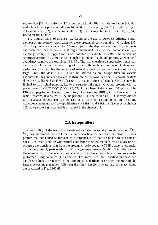
The availability of the isotopically enriched samples (especially protein samples,15N /13C) has introduced the need for methods which allow selective detection of eitherprotons that are bound to the labeled heteronucleus or that are bound to non-labeledones. Also when working with natural abundance samples, methods which allow one tosuppress the signals arising from the protons directly bound to NMR-active heteronucleican be very useful, particularly in HMBC-type experiments [60, 65]. The selection, orthe elimination, of the magnetization arising from the directly bound protons can beperformed using so-called X half-filters. The most usual are so-called in-phase andantiphase filters. The names of the aforementioned filters arise from the state of theheteronuclear magnetization following the filter. Simple in-phase and antiphase filtersare presented in Fig. 2 [66-68].

21
Fig. 2. Pulse sequences [66-68] for a simple in-phase (A) and an antiphase X half-filters (B).Narrow white bars and filled black bars indicate 90°°°° and 180°°°° pulses, respectively. All pulseshave x-phase unless otherwise indicated. Phases for the pulses: Filtration of X-boundprotons, φφφφ1 = x, -x; receiver = x, x and filtration of non-X-bound protons, φφφφ1 = x, -x; receiver =x, -x. The delay∆∆∆∆XH = 1/(21JXH).
The implementation of the X half-filters into a variety of pulse sequences isstraightforward. The desired filter selects or rejects protons that are coupled to X-spins inthe time domain that follows the filter period. The X half-filters are based on specialdelay periods during which the heteronuclear coupling over one bond1JXH is allowed toevolve to create a proton magnetization antiphase with respect to1JXH, i.e. HxXz [67].The time needed to create such a HxXz-magnetization starting from Hy is 1/(21JXH). Asthere is usually more than one value for1JXH in real molecules, the delay∆XH is set foran average1JXH ( for instance 145 Hz for1JCH:s) [68]. In order to obtain peaks with goodlineshape features, the delay∆XH should be sufficiently short to prevent extensiveevolution of the homonuclear proton-proton couplings. This in turn means for thesefilters to work properly, that the heteronuclear coupling constant1JXH should besignificantly larger than the homonuclear coupling constants JHH [2]. This condition isadequately fulfilled with1JNH and1JCH. In 2D experiments, the half-filter can be appliedto either t1 or t2 domain. The spectrum can also be acquired with half-filtering in both t1
and t2 domains (so-called double half-filtration) [67].The simplest filter to selectively detect the non-X-bound protons is the low-pass filter
commonly used in HMBC-experiment [65, 70]. Because in-phase magnetization duringthe indirectly or directly detected period is often preferred, the antiphase X half-filtersare suitable for elimination of the directly X-bound proton magnetization, if separaterefocusing periods are not applied. The in-phase X half-filter presented can in turn beused for both purposes [67]. The PFG z-filter (pulsed field gradient z-filter) is designedonly for detection of the non-bound proton magnetization [69]. The sequence for PFG
1H:∆XH
A)∆XH
X:
φ1
1H:∆XH/2
B)∆XH/2
X:
φ1
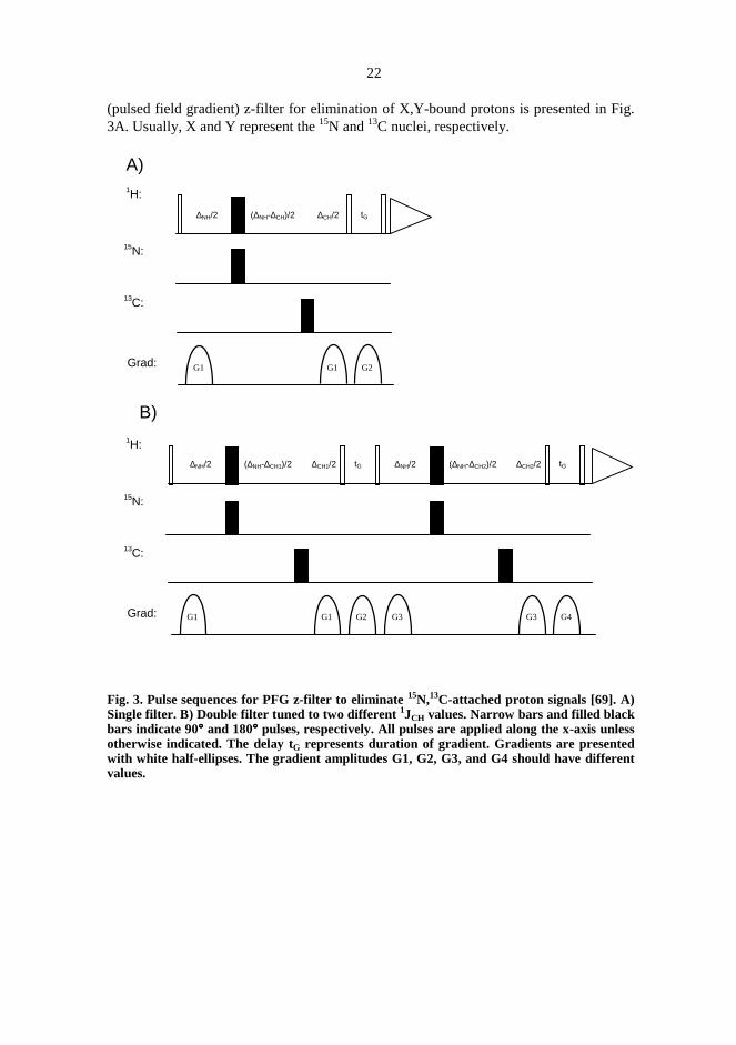
22
(pulsed field gradient) z-filter for elimination of X,Y-bound protons is presented in Fig.3A. Usually, X and Y represent the15N and13C nuclei, respectively.
Fig. 3. Pulse sequences for PFG z-filter to eliminate15N,13C-attached proton signals [69]. A)Single filter. B) Double filter tuned to two different 1JCH values. Narrow bars and filled blackbars indicate 90°°°° and 180°°°° pulses, respectively. All pulses are applied along the x-axis unlessotherwise indicated. The delay tG represents duration of gradient. Gradients are presentedwith white half-ellipses. The gradient amplitudes G1, G2, G3, and G4 should have differentvalues.
Grad:
A)1H:
13C:
Grad:
15N:
G1 G2G1
tG∆NH/2 (∆NH-∆CH)/2 ∆CH/2
B)
G3 G4G3
tG∆NH/2 (∆NH-∆CH2)/2 ∆CH2/2
G1 G2G1
tG∆NH/2 (∆NH-∆CH1)/2 ∆CH1/2
1H:
13C:
15N:

23
The total length of the J-evolution period of the filter is determined by the smallerheteronuclear coupling constant, i.e. when the nuclei involved are15N and 13C, thisduration is determined by1JNH, ∆NH = 1/(21JNH) [69]. The difference between1JNH and1JCH can be taken into account if the carbon 180° pulse is shifted by (∆NH-∆CH)/2 fromthe midpoint of the PFG z-filter element [69]. Similar trick can be applied to other Xhalf-filters as well. Now the evolution times for1JNH and 1JCH are ∆NH and ∆CH,respectively. The magnetization of15N- and 13C-bound protons just before the z-filterelement will be in the antiphase form (HN
xNz and HCxCz), whereas the magnetization of
the non-bound protons are along the y-axis (Hy). The proton 90°(x) pulse flips the Hy-magnetization to z-axis without disturbing HN
xNz and HCxCz magnetization, which then
will be effectively destroyed by the B0-gradient applied during the z-filter delay. Thesucceeding 90°(x) pulse then flips the z-magnetization of the non-bound protons toobservable transverse magnetization.
The fact that there is more than just one value for1JXH leads to phenomenon called J-leaking [68, 69]. A single X half-filter, tuned to some average value JXH, is used toeliminate protons bound to nucleus X will pass a small amount of undesiredmagnetization with an intensity depending on the difference between the actual value ofheteronuclear coupling constant, and the value used to tune the filter (so-called J-mismatch) [68]. If the intensity of the leaking signals is comparable to the desired ones,the analysis of the spectrum may lead to erroneous interpretations. As an example, theproduct operator calculations [30] were performed for the PFG z-filter (Fig. 3A), whichfilters out X-bound proton magnetization. Results for A- and AX-spin systems arepresented in Eq. 2 and Eq. 3. The effects of relaxation are not taken into account incalculations.
A-spin system:
Hz90x
H
→ -Hy∆ ∆/ /2 180 180 2− − →x
HxX
Hy90x
H
→Hz
B gradient0 − → 90xH
→ -Hy (2)
AX-spin system:
Hz90x
H
→ -Hy∆ ∆/ /2 180 180 2− − →x
HxX
Hycos(π1JXH∆XH) - HxXzsin(π1JXH∆XH)90x
H
→ Hzcos(π1JXH∆XH) - HxXz sin(π1JXH∆XH)B gradient0 − → 90x
H
→ -Hy cos(π1JXH∆XH) (3)
The desired magnetization component is presented in Eq. 2 and the component due toJ-leakage is presented in Eq. 3. Both the leaking and the desired magnetization appear asabsorptive, in-phase signals. If the delay∆XH is correctly matched, the intensity of theleaking magnetization -Hycos(π1JXH∆XH) will be zero. However, any deviation from theideal ∆XH-value will introduce leaking. The performance of the filters can be improvedby applying two (or more) filter steps with different∆XH-values in series. The filteringproperties of double tuned filters are superior to single stage filters, and therefore thesetwo stage filters have found widespread use in modern NMR-spectroscopy [68, 69]. Thepulse sequence for double tuned PFG z-filter is presented in Fig. 3B. The intensity of

24
leaking magnetization obtained using double tuned PFG z-filter (tuned with delays∆XH1
and∆XH2) is presented in Eq. 4.
Ileaking(double tuned)∝ -Hycos(π1JXH∆XH1)cos(π1JXH∆XH2) (4)
Figure 4 presents theoretical curves for intensity of the leaking magnetization as afunction of heteronuclear coupling constant. The intensities of the leaking magnetizationfor single PFG z-filter and double tuned PFG z-filter were calculated using Eq. 3 and Eq.4, respectively. The single filter was tuned to1JXH=145 Hz whereas values1JXH1=135 Hzand1JXH2=170 Hz were used to tune the double filter.
Fig. 4. Theoretical leaking intensity (percentage of intensity obtained without filtering) as afunction of 1JXH for single PFG z-filter (open rectangles) and double tuned PFG z-filter(filled rectangles) applied to isolated HX-system. The single PFG z-filter was tuned tocoupling 145 Hz. Two values, 135 Hz and 170 Hz, were used for the double tuned PFG z-filter.
As is the case with all gradient-purged methods with more than one purging step, onemust be careful with gradient polarizations, levels and durations to avoid inadvertentrefocusing of the previously dephased, unwanted coherences.
J-leaking is of major concern when the protons bound to13C are to be filtered out.This rises from the fact that the range of1JCH is relatively large [1] and thus a filter tunedfor a single1JCH-value will not usually have acceptable filtering properties [68]. Twofilter elements tuned to different1JCH-values for example 135 Hz (aliphatic protons) and
-40%
-30%
-20%
-10%
0%
10%
20%
30%
40%
120
130
140
150
160
170
180
190
200
210
1JXH
The
inte
nsity
ofle
akin
gsi
gnal

25
175 Hz (aromatic protons) can be applied in succession. This will significantly improvethe suppression of the13C-bound protons over a considerable range of coupling constantvalues. The effective elimination of15N-bound protons in peptides and proteins (inisotropic solutions) does not require double tuning as the1JNH:s fall into a narrow range(1JNH = 89 - 97 Hz). If both 15N- and 13C-bound protons are to be filtered outsimultaneously using the double tuned filter, usually each of the filters are tuned todifferent1JCH:s whereas a single value for1JNH is used for both elements (Fig. 3B) [69].
In contrast to filtration of X-bound protons, the mismatches in∆XH value do notcause J-leaking when X half-filters are used to filter the non-X-bound protonmagnetization. The mismatches in the delay∆XH will only cause a drop in the signalintensities. In addition to the usual X half-filters the selection of the X-bound protonscan also be done using HSQC [61-64] and HMQC [56-61] sequences utilizing gradientsor phase cycling for coherence selection. It should be noted that phase cycled HMQCwithout time incrementation is in fact an in-phase X half-filter (Fig. 2A).

26
3. Applications
3.1. RF-gradients in some homonuclear 1D- and 2D-experiments
The following two chapters present applications of RF-gradients in homonuclear,selective 1D-, and non-selective 2D TOCSY- and NOESY-experiments. The RF-gradients in the presented methods are produced using the z-rotation approach discussedin chapter 2.1.1 and thus mimic the effect of B0-gradients. A similar RF-gradientapproach was also applied to isotope filtration (isotope edited) experiments with BIRDand BIRDR, and these are discussed separately in chapters 3.2.1 and 3.2.2.
3.1.1. Selective one dimensional experiments with RF-gradients
The simplest way to selectively excite a particular resonance is to apply a frequencyselective 90° pulse. The evolution of couplings and chemical shifts during the pulse mayeasily lead to phase distortions in the resulting spectrum. These problems can be to someextent circumvented with properly shaped excitation pulses which result in pure phasespectra [71-74]. Application of this kind of excitation pulses requires waveformgenerator which may not be available in older spectrometers. Due to aforementionedfact, and reasons concerning differences between selective 90° and 180° pulsesdiscussed earlier (chapter 2.2), approaches which utilize selective inversion rather thanselective excitation are usually recommended. If the capability of the spectrometer inproducing selective pulses is restricted to long rectangular pulses or DANTE shaping[75, 76], they are also the only sensible possibility.
A selective 180° pulse embraced by two gradients can be used either to destroy allthe magnetization not inverted by selective pulse (i.e. excitation sculpting method [37-39]), or to selectively phase encode the inverted magnetization by the gradients [18, 41,77-81]. In the excitation sculpting approach, the gradients on both side of the selectivepulse have the same polarity. If the B0-gradients are replaced with spin-lock pulsesarranged in the z-rotation sandwich [22, 31], the concept of equal polarity means that thephases of the spin-lock pulses must be the same. If, in turn, selective phase encoding is

27
desired, the phases of the spin-lock pulses in the z-rotation sandwiches on both sides ofthe selective pulse should differ by 180°. Now the inverted magnetization will be labeledby the dephasing action of the both gradients and all other magnetization is not affected.The gradient labeled magnetization can be refocused later in the pulse sequence byapplying a gradient with an amplitude equal to the sum of amplitudes of the dephasinggradients.
When RF-gradients are used, the most convenient way is to apply a similar RF-gradient with duration equal to the sum of the durations of the dephasing gradients. Asthe evolution of the chemical shifts is not active during the RF-gradients [22], therephasing can be done without an additional delay and a 180° pulse, which are oftenneeded to compensate for the evolution of the chemical shifts if B0-gradients are used[4]. When gradients are used for coherence selection, half of the signal intensity will belost due to the fact that gradients can select only one of the two coherence pathways [4].Figures 5A-B present single and double echo excitation sculpting pulse sequenceelements. The pulse sequences presented in Fig. 5C-D are based on RF-gradientselection. The white box marked "mixing" can be virtually any mixing element, forexample MLEV-17 [82] for a selective 1D TOCSY [I], or 90°-(spoil gradient)-delay-90°for a selective1D NOESY [I].
Selective 1D versions of NOESY [43, 45, 78, 83, 84] and TOCSY [41, 83-85] can beuseful for relatively small organic compounds. The selective 1D TOCSY extracts outproton signals of a selected spin system. This can be important when analysis of theproton spectrum is hampered by overlapping signals. The selective 1D TOCSYexperiment can be performed using the pulse sequences presented in Fig. 5A-D. Becausehalf of the magnetization is lost in the gradient selected methods (Fig. 5C-D), excitationsculpting based methods are naturally preferred.
Figure 6 presents anomeric proton selective 1D TOCSY spectra of sucrose recordedusing pulse sequence presented in Fig. 5B (excitation sculpting method) and thecorresponding sequence with B0-gradients [I]. Both sets of spectra were recorded with 1,2, and 4 scans. If a single-scan spectrum is needed, the spectrum recorded using the B0-gradient method is better but the suppression of the unwanted signals is not complete,though. As two scans are acceptable (the first two steps of EXORCYCLE [40]), bothmethods work well.
Figure 7 presents anomeric proton selective 1D TOCSY using RF-gradient selectionapproach (pulse sequence presented in Fig. 5C) [I]. The spectrum was recorded with twoscans. The quality of the spectrum is the same as for spectrum recorded with theexcitation sculpting method utilizing RF-gradients (Fig. 6, two scans)

28
Fig. 5. Selective RF-gradient 1D TOCSY and NOESY using the excitation sculptingapproach (single and double echoes, A and B) and gradient selection approach (single anddouble echo, C and D). Narrow black and white bars indicate 90°°°° hard rectangular pulses inthe basic sequences and in RF-gradients using z-rotation approach, respectively. The longspin lock pulses in z-rotation clusters are presented with wide gray bars denoted “SL”.Selective 180°°°° pulses are presented by dark half ellipses. Wide gray bars denoted “trim”present low power trim pulses in the TOCSY mixing propagator. All pulses have x-phasesunless otherwise indicated. The RF-gradient denoted with SL3 during the NOESY mixingperiod serves as a spoil-gradient, and tmix represents mixing time. The basic phase cycle isφφφφ1= x, y, -x, -y; φφφφ2 = -x, φφφφ3 = 4(-y), 4(y), receiver = x, -x, x, -x. Phaseφφφφ4 of NOESY mixingpropagator should be set to -x for sequence A, if a single scan spectrum is desired. For othersequencesφφφφ4 = x.
1H:
y --
y -y
-x
SL1
φ1
SL1
y -y
-x
MIXING
A)
1H:
y --
y -y
-x
SL1
φ1
SL1
y -y
x
MIXING
C)
2×SL1
y -y
x
1H:
B)
MIXING
y --
y -y
-x
SL1
y
-x
-y
SL1 + SL2 SL2
φ1y -y
-x
φ2
1H:
D)
MIXING
y --
y -y
-x
SL1
y
x
-y
SL1 + SL2 SL2
φ1y -y
-x
φ2
2×SL1+2×SL2
y -y
x
φ3 φ3 φ3
=MIXINGy -y
x
SL3 tmix
MLEV-17trim trim : TOCSY
: NOESY
φ4

29
Fig. 6. The quality of anomeric proton selective 1D TOCSY spectra with 1, 2, and 4 scans.The spectra were recorded using a 0.5 M sucrose sample in D2O utilizing the pulse sequencepresented in Fig. 5B and the corresponding sequence with B0-gradients. The spectra wererecorded on a Bruker DRX-500 spectrometer equipped with a triple-resonance probeheadincorporating a single shielded gradient coil at 298 K. Relaxation delay = 3.0 s, acquisitiontime = 1.36 s, selective 180°°°° pulse = 20 ms Gaussian, proton 90°°°° pulse = 5.6 µµµµs, RF-power fortrim-pulses and MLEV-17 = 5.48 kHz, trim-pulse length = 2.5 ms, isotropic mixing time =152.5 ms; an exponential weighting function (0.3 Hz) was applied prior to Fourier transform.RF-gradient method: SL1 = 1.8 ms, SL2 = 2.2 ms. B0-gradient method: gradient shape =sinusoid, gradient pulse length = 1 ms, recovery delay = 200 µµµµs, gradient amplitudes = 7.2and 3.0 G/cm. The small signals at 4.05, 3.90, and 3.50 ppm belong to the fructose ring andare due to incomplete suppression by the RF-gradients. Similar residual signals (althoughsmaller) can also be found in B0-gradient based experiments [I].
Fig. 7. The anomeric proton selective 1D TOCSY spectrum of sucrose recorded using theRF-gradient selected method presented in Fig. 5C. Number of scans = 2, proton 90°°°° pulse =16 µµµµs, relaxation delay = 4.0 s, SL1 = 2.8 ms. Other parameters are the same as for spectra inFig. 6 [I].

30
The NOE spectra contain important information about the three dimensionalstructure of a molecule [86-88]. The classic 1D-method to obtain NOE information isNOE-difference spectroscopy [88]. Although this experiment is very simple to perform,the results can be disappointing. As the experiment is based on the difference of twospectra, the subtraction artefacts may overrun the NOE-enhancement. In principle, aselective 1D NOESY experiment can be performed using either excitation sculpting(Fig. 5A-B), or the gradient selected method (Fig. 5C-D). Although the excitationsculpting preserves both coherence transfer pathways [43-45], and so offers better signalintensity, the relaxation during the NOESY mixing time can lead to spurious peaks [43-45]. After the excitation sculpting step, all the magnetization not inverted by theselective 180° pulse is destroyed by the gradients. The selected magnetization goesthrough the NOESY step and the resulting spectrum contains the signal of the selectedproton (corresponds the diagonal peak in 2D NOESY), and also signals of those protons,which have NOE with the initially selected proton. When the excitation sculptingapproach is used, difficulties will be introduced due to relaxation during the NOESYmixing time. The unwanted magnetization dephased by the gradients will relax towardsthe equilibrium state during the NOESY mixing time. The relaxed magnetization has lostgradient induced phase dispersion and the last pulse of NOESY flips this magnetizationinto the transverse plane resulting in extra peaks in the spectrum. Elimination of theseartefact peaks can be performed by applying EXORCYCLE[40] to the selective pulse.This, however, works only when the intensity of the magnetization responsible for theartefact peaks is not large. When long mixing times are used, almost full intensity of theequilibrium magnetization can be reached and correspondingly successfulEXORCYCLE-based difference spectroscopy becomes more difficult [44]. Thesuppression of the relaxation induced artefact peaks via EXORCYCLE can be improvedby adding extra 180° pulse(s) within the mixing time resulting in decrease of theintensity of the forming equilibrium magnetization [44]. In practice the positioning ofthese 180° pulses can be rather difficult due to the variation in T1-relaxation times.
A more straightforward way to obtain a selective 1D NOESY is to utilize RF-gradient selection [I], as presented in Fig. 5C-D. This method is a modification of the B0-gradient based GOESY-experiment [78]. In RF-gradient selected NOESY, theselectively inverted magnetization is phase encoded due to the gradients. Thismagnetization goes through the NOESY step, during which the phase encoding istransferred to other nuclei via NOE-interaction. The phase encoding is decoded back bythe final RF-gradient just before the acquisition. If the first pulse of the mixingpropagator has phase x, only the magnetization parallel to y-axis needs to be considered,as the x-magnetization will be further dephased by the spoil-gradient gradient, andtherefore is not refocused by the final RF-gradient. The destruction of the x-magnetization leads to the fact that half of the magnetization is lost during the gradientselected methods i.e. only one of the two coherence pathways is selected [43-45]. Thegradient labeled y-magnetization has the gradient-induced, spatially dependent phase(cosine function). The 90° pulse prior to the NOESY mixing time creates a longitudinalmagnetization, which is aligned along the -z or the +z-axis. Now, an additional signalloss of factor 0.5 is introduced, as only half of the longitudinal magnetization createsNOESY-peaks [I, 44]. Diffusion effects will also cause some decrease in signal
31
intensity. This may be problematic for small molecules in non-viscous solvents, whenrelatively long NOESY mixing times are used [I, 44].
The main advantage of RF-gradient selected 1D NOESY (and GOESY) is that theresulting spectrum contains only the resonances of the target proton and resonances thatare formed during mixing time via NOE-interaction. Some spurious small anti-phasesignals (i.e. COSY-type signals) can appear in spectrum as a result of terms havingcoherence order zero during the NOESY mixing time. Zero-quantum coherences and so-called “zz terms” present during mixing time survive the dephasing caused by the spoil-gradient and may be converted into observable signals. Uneven excitation of the targetmultiplet and imperfect 90° pulses of the NOESY mixing propagator may give rise tothese terms [44].
Figure 8 presents anomeric proton selective 1D NOESY spectra of sucrose recordedusing the RF-gradient selected method (pulse sequence presented in Fig. 5C) and mixingtimes of 4µs, 125 ms, 500 ms, and 1000 ms [I]. For a comparison also anomeric protonslice of 2D NOESY spectrum (mixing time = 1000 ms) is presented. The spectrum inFig. 8A (mixing time = 4µs) shows an antiphase J peak at 3.4 ppm due to magnetizationcomponents with coherence order zero during the mixing time. As can be seen from thespectra in Fig. 8A-D, the magnitude of J peak remains constant and the developing in-phase NOE-peak overruns this J peak when the proper mixing time (1.0 s in this case) isused.

32
Fig. 8. Four anomeric proton selective 1D NOESY (pulse sequence in Fig. 5C) spectra withdifferent NOESY mixing times (A-D) and the anomeric proton slice of non-selective 2DNOESY spectrum of 0.5 M sucrose at 298 K. 1D spectra (A-D): Number of scans = 32,relaxation delay = 10 s, acquisition time = 1.36 s, selective 180°°°° pulse = 20 ms Gaussian, SL1= 1.4 ms, SL3 = 1.7 ms, tmix = 4 µµµµs (A), 125 ms (B), 500 ms (C), and 1000 ms (D); anexponential weighting function (0.3 Hz) was applied prior to Fourier transform. 2D phase-sensitive NOESY (E): Relaxation delay = 2.0 s, number of transients = 16, number ofincrements = 256, tmix = 1.0 s, resolution in f2-dimension 7.82 Hz/pt [I].

33
3.1.2. Two dimensional TOCSY and NOESY with RF-gradient selection
The pulse sequences for 2D RF-gradient NOESY and TOCSY are shown in Fig. 9 [I].No trim-pulses were applied in TOCSY. Again, as the chemical shifts are not activeduring the RF-gradients, there is no need for extra echo-periods in order to compensatefor chemical shift evolution.
Phase sensitive spectra can be obtained by applying the echo-antiecho method [89,90] to the rephasing gradient. The phase of the spin-lock pulse in the z-rotation clusterprior to detection (rephasing gradient) was inverted to record both P- and N-type spectrafor the same t1-increment value. The RF-gradient versions of these common 2D methodsoffer good quality spectra in a short time, as there is no need for phase cycling. Inpractice, a two-step cycle applied to the excitation pulse is preferable to ensureelimination of axial peaks.
Figure 10 shows phase-sensitive 2D TOCSY an NOESY spectra of 0.5 M sucrose inD2O recorded using pulse sequences presented in Fig. 9 [I]. A two-step cycle was usedto avoid axial peaks.
Fig. 9. Pulse sequences for phase-sensitive 2D RF-gradient TOCSY (A) and RF-gradientNOESY (B). Notation is the same as in Fig. 5. All pulses are in the x-phase unless otherwiseindicated. Phase-sensitive spectra are obtained by inverting the phase of the spin-lock pulsein the z-rotation cluster prior to detection (refocusing RF-gradient) to record both echo- andantiecho-type spectra (denoted by “e/ae”) for the same time increment (t1). A) Basic phasecycle isφφφφ1 = x, -x; φφφφ2 = x, -x; receiver = x, -x. B) Basic phase cycleφφφφ3 = x, -x; receiver = x, -x.
1H:SL1
y -y
-x
MLEV-17
φ2
A)
e/ae-x / x
t1
φ1
SL1
y -y
1H:SL1
y -y
-x
SL2
y -y
-x
tmix
B)
t1
e/ae-x / x
SL1
y -yφ3

34
Fig. 10. Phase-sensitive RF-gradient selected TOCSY (A) and NOESY (B) spectra of 0.5 Msucrose in D2O at 298 K recorded using the pulse sequences presented in Fig. 9.Experimental parameters: Bruker DRX-500 NMR spectrometer equipped with Brukertriple-resonance probe and z-axis gradient system, relaxation delay = 4.0 s, t1max = t2 = 85.2ms, number of transients = 2, number of increments = 256, number of sampled t2-points =256, (A) SL1 = 1.8 ms, length of MLEV-17 mixing = 147.4 ms, RF-power for MLEV-17 =5.48 kHz, (B) SL1 = 1.8 ms, SL2 (spoil) = 1.4 ms, tmix = 1.0 s. The t1 and t2 domains were zero-filled once, and both dimensions were multiplied with squared cosine function prior toFourier transform [I].

35
3.2. Isotope filtration using excitation sculpting
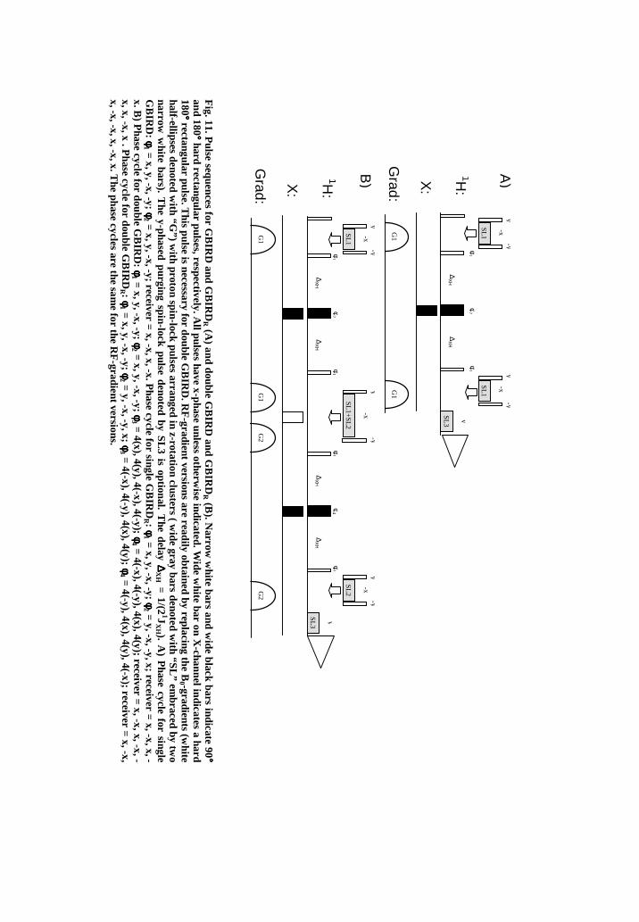
The excitation sculpting method is readily applicable to isotope filtration, as there is amethod capable of selectively inverting X-bound/non-bound proton magnetization [38,39]. Such inversions can be accomplished with the aid of bilinear pulse clusters BIRDand BIRDR [55]. All the proton pulses in BIRD are applied along the x-axis whereas forBIRDR, the central proton 180° pulse has y-phase (or alternatively the last 90° pulse has–x-phase). The total length (2∆XH) of the BIRD- or BIRDR-cluster is 1/1JXH. If either ofthe aforementioned BIRD-clusters is applied in between two gradient pulses of equallengths and dephasing properties, and such a pulse sequence element is applied after a90° excitation pulse on protons, the inverted proton magnetization is refocused at the endof gradient-BIRD-gradient/gradient-BIRDR-gradient element. The magnetization notexperiencing inversion is, in turn, effectively dephased. Both BIRD and BIRDR caneasily be modified to simultaneously select or reject the protons bound to X- or Y-nuclei[IV] by applying the method used for PFG z-filter discussed in chapter 2.3. The singleand doublegradient-BIRD/gradient-BIRDR elements are presented in Fig. 11. Thenormal B0-gradients can be conveniently replaced with RF-gradients using z-rotationapproach discussed in chapter 2.1.1 (Fig. 7) [III, 22, 31]. The abbreviations GBIRD orGBIRDR refer to B0-gradient versions. The pulse sequence for double GBIRD/GBIRDR
for the simultaneous selection/rejection of protons bound to15N and 13C is presented inFig. 12.
36
Fig.11.P
ulsesequences
forG
BIR
Dand
GB
IRDR
(A)
anddouble
GB
IRD
andG
BIR
DR
(B).N
arroww
hitebars
andw
ideblack
barsindicate
90°°° °and
180°°° °hard
rectangularpulses,respectively.A
llpulseshave
x-phaseunless
otherwise
indicated.Wide
white
baron
X-channelindicates
ahard
180°°° °rectangular
pulse.This
pulseis
necessaryfor
doubleG
BIR
D.R
F-gradientversions
arereadily
obtainedby
replacingthe
B0 -gradients(w
hitehalf-ellipses
denotedw
ith“G
”)w
ithproton
spin-lockpulses
arrangedin
z-rotationclusters
(w
idegray
barsdenoted
with
“SL”
embraced
bytw
onarrow
white
bars).T
hey-phased
purgingspin-lock
pulsedenoted
byS
L3is
optional.T
hedelay∆∆∆ ∆
XH
=1/(2
1JX
H ).A
)P
hasecycle
forsingle
GB
IRD
:φφφ φ
1=
x,y,-x,-y;φφφ φ2
=x,y,-x,-y;receiver
=x,-x,x,-x.P
hasecycle
forsingle
GB
IRDR :φφφ φ
1=
x,y,-x,-y;φφφ φ2
=y,-x,-y,x;receiver
=x,-x,x,-
x.B)
Phase
cyclefor
doubleG
BIR
D:φφφ φ
1=
x,y,-x,-y;φφφ φ2
=x,y,-x,-y;φφφ φ
3=
4(x),4(y),4(-x),4(-y);φφφ φ4
=4(-x),4(-y),4(x),4(y);receiver
=x,-x,x,-x,-
x,x,-x,x.P
hasecycle
fordouble
GB
IRDR :φφφ φ
1=
x,y,-x,-y;φφφ φ2
=y,-x,-y,x;φφφ φ
3=
4(-x),4(-y),4(x),4(y);φφφ φ4
=4(-y),4(x),4(y),4(-x);receiver
=x,-x,
x,-x,-x,x,-x,x.The
phasecycles
arethe
same
forthe
RF
-gradientversions.
y-y
-x
Grad:
G1
1H:
φ1
G1
X:
∆X
H∆
XH
y-y
-xSL1
SL1
Grad:
G1
1H:
X:
G2
G1
∆X
H∆
XH
G2
∆X
H∆
XH
SL1
y-y
-x
SL1
+S
L2S
L2
A)
B)
y
SL3
y
SL3
y-y
-xy
-y-x
φ2
φ1
φ1
φ2
φ1
φ3
φ4
φ3

37
Fig.
12.T
hepulse
sequencefor
simultaneous
selection(double
GB
IRD
)or
rejection(double
GB
IRD
R )of
protonsbound
to15N
and13C
.T
henotation
isthe
same
asin
Fig.11.P
hasecycles
arethe
same
asindicated
forsequences
presentedin
Fig.11B
.Delay
∆∆∆ ∆N
H=
1/(21J
NH )
anddelay∆∆∆ ∆
CH
=1/(2
1JC
H ).
G2
1H:
13C:
Grad:
G1
G1
15N:
∆N
H∆
CH
G2
∆N
H -∆C
H∆
NH
∆C
H∆
NH -∆
CH
y
SL3
φ1
φ2
φ1
φ3
φ4
φ3

38
3.2.1 Gradient-BIRD and selection of X-bound magnetization
The pulse sequences for single and double gradient-BIRD are presented in Fig. 11 [II,38, 39]. After the excitation pulse the first gradient (B0-gradient or RF-gradient using z-rotation approach) defocuses all the magnetization in the transverse plane. SubsequentBIRD causes complete inversion for X-bound protons, if the BIRD-delay (2∆XH) iscorrectly matched to the 1/1JXH. The non-bound magnetization experiences a rotation of360°. Thus, the second gradient refocuses the inverted magnetization while themagnetization not inverted by the BIRD is further dephased. Because both the coupledprotons and the X-nuclei are flipped by the BIRD, the evolution of1JXH during the B0-gradient pulses is not refocused. This will result in lineshapes with mixed phases,making the single GBIRD approach not preferable. However, the evolution of1JXH
during the gradient delays can be refocused when double GBIRD is used (Fig. 11B) [38,39]. The refocusing is done by applying a 180° pulse on the X-channel in between thetwo GBIRD-elements. If the X-nucleus is low natural abundance species, (for instance13C) other homonuclear JHH-coupling evolution during B0-gradient pulses, except theevolution of geminal couplings, will be refocused. By changing the B0-gradient pulses toRF-gradients (Fig. 11A), evolution of homonuclear JHH- and heteronuclear JXH-couplingsduring the gradients is avoided and thus a single RF-gradient BIRD can be used. In fact,RF-gradients make it also possible to leave out the 180° pulse on X-channel in betweenthe two BIRDs [II]. The 180° proton pulse of the second BIRD has phase –x resulting in0° net rotation for non-bound protons. This partially compensates for the offset- and B1-inhomogenity effects on suppression [38].
As a consequence of the misset of delay 2∆XH, some decrease in the intensity of therefocused signal will take place, whereas homonuclear JHH-couplings lead to leakage ofthe undesired signals into the spectrum. Homonuclear JHH-couplings (that are activeduring the BIRD-delay 2∆XH) result in incomplete suppression of the non-bound protons[39]. While the suppression for singlets is virtually complete, the suppression ratiosdecrease with increasing width of the proton multiplet. The intensities of the leaking in-phase magnetization for a proton with one homonuclear JHH-coupling after single anddouble GBIRD-elements are presented in Eq. 5 and Eq. 6, respectively.
IsingleGBIRD ∝ -0.5{ 1 – cos(2πJHH∆XH) } (5)
IdoubleGBIRD ∝ -0.25{ 1- 2cos(2πJHH∆XH) + cos(4πJHH∆XH) } (6)
Suppression ratios of 50-200 for non-13C bound proton multiplets can be achievedusing the double GBIRD approach, whereas sharp singlets can be suppressed up to afactor of 20000 [39]. The obtained ratio depends on the number and magnitude ofhomonuclear proton-proton couplings. Naturally, heteronuclear long-range couplingswill also lead to leaking. However, in the case of natural abundance samples, where thepercentage for NMR-active isotope of heteronucleus is low, the contribution of theresulting leaking is low compared to the leaking arising from homonuclear JHH. The

39
suppression ratio of 50-200 is not enough for13C half-filter purposes, when weaksatellite signals in natural abundance samples are to be selected, but is sufficient topresuppress the signals of the12C-bound protons prior to HMQC or HSQC [II, 39]. Dueto the presuppression, the signal intensity of the non-bonded protons is of about the samemagnitude as the desired signals, and a conventional two-step phase cycle of theHMQC/HSQC will be efficient in eliminating the rest of the12C-bound protons [II, 39].Application of four consecutive GBIRD elements after excitation pulse suppressesstrong parent signals to a negligible level and even manages to minimize the strongwater peak of H2O-solutions [38, 39]. The relatively low suppression ratio of the doubleGBIRD is not of major concern when both selected and rejected signals areapproximately of the same order of magnitude, which is often the case with mixtures ofisotopically enriched and natural abundance molecules prepared for interaction studies.However, as the gradient selected HSQC and HMQC [60, 61] efficiently eliminate allnon-X-bound signals, while selecting X-bound proton signals of the isotopicallyenriched molecule, the double GBIRD approach is not usually preferred. In addition, thedouble GBIRD-element is longer than HSQC or HMQC.
Probably the best application of double GBIRD is the presuppression of the non-X-bound protons in natural abundance samples prior to phase cycled HMQC or HSQC. Onthe other hand, this is not necessary if there is existing hardware for B0-gradients that canbe used for heteronuclear gradient selection. In this context, the RF-gradient basedapproach seems more valuable. The pulse sequences for double RF-gradient-BIRD-HSQC are presented in Fig.13 [II]. The double RF-gradient-BIRD sequence presented inFig. 13B is a direct modification of the original double GBIRD-sequence. All the B0-gradients of the double GBIRD are replaced by RF-gradients utilizing the z-rotationapproach. The pulse sequence presented in Fig. 13A contains combined RF-gradient inbetween the two BIRD-clusters. In addition the 180° pulse on13C-channel is omitted.
Recording of a good quality HMQC/HSQC spectrum of low natural abundanceheteronucleus, like13C or 15N, using phase cycling to eliminate strong parent signalwhile selecting for weak satellite signal is a difficult task. This is due to the fact that theelimination of very strong signals using the phase cycling fails quite frequently [II, 39].In addition, very low receiver gain setting must be used. For those spectrometers lackingthe B0-gradient capabilities, double RF-gradient-BIRD-HMQC/HSQC offers the spectrawith a quality close to B0-gradient selected experiments [II].
Figure 14 presents a series of 2D HSQC spectra of 0.5 M sucrose in D2O. Spectra Aand B were recorded using pulse sequences presented in Fig. 13B and 13A, respectively[II]. The spectrum C was recorded using the B0-gradient version of the sequencepresented in Fig. 13B (double GBIRD-HSQC) [38, 39]. The spectra presented in Fig.14D-14F were recorded with double-spin-locked HSQC [29, 33], conventional phase-cycled HSQC [62, 63], and gradient selected HSQC pulse sequences [19], respectively.Clearly, the magnitude of t1-noise is lowest in the spectra presented in Fig. 14B, 14C,and 14F. The spectrum recorded with pulse sequence presented in Fig. 13A is ofcomparable quality to the spectrum obtained with B0-gradient selected method (Fig.14F). The dynamic range properties of double RF-gradient-BIRD-HSQC sequences werealso good, allowing the use of about 44 dB larger receiver gain than could be used forconventional phase-cycled HSQC.

40
Fig.13.P
ulsesequence
fordouble
RF
-gradient-BIR
D-H
SQ
C.N
arroww
hitebars
andw
ideblack
barsindicate
90°°° °
and180°°° °
pulses,respectively.A
llpulsesare
inthe
x-phaseunless
otherwise
indicated.Spin-lock
pulsesare
denotedw
ith“S
L”.Spin-lock
pulsesS
L1and
SL2
arearranged
inz-
rotationclusters
,and
thusact
asB0 -gradients.
The
spin-lockpulse
SL3
isused
forpurging
anym
agnetizationthat
isnot
alongy-axis
priorto
HS
QC
.Com
positepulse
decouplingon
carbon(G
AR
P-1)
[91]is
indicatedby
CP
D.
Basic
phasecycle:
φφφ φ1
=x,
y,-x,-y;φφφ φ
2=
4(x),4(-x);φφφ φ
3=
8(x),8(-x);
receiver=
(x,-x,
x,-x),
2(-x,x,
-x,x),
(x,-x,
x–x).
The
delay∆∆∆ ∆C
H=
1/(21J
CH ).
The
phase-sensitivespectrum
isobtained
byincre
menting
phaseφφφ φ2
inT
PP
I-manner
[92].A)
Com
binedR
F-gradient
periodin
between
thetw
oB
IRD
-clusters.B)
Separate
RF
-gradients.T
hisis
similar
tothe
originaldoubleG
BIR
Dsequence,only
theB0 -gradients
arereplaced
byR
F-gradients
utilizingthe
z-rotationapproach.
13C:
1H:
yy
t1 /2t1 /2
- φ1
y-y
-xSL1
y
-x
-y
SL1
+S
L2
-xy
-y
-xy
SL2
SL3
CP
D
∆C
H /2∆
CH /2
∆C
H /2∆
CH /2
∆C
H∆
CH
∆C
H∆
CH
y-y
-x
SL1
y-y
-x
SL2
1H:
13C:
A)
B)
φ1
φ1
φ2
φ3

41
Fig. 14. Contour plots of phase sensitive 2D HSQC spectra of 0.5 M sucrose in D2O at 298 K.(A) Double RF-gradient-BIRD-HSQC (pulse sequence in Fig. 13B), (B) double RF-gradient-BIRD-HSQC (pulse sequence in Fig. 13A), (C) double GBIRD-HSQC [38, 39], (D) double-spin-locked HSQC [29, 33], (E) conventional HSQC[62, 63], and (F) gradient selected HSQC[19]. All the spectra are plotted with the same relative scale. Relaxation delay = 1.5 s, ∆∆∆∆CH =3.45 ms, t1max = 3.2 ms, t2 = 681.6 ms, carbon decoupling during acquisition by GARP-1 [91],number of transients = 16, number of increments = 64, B0-gradient pulse length = 1 ms,gradient recovery delay = 200 µµµµs, gradient shape = sinusoid; (A, B) SL1 = 1.4 ms, SL2 = 1.8ms, SL3 = 1.0 ms; (C) gradient amplitudes 7.2, 7.2, 3.0, and 3.0 G/cm; (D) spin-lock pulses =1.4 ms and 1.8 ms; (F) gradient amplitudes = 24.0, 9.0 (z-filter), and 6.0 G/cm. The t1 domainwas zero filled twice and the t2 domain was zero filled once, and both domains weremultiplied with a cosine function prior to Fourier transform.
3.2.2. Gradient-BIRDR and selection of non-X-bound magnetization
The pulse sequence elements GBIRDR and double GBIRDR are presented in Fig. 11. The180° proton pulse has now phase y, whereas for the BIRD-element the correspondingpulse was applied along the x-axis. This results in inversion of the non-bound protonsand zero-rotation for protons coupled to the X-nuclei. Similar effect can also beachieved, if the phase of the last 90° proton pulse of BIRD-elements is changed from xto –x [55].
The selective inversion for non-bound protons via BIRDR can be utilized inexcitation sculpting based filtering. The leaking properties of GBIRDR (Fig. 11A) anddouble GBIRDR (Fig. 11B) for isolated HX-system are presented in Eq. 7 and 8. Therelaxation is neglected and gradient pulses are assumed to be infinitely short. The totalduration of BIRDR is 2∆XH=1/1JXH.
42
IsingleGBIRDR∝ -0.5{ 1 + cos(2π∆1JXH) } H Y = { -cos2(π∆1JXH) }H Y (7)
IdoubleGBIRDR∝ -0.25{ 1 + cos(2π∆1JXH) } 2 HY = { -cos4(π∆1JXH) }H Y (8)
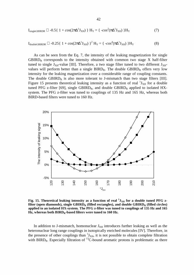
As can be seen from the Eq. 7, the intensity of the leaking magnetization for singleGBIRDR corresponds to the intensity obtained with common two stage X half-filtertuned to single JXH-value [III]. Therefore, a two stage filter tuned to two different JXH-values will perform better than a single BIRDR. The double GBIRDR offers very lowintensity for the leaking magnetization over a considerable range of coupling constants.The double GBIRDR is also more tolerant to J-mismatch than two stage filters [III].Figure 15 presents theoretical leaking intensity as a function of real1JXH for a doubletuned PFG z-filter [69], single GBIRDR, and double GBIRDR applied to isolated HX-system. The PFG z-filter was tuned to couplings of 135 Hz and 165 Hz, whereas bothBIRD-based filters were tuned to 160 Hz.
Fig. 15. Theoretical leaking intensity as a function of real1JXH for a double tuned PFG z-filter (open diamonds), single GBIRDR (filled rectangles), and double GBIRDR (filled circles)applied to an isolated HX-system. The PFG z-filter was tuned to couplings of 135 Hz and 165Hz, whereas both BIRDR-based filters were tuned to 160 Hz.
In addition to J-mismatch, homonuclear JHH introduces further leaking as well as theheteronuclear long range couplings in isotopically enriched molecules [IV]. Therefore, inthe presence of other couplings than1JXH, it is not possible to obtain complete filtrationwith BIRDR. Especially filtration of13C-bound aromatic protons is problematic as there
-5%
0%
5%
10%
15%
20%
120
130
140
150
160
170
180
190
200
210
1JXH
The
inte
nsity
ofle
akin
gsi
gnal

43
are usually more than one large homonuclear couplings present as well as severalheteronuclear long-range couplings [IV]. In a similar manner to GBIRD, the resultingleaking proton magnetization has experienced flipping by the BIRDR-element and thus,as the magnetization of the heteronucleus is also flipped, the heteronuclear couplingevolution during the first gradient pulse is not refocused during the second gradient. Thisis not the case if RF-gradients are used. The evolution of the heteronuclear couplingsduring B0-gradient pulses can be utilized if additional suppression of the unwantedmagnetization is desired. If the length of the B0-gradient pulse, tG, is set to tG=1/(41JXH),the created antiphase magnetization, HxXz, can be purged using spin-lock pulse with y-phase [III] or by applying PFG z-filter element after GBIRDR-cluster [IV]. The antiphasemagnetization can also be removed with a low-pass filter, i.e. by applying a 90° pulse onX-channel after GBIRDR element [IV, 65, 70]. The elimination of the resulting antiphasemagnetization with X-pulses can be based on the purging principle, or phase cycling. If asingle GBIRDR is used, the 90° X-pulse converts the created heteronuclear antiphasecoherence into unobservable heteronuclear multiple quantum coherence.
In the double GBIRDR case, if the purging principle on X-nucleus channel is used,the two 90° X-pulses should have a phase difference of 90° to avoid the conversion ofthe multiple quantum coherence created by the first X-pulse into an observable singlequantum coherence by the second 90° X-pulse. During the pulse sequence, any 90° X-pulse (or imperfect 180° pulse) after the filter element could convert the purgedmagnetization into an observable signal. Therefore, the elimination of the antiphasemagnetization using phase cycling for these low-pass filter pulses could be beneficial.The sign of the created antiphase magnetization follows the phase of the X-pulse andtherefore the undesired signal can be eliminated by simply inverting the phase of the X-pulse in every second scan. This phase alternation is performed separately on both 90°X-pulses. A double GBIRDR filter utilizing additional low-pass filtration forsimultaneous dephasing the magnetization of protons bound to15N and 13C is presentedin Fig. 16A (double GBIRDR-LP). As the range of1JCH:s is large, the coupling evolutionduring the B0-gradient pulses is utilized to further suppress the magnetization of13C-bound protons. This improves the performance of the double GBIRDR-filter withoutsignificantly increasing the filter duration. As the range for1JNH is rather narrow, thesuppression of the coupled magnetization with double GBIRDR alone is usuallysufficient enough[IV].
The pulse sequence for double GBIRDR with additional PFG z-filter is presented inFig. 16B (double GBIRDR-PFG-z-filter). Analogously to double GBIRDR-LP-filter, thisfilter is also designed for simultaneous suppression of protons bound to15N and13C. Theevolution of heteronuclear couplings during the B0-gradient pulses is utilized to improvethe suppression of13C-bound protons.
The filtration properties of the double GBIRDR can be utilized in suppression of thedirectly 13C-bound protons in the HMBC spectrum (Fig 16C) [III]. Also the eliminationof both 15N and 13C-bound protons has been found to be quite efficient [IV]. Figure 17presents slices taken from the 2D HMBC spectra of 0.5 M sucrose at anomeric carbonfrequency recorded using double GBIRDR-filtered HMBC (pulse sequence in Fig. 16C)[III] and conventional low-pass filtered, gradient selected HMBC [19]. The filters weretuned for1JCH values of 125, 145, 165, and 185 Hz. The signals from directly13C-bondedprotons are marked with arrows. As can be seen from Fig. 17A-D, low-pass filtration

44
using double GBIRDR tolerates tuning mismatches rather well, whereas conventionallow-pass filter results in a significant amount of leaking even if the mismatch is of theorder of 5 Hz (Fig. 17G,1JCH = 170 Hz for anomeric proton).
The pulse sequences for f215N,13C half filtered NOESY are presented in Fig. 16D
(NOESY-double-GBIRDR-LP) and 16E (NOESY-double-GBIRDR-PFG-z-filter) [IV].These two pulse sequences produce 2D NOESY spectra where all the resonancesdetected during the t2-period belong to protons not bound to15N or 13C [IV]. As theduration of the double GBIRDR element is relatively long intensity loss due to T2-relaxation cannot be avoided. In addition, as homonuclear JHH:s are active during therelatively long filtration period, the intensity of the detected NOESY-correlation can bereduced. This can be a major issue when the NOE is transferred to a proton havingseveral coupling partners. This can be seen in the NOESY-double-GBIRDR-LP-spectraof uniformly 15N, 13C-labeled human ubiquitin (VLI Research) recorded at 303 K (Fig.18) (ubiquitin sequence: Met – Gln – Ile – Phe – Val – Lys – Thr – Leu – Thr – Gly –Lys - Thr – Ile – Thr – Leu – Glu – Val – Glu – Pro – Ser – Asp – Thr – Ile – Glu – Asn– Val – Lys – Ala – Lys – Ile – Gln – Asp – Lys – Glu – Gly – Ile – Pro – Pro – Asp –Gln – Gln – Arg – Leu – Ile – Phe – Ala – Gly – Lys – Gln – Leu – Glu – Asp – Gly –Arg – Thr – Leu – Ser – Asp – Tyr – Asn – Ile – Gln – Lys – Glu – Ser – Thr – Leu –His – Leu – Val – Leu – Arg – Leu – Arg – Gly - Gly). The spectrum in Fig. 18A wasrecorded omitting all the15N-pulses of NOESY-double-GBIRDR-LP-sequence afterNOESY-mixing period. In addition the WALTZ-16 decoupling scheme [94] was appliedto 15N-nuclei during the detection period. Thus, the resulting spectrum shouldexclusively contain signals detected at amide proton frequencies (chemical shift range≈6.0 –10.0 ppm). However, due to imperfect filtration, a residual diagonal of protonsbound to13C is visible (chemical shift range of aliphatic protons≈ 0.0 – 6.0 ppm) .Importantly, the NOESY-correlations detected at amide proton frequencies in the f2-dimension do not have counterparts (cross peaks) at the aliphatic proton frequencies.This indicates successful filtering. The spectrum in Fig. 18B was recorded omitting allthe 13C-pulses of NOESY-double-GBIRDR-LP-sequence after NOESY-mixing period.Carbon decoupling during the acquisition period was performed using GARP-1 [91].Now only those protons bound to13C are detected during the t2-period. As is clearlyvisible in the spectrum, the NOESY-correlations between amide and aliphatic protonsare much weaker than corresponding correlations in the spectrum presented in Fig. 18A.This phenomenon is due to the evolution of homonuclear JHH-coupling during thefiltration period. A clear indication of this is also seen in the intensities of correlationsbetween alpha and other aliphatic protons. The correlations detected at alpha protonfrequencies (chemical shift range≈ 3.5 – 6.0 ppm) during t2-period are more intense thancorresponding ones below the diagonal. This is possibly due to the fact that alphaprotons usually have less protons to couple with than side-chain protons.
Figure 19 presents NOESY-double-GBIRDR-PFG-z-filter-spectrum (pulse sequencepresented in Fig. 16E) of 1 mM :1 mM mixture of uniformly15N, 13C-labeled proteinTnC (N-terminal domain of cardiac Troponin C, 93 amino acid residues) and non-labeled peptide TnI (Troponin I, 28 amino acid residues) in 7% D2O/H2O-solution at 303K [IV]. The resulting NOESY spectrum shows intramolecular NOESY-correlations ofTnI. In addition, if there are any intermolecular NOESY-correlations, they are alsoobserved. Such a correlation would be observed at a frequency of proton of isotopically

45
enriched TnC in the f1-dimension and correspondingly at frequency of proton of non-labeled TnI in the f2-dimension. The overall outlooks of the spectrum i. e. the amount ofthe correlation peaks indicates successful filtration as significant leaking of signalsbelonging to a relatively large protein would have been indicated by much more“crowded” 2D-correlation map.
Figure 20 presents expansions of two 1D-spectra of the TnC/TnI-sample describedabove, recorded using the pulse sequence presented in Fig. 16B. The delays∆NH and∆CH
were optimized for couplings of 92 Hz and 135 Hz, respectively. The gradient delays tG1
and tG3 were optimized for1JCH-couplings of 155 Hz and 175 Hz. The lower spectrumwas recorded without heteronuclear pulses and thus contains all the proton resonances(both TnC and TnI). When heteronuclear pulses are applied (upper spectrum), thenumber of resonances is significantly reduced as the proton signals of uniformly15N,13C-enriched TnC are effectively suppressed [IV].
Due to the limitations of the double GBIRDR it should be applied with caution if theproton signals of a very large molecule with short T2-times are to be selected. Inaddition, if the selected protons belong to an extensively coupled spin-system, a decreasein the signal intensities will be expected. Quite often, however, the situation forinteraction studies is such that the larger molecule in the sample mixture is isotopicallyenriched protein and the smaller one is synthetic compound or peptide at naturalabundance. In these kinds of cases the double GBIRDR approach may be useful.
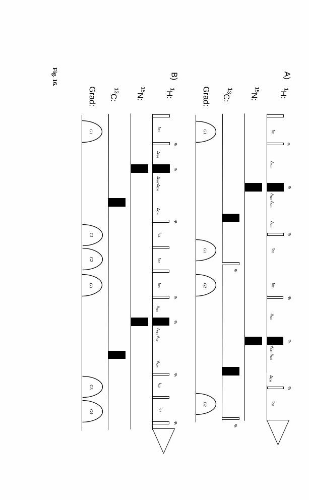
46
Fig.16.
G1
G1
G3
G3
G2
G4
B)
Grad:
G1
G1
G2
G2
1H:
13C
:
15N:
φ1
tG1
∆N
H∆
CH
tG1
tG2
tG2
∆N
H -∆C
H∆
NH
∆C
H∆
NH -∆
CH
A)
Grad:
1H:
13C:
15N:
tG1
∆N
H∆
CH
tG1
tG3
tG3
∆N
H -∆C
H∆
NH
∆C
H∆
NH -∆
CH
tG2
tG4
φ2
φ1
φ4
φ5
φ4
φ3
φ6
φ1
φ2
φ1
φ3
φ4
φ3
φ5

47
Fig.16.
C)
D)
Grad:
G1
1H:
tm
13C
:
G0
Grad:
1H:
13C:
15N:
tG0
CP
D
t1tm
ix
φ7
GS
G1
G2
G3
G4
G5
G2
y
∆C
H∆
CH
∆C
H∆
CH
t1 /2t1 /2
G1
G1
G2
G2
tG1
∆N
H∆
CH
tG1
tG2
tG2
∆N
H -∆C
H∆
NH
∆C
H∆
NH -∆
CH
φ1
φ2
φ1
φ3
φ8
φ1
φ2
φ1
φ3
φ4
φ5
φ4
φ6

48
Fig.16.P
ulsesequences
fordouble
GB
IRDR
with
low-pass
filtersfor
simultaneous
dephasingof
magnetization
ofprotons
directlyattached
to 15Nand
13C(double
GB
IRD
R -LP)
(A),and
(doubleG
BIR
DR -P
FG
-z-filter)(B
),absolutevalue
doubleG
BIR
DR -filtered
HM
BC
(C),t
2 -filteredN
OE
SY
(NO
ES
Y-
double-GB
IRD
R -LP)
(D),and
t2 -filteredN
OE
SY
(NO
ES
Y-double-G
BIR
DR -P
FG
-z-filter)(E
).The
narroww
hitebars
andw
ideblack
barsindicate
90°°° °and
180°°° °hard
rectangularpulses,
respectively.A
llpulsesare
appliedalong
thex-axis
unlessotherw
iseindicated.
B0 -gradients
arepresented
asw
hitehalf-ellipses
denotedw
ith“G
”,andtheir
durationsare
indicatedw
ithtG
0 ,tG1 ,and
tG2 .C
omposite
pulsedecoupling
isindicated
bya
boxlabeled
CP
D.
Sequences
A,B
,D,and
Ecan
betuned
tothree
differentvalues 1JC
H1 ,
1JC
H2 ,
1JC
H3
sothat∆∆∆ ∆
CH
=1/(2
1JC
H1 ),tG
1=
1/(41J
CH
2 ),tG2
=1/(4
1JC
H3 )
(sequencesA
andD
),and
tG3
=1/(4
1JC
H3 )
(sequencesB
andE
).D
elay∆∆∆ ∆N
H=
1/(21J
NH ).
Sequence
(B)
istuned
tosingle 1J
CH
value.T
hedelay
tmin
sequence(C
)represents
thedelay
forlong-range
couplingevolution.
The
delaytmix
insequences
(D,
E)
representsN
OE
SY
mixing
time.
The
water
suppressionsche
me
WE
T[93]can
beincluded
within
theN
OE
SY
mixing
time.In
thatcasespoiling
gradientGScan
beom
itted.(A)
The
basicphase
cycle:φφφ φ1 =x,y,
-x,-y;φφφ φ2 =
y,-x,-y,x;φφφ φ3 =
4(x),4(-x);φφφ φ4 =
-x;φφφ φ
5 =-y
;φφφ φ6 =
8(x),8(-x);receiver=
x,-x.This
phasecycle
requires16
scansfor
low-pass
filtrationto
work.
The
phasecycle
canbe
arrangedin
foursteps
ifEX
OR
CY
LE[40]ofB
IRDR -propagator
isom
itted.(B)
The
basicphase
cycle:φφφ φ1 =x,y,-x,-y;φφφ φ
2 =y,-x,
-y,x;
φφφ φ3 =
-x;φφφ φ
4 =-y;
φφφ φ5
=4(x),
4(-x);receiver
=(x,
-x,x,
-x),(-x,
x,-x,
x).(C
)T
hebasic
phasecycle:φφφ φ1
=x,
y,-x,
-y;φφφ φ2
=-y,
x,y,
-x;φφφ φ3
=4(x),4(-x);
receiver=
.(x,-x,x,-x),(-x,x,-x,x).(D)
Basic
phasecycle:φφφ φ1 =
16(x),16(y),16(-x),16(-y);φφφ φ2 =
16(y),16(-x),16(-y),16(x);φφφ φ3 =
x,-x;φφφ φ4 =
-x;φφφ φ5
=-y;φφφ φ
6=
x,x,-x,
-x;φφφ φ
7=
4(x),4(-x);φφφ φ
8=
8(x),8(-x);
receiver=
4(x),4(-x),
4(-x),4(x),4(-x),
4(x),4(x),
4(-x).T
hephase-sensitive
spectrumis
obtainedby
applyingS
tates-TP
PI[95]to
phaseφφφ φ7 .A
minim
umoffour
scansis
neededfor
low-pass
filtration.(E)
The
basicphase
cycle:φφφ φ1 =x,y,-x,-y;φφφ φ
2 =y,-x
,-y,x;φφφ φ3 =
-x;φφφ φ
4 =-y;φφφ φ
5=
4(x),4(-x);φφφ φ6
=8(x),8(-x);φφφ φ
7=
16(x),16(-x);receiver=
(x,-x,x,-x),2(-x,x,-x,x),(x,-x,x,-x),(-x,x,-x,x),2(x,-x,x,-x),(-x,x,-x,x).T
hephase-sensitive
spectrumis
obtainedby
applyingS
tates-TP
PI[95]to
phaseφφφ φ
6 .
E)
G1
G1
G0
G3
GS
G2
G4
G3
Grad:
1H:
13C:
15N:
tG1
∆N
H∆
CH
tG1
tG3
tG4
∆N
H -∆C
H∆
NH
∆C
H∆
NH -∆
CH
tG0
CP
D
t1tm
ixtG
2tG
3
φ6
φ7
φ1
φ2
φ1
φ3
φ4
φ3
φ5

49
Fig. 17. The anomeric carbon slices of HMBC-spectra of sucrose in D2O at 298 K. Spectra A-D were recorded with sequence presented in Fig. 16B. Spectra E-H were recorded usingnormal low-pass filtered, gradient selected HMBC-sequence [19]. The filters were tuned for125, 145, 165, and 185 Hz in spectra A and E, B and F, C and G, D and H, respectively.Experimental parameters: Bruker DRX-500 spectrometer equipped with two-channelmultiprobe incorporating a single shielded gradient coil, 500 MHz 1H frequency, relaxationdelay = 2.0 s, delay for long-range coupling evolution tm = 50 ms, number of transients = 8,number of time increments = 64 , number of f2-points = 2 k, 90°°°° (1H) pulse = 5.90 µµµµs, 180°°°°(13C) pulse = 13.00 µµµµs, gradient pulse length = 1 ms, gradient recovery delay = 100 µµµµs,gradient shape = sinusoid, (A-D) gradient amplitudes G1, G2, G3, G4, G5 = 10.2, 4.2, 30.0,18.0, and 24.0 G/cm, (E-H) gradient amplitudes G3, G4, G5 = 30.0, 18.0, and 24.0 G/cm. Thet1 and t2 domains were zero-filled and multiplied by a sine function prior to Fouriertransformation. Signals arising from directly 13C-bonded proton are marked with arrows.The signals at 3.5 – 4.1 ppm are true HMBC correlations to the anomeric carbon [III] .

50
Fig.18.
2DN
OE
SY-double-G
BIR
DR -L
P-spectra
ofuniform
ly15N
, 13C-labeled
human
ubiquitinrecorded
at303
K.Sam
pleconcentration
was
1m
M(50
mM
sodiumphosphate
bufferin
90%D
2 O/10%
H2 O
atpH
5.3).P
ulses:1H
90°°° °=
7.8µµµ µs,
15N90°°° °
=44
µµµ µs,and
13C90°°° °=
13µµµ µs.
All
thegradients
were
rectangularshaped.
Gradient
amplitudes:
G0
=8.8
G/cm
,G
1=
7.5G
/cm,
andG
2=
11.0G
/cm.
Num
berof
dumm
yscans
=64,
number
oftransients
=64,num
berof
increments
=200,spectralw
idthin
bothdim
ensions=
5999.7H
z,acquisition
time
inf2
=85.3
ms.D
elays:relaxation
delay=
A)
750m
sand
B)
5s,tG
0 =1.00
ms
tG1 =
1.52m
s( 1J
CH
2=
155H
z),tG2 =
1.43m
s( 1J
CH
3=
175H
z),tmix
=150
ms,∆∆∆ ∆
NH =5.43
ms
( 1JN
H=
92H
z),and∆∆∆ ∆
CH =3.45
ms
( 1JC
H1
=145
Hz).T
het1
andt2
domains
were
multiplied
bysquared
cosinefunctions
andt1 -dom
ainw
aszero-
filledto
1Kprior
toF
ouriertransform
ation.Linear
predictionw
asused
tocorrect
thefirst
data-pointin
bothdim
ensions.The
water
suppressionschem
eW
ET
was
appliedduring
them
ixingtim
e[93].A
)N
onitrogen
pulsesw
ereapplied
duringthe
filtrationperiod,enabling
theobservation
of15N
-boundprotons.
The
WA
LT
Z-16
decouplingschem
e[94]
was
appliedto
the15N
-nucleiduring
theacquisition.
[IV]
B)
No
carbonpulses
were
appliedduring
thefiltration
period,enabling
theobservation
of13C
-boundprotons.
The
GA
RP
-1decoupling
scheme
[91]w
asapplied
to13C
-nucleiduringthe
acquisition.Additionallow
power
presaturationw
asapplied
duringthe
relaxationdelay.

51
Fig. 19. 2D NOESY-double-GBIRDR-PFG-z-filter-spectrum of 1 mM : 1 mM mixture ofuniformly 15N,13C-labeled protein TnC and non-labeled peptide TnI in 7% D2O/H2Orecorded at 303 K. Pulses: 1H 90°°°° = 8.2 µµµµs, 15N 90°°°° = 44.0 µµµµs, and 13C 90°°°°= 13.0 µµµµs. All thegradients were rectangular shaped. Gradient amplitudes: G0 = 8.8 G/cm, G1 = 7.5 G/cm, G2= 7.0 G/cm, G3 = 11.0 G/cm, G4 = 9.0 G/cm. Number of dummy scans = 32, number oftransients = 96, number of increments=256, spectral width in both dimensions = 5999.7 Hz,acquisition time in f2 = 170.7 ms. Delays: relaxation delay = 750 ms, tG0 = 2.00 ms tG1 = 1.72ms (1JCH2 = 145 Hz), tG2 = 1.00 ms tG3 = 1.43 ms (1JCH3 = 175 Hz), tG4 = 1.00 ms, tmix = 150 ms,∆∆∆∆NH = 5.43 ms (1JNH = 92 Hz), and ∆∆∆∆CH =3 .70 ms (1JCH1 = 135 Hz). The t1 and t2 domains weremultiplied by combined cosine and Gaussian functions and t1-domain was zero-filled to 1Kprior to Fourier transformation. The water suppression scheme WET was applied duringthe mixing time [93]. The GARP-1 decoupling scheme [91] was applied to 13C-nuclei duringthe t1-period [IV].

52
Fig. 20. Expansions of spectra of 1 mM : 1 mM mixture of uniformly 15N,13C-labeled proteinTnC and non-labeled peptide TnI in 7% D2O/H2O recorded at 303 K using pulse sequencepresented in Fig. 16B. Lower spectrum was recorded without heteronuclear pulses, thus nofiltering takes place. When heteronuclear pulses are applied (upper spectrum), the protonresonances belonging to the labeled TnC are significantly suppressed. Pulses: 1H 90°°°° = 8.2 µµµµs,15N 90°°°° = 44.0 µµµµs, and 13C 90°°°°= 13.0 µµµµs. All the gradients were rectangular shaped. Gradientamplitudes: G1 = 7.5 G/cm, G2 = 7.0 G/cm, G3 = 11.0 G/cm, G4 = 9.0 G/cm. Number ofdummy scans = 4, number of transients = 32, spectral width = 5999.7 Hz , acquisition time =682.7 ms, relaxation delay = 750 ms, tG1=1.61 ms (1JCH1 = 155 Hz), tG2=1.00 ms, tG3=1.43 ms(1JCH3 = 175 Hz), tG4=1.00 ms, ∆∆∆∆NH=5.43 ms (1JNH = 92 Hz), and ∆∆∆∆CH=3.70 ms (1JCH1 = 135 Hz).Exponential weighting function (line broadening = 3.0 Hz) was applied prior to Fouriertransform. The water suppression scheme WET [93] was applied prior to the excitation pulse[IV].

53
4. Conclusions
Radio-frequency gradients (RF-gradients) can be used to replace B0-gradients in variousexperiments. Because RF-gradients can be produced utilizing the B1-inhomogenity ofthe normal transmitter/receiver coil, no extra hardware is needed and therefore many ofthe B0-based experiments can be made accessible also for those older spectrometerslacking B0-gradient capabilities. Especially when RF-gradients are created using the z-rotation approach, the transformation of the B0-gradient based pulse sequence into RF-gradient version is very straightforward as the principal action of on magnetization isequal for both B0-gradient and z-rotation based RF-gradient. The RF-gradient versionsusually work well when the desired signal is not several magnitudes smaller than thesignal to be destroyed. This is due to the fact that the residual inhomogenity of the B1-field is not enough to suppress extremely intensive signals. Therefore, the suppressionof, for instance, water signal using RF-gradients without separate RF-gradient coil is notadequate. In addition, experiments where the heteronuclear magnetization is phaseencoded by the gradients and decoded back during the proton transverse magnetization,RF-gradients do not necessarily succeed. In experiments where the B0-gradients are usedto purge proton magnetization or to encode and decode homonuclear magnetization, RF-gradients usually perform well and therefore homonuclear proton experiments utilizingB0-gradients (gradient selection or excitation sculpting) can readily be converted intoRF-gradient versions just by changing conventional gradients with spin lock pulsesarranged in z-rotation cluster. In addition, as the recording of good quality inversedetected, phase-cycled HMQC/HSQC is difficult at low natural abundance, theexcitation sculpting with the double RF-gradient BIRD to presuppress the large parentsignals can be very useful.
As the dephasing performance of RF-gradients produced with normal coil is worsethan for B0-gradients, RF-gradients should not be considered as an alternative to B0-gradients, but rather as substitute for those older spectrometers lacking the B0-gradientaccessory. However, the frequency selectivity of the RF-gradients, and the fact thatchemical shift and coupling evolution can be practically eliminated during the RF-gradients, can be of advantage in some cases.
The excitation sculpting method can be used for isotope filtration. Isotope filtersbased on double GBIRD or double GBIRDR have been introduced. When working withisopically enriched molecules both GBIRD and GBIRDR tend to perform well without

54
significant leakage of the undesired magnetization. The double GBIRD filter passes themagnetization of protons directly bound to heteronucleus. Similar spectrum can beefficiently recorded without J-leaking using common inverse detected experiments(HMQC or HSQC). Therefore, the application of double GBIRD as a filter is nottempting. However, the RF-gradient version can be useful as a presuppression step priorto HMQC/HSQC for spectrometers lacking B0-gradient accessories. The doubleGBIRDR is very tolerant to J-mismatch. Therefore, it can be quite efficiently used as anisotope filter to eliminate resonances of those protons directly bound to heteronuclei. Inaddition, it is easy to simultaneously eliminate protons bound to different heteronuclei.

55
5. References
1. Cavanagh J, Fairbrother WJ, Palmer AG & Skelton NJ (1996) Protein NMR Spectroscopy.Academic Press, San Diego.
2. Croasmun WR & Carlson RMK (eds) (1994) Two-Dimensional NMR Spectroscopy.Applications for Chemists and Biochemists. 2nd. Ed. VCH Publishers, New York.
3. Canet D (1996) Nuclear Magnetic Resonance Concepts and Methods. Wiley, Chichester.4. Parella T (1998) Magn Reson Chem 36: 467.5. a) Levitt MH (1988) In: Brey WS (ed) Pulse Methods in 1D and 2D Liquid-Phase NMR.
Academic Press, San Diego, p 111-147 b) Brey WS (1988) In: Brey WS (ed) Pulse Methodsin 1D and 2D Liquid-Phase NMR. Academic Press, San Diego, p 1-109.
6. Bodenhausen G, Kogler H & Ernst RR (1984) Chem Phys Lett 69: 370.7. Bain AD (1984) J Magn Reson 56: 418.8. Ernst RR, Bodenhausen G & Wokaun A (1987) Principles of Nuclear Magnetic Resonance in
One and Two Dimensions. Oxford University Press, Oxford.9. Maudsley AA, Wokaun A & Ernst RR (1978) Chem Phys Lett 55: 9.
10. Bax A, DeJong PG, Mehlkopf AF & Smidt J (1980) Chem Phys Lett 69: 567.11. Barker P & Freeman R (1985) J Magn Reson 64: 334.12. Hurd RE (1990) J Magn Reson 87: 422.13. Sotak CH, Freeman DM & Hurd RE (1988) J Magn Reson 78: 355.14. Davis AL, Laue ED, Keeler J, Moskau D & Lohman J (1991) J Magn Reson 94: 637.15. Hurd RE (1996) In: Harris RK & Grant DM (eds) Encyclopedia of NMR 3: 1990. Wiley,
Chichester.16. Johnson CS Jr (1996) In: Harris RK & Grant DM (eds) Encyclopedia of NMR 3: 1626.
Wiley, Chichester.17. Berger S (1997) Proc Nucl Magn Reson Spectrosc 30: 137.18. Parella T (1996) Magn Reson Chem 34: 329.19. Willker W, Leibfritz D, Kerssebaum R & Bermel W (1993) Magn Reson Chem 31: 287.20. Solomon I (1959) Phys Rev Lett 2: 301.21. Hoult DI (1979) J Magn Reson 33: 183.22. Counsell CJR, Levitt MH & Ernst RR (1985) J Magn Reson 64: 470.23. Canet D, Diter B, Belmajdoub A, Brondeau J, Boubel JC & Elbayed K (1989) J Magn Reson
81: 1.24. Brondeau J, Boudot D, Mutzenhardt P & Canet D (1992) J Magn Reson 100: 611.25. Maas WE, Laukien F & Cory DG (1993) J Magn Reson A103: 115.26. Eads CD (1994) J Magn Reson A107: 109.27. Sodickson A & Cory DG (1997) J Magn Reson 125: 340.28. Maas WE & Cory DG (1994) J Magn Reson A106: 256.29. Maas WE & Cory DG (1995) J Magn Reson A112: 229.

56
30. Sørensen OW, Eich GW, Levitt MH, Bodenhausen G & Ernst RR (1983) Proc Nucl MagnReson Spectrosc 16: 163.
31. Freeman R, Frenkiel TA & Levitt MH (1981) J Magn Reson 44: 409.32. Mattila S, Koskinen AMP & Otting G (1995) J Magn Reson B109: 326.33. Otting G & Wüthrich K (1988) J Magn Reson 76: 569.34. Canet D, Tekely P, Mahieu N & Boudot D (1991) Chem Phys Lett 182: 541.35. Cory DG, Laukien FH & Maas WE (1993) Chem Phys Lett 212: 487.36. Cory DG, Laukien FH & Maas WE (1993) J Magn Reson A105: 223.37. Hwang T-L & Shaka AJ (1995) J Magn Reson A112: 275.38. Emetarom C, Hwang T-L, Mackin G & Shaka AJ (1995) J Magn Reson A115: 137.39. Mackin G & Shaka AJ (1996) J Magn Reson A118: 247.40. Bodenhausen G, Freeman R & Turner DL (1977) J Magn Reson 27: 511.41. Fäcke T & Berger S (1995) J Magn Reson A113: 257.42. Callihan D, West J, Kumar S, Schweitzer BI & Logan TM (1996) J Magn Reson B112: 82.43. Stott K, Stonehouse J, Keeler J, Hwang T-L & Shaka AJ (1995) J Am Chem Soc 117: 4199.44. Stott K, Keeler J, Van QN & Shaka AJ (1997) J Magn Reson 125: 302.45. Van QN & Shaka AJ (1996) J Magn Reson A119: 295.46. Gradwell MJ, Kogelberg H & Frenkiel TA (1997) J Magn Reson 124: 267.47. Parella T, Sánches-Ferrando F & Virgili A (1998) J Magn Reson 135: 50.48. Bourg S & Nuzillard J-M (1998) J Magn Reson 133: 173.49. Dalvit C, Ko SY & Böhlen JM (1996) J Magn Reson B110: 124.50. Krishnamurty VV (1996) J Magn Reson B113: 46.51. Krishnamurty VV (1996) J Magn Reson A121: 33.52. Roumestand C, Mutzenhardt P, Delay C & Canet D (1996) Magn Reson Chem 34: 807.53. Zangger K & Armitage IM (1998) J Magn Reson 135: 70.54. Xu G & Evans JS (1996) J Magn Reson A123: 105.55. Garbow J, Weitekamp DP & Pines A (1982) Chem Phys Lett 93: 504.56. Müller L (1979) J Am Chem Soc 101: 4481.57. Bendall MR, Pegg DT & Doddrell DM (1983) J Magn Reson 52: 81.58. Bax A, Griffey RH & Hawkins BL (1983) J Am Chem Soc 105: 7188.59. Bax A, Griffey RH & Hawkins BL (1983) J Magn Reson 55: 301.60. Hurd RE & John BK (1991) J Magn Reson 91: 648.61. Davis AL, Keeler J, Laue ED & Moskau D (1992) J Magn Reson 98: 207.62. Bodenhausen G & Ruben DJ (1980) Chem Phys Lett 69: 185.63. Brühwiler D & Wagner G (1986) J Magn Reson 69: 546.64. Wider G & Wüthrich K (1993) J Magn Reson B102: 239.65. Bax A & Summers MF (1986) J Am Chem Soc 108: 2093.66. Otting G, Senn H, Wagner G & Wüthrich K (1986) J Magn Reson 70: 500.67. Otting G & Wüthrich K (1989) J Magn Reson 85: 586.68. Gemmecker G, Olejniczak ET & Fesik SW (1992) J Magn Reson 96: 199.69. Ogura K, Terasawa H & Inagaki F (1996) J Biomol NMR 8: 492.70. Kogler H, Sørensen OW, Bodenhausen G & Ernst RR (1983) J Magn Reson 55: 157.71. Geen H & Freeman R (1991) J Magn Reson 93: 93.72. Nuzillard J-M & Freeman R (1994) J Magn Reson A110: 252.73. Kupce E & Freeman R (1995) J Magn Reson A112: 134.74. Lunati E, Cofrancesco P, Villa M, Marzola P & Osculati F (1998) J Magn Reson 134: 223.75. Bodenhausen G, Freeman R & Morris GA (1976) J Magn Reson 23: 171.76. Morris GA & Freeman R (1978) J Magn Reson 29: 433.77. Bax A & Pochapsky SS (1992) J Magn Reson 99: 638.78. Stonehause J, Adell P, Keeler J & Shaka AJ (1994) J Am Chem Soc 116: 6037.79. Dalvit C (1995) Magn Reson Chem 33: 570.80. Dalvit C (1995) J Magn Reson A113: 120.81. Adell P, Parella T, Sánches-Ferrando F & Virgili A (1995) J Magn Reson A113: 124.82. Bax A & Davis DG (1985) J Magn Reson 65: 355.83. Kessler H, Oschkinat C, Griesinger C & Bermel W (1986) J Magn Reson 70: 106.

57
84. Kessler H, Anders U, Gemmecker G & Steuernagel S (1989) J Magn Reson 85: 1.85. Xu G & Evans JS (1996) J Magn Reson B111: 183.86. Jeener J, Meier BH, Bachman P & Ernst RR (1977) J Chem Phys 71: 4546.87. Wüthrich K (1986) NMR of Proteins and Nucleic Acids. Wiley, New York.88. Sanders JKM & Mersh JD (1982) Proc Nucl Magn Reson 15: 353.89. Ross A, Czisch M, Cieslar C & Holak TA (1993) J Biomol NMR 3: 215.90. States DJ, Haberkorn RA & Ruben PJ (1982) J Magn Reson 48: 286.91. Shaka AJ, Barker PB & Freeman R (1985) J Magn Reson 64: 547.92. Marion D & Wüthrich K (1983) Biochem Biophys Res Common 113: 967.93. Smallcombe SH, Patt SL & Kiefer PA (1995) J Magn Reson A117: 295.94. Shaka AJ, Keeler J, Frenkiel T & Freeman R (1983) J Magn Reson 52: 334.95. Marion D, Ikura M, Tschudin R & Bax A (1989) J Magn Reson 85: 393.


![[hal-00945905, v2] Crowd Sculpting: A space-time sculpting](https://img.pdfslide.us/doc/110x75/61d00833c69c8e549e339118/hal-00945905-v2-crowd-sculpting-a-space-time-sculpting-.jpg)