Embed Size (px)
Citation preview
Saudi Journal of Biological Sciences (2011) 18, 369–380
King Saud University
Saudi Journal of Biological Sciences
www.ksu.edu.sawww.sciencedirect.com
ORIGINAL ARTICLE
Somatic embryogenesis, scanning electron microscopy,
histology and biochemical analysis at different developing
stages of embryogenesis in six date palm
(Phoenix dactylifera L.) cultivars
Junaid Aslam a,c,*, Saeed Ahmad Khan a, Abdul Jaleel Cheruth b, Abdul Mujib d,
Maheshwar Pershad Sharmad, Prem Shanker Srivastava
c
a Dubai Pharmacy College, Al-Muhaisanah 1, Al Mizhar, P.O. Box 19099, Dubai, United Arab Emiratesb Department of Aridland Agriculture, College of Food and Agriculture, UAE University, P.O. Box 17555, Al-Ain, United ArabEmiratesc Department of Biotechnology, Jamia Hamdard (Hamdard University), New Delhi 110062, Indiad Department of Botany, Jamia Hamdard (Hamdard University), New Delhi 110062, India
Received 29 March 2011; revised 31 May 2011; accepted 15 June 2011Available online 22 June 2011
A
2,
ya
in
an*
H
E
13
El
Pe
do
KEYWORDS
Amino acid;
Date palm cultivars;
Somatic embryogenesis;
Histological analysis;
bbreviations: ANOVA, analy
4-D, 2,4-dichlorophenoxyace
cetic acid; CPA, chloropheno
dole-3-acetic acid; NAA, a-n
d Skoog’s (1962) medium; SCorresponding author at:
amdard (Hamdard Universit
-mail address: Junaidg1@gm
19-562X ª 2011 King Saud
sevier B.V. All rights reserve
er review under responsibilit
i:10.1016/j.sjbs.2011.06.002
Production and h
sis of var
tic acid; 2
xyacetic
aphthale
E, somatDepartm
y), New D
ail.com
Universit
d.
y of King
osting by E
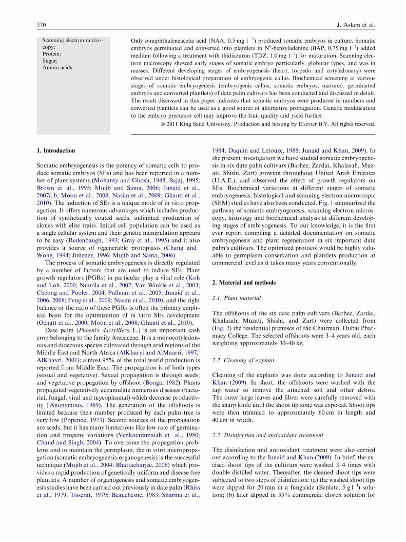
Abstract An efficient somatic embryogenesis system has been established in six date palm (Phoenix
dactylifera L.) cultivars (Barhee, Zardai, Khalasah, Muzati, Shishi and Zart). Somatic embryogen-
esis (SE) was growth regulators and cultivars dependent. Friable embryogenic callus was induced
from excised shoot tips on MS medium supplemented with various auxins particularly 2,4-dichlo-
rophenoxyacetic acid (2,4-D, 1.5 mg 1�l). Suspension culture increased embryogenesis potentiality.
iance; BA, N6-benzyladenine;
,4,5-T, 2,4,5-trichlorophenox-
acid; TDZ, thidiazuron; IAA,
neacetic acid; MS, Murashige
ic embryogenesis.ent of Biotechnology, Jamia
elhi 110062, India.
(J. Aslam).
y. Production and hosting by
Saud University.
lsevier
370 J. Aslam et al.
Scanning electron micros-
copy;
Protein;
Sugar;
Amino acids
Only a-naphthaleneacetic acid (NAA, 0.5 mg 1�1) produced somatic embryos in culture. Somatic
embryos germinated and converted into plantlets in N6-benzyladenine (BAP, 0.75 mg 1�l) added
medium following a treatment with thidiazuron (TDZ, 1.0 mg 1�l) for maturation. Scanning elec-
tron microscopy showed early stages of somatic embryo particularly, globular types, and was in
masses. Different developing stages of embryogenesis (heart, torpedo and cotyledonary) were
observed under histological preparation of embryogenic callus. Biochemical screening at various
stages of somatic embryogenesis (embryogenic callus, somatic embryos, matured, germinated
embryos and converted plantlets) of date palm cultivars has been conducted and discussed in detail.
The result discussed in this paper indicates that somatic embryos were produced in numbers and
converted plantlets can be used as a good source of alternative propagation. Genetic modification
to the embryo precursor cell may improve the fruit quality and yield further.
ª 2011 King Saud University. Production and hosting by Elsevier B.V. All rights reserved.
1. Introduction
Somatic embryogenesis is the potency of somatic cells to pro-duce somatic embryos (SEs) and has been reported in a num-ber of plant systems (Mohanty and Ghosh, 1988; Bajaj, 1995;Brown et al., 1995; Mujib and Sama, 2006; Junaid et al.,
2007a,b; Moon et al., 2008; Nasim et al., 2009; Ghanti et al.,2010). The induction of SEs is a unique mode of in vitro prop-agation. It offers numerous advantages which includes produc-
tion of synthetically coated seeds, unlimited production ofclones with elite traits. Initial cell population can be used asa single cellular system and their genetic manipulation appears
to be easy (Redenbaugh, 1993; Gray et al., 1995) and it alsoprovides a source of regenerable protoplasts (Chang andWong, 1994; Jimenez, 1996; Mujib and Sama, 2006).
The process of somatic embryogenesis is directly regulated
by a number of factors that are used to induce SEs. Plantgrowth regulators (PGRs) in particular play a vital role (Kohand Loh, 2000; Nuutila et al., 2002; Van Winkle et al., 2003;
Cheong and Pooler, 2004; Pullman et al., 2005; Junaid et al.,2006, 2008; Feng et al., 2009; Nasim et al., 2010), and the rightbalance or the ratio of these PGRs is often the primary empir-
ical basis for the optimization of in vitro SEs development(Ochatt et al., 2000; Moon et al., 2008; Ghanti et al., 2010).
Date palm (Phoenix dactylifera L.) is an important cash
crop belonging to the family Arecaceae. It is a monocotyledon-ous and dioecious species cultivated through arid regions of theMiddle East and North Africa (AlKharyi andAlMaarri, 1997;AlKhayri, 2001); almost 95% of the total world production is
reported from Middle East. The propagation is of both types(sexual and vegetative). Sexual propagation is through seeds;and vegetative propagation by offshoot (Bonga, 1982). Plants
propagated vegetatively accumulate numerous diseases (bacte-rial, fungal, viral and mycoplasmal) which decrease productiv-ity ( Anonymous, 1969). The generation of the offshoots is
limited because their number produced by each palm tree isvery low (Popenoe, 1973). Second sources of the propagationare seeds, but it has many limitations like low rate of germina-
tion and progeny variations (Venkataramaiah et al., 1980;Chand and Singh, 2004). To overcome the propagation prob-lems and to maintain the germplasm, the in vitro micropropa-gation (somatic embryogenesis/organogenesis) is the successful
technique (Mujib et al., 2004; Bhattacharjee, 2006) which pro-vides a rapid production of genetically uniform and disease freeplantlets. A number of organogenesis and somatic embryogen-
esis studies have been carried out previously in date palm (Rhisset al., 1979; Tisserat, 1979; Beauchesne, 1983; Sharma et al.,
1984; Daquin and Letouze, 1988; Junaid and Khan, 2009). Inthe present investigation we have studied somatic embryogene-sis in six date palm cultivars (Barhee, Zardai, Khalasah, Muz-
ati, Shishi, Zart) growing throughout United Arab Emirates(U.A.E.), and observed the effect of growth regulators onSEs. Biochemical variations at different stages of somatic
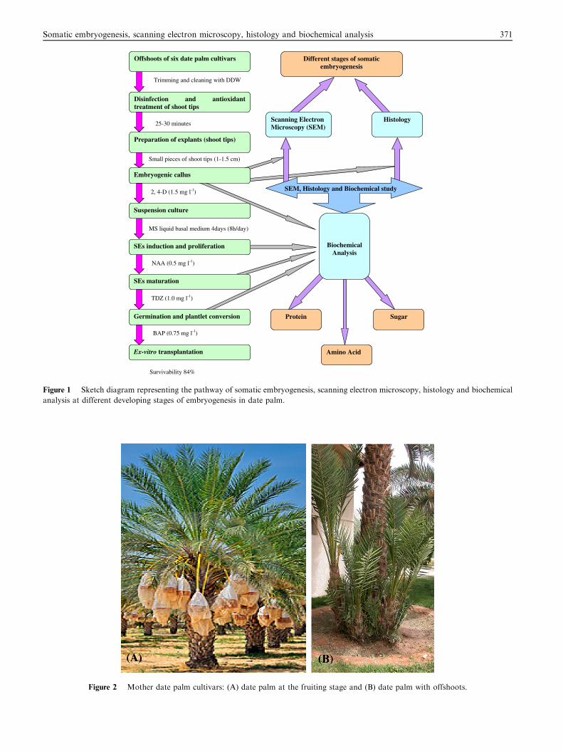
embryogenesis, histological and scanning electron microscopic(SEM) studies have also been conducted. Fig. 1 summarized thepathway of somatic embryogenesis, scanning electron micros-
copy, histology and biochemical analysis at different develop-ing stages of embryogenesis. To our knowledge, it is the firstever report compiling a detailed documentation on somaticembryogenesis and plant regeneration in six important date
palm’s cultivars. The optimized protocol would be highly valu-able to germplasm conservation and plantlets production atcommercial level as it takes many years conventionally.
2. Material and methods
2.1. Plant material
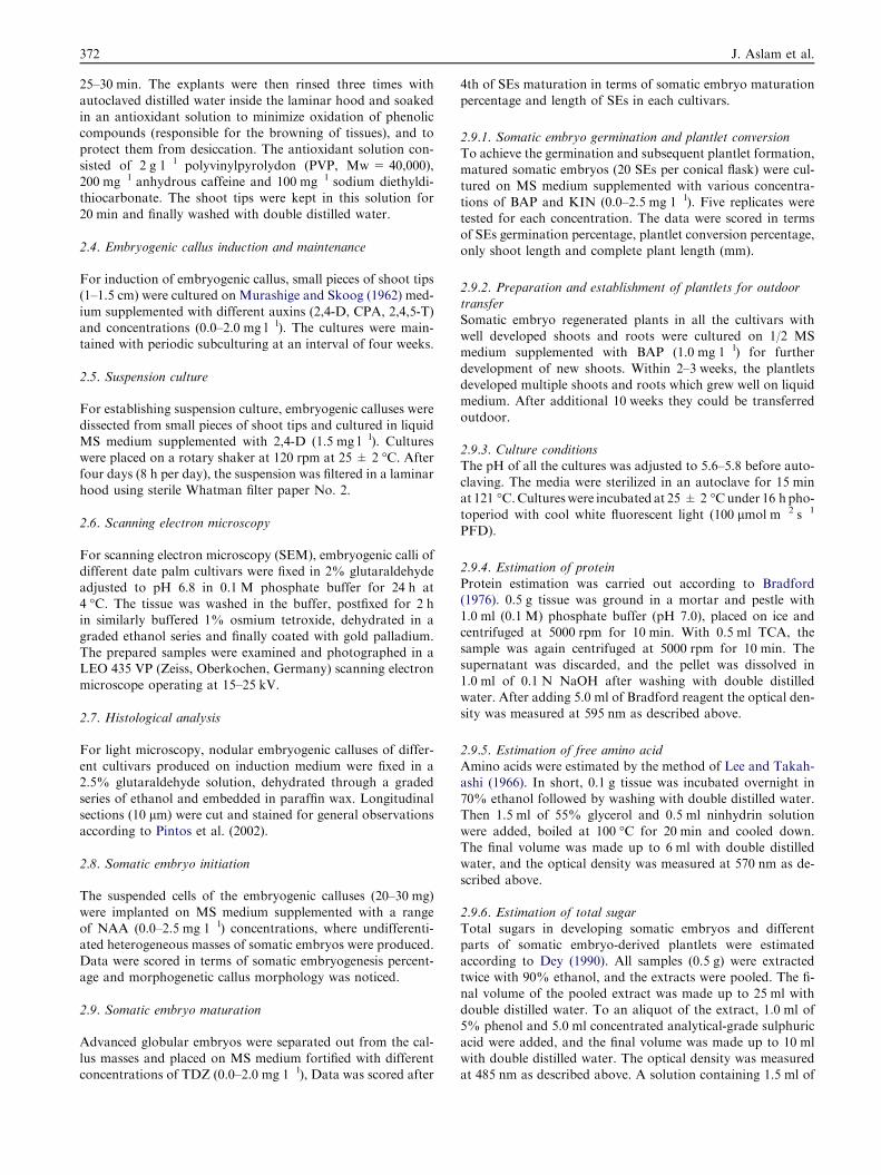
The offshoots of the six date palm cultivars (Barhee, Zardai,Khalasah, Muzati, Shishi, and Zart) were collected from(Fig. 2) the residential premises of the Chairman, Dubai Phar-
macy College. The selected offshoots were 3–4 years old, eachweighting approximately 30–40 kg.
2.2. Cleaning of explant
Cleaning of the explants was done according to Junaid andKhan (2009). In short, the offshoots were washed with the
tap water to remove the attached soil and other debris.The outer large leaves and fibres were carefully removed withthe sharp knife until the shoot tip zone was exposed. Shoot tips
were then trimmed to approximately 60 cm in length and40 cm in width.
2.3. Disinfection and antioxidant treatment
The disinfection and antioxidant treatment were also carriedout according to the Junaid and Khan (2009). In brief, the ex-
cised shoot tips of the cultivars were washed 3–4 times withdouble distilled water. Thereafter, the cleaned shoot tips weresubjected to two steps of disinfection: (a) the washed shoot tips
were dipped for 20 min in a fungicide (Benlate, 5 g l�l) solu-tion; (b) later dipped in 33% commercial clorox solution for
SEM, Histology and Biochemical study
Scanning Electron Microscopy (SEM)
Histology
Biochemical Analysis
Protein Sugar
Amino Acid
Offshoots of six date palm cultivars
Disinfection and antioxidant treatment of shoot tips
Preparation of explants (shoot tips)
Different stages of somatic embryogenesis
Embryogenic callus
Suspension culture
SEs induction and proliferation
SEs maturation
Germination and plantlet conversion
Ex-vitro transplantation
Trimming and cleaning with DDW
25-30 minutes
Small pieces of shoot tips (1-1.5 cm)
2, 4-D (1.5 mg l-1)
MS liquid basal medium 4days (8h/day)
NAA (0.5 mg l-1)
TDZ (1.0 mg l-1)
BAP (0.75 mg l-1)
Survivability 84%
Figure 1 Sketch diagram representing the pathway of somatic embryogenesis, scanning electron microscopy, histology and biochemical
analysis at different developing stages of embryogenesis in date palm.
Figure 2 Mother date palm cultivars: (A) date palm at the fruiting stage and (B) date palm with offshoots.
Somatic embryogenesis, scanning electron microscopy, histology and biochemical analysis 371
372 J. Aslam et al.
25–30 min. The explants were then rinsed three times with
autoclaved distilled water inside the laminar hood and soakedin an antioxidant solution to minimize oxidation of phenoliccompounds (responsible for the browning of tissues), and toprotect them from desiccation. The antioxidant solution con-
sisted of 2 g 1�l polyvinylpyrolydon (PVP, Mw = 40,000),200 mg�l anhydrous caffeine and 100 mg�l sodium diethyldi-thiocarbonate. The shoot tips were kept in this solution for
20 min and finally washed with double distilled water.
2.4. Embryogenic callus induction and maintenance
For induction of embryogenic callus, small pieces of shoot tips(1–1.5 cm) were cultured on Murashige and Skoog (1962) med-
ium supplemented with different auxins (2,4-D, CPA, 2,4,5-T)and concentrations (0.0–2.0 mg l�l). The cultures were main-tained with periodic subculturing at an interval of four weeks.
2.5. Suspension culture
For establishing suspension culture, embryogenic calluses were
dissected from small pieces of shoot tips and cultured in liquidMS medium supplemented with 2,4-D (1.5 mg l�l). Cultureswere placed on a rotary shaker at 120 rpm at 25 ± 2 �C. After
four days (8 h per day), the suspension was filtered in a laminarhood using sterile Whatman filter paper No. 2.
2.6. Scanning electron microscopy
For scanning electron microscopy (SEM), embryogenic calli ofdifferent date palm cultivars were fixed in 2% glutaraldehyde
adjusted to pH 6.8 in 0.1 M phosphate buffer for 24 h at4 �C. The tissue was washed in the buffer, postfixed for 2 hin similarly buffered 1% osmium tetroxide, dehydrated in a
graded ethanol series and finally coated with gold palladium.The prepared samples were examined and photographed in aLEO 435 VP (Zeiss, Oberkochen, Germany) scanning electron
microscope operating at 15–25 kV.
2.7. Histological analysis
For light microscopy, nodular embryogenic calluses of differ-ent cultivars produced on induction medium were fixed in a2.5% glutaraldehyde solution, dehydrated through a graded
series of ethanol and embedded in paraffin wax. Longitudinalsections (10 lm) were cut and stained for general observationsaccording to Pintos et al. (2002).
2.8. Somatic embryo initiation
The suspended cells of the embryogenic calluses (20–30 mg)
were implanted on MS medium supplemented with a rangeof NAA (0.0–2.5 mg 1�l) concentrations, where undifferenti-ated heterogeneous masses of somatic embryos were produced.
Data were scored in terms of somatic embryogenesis percent-age and morphogenetic callus morphology was noticed.
2.9. Somatic embryo maturation
Advanced globular embryos were separated out from the cal-
lus masses and placed on MS medium fortified with differentconcentrations of TDZ (0.0–2.0 mg 1�l), Data was scored after
4th of SEs maturation in terms of somatic embryo maturation
percentage and length of SEs in each cultivars.
2.9.1. Somatic embryo germination and plantlet conversionTo achieve the germination and subsequent plantlet formation,matured somatic embryos (20 SEs per conical flask) were cul-tured on MS medium supplemented with various concentra-
tions of BAP and KIN (0.0–2.5 mg 1�l). Five replicates weretested for each concentration. The data were scored in termsof SEs germination percentage, plantlet conversion percentage,only shoot length and complete plant length (mm).
2.9.2. Preparation and establishment of plantlets for outdoortransferSomatic embryo regenerated plants in all the cultivars withwell developed shoots and roots were cultured on 1/2 MSmedium supplemented with BAP (1.0 mg 1�l) for further
development of new shoots. Within 2–3 weeks, the plantletsdeveloped multiple shoots and roots which grew well on liquidmedium. After additional 10 weeks they could be transferred
outdoor.
2.9.3. Culture conditionsThe pH of all the cultures was adjusted to 5.6–5.8 before auto-claving. The media were sterilized in an autoclave for 15 minat 121 �C.Cultureswere incubated at 25 ± 2 �Cunder 16 hpho-
toperiod with cool white fluorescent light (100 lmol m�2 s�1
PFD).
2.9.4. Estimation of proteinProtein estimation was carried out according to Bradford(1976). 0.5 g tissue was ground in a mortar and pestle with
1.0 ml (0.1 M) phosphate buffer (pH 7.0), placed on ice andcentrifuged at 5000 rpm for 10 min. With 0.5 ml TCA, thesample was again centrifuged at 5000 rpm for 10 min. Thesupernatant was discarded, and the pellet was dissolved in
1.0 ml of 0.1 N NaOH after washing with double distilledwater. After adding 5.0 ml of Bradford reagent the optical den-sity was measured at 595 nm as described above.
2.9.5. Estimation of free amino acidAmino acids were estimated by the method of Lee and Takah-
ashi (1966). In short, 0.1 g tissue was incubated overnight in70% ethanol followed by washing with double distilled water.Then 1.5 ml of 55% glycerol and 0.5 ml ninhydrin solution
were added, boiled at 100 �C for 20 min and cooled down.The final volume was made up to 6 ml with double distilledwater, and the optical density was measured at 570 nm as de-
scribed above.
2.9.6. Estimation of total sugarTotal sugars in developing somatic embryos and differentparts of somatic embryo-derived plantlets were estimatedaccording to Dey (1990). All samples (0.5 g) were extractedtwice with 90% ethanol, and the extracts were pooled. The fi-
nal volume of the pooled extract was made up to 25 ml withdouble distilled water. To an aliquot of the extract, 1.0 ml of5% phenol and 5.0 ml concentrated analytical-grade sulphuric
acid were added, and the final volume was made up to 10 mlwith double distilled water. The optical density was measuredat 485 nm as described above. A solution containing 1.5 ml of
Somatic embryogenesis, scanning electron microscopy, histology and biochemical analysis 373
55% glycerol, 0.5 ml ninhydrin and 4.0 ml double distilled
water was used as a calibration standard.
2.9.7. Statistical analysisThe data on the effects of growth regulators on different stagesof somatic embryogenesis and other parameters were analysedby one-way analysis of variance (ANOVAs). Values are means
of six replicates from two experiments, and the presented meanvalues were separated using Duncan’s Multiple Range Test(DMRT) at at P < 0.05.
3. Results
3.1. Embryogenic callus induction and maintenance
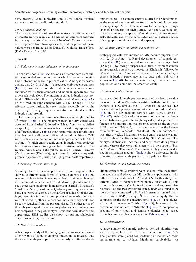
The excised shoot (Fig. 3A) tips of six different date palm cul-
tivars responded well in culture on which three tested auxinshad profound influence to produce callus. Amongst the testedauxins, only 2,4-D (1.5 mg 1�l) produced embryogenic calluses
(Fig. 3B), however, callus induced at the higher concentrationscharacterized by their compact and nodular appearance, aregrown relatively slow. The maximum callus induction was no-
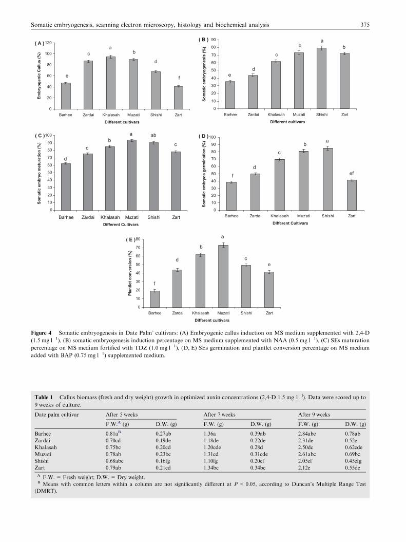
ticed in ‘Khalasah’ followed by ‘Zadai’ and ‘Muzati’ (Fig. 4A)on MS medium supplemented with 2,4-D (1.5 mg 1�l). Theeffective concentration, however, varied generally lay within0.5–1.5 mg 1�l range; higher concentration inhibited callus
induction and growth.Fresh and dry callus masses of cultivars were weighted up to
9th weeks (Table 1). The maximum fresh and dry weight was
observed from ‘Barhee’ followed by ‘Muzati’ cultivar. A visiblemorphological variation was noticed in callusing appearanceof different cultivars. Table 2 showing morphological variations
in embryogenic calluses of different date palm cultivars. Calliwere routinely maintained on media supplemented with 2,4-D(1.5 mg 1�l). High embryogenic callus induction was achievedby continuous subculturing on fresh nutrient medium. The
calluses were friable light yellow greenish (Barhee), creamy(Zardai), yellow (Khalasah), light green (Muzati), creamy withgreenish appearances (Shishi) and light green (Zart) respectively.
3.2. Scanning electron microscopy
Scanning electron microscopic study of embryogenic callusshowed undifferentiated forms of somatic embryos (Fig 3D).A remarkable variation in somatic embryo origin was observed
in different cultivars. In ‘Barhee’ and ‘Muzati’, globular and tor-pedo types were maximum in numbers; in ‘Zardai’, ‘Khalasah’,‘Shishi’ and ‘Zart’, heart and cotyledonary were highest in num-bers. Theywere developed on the surface of callus.Globular em-
bryo, were high in number and produced regularly. Embryoswere clustered together in a common mass, but they could notbe easily detached from the parental tissue. The other forms of
the embryos (torpedo, heart and cotyledonary)were in less num-bers compared to the globular type.Beside the normal forms andappearance, SEM studies also show various morphological
deviations in embryos structure.
3.3. Histological analysis
A histological study of the embryogenic callus was performedafter 4 weeks of somatic embryos induction. It revealed thatthe somatic embryos appear simultaneously at different devel-
opment stages. The somatic embryos started their development
at the stage of meristematic centres through globular to coty-ledonary shape. Most of the embryos formed a typical singlelayer of protoderm on their surface very soon. Somatic em-bryos are mainly composed of small compact meristematic
cells, characterized by the dense cytoplasm and dense nucleus(photographs not shown due to the space).
3.4. Somatic embryo initiation and proliferation
Embryogenic calli was induced on MS medium supplemented
with 2,4-D (1.5 mg 1�l). Rapid development of somatic em-bryos (Fig. 3C) was observed on medium containing NAA(1.5 mg 1�l) following a suspension culture. Maximum somatic
embryogenesis potency was noticed in ‘Khalasah’ followed by‘Muzati’ cultivar. Comparative account of somatic embryo-genesis induction percentage in six date palm cultivars isshown in Fig. 4B. Induced somatic embryos were in mixed
population and could not be separated easily.
3.5. Somatic embryo maturation
Advanced globular embryos were separated out from the callusmass and placed onMSmedium fortified with different concen-
trations of TDZ (0.0–2.0 mg l�l). Amongst the various TDZconcentrations highest SEs maturation was noticed in ‘Muzati’(Fig. 3D) on MS medium fortified with TDZ (1.0mg 1�l)(Fig. 4C). After 2–3 weeks in maturation medium embryos
started to become greenish morphologically, but significant dif-ference in SE maturation was observed amongst the cultivars.In ‘Barhee’ and ‘Muzati’, maturation started after the 2 weeks
of implantation; in ‘Zardai’, ‘Khalasah’, ‘Shishi’ and ‘Zart’ itwas after 3 weeks. Maximum somatic embryogenesis was no-ticed in ‘Muzati’ cultivars followed by ‘Shishi’ cultivars. The
embryos of ‘Khalash’, ‘Zardai’ and ‘Zart’ were dark green incolour, whereas they were light green with brown spots in ‘Bar-hee’, ‘Muzati’, ‘Khalasah’. The somatic embryos increased in
size in maturation medium. Table 3 shows differences in sizeof matured somatic embryos of six date palm’s cultivars.
3.6. Germination and plantlet conversion
Highly green somatic embryos were isolated from the matura-tion medium and placed on MS medium supplemented with
different concentrations of BAP and KN. In this study, twodifferent types of responses were mainly observed: (1) onlyshoot (without root); (2) plants with shoot and root (complete
plantlets). Of the two cytokinins tested, BAP was found to bemore active as compared to KN in SEs germination and plant-let conversion. BAP (0.75 mg 1�l) proved to be highly effectivecompared to the other concentrations (Fig. 3E). The highest
SE germination was in ‘Shishi’ (Fig. 4D), however, plantletconversion was noticed in ‘Muzati’ (Fig. 4E). A comparativeaccount of only shoot and complete plantlet length raised
through somatic embryos is shown in Tables 4 and 5.
3.7. Acclimatization
A large number of somatic embryos derived plantlets weresuccessfully acclimatized to ex vitro conditions (Fig. 3F).
The plantlets were placed for hardening at incubation roomtemperature up to 45 days. Maximum survivability was
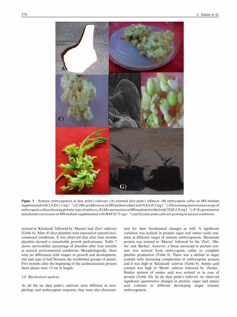
Figure 3 Somatic embryogenesis in date palm’s cultivars: (A) trimmed date palm’s offshoot, (B) embryogenic callus on MS medium
supplementedwith2,4-D(1.5 mg l�1), (C)SEsproliferationonMSmediumaddedwithNAA(0.5 mg l�1), (D)scanningelectronmicroscopyof
embryogenic calluse showingglobular typeof embryos, (E)SEsmaturationonMSmediumfortifiedwithTDZ(1.0 mg l�1), (F)Esgermination
and plantlet conversion onMSmedium supplemented with BAP (0.75 mg l�1) and (G) date palm cultivars growing in natural conditions.
374 J. Aslam et al.
noticed in ‘Khalasah’ followed by ‘Muzati’ and ‘Zart’ cultivars(Table 6). After 45 days plantlets were exposed at natural envi-ronmental conditions. It was observed that after four monthsplantlets showed a remarkable growth performance. Table 7
shows survivability percentage of plantlets after four monthsat natural environmental conditions. Morphologically, therewere no differences with respect to growth and development,
size and type of leaf between the established groups of plants.Five months after the beginning of the acclimatization processthese plants were 15 cm in length.
3.8. Biochemical analysis
As all the six date palm’s cultivars were different in mor-
phology and embryogenic response, they were also character-
ized for their biochemical changes as well. A significantvariation was noticed in protein sugar and amino acids con-tents at different stages of somatic embryogenesis. Maximumprotein was noticed in ‘Muzati’ followed by the ‘Zart’, ‘Shi-
shi’ and ‘Barhee’, however, a linear increased in protein con-tent was noticed from embryogenic callus to completeplantlet production (Table 8). There was a decline in sugar
content with increasing complexities in embryogenic processand it was high in ‘Khalasah’ cultivar (Table 9). Amino acidcontent was high in ‘Shishi’ cultivar followed by ‘Zardai’.
Similar pattern of amino acid was noticed as in case ofprotein (Table 10). In six date palm’s cultivars we observedsignificant quantitative changes in protein, sugar and aminoacid contents at different developing stages somatic
embryogenesis.
0
20
40
60
80
100
120
Barhee Zardai Khalasah Muzati Shishi Zart
Different cultivars
Embr
yoge
nic
Cal
lus
(%)
abc
d
e f
( A )
0
10
20
30
40
50
60
70
80
90
Barhee Zardai Khalasah Muzati Shishi Zart
Different cultivars
Som
atic
em
bryo
gene
sis
(%)
abb
c
de
( B )
0
10
20
30
40
50
60
70
80
90
100
Barhee Zardai Khalasah Muzati Shishi ZartDifferent Cultivars
Som
atic
em
bryo
met
urat
ion
(%)
a abb
cc
d
( C )
0
10
20
30
40
50
60
70
80
90
100
Barhee Zardai Khalasah Muzati Shishi Zart
Different Cultivars
Som
atic
em
bryo
s ge
rmin
atio
n (%
)
( D )a
bc
deff
0
10
20
30
40
50
60
70
80
Barhee Zardai Khalasah Muzati Shishi Zart
Different cultivars
Plan
tlet c
onve
rsio
n (%
)
( E ) a
b
cde
f
Figure 4 Somatic embryogenesis in Date Palm’ cultivars: (A) Embryogenic callus induction on MS medium supplemented with 2,4-D
(1.5 mg l�1), (B) somatic embryogenesis induction percentage on MS medium supplemented with NAA (0.5 mg l�1), (C) SEs maturation
percentage on MS medium fortified with TDZ (1.0 mg l�1), (D, E) SEs germination and plantlet conversion percentage on MS medium
added with BAP (0.75 mg l�1) supplemented medium.
Table 1 Callus biomass (fresh and dry weight) growth in optimized auxin concentrations (2,4-D 1.5 mg 1�l). Data were scored up to
9 weeks of culture.
Date palm cultivar After 5 weeks After 7 weeks After 9 weeks
F.W.A (g) D.W. (g) F.W. (g) D.W. (g) F.W. (g) D.W. (g)
Barhee 0.81aB 0.27ab 1.36a 0.39ab 2.84abc 0.78ab
Zardai 0.70cd 0.19de 1.18de 0.22de 2.31de 0.52e
Khalasah 0.75bc 0.20cd 1.20cde 0.28d 2.50dc 0.62cde
Muzati 0.78ab 0.23bc 1.31cd 0.31cde 2.61abc 0.69bc
Shishi 0.68abc 0.16fg 1.10fg 0.20ef 2.05ef 0.45efg
Zart 0.79ab 0.21cd 1.34bc 0.34bc 2.12e 0.55de
A F.W. = Fresh weight; D.W. = Dry weight.B Means with common letters within a column are not significantly different at P < 0.05, according to Duncan’s Multiple Range Test
(DMRT).
Somatic embryogenesis, scanning electron microscopy, histology and biochemical analysis 375
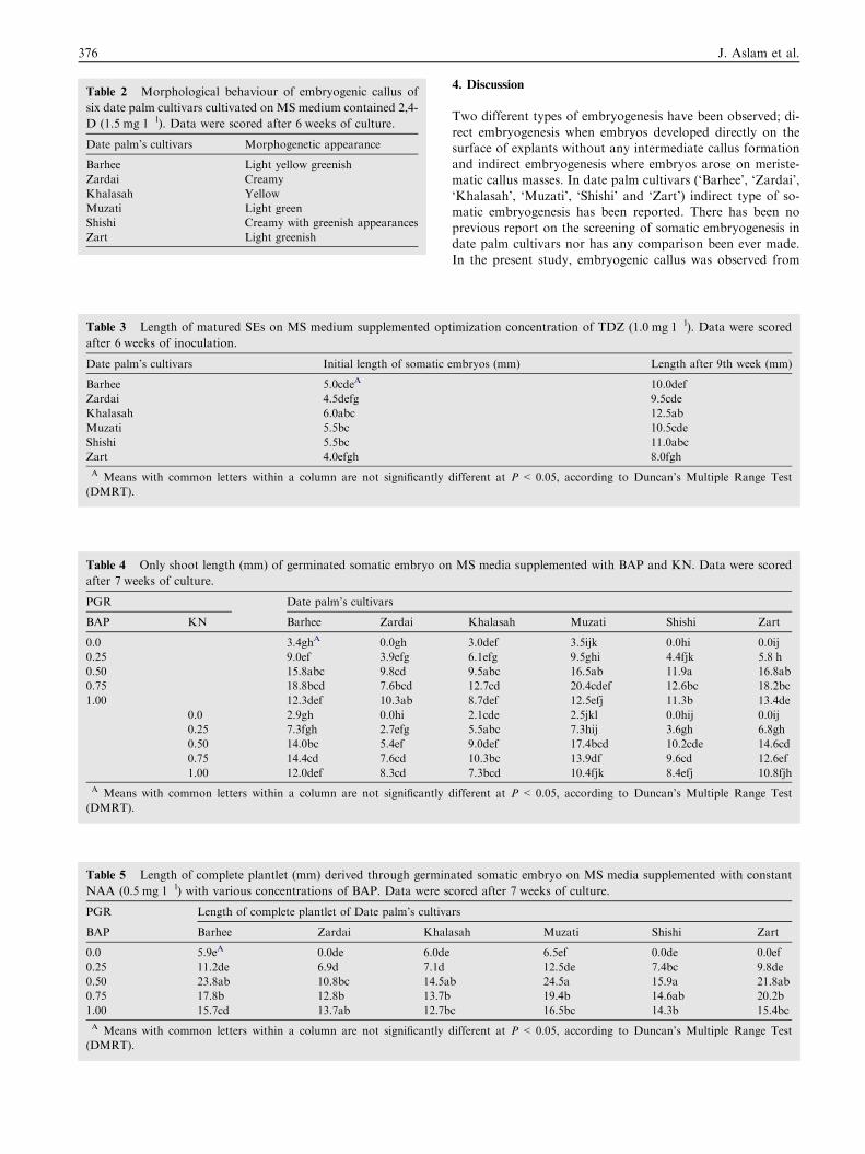
Table 3 Length of matured SEs on MS medium supplemented opt
after 6 weeks of inoculation.
Date palm’s cultivars Initial length of somatic e
Barhee 5.0cdeA
Zardai 4.5defg
Khalasah 6.0abc
Muzati 5.5bc
Shishi 5.5bc
Zart 4.0efgh
A Means with common letters within a column are not significantly
(DMRT).
Table 4 Only shoot length (mm) of germinated somatic embryo on
after 7 weeks of culture.
PGR Date palm’s cultivars
BAP KN Barhee Zardai
0.0 3.4ghA 0.0gh
0.25 9.0ef 3.9efg
0.50 15.8abc 9.8cd
0.75 18.8bcd 7.6bcd
1.00 12.3def 10.3ab
0.0 2.9gh 0.0hi
0.25 7.3fgh 2.7efg
0.50 14.0bc 5.4ef
0.75 14.4cd 7.6cd
1.00 12.0def 8.3cd
A Means with common letters within a column are not significantly
(DMRT).
Table 5 Length of complete plantlet (mm) derived through germin
NAA (0.5 mg 1�l) with various concentrations of BAP. Data were s
PGR Length of complete plantlet of Date palm’s cultiva
BAP Barhee Zardai Khal
0.0 5.9eA 0.0de 6.0de
0.25 11.2de 6.9d 7.1d
0.50 23.8ab 10.8bc 14.5a
0.75 17.8b 12.8b 13.7b
1.00 15.7cd 13.7ab 12.7b
A Means with common letters within a column are not significantly
(DMRT).
Table 2 Morphological behaviour of embryogenic callus of
six date palm cultivars cultivated on MS medium contained 2,4-
D (1.5 mg 1�l). Data were scored after 6 weeks of culture.
Date palm’s cultivars Morphogenetic appearance
Barhee Light yellow greenish
Zardai Creamy
Khalasah Yellow
Muzati Light green
Shishi Creamy with greenish appearances
Zart Light greenish
376 J. Aslam et al.
4. Discussion
Two different types of embryogenesis have been observed; di-
rect embryogenesis when embryos developed directly on thesurface of explants without any intermediate callus formationand indirect embryogenesis where embryos arose on meriste-matic callus masses. In date palm cultivars (‘Barhee’, ‘Zardai’,
‘Khalasah’, ‘Muzati’, ‘Shishi’ and ‘Zart’) indirect type of so-matic embryogenesis has been reported. There has been noprevious report on the screening of somatic embryogenesis in
date palm cultivars nor has any comparison been ever made.In the present study, embryogenic callus was observed from
imization concentration of TDZ (1.0 mg 1�l). Data were scored
mbryos (mm) Length after 9th week (mm)
10.0def
9.5cde
12.5ab
10.5cde
11.0abc
8.0fgh
different at P< 0.05, according to Duncan’s Multiple Range Test
MS media supplemented with BAP and KN. Data were scored
Khalasah Muzati Shishi Zart
3.0def 3.5ijk 0.0hi 0.0ij
6.1efg 9.5ghi 4.4fjk 5.8 h
9.5abc 16.5ab 11.9a 16.8ab
12.7cd 20.4cdef 12.6bc 18.2bc
8.7def 12.5efj 11.3b 13.4de
2.1cde 2.5jkl 0.0hij 0.0ij
5.5abc 7.3hij 3.6gh 6.8gh
9.0def 17.4bcd 10.2cde 14.6cd
10.3bc 13.9df 9.6cd 12.6ef
7.3bcd 10.4fjk 8.4efj 10.8fjh
different at P< 0.05, according to Duncan’s Multiple Range Test
ated somatic embryo on MS media supplemented with constant
cored after 7 weeks of culture.
rs
asah Muzati Shishi Zart
6.5ef 0.0de 0.0ef
12.5de 7.4bc 9.8de
b 24.5a 15.9a 21.8ab
19.4b 14.6ab 20.2b
c 16.5bc 14.3b 15.4bc
different at P< 0.05, according to Duncan’s Multiple Range Test
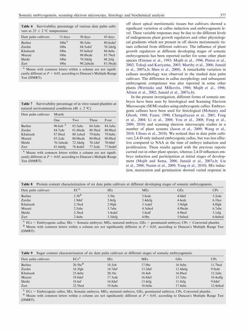
Table 7 Survivability percentage of in vitro raised plantlets at
natural environmental conditions (48 ± 2 �C).
Date palm cultivars Month
One Two Three Four
Barhee 90.6ab A 85.5abc 84.5abc 84.5abc
Zardai 84.7cde 81.6bcde 80.5bcd 80.0bcd
Khalasah 87.8bcd 80.1abcd 79.0abc 79.0abc
Muzati 83.2cde 80.0bcde 80.0bcd 80.0bcd
Shishi 76.5abcde 72.3defg 70.1def 70.0def
Zart 81.6defg 78.4cdef 77.5cde 77.0cdef
A Means with common letters within a column are not signifi-
cantly different at P< 0.05, according to Duncan’s Multiple Range
Test (DMRT).
Table 8 Protein content characterization of six date palm cultivars
Date palm cultivars ECA SEs
Barhee 2.3bB 3.5bc
Zardai 1.9def 3.0efg
Khalasah 2.1bcd 2.9fgh
Muzati 2.5abc 3.7abc
Shishi 2.3bcd 3.4cdef
Zart 2.4abc 3.3defg
A ECs = Embryogenic callus; SEs = Somatic embryos; MEs, maturedB Means with common letters within a column are not significantly
(DMRT).
Table 9 Sugar content characterization of six date palm cultivars a
Date palm cultivars ECsA SEs
Barhee 20.5bcB 18.5ab
Zardai 16.3fgh 14.7def
Khalasah 23.4abc 20.1bc
Muzati 19.8def 17.3cde
Shishi 18.6ef 16.8def
Zart 22.5bcd 19.8abc
A ECs = Embryogenic callus, SEs, Somatic embryos, MEs, matured emB Means with common letters within a column are not significantly
(DMRT).
Table 6 Survivability percentage of various date palm culti-
vars at 25 ± 2 �C temperature.
Date palm cultivars 15 days 30 days 45 days
Barhee 100aA 90.5abc 80.6cdef
Zardai 100a 84.5cdef 70.2defg
Khalasah 100a 95.6abcd 86.4abc
Muzati 100a 90.0bcde 83.7bcd
Shishi 100a 70.5defg 68.2efg
Zart 100a 90.2abcde 83.5bcde
A Means with common letters within a column are not signifi-
cantly different at P< 0.05, according to Duncan’s Multiple Range
Test (DMRT).
Somatic embryogenesis, scanning electron microscopy, histology and biochemical analysis 377
off shoot apical meristematic tissues but cultivars showed a
significant variation at callus induction and embryogenesis le-vel. These variable responses may be due to the different levelsof endogeneous plant growth regulators and other physiologi-cal gradients which are present in off shoots meristematic tis-
sues collected from different cultivars. The influence of plantgrowth regulators at different developing stages of somaticembryogenesis has been reported earlier for some other plant
species (Etienne et al., 1993; Mujib et al., 1996; Pintos et al.,2002; Tokuji and Kuriyama, 2003; Murthy et al., 2006; Junaidet al., 2007a,b; Shen et al., 2008). A remarkable variation in
calluses morphology was observed in the studied date palmcultivars. The difference in callus morphology and subsequentembryogenic competence was also reported in some other
plants (Wernicke and Milkovits, 1986; Mujib et al., 1996;Afreen et al., 2002; Junaid et al., 2007a,b).
In the present investigation, different forms of somatic em-bryos have been seen by histological and Scanning Electron
Microscopic (SEM) studies using embryogenic callus. Embryo-genic calluses have been used for histological (Mohanty andGhosh, 1988; Faure, 1990; Chengalrayan et al., 2001; Feng
et al., 2004; Li et al., 2008; You et al., 2008; Feng et al.,2009, 2010) and scanning electron microscopic studies in anumber of plant systems (Jason et al., 2009; Wang et al.,
2010; Ulisses et al., 2010). We noticed that in date palm culti-vars 2,4-D only induced embryogenic callus, but was less effec-tive compared to NAA at the time of embryo induction andproliferation. These results agreed with the previous reports
carried out in other plant species, whereas 2,4-D influences em-bryo induction and participation at initial stages of develop-ment (Mujib and Sama, 2006; Junaid et al., 2007a,b; Liu
et al., 2008; Nasim et al., 2009; Yang et al., 2010). SEs induc-tion, maturation and germination showed varied response in
at different developing stages of somatic embryogenesis.
MEs GEs CPs
3.8cde 4.0def 5.2cde
3.4defg 4.6cde 6.1bce
3.1edef 3.9efgh 4.8fgh
4.3abcd 4.9bcde 6.7abc
3.8cdf 4.9bcd 5.1efg
4.0bc 5.0abcd 6.0abcd
embryos, GEs = germinated embryos; CPs = Converted plantlet.
different at P < 0.05, according to Duncan’s Multiple Range Test
t different stages of somatic embryogenesis.
MEs GEs CPs
17.0bc 16.9abc 11.7bcd
13.2def 12.4defg 9.9cde
18.4ab 16.8bcd 12.2abc
16.8def 13.7abc 10.4cdfg
13.4efg 11.9efg 9.8def
18.0abc 17.4abc 12.4abcd
bryos, GEs, germinated embryos, CPs, Converted plantlet.
different at P < 0.05, according to Duncan’s Multiple Range Test
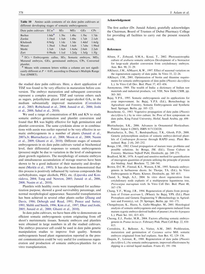
Table 10 Amino acids contents of six date palm cultivars at
different developing stages of somatic embryogenesis.
Date palm cultivars ECssA SEs MEs GEs CPs
Barhee 1.0deB 1.3bc 1.4bc 1.5bc 1.7de
Zardai 1.1bcd 1.5ab 1.6bc 1.7ab 2.1ab
Khalasah 0.9efg 1.1dc 1.3de 1.4def 1.6efg
Muzati 1.3bcd 1.5bcd 1.6ab 1.7abc 1.9bcd
Shishi 1.5ab 1.8ab 1.8abc 2.0ab 2.2ab
Zart 0.9bcde 1.1cd 1.2efg 1.3efg 1.5fg
A ECs = Embryogenic callus, SEs, Somatic embryos, MEs,
Matured embryos, GEs, germinated embryos, CPs, Converted
plantlet.B Means with common letters within a column are not signifi-
cantly different at P < 0.05, according to Duncan’s Multiple Range
Test (DMRT).
378 J. Aslam et al.
the studied date palm cultivars. Here, a short application ofTDZ was found to be very effective in maturation before con-
version. The embryo maturation and subsequent conversionrepresent a complex process. TDZ and various others suchas sugars, polyethylene glycol and sugar-alcohol GA3 in the
medium substantially improved maturation (Corredoiraet al., 2003; Robichaud et al., 2004; Junaid et al., 2006; Joshiet al., 2008; Sahal et al., 2010).
We used a range of concentration of BA and KN to studysomatic embryo germination and plantlet conversion andfound that BA was highly effective for high germination and
plantlet conversion. The same cytokinin alone or in combina-tions with auxin was earlier reported to be very effective in so-matic embryogenesis in a number of plants (Junaid et al.,2007a,b; Bhattacharya et al., 2008; Moon et al., 2008; Nasim
et al., 2009; Ghanti et al., 2010). Different stages of somaticembryogenesis in six date palm cultivars varied at biochemicallevel, their differential responses to somatic embryogenesis
(process) might be due to variation in the level of endogenousplant growth regulators. The morphology of somatic embryosand simultaneous accumulation of storage reserves have been
shown to be a good indicator of their maturity and develop-ment (Merkle et al., 1995). It has also been demonstrated thatthis process is positively influenced by various compounds like
carbohydrates, sugar alcohols, PEG, etc. (Lipavska and Kon-radova, 2004; Tang and Newton, 2005; Junaid et al., 2006,2008; Nasim et al., 2010).
Plantlets with healthy roots were transplanted for acclima-
tization purpose, showed a good survivability percentage, andnormal morphological appearance. Successful transplantationhas been achieved in several other plants earlier (Zhang and
Davis, 1986; Debergh and Read, 1991; Preece and Sutter,1991; Shibli and Smith, 1996; Kim et al., 1997; Dhar and Joshi,2005; Junaid et al., 2008; Ghanti et al., 2010).
In date palm cultivars, we have been able to demonstrate anefficient somatic embryogenesis system originating from offshoot’s meristematic tissues. Somatic embryos and plantletswere obtained in large numbers in all the studied cultivars.
The embryo precursor cell could be used in date palm geneticmanipulation studies to improve fruit quality. Somaticembryogenesis based plant regeneration reported in the pres-
ent communication could be very useful for continuous regen-eration and production of somatic embryos/plantlets for exvitro transplantation.
Acknowledgement
The first author (Dr. Junaid Aslam), gratefully acknowledgesthe Chairman, Board of Trustees of Dubai Pharmacy Collegefor providing all facilities to carry out the present research
work.
References
Afreen, F., Zobayed, S.M.A., Kozai, T., 2002. Photoautotrophic
culture of arabusta somatic embryos Development of a bioreactor
for large-scale plantlet conversion from cotyledonary embryos.
Ann. Bot. 90, 21–29.
AlKharyi, J.M., AlMaarri, K.W., 1997. Effect of seasonal variation on
the regeneration capacity of date palm. In Vitro 33, 22–26.
AlKhayri, J.M., 2001. Optimization of biotin and thiamine require-
ments for somatic embryogenesis of date palm (Phoenix dactylifera
L.). In Vitro Cell Dev. Biol. Plant 37, 453–456.
Anonymous, 1969. The wealth of India: a dictionary of Indian raw
materials and industrial products, vol. VIII, New Delhi CSIR, pp.
303–305.
Bajaj, Y.P.S., 1995. Somatic embryogenesis and its applications for
crop improvement. In: Bajaj, Y.P.S. (Ed.), Biotechnology in
Agriculture and Forestry, Somatic Embryogenesis and Synthetic
Seed. Springer, Berlin, pp. 105–125.
Beauchesne, G., 1983. Vegetative propagation of date palm (Phoenix
dactylifera L.) by in vitro culture. In: Proc of first symposium on
date palm, King Faisal University, Hofuf, Saudi Arabia, pp. 698–
699.
Bhattacharjee, S.K., 2006. Advances in Ornamental Horticulture.
Pointer Jaipur 6 (2065), ISBN 8171324320.
Bhattacharya, S., Dey, T., Bandopadhyay, T.K., Ghosh, P.D., 2008.
Genetic polymorphism analysis of somatic embryo-derived plant-
lets of Cymbopogon flexuosus through RAPD assay. Plant Bio-
technol. Rep. 2 (4), 245–252.
Bonga, J.M., 1982. Clonal propagation of mature trees: problems and
possible solutions. In: Bonga, JM. (Ed.), Tissue Culture in
Forestry. Martinus Nijhoff Publ, Dordrecht, pp. 249–271.
Bradford, M.M., 1976. A rapid and sensitive method for quantification
of microgram quantities of protein utilizing the principle of protein
dye binding. Anal. Biochem. 72, 248–541.
Brown, D.C.W., Finstad, K.I., Watson, E.M., 1995. Somatic embryo-
genesis in herbaceous dicots. In: Thorpe, TA. (Ed.), In Vitro
Embryogenesis in Plants. Kluwer, Dordrecht, pp. 345–415.
Chand, S., Singh, A.J., 2004. In vitro shoot regeneration from
cotyledonary node explants of a multipurpose leguminous tree,
Pterocarpus marsupium roxb. In Vitro Cell Dev. Biol. Plant 40,
167–170.
Chang, Y.F., Wong, J.R., 1994. Regeneration of plants from protop-
lasts of Tritium aestivum L. (Wheat). In: Bajaj, YPS. (Ed.), Plant
Protoplasts and Genetic Engineering V (Biotechnology in Agricul-
ture and Forestry), vol. 29. Springer, Berlin, pp. 161–171.
Chengalrayan, K., Hazra, S., Gallo-Meagher, M., 2001. Histological
analysis of somatic embryogenesis and organogenesis induced from
mature zygotic embryo-derived leaflets of peanut (Arachis hypogaea
L.). Plant Sci. 161, 415–421.
Cheong, E.J., Pooler, M.R., 2004. Factors affecting somatic embryo-
genesis in Prunus incisa cv. February Pink. Plant Cell Rep. 22, 810–
815.
Corredoira, E., Ballester, A., Vieitez, A.M., 2003. Proliferation,
maturation and germination of Castanea sativa Mill. somatic
embryos originated from leaf explants. Ann Bot. 92, 129–136.
Daquin, F., Letouze, R., 1988. Regeneration of date palm (Phoenix
dactylifera L.) by somatic embryogenesis, improved effectiveness by
dipping in a stirred liquid medium. Fruits 43, 191–194.
Somatic embryogenesis, scanning electron microscopy, histology and biochemical analysis 379
Debergh, P.C., Read, P.E., 1991. Micropropagation. In: Debergh, PC.,
Zimmerman, RH. (Eds.), Micropropagation: Technology
and Application. Kluwer Acad Publishers, Dordrecht,
pp. 1–3.
Dey, P.M., 1990. Methods in plant biochemistry. Carbohydrates, vol.
2. Acad. Press, London.
Dhar, U., Joshi, M., 2005. Efficient plant regeneration protocol
through callus for Saussurea obvallata (DC) Edgew. (Asteraceae):
effects of explant type, age and plant growth regulators. Plant Cell
Rep. 24, 195–200.
Etienne, H., Sotta, B., Montoro, P., Miginiac, E., Carron, M.P., 1993.
Relations between exogenous growth regulators and endogenous
indole-3-acetic acid and abscisic acid in the expression of somatic
embryogenesis in Hevea brasiliensis (Mull Arg.). Plant Sci. 88 (1),
91–96.
Faure, O., 1990. Somatic embryos of Vitis rupestris and zygotic
embryos of Vitis species: morphology, histology, histochemistry
and development. Candian J. Bot. 68 (3), 2305–2315.
Feng, D.L., Li, Y., Sun, Z.Y., 2004. Histology and somatic embryo-
genesis of Parthenocissus tricuspidata. J Beij For Univer 26 (4), 97–
99.
Feng, D.L., Li, W., Li, J., Li, P.T., Zhao, M., Zhao, S.G., Shi, B.S.,
Peng, W.X., 2009. Observation of somatic embryogenesis and
histology in Koelreuteria bipinnata Franch var integrifoliola. Chen.
Plant Physiol. Commun. 45 (9), 855–858.
Feng, D.L., Meng, Q.R., Li, W.P., Hu, Y.H., Li, M., Gu, A.X., 2010.
Morphology of somatic embryogenesis and plantlet formation in
tissue cultures of lantern tree (Koelreuteria bipinnata var integrifo-
liola). For Stud. China 12 (1), 31–36.
Ghanti, S.K., Sujata, K.G., Rao, S.M., Kishor, P.B.K., 2010. Direct
somatic embryogenesis and plant regeneration from immature
explants of chickpea. Biol. Plant 54 (1), 121–125.
Gray, D.J., Compton, M.E., Harrell, R.C., Cantliffe, D.J., 1995.
Somatic embryogenesis and the technology of synthetic seed. In:
Bajaj, Y.P.S. (Ed.), . In: Biotechnology in Agriculture and Forestry
Somatic Embryogenesis and Seed I, vol. 30. Springer, Berlin, pp.
126–151.
Jason, N., Burris, David, G.J., Mann, Blake L., Joyce, C., Neal
Stewart Jr., C., 2009. An improved tissue culture system for
embryogenic callus production and plant regeneration in switch-
grass (Panicum virgatum L.). Bioener. Res. 2, 267–274.
Jimenez, V.M., 1996. El cultivo de protoplastos en citricos y su
potencial para el mejoramiento genetico. Agron Costarric 20, 187–
204.
Joshi, M., Sujatha, K., Hazra, S., 2008. Effect of TDZ and 2,4-D on
peanut somatic embryogenesis and in vitro bud development. Plant
Cell Tiss. Org. Cult. 94 (1), 85–90.
Junaid, A., Khan, S.A., 2009. In vitro micropropagation of Khalasah
date palm (Phoenix dactylifera L.). An important fruit plant. J.
Fruit Orna. Plant Res. 17 (1), 5–17.
Junaid, A., Bhatt, M.A., Mujib, A., Sharma, M.P., 2006. Somatic
embryo proliferation maturation and germination in Catharanthus
roseus. Plant Cell Tiss. Org. Cult. 84, 325–332.
Junaid, A., Mujib, A., Sharma, M.P., Tang, W., 2007a. Growth
regulators affect primary and secondary somatic embryogenesis in
Madagaskar periwinkle (Catharanthus roseus (L) GDon) at mor-
phological and biochemical levels. Plant Growth Regul. 51, 271–
281.
Junaid, A., Mujib, A., Bhat, M.A., Sharma, M.P., Samaj, J., 2007b.
Somatic embryogenesis and plant regeneration in Catharanthus
roseus. Biol. Plant 51 (4), 641–646.
Junaid, A., Mujib, A., Fatima, S., Sharma, M.P., 2008. Cultural
conditions affect somatic embryogenesis in Catharanthus roseus L.
(G.) Don. Plant Biotechnol. Rep. 2, 179–189.
Kim, M.K., Sommer, H.E., Bongarten, B.C., Merkle, S.A., 1997. High
frequency induction of adventitious shoots from hypocotyl seg-
ments of Liquidambar styraciflua L. by thidiazuron. Plant Cell Rep.
16, 536–540.
Koh, W.L., Loh, C.S., 2000. Direct somatic embryogenesis, plant
regeneration and in vitro flowering in rapid-cycling Brassica napus.
Plant Cell Rep. 19, 1177–1183.
Lee, Y.P., Takahashi, T., 1966. Improved calorimetric determination
of amino acids with the use of ninhydrin. Anal. Biochem. 24, 71–77.
Li, H., Li, F.L., Du, C.J., Lu, H., He, Z.Q., 2008. Somatic
embryogenesis and histological analysis from zygotic embryos in
Vitis vinifera L. ‘Moldova’. For Stud China 10 (4), 253–258.
Lipavska, ´ H., Konradova, ´ H., 2004. Somatic embryogenesis in
conifers: the role of carbohydrate metabolism. In Vitro Cell Dev.
Biol. Plant 40, 23–30.
Liu, M.Y.J., Lu, S., Guo, Z., Lin, X., Wu, H., 2008. Somatic
embryogenesis and plant regeneration in centipede grass (Eremo-
chloa ophiuroides [Munro] Hack). In Vitro Cell Dev. Biol. Plant 44
(2), 100–104.
Merkle, S.A., Parrot, W.A., Flinn, B.S., 1995. Morphogenetic aspects
of somatic embryogenesis. In: Thorpe, TA. (Ed.), In Vitro
Embryogenesis in Plants. Kluwer Acad Publ, Dordrecht, pp.
155–203.
Mohanty, B.D., Ghosh, P.D., 1988. Somatic embryogenesis and plant
regeneration from leaf callus of Hordeum vulgare. Ann. Bot. 61,
551–555.
Moon, H.K., Park, S.Y., Kim, Y.W., Kim, S.H., 2008. Embryogenesis
and plantlet production using rejuvenated tissues from serial
grafting of a mature Kalopanax septemlobus tree. In Vitro Cell
Dev. Biol. Plant 44 (2), 119–127.
Mujib, A., Sama, J., 2006. Somatic Embryogenesis. Springer, Berlin.
Mujib, A., Bandyopadhyay, S., Jana, .BK., Ghosh, P.D., 1996.
Growth regulator involvement and somatic embryogenesis in
Crinum asiaticum. Indian J. Plant Physiol. 1, 84–86.
Mujib, A., Cho, M.J., Predieri, S., Banerjee, S., 2004. In Vitro
Application in Crop Improvement. Oxford IBH Publ. Co. Pvt. Ltd,
New Delhi.
Murashige, T., Skoog, F., 1962. A revised medium for rapid growth
and bioassays with tobacco tissue cultures. Physiol. Plant 15, 473–
497.
Murthy, B.N.S., Murch, S.J., Saxena, P.K., 2006. Thidiazuron-
induced somatic embryogenesis in intact seedlings of peanut
(Arachis hypogaea): Endogenous growth regulator levels and
significance of cotyledons. Physiol. Plant 94 (2), 268–276.
Nasim, S.A., Mujib, A., Rashmi, K., Fatima, S., Junaid, A.,
Mahmooduzzafar, 2009. Improved Allin yield in somatic embryo-
genesis of Allium sativum L (CV. YAMUNA SAFED) as analyzed
by HPTLC. Acta Biol. Hungarica 60 (4), 441–454.
Nasim, S.A., Mujib, A., Kapoor, R., Fatima, S., Junaid, A.,
Mahmooduzzafar, 2010. Somatic embryogenesis in Allium sativum
L. (cv. Yamuna Safed 3): Improving embryo maturation and
germination with PGRs and carbohydrates. Anal. Biol. 32, 1–9.
Nuutila, A.M., Villiger, C., Oksman-Caldentey, K.M., 2002. Embryo-
genesis and regeneration of green plantlets from oat (Avena sativa
L.) leaf-base segments: influence of nitrogen balance, sugar and
auxin. Plant Cell Rep. 20, 1156–1161.
Ochatt, S.J., Pontecaille, C., Rancillac, M., 2000. The growth
regulators used for bud regeneration and shoot rooting affect the
competence for flowering and seed set in regenerated plants of
protein peas. In Vitro Cell Dev. Biol. Plant 36, 188–193.
Pintos, B., Martin, J.P., Centeno, M.L., Villalobos, N., Guerra, H.,
Martin, L., 2002. Endogenous cytokinin levels in embryogenic and
non-embryogenic calli of Medicago arborea L.. Plant Sci. 163, 955–
960.
Popenoe, P., 1973. The date palm. Field Research Projects Coconut
Grove Miami, 274.
Preece, J.E., Sutter, E.G., 1991. Acclimatization of micropropagated
plants to the greenhouse and field. In: Debergh, PC., Zimmerman,
RH. (Eds.), Micro Propagation: Technology and Application.
Kluwer Acad Publ, Dordrecht, pp. 71–94.
Pullman, G.S., Gupta, P.K., Timmis, R., Carpenter, C., Kreitinger,
M., Welty, E., 2005. Improved Norway spruce somatic embryo
380 J. Aslam et al.
development through the use of abscisic acid combined with
activated carbon. Plant Cell Rep. 24, 271–279.
Redenbaugh, K., 1993. Synseeds. Application of Synthetic Seeds to
Crop Improvement. CRC Press, Boca Raton, FL.
Rhiss, A., Poulain, C., Beauchesne, G., 1979. La culture in vitro
applique ala multiplication vegetative du palmier dattier (Phoenix
dactylifera L.). Fruits 34, 551–554.
Robichaud, R.L., Lessard, V.C., Merkle, S.A., 2004. Treatments
affecting maturation and germination of American chestnut
somatic embryos. J. Plant Physiol. 161, 957–969.
Sahal, A., Shahzad, A., Anis, M., 2010. High frequency plant
production via shoot organogenesis and somatic embryogenesis
from callus in Tylophora indica, an endangered plant species. Tur.
J. Bot. 34, 11–20.
Sharma, D.R., Sunita, D., Chowdhury, J.R., 1984. Somatic embryo-
genesis and plant regeneration in date palm (Phoenix dactylifera L.)
cv. 437 ‘‘Khadravi’’ through tissue culture. Indian J. Exp. Biol. 22,
596–598.
Shen, X., Kane, M.E., Chen, J., 2008. Effects of genotype, explant
source, and plant growth regulators on indirect shoot organogen-
esis in Dieffenbachia cultivars. In Vitro Cell Dev. Biol. Plant. 44 (4),
282–288.
Shibli, R.A., Smith, M.A.L., 1996. Direct shoot regeneration from
Vaccinium pahalae (ohelo) and Vaccinium myrtillus (bilberry) leaf
explants. Hort Sci 31, 1225–1228.
Tang, W., Newton, R.J., 2005. Plant regeneration from callus cultures
derived from mature zygotic embryos in white pine (Pinus strobes
L.). Plant Cell Rep. 24, 1–9.
Tisserat, B., 1979. Propagation of date palm (Phoenix dactylifera L.)
in vitro. J. Exp. Bot. 30, 1275–1283.
Tokuji, Y., Kuriyama, K., 2003. Involvement of gibberellin and
cytokinin in the formation of embryogenic Cell clumps in carrot
(Daucus carota). J. Plant Physiol. 160 (2), 133–141.
Ulisses, C., Camara, T.R., Willadino, L., Cavalcanti de Albuquerque,
C., Zoe de Brito, J., 2010. Early somatic embryogenesis in
Heliconia chartacea Lane ex Barreiros cv Sexy Pink ovary section
explants. Brazil Arch. Biol. Technol. 53 (1), doi: 10.1590/S1516-
89132010000100002.
Van Winkle, S.C., Johnson, S., Pullman, G.S., 2003. The impact of
gelrite and activated carbon on the elemental composition of two
conifer embryogenic tissue initiation media. Plant Cell Rep. 21,
1175–1182.
Venkataramaiah, V., Prasad, S.V., Rajeswara, R.G., Swamy, P.M.,
1980. Levels of phenolic acids in Pterocarpus santalinus L.. Indian
J. Exp. Biol. 18, 887–889.
Wang, H.C., Chen, J.T., Chang, W.C., 2010. Morphogenetic routes of
long-term embryogenic callus culture of Areca catechu. Biol. Plant
54 (1), 1–5.
Wernicke, W., Milkovits, L., 1986. Development gradient in wheat
leaves Responses of leaf segments in different genotypes cultured
in vitro. J. Plant Physiol. 115, 49–58.
Yang, J.L., Seong, E.S., Kim, M.J., Ghimire, B.K., Kang, W.H., Yu,
C.Y., Li, C.H., 2010. Direct somatic embryogenesis from pericycle
cells of broccoli (Brassica oleracea L. var. italica) root explants.
Plant Cell Tiss. Org. Cult. 100 (1), 49–58.
You, C.R., Fan, L.J., Qu, F.N., Bian, F.H., Liang, L.K., Gong, X.Q.,
2008. Somatic embryogenesis and study on histology and cytology
in Cyclamen persicumMill. J. Sichuan University 45 (6), 1477–1484.
Zhang, Z.M., Davis, F.T., 1986. In vitro culture of Crape myrtle. Hort.
Sci. 21, 1044–1045.