Embed Size (px)
Citation preview

Somatic and Special Senses
*Somatic: touch, pressure, pain, & temperature
*Special: smell, taste, hearing, vision, & equilibrium
• All senses occur when “stimuli” are received through sensory receptors.
• These stimuli are converted to an electrical message that is carried to the brain and interpreted.
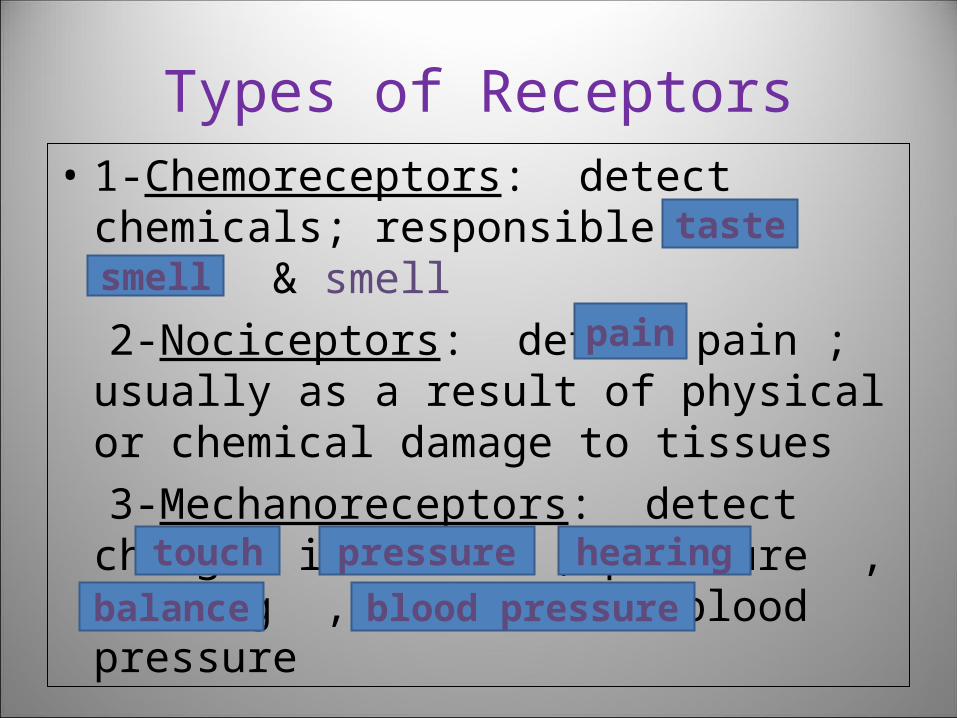
Types of Receptors• 1-Chemoreceptors: detect
chemicals; responsible for taste & smell
2-Nociceptors: detect pain ; usually as a result of physical or chemical damage to tissues
3-Mechanoreceptors: detect changes in touch , pressure , hearing , balance , & blood pressure
tastesmell
pain
touch pressure hearing
balance blood pressure
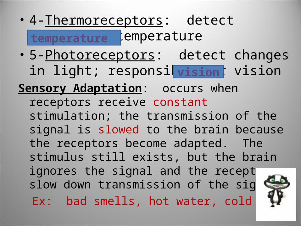
• 4-Thermoreceptors: detect changes in temperature
• 5-Photoreceptors: detect changes in light; responsible for vision
Sensory Adaptation: occurs when receptors receive constant stimulation; the transmission of the signal is slowed to the brain because the receptors become adapted. The stimulus still exists, but the brain ignores the signal and the receptors slow down transmission of the signal.
Ex: bad smells, hot water, cold temps
temperature
vision

Touch & Pressure• 1-Merkel disk: found in the stratum
basale and perceives very light touch. Ex: lips and fingertips
• 2-Meissner corpuscle: found in the dermis of hairless skin and perceives light touch.Ex:fingertips, palms,& soles
• 3-Ruffini corpuscle: found in the lower dermis and perceive stretch
Ex: digits and limbs • 4-Pacinian corpuscles: found in lower
dermis, subcutaneous, and around joints; respond to heavy pressure
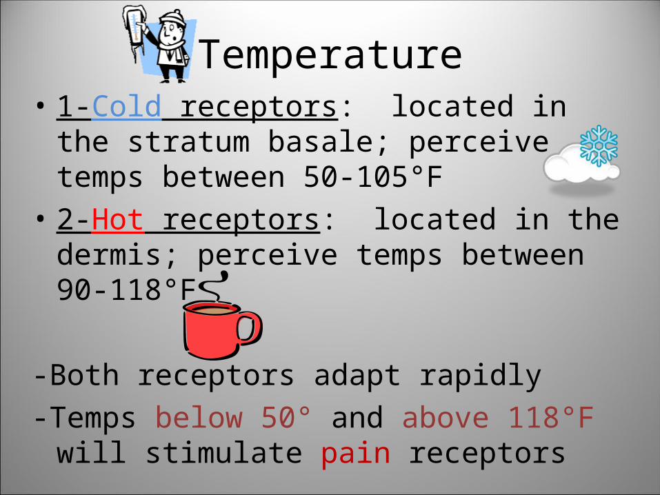
Temperature• 1-Cold receptors: located in the
stratum basale; perceive temps between 50-105°F
• 2-Hot receptors: located in the dermis; perceive temps between 90-118°F
-Both receptors adapt rapidly-Temps below 50° and above 118°F will
stimulate pain receptors
Pain
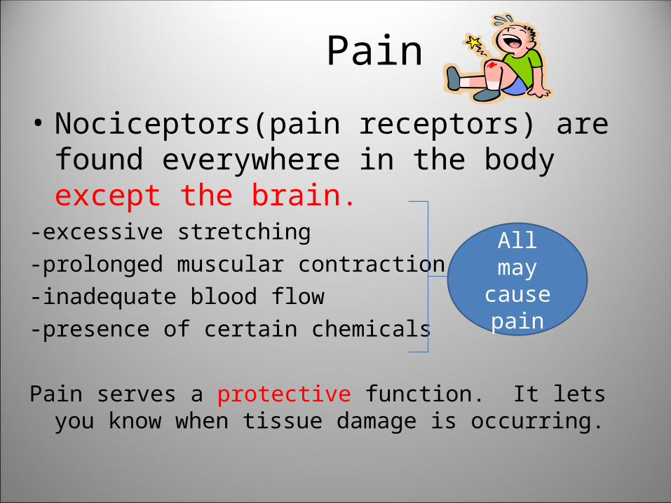
• Nociceptors(pain receptors) are found everywhere in the body except the brain.
-excessive stretching-prolonged muscular contraction-inadequate blood flow-presence of certain chemicals
Pain serves a protective function. It lets you know when tissue damage is occurring.
All may cause pain
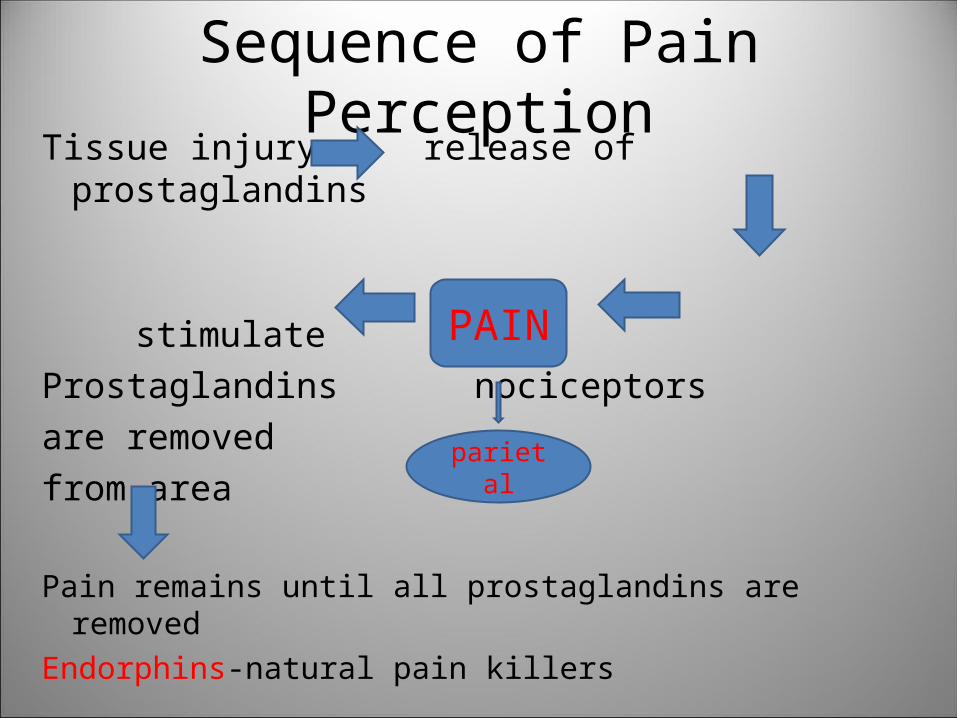
Sequence of Pain PerceptionTissue injury release of prostaglandins
stimulateProstaglandins nociceptorsare removed from area
Pain remains until all prostaglandins are removed
Endorphins-natural pain killers
PAIN
parietal

• Referred Pain: tissue damage that takes place in the organs of the body may cause pain in a different area. This happens because pain impulses travel along common pathways to the brain. The brain is uncertain to where it should reflect the impression of pain. Ex. Tissue damage in the heart can be “felt” in the arm or back

Smell
• aka olfaction• nose contains 10-100 million smell receptors• most memories are stored in association with a
smell• Humans can recognize ~10,000 scents• Olfactory equipment is found in the upper part of
nasal cavity. Olfactory hairs pick up chemicals in the air we breathe. These chemicals bond with a complimentary-shaped receptor. This sends a message to the temporal lobe via olfactory nerve.
Taste
• aka gustation• 5 primary tastes(sweet, salty, sour, bitter, &
umami)• ~10,000 taste buds on adult tongue( as you
age)• taste buds are found on tongue, roof of
mouth, throat, & larynx• taste receptors are located inside papillae• gustatory hairs direct food into taste pores
where taste buds are located• chemicals in food bind with complimentary-
shaped receptors

Hearing• Outer:– auricle: aka pinna; collects sounds with its funnel shape-external auditory meatus: canal that carries sound from the auricle to the middle ear
• Middle:– tympanic membrane: aka eardrum; cone-shaped; responds to sound waves by moving back and forth
-auditory ossicles: malleus, incus & stapes; these bones form a bridge that connects the eardrum to the inner ear
-oval window: an opening that connects the middle ear to the inner ear; vibrations pass through the oval window and cause movement of a fluid inside the inner ear
-Eustachian tube: connects middle ear to the throat; maintains pressure balance
• Inner Ear:– labyrinth: system of chambers and tubes that are filled with fluid-semicircular canals: loops that extend
into each plane; full of thick
fluid and hairs; as you movethe fluid pulls on the
hairs and sends a message to your brain about your position

-cochlea: shell-shaped organ that sends sound vibrations
through the round window to the… -Organ of Corti: contains sensory receptors that transmit “hearing” impulses to brain

Equilibrium
Static-helps you maintain
stability and posture of the head and body when not in motion; occurs in the vestibule
Dynamic -helps you maintain balance when you are in motion; occurs in the semicircular canals
Sight• Photoreceptors receive the stimuli• Accessory organs assist the eye:
– eyelid: covers the eye; moistens and protects the eye-conjuctiva: lines the inner surface of the eyelids and outer surface of eye-lacrimal gland: secretes tears; found above eye; sends tears into
lacrimal ducts which carry tears into the nasal cavity-tears: keep eye moist; contain lysozyme (enzyme
that reduces eye infections)-extrinsic muscles: attach from bones of orbit to
eye; move the eye in various directions

Structure of the Eye• The eye is a hollow muscular structure about 2.5
cm in diameter; filled with fluid that gives shape -cornea: transparent tissue that serves as a window to allow light to enter; contains few cells and NO blood vessels
-sclera: white portion of the eye; provides
protection; serves as an attachment point for the extrinsic muscle-optic nerve: carries visual impulses to the brain-choroid coat: middle layer of the eye; contains
lots of blood vessels

– lens: a transparent ball of elastic material; ligaments pull on the lens to change its shape. *to view objects close convex
close-convex
*to view objects far away concave
far-flat

– iris: a ring of muscle that forms the opening of the eye to determine amt of light that enters *lots of light=small pupil *low light=large pupil-aqueous humor: a thin fluid that fills the anterior chamber of the eye (between the cornea and lens); constantly being formed/drained-retina: the inner lining of the eye; contains rods and cones-rods: type of photoreceptor that is active during low-light conditions; contain rhodopsin- pigment that absorbs light/helps you see in the
dark
– cones: type of photoreceptor that is active in bright light conditions; helps perceive color; area where cones are most highly-concentrated is called the fovea(most acute vision) this is where the lens tries to focus light(focal point)- vitreous humor: gelatin-like substance that fills the posterior chamber of the eye; maintains shape; born with
it