Embed Size (px)
Citation preview
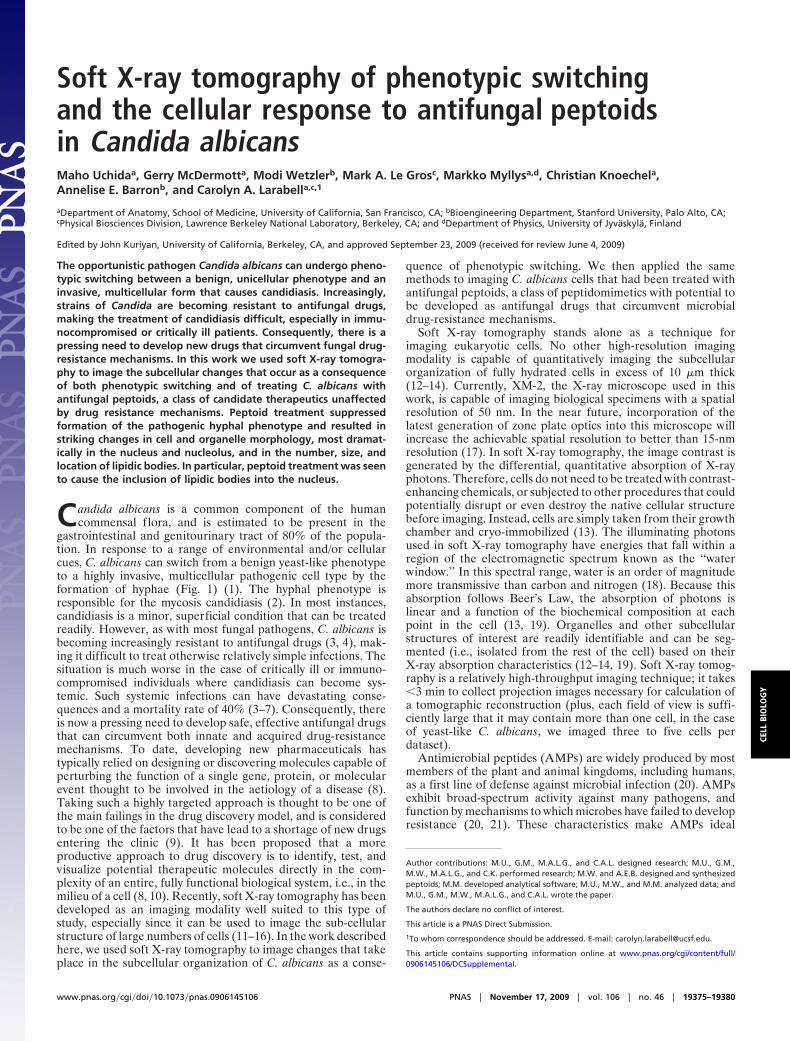
Soft X-ray tomography of phenotypic switchingand the cellular response to antifungal peptoidsin Candida albicansMaho Uchidaa, Gerry McDermotta, Modi Wetzlerb, Mark A. Le Grosc, Markko Myllysa,d, Christian Knoechela,Annelise E. Barronb, and Carolyn A. Larabella,c,1
aDepartment of Anatomy, School of Medicine, University of California, San Francisco, CA; bBioengineering Department, Stanford University, Palo Alto, CA;cPhysical Biosciences Division, Lawrence Berkeley National Laboratory, Berkeley, CA; and dDepartment of Physics, University of Jyvaskyla, Finland
Edited by John Kuriyan, University of California, Berkeley, CA, and approved September 23, 2009 (received for review June 4, 2009)
The opportunistic pathogen Candida albicans can undergo pheno-typic switching between a benign, unicellular phenotype and aninvasive, multicellular form that causes candidiasis. Increasingly,strains of Candida are becoming resistant to antifungal drugs,making the treatment of candidiasis difficult, especially in immu-nocompromised or critically ill patients. Consequently, there is apressing need to develop new drugs that circumvent fungal drug-resistance mechanisms. In this work we used soft X-ray tomogra-phy to image the subcellular changes that occur as a consequenceof both phenotypic switching and of treating C. albicans withantifungal peptoids, a class of candidate therapeutics unaffectedby drug resistance mechanisms. Peptoid treatment suppressedformation of the pathogenic hyphal phenotype and resulted instriking changes in cell and organelle morphology, most dramat-ically in the nucleus and nucleolus, and in the number, size, andlocation of lipidic bodies. In particular, peptoid treatment was seento cause the inclusion of lipidic bodies into the nucleus.
Candida albicans is a common component of the humancommensal f lora, and is estimated to be present in the
gastrointestinal and genitourinary tract of 80% of the popula-tion. In response to a range of environmental and/or cellularcues, C. albicans can switch from a benign yeast-like phenotypeto a highly invasive, multicellular pathogenic cell type by theformation of hyphae (Fig. 1) (1). The hyphal phenotype isresponsible for the mycosis candidiasis (2). In most instances,candidiasis is a minor, superficial condition that can be treatedreadily. However, as with most fungal pathogens, C. albicans isbecoming increasingly resistant to antifungal drugs (3, 4), mak-ing it difficult to treat otherwise relatively simple infections. Thesituation is much worse in the case of critically ill or immuno-compromised individuals where candidiasis can become sys-temic. Such systemic infections can have devastating conse-quences and a mortality rate of 40% (3–7). Consequently, thereis now a pressing need to develop safe, effective antifungal drugsthat can circumvent both innate and acquired drug-resistancemechanisms. To date, developing new pharmaceuticals hastypically relied on designing or discovering molecules capable ofperturbing the function of a single gene, protein, or molecularevent thought to be involved in the aetiology of a disease (8).Taking such a highly targeted approach is thought to be one ofthe main failings in the drug discovery model, and is consideredto be one of the factors that have lead to a shortage of new drugsentering the clinic (9). It has been proposed that a moreproductive approach to drug discovery is to identify, test, andvisualize potential therapeutic molecules directly in the com-plexity of an entire, fully functional biological system, i.e., in themilieu of a cell (8, 10). Recently, soft X-ray tomography has beendeveloped as an imaging modality well suited to this type ofstudy, especially since it can be used to image the sub-cellularstructure of large numbers of cells (11–16). In the work describedhere, we used soft X-ray tomography to image changes that takeplace in the subcellular organization of C. albicans as a conse-
quence of phenotypic switching. We then applied the samemethods to imaging C. albicans cells that had been treated withantifungal peptoids, a class of peptidomimetics with potential tobe developed as antifungal drugs that circumvent microbialdrug-resistance mechanisms.
Soft X-ray tomography stands alone as a technique forimaging eukaryotic cells. No other high-resolution imagingmodality is capable of quantitatively imaging the subcellularorganization of fully hydrated cells in excess of 10 �m thick(12–14). Currently, XM-2, the X-ray microscope used in thiswork, is capable of imaging biological specimens with a spatialresolution of 50 nm. In the near future, incorporation of thelatest generation of zone plate optics into this microscope willincrease the achievable spatial resolution to better than 15-nmresolution (17). In soft X-ray tomography, the image contrast isgenerated by the differential, quantitative absorption of X-rayphotons. Therefore, cells do not need to be treated with contrast-enhancing chemicals, or subjected to other procedures that couldpotentially disrupt or even destroy the native cellular structurebefore imaging. Instead, cells are simply taken from their growthchamber and cryo-immobilized (13). The illuminating photonsused in soft X-ray tomography have energies that fall within aregion of the electromagnetic spectrum known as the ‘‘waterwindow.’’ In this spectral range, water is an order of magnitudemore transmissive than carbon and nitrogen (18). Because thisabsorption follows Beer’s Law, the absorption of photons islinear and a function of the biochemical composition at eachpoint in the cell (13, 19). Organelles and other subcellularstructures of interest are readily identifiable and can be seg-mented (i.e., isolated from the rest of the cell) based on theirX-ray absorption characteristics (12–14, 19). Soft X-ray tomog-raphy is a relatively high-throughput imaging technique; it takes�3 min to collect projection images necessary for calculation ofa tomographic reconstruction (plus, each field of view is suffi-ciently large that it may contain more than one cell, in the caseof yeast-like C. albicans, we imaged three to five cells perdataset).
Antimicrobial peptides (AMPs) are widely produced by mostmembers of the plant and animal kingdoms, including humans,as a first line of defense against microbial infection (20). AMPsexhibit broad-spectrum activity against many pathogens, andfunction by mechanisms to which microbes have failed to developresistance (20, 21). These characteristics make AMPs ideal
Author contributions: M.U., G.M., M.A.L.G., and C.A.L. designed research; M.U., G.M.,M.W., M.A.L.G., and C.K. performed research; M.W. and A.E.B. designed and synthesizedpeptoids; M.M. developed analytical software; M.U., M.W., and M.M. analyzed data; andM.U., G.M., M.W., M.A.L.G., and C.A.L. wrote the paper.
The authors declare no conflict of interest.
This article is a PNAS Direct Submission.
1To whom correspondence should be addressed. E-mail: [email protected].
This article contains supporting information online at www.pnas.org/cgi/content/full/0906145106/DCSupplemental.
www.pnas.org�cgi�doi�10.1073�pnas.0906145106 PNAS � November 17, 2009 � vol. 106 � no. 46 � 19375–19380
CELL
BIO
LOG
Y

candidates for development as drug molecules. Unfortunately,AMPs are prone to rapid proteolysis in vivo; thus, hindering theirclinical applications (22). However, peptoids, a class of peptido-mimetics (23), are extremely resistant to proteolysis (24), andpossess broad-spectrum antimicrobial activity against manypathogenic organisms, including C. albicans (25). Consequently,peptoids are a much more attractive system for studying anddeveloping novel antimicrobial and antifungal therapies. In thisstudy, we treated C. albicans with two peptoids, termed peptoid1 and peptoid 2 (Fig. S1), and imaged the subcellular conse-quences.
Peptoid 1 was designed to mimic magainin 2, one of the mostestablished and widely studied AMPs (26). Peptoid 2 wasdesigned to have homology with a family of AMPs previouslyshown to have strong antifungal activity against C. albicans (27).The mode of action by which these peptoids function is thesubject of ongoing debate, but the evidence indicates thatpeptoids likely function similar to AMPs. However, even in thecase of the more studied AMPs, there are still a number ofunanswered questions regarding the precise mechanism(s) bywhich they act. One of the most widely accepted views being thatAMPs exert at least part of their effects through membranedisruption (28). For example, a family of AMPs with highstructural similarity to peptoid 2 was reported to cause disrup-tion of C. albicans membranes (27). Based on the activity ofpeptoid 1 in membrane-mimetic vesicles and monolayers, it isreasonable to expect it to also cause membrane disruption (25).However, magainin 2 has also been shown to damage DNA inyeast (29); thus, demonstrating that AMPs and peptoids mayhave both intracellular and intranuclear effects.
ResultsWe began this study by establishing the subcellular architectureof the three phenotypes displayed by C. albicans, namely,yeast-like, germ-tube, and hyphal cells. Live cells were mountedin thin-walled glass capillary tubes, cryo-immobilized, and thenimaged by soft X-ray microscopy. A tomographic dataset foreach was obtained by collecting projection images at 2° incre-
ments around a rotation axis. The projection data were thencomputationally reconstructed to give the 3D cell volumes,which were then segmented to isolate, visualize, and quantifyorganelles and other subcellular structures (Fig. 2; MoviesS1–S3). For the pathogenic form of C. albicans cells (i.e., thehyphal cell shown in Fig. 2 E and F and Movies S3 and S4) withcell lengths greater than the microscope field of view, projectionseries were collected from sequentially overlapping fields ofview. Each series was individually reconstructed and then com-putationally ‘‘stitched together’’ to form the single, seamlesstomographic reconstructions shown in Fig. 2E. For smaller cells,such as the yeast-like or germ-tube cells, each field of viewcontained between one and four cells, each of which could bereconstructed and segmented. Consequently, significant num-bers of cells could be imaged tomographically for each peptoidtreatment.
Subcellular structures such as the nucleus, nucleolus, mito-chondria, vacuole, and lipid bodies were segmented based on thecalculated linear absorption coefficients (LAC) at each voxel(Table S1). The measured LAC value for each voxel is deter-mined by the biochemical composition at that point in the cell.Regions of the cell with relatively similar biochemical compo-sition (at the level of spatial resolution observed) have charac-teristic LAC values (13, 19). For example, structures such as lipidbodies have consistent LAC values within the same cell, betweencells, and can even be consistent between different cell types.That said, within a particular organelle, such as a nucleus, theLAC values vary in accordance with the internal structures andmolecular densities. In this work, it was possible to segmentorganelles on their measured LAC values alone, and subse-quently, assign the identity based on LAC and organelle shape
Fig. 1. Schematic representation of the three phenotypes adopted by C.albicans. Blue, nucleus; dark gray, septum.
Fig. 2. Soft X-ray tomographic reconstruction of phenotypically distinct C.albicans cells. A representative orthoslice from each tomographic reconstruc-tion is shown for a yeast-like (A), germ-tube (C) and hyphal (E) cell. Volumerendered views of the same cells are shown in (B, D, and F) respectivelyshowing selected organelles that have been segmented and color-coded foridentification. Blue, nucleus; orange, nucleolus; gray, mitochondria; yellow,vacuole; green, lipid bodies. (Scale bar for A–D, 0.5 �m, for E and F, 2.0 �m.)
19376 � www.pnas.org�cgi�doi�10.1073�pnas.0906145106 Uchida et al.

established by other modalities (in particular, light and electronmicroscopy (30–32)). In this regard, segmentation of organellesin a cell imaged by soft X-ray tomography is analogous todifferentiating liver from kidney, fat from muscle, or normalfrom abnormal tissue, in a patient imaged using clinical com-puted axial tomography (CAT).
Initially, the reconstructed cells were manually segmented.However, the excellent signal-to-noise inherent in soft X-raytomographic data allowed us to apply automated segmentationmethods, and thus, greatly speed up this process. To check theaccuracy of the automated method, we segmented cells that hadpreviously been segmented manually. A comparison of organellevolumes showed that the automated methods produced valuesthat were within 10% of those obtained by manual segmentation.
Overall, we found that cells with the germ-tube phenotypeclosely resembled those of yeast-like cells in terms of organelleorganization and structures. A number of dense objects withaverage volume of 0.07 �m3 were assigned as lipid droplets basedon their average LAC values of 0.75 � 0.09 �m�1 (for a list ofsegmented organelle LAC values, see Table S1). The mitochon-dria appeared as single, continuous branched structures thatcurved around the nucleus and the vacuole. The segmentedhyphal cells showed marked structural differences comparedwith yeast-like and germ-tube phenotypes. The nuclei andnucleoli were less spherical, and much more elongated, andmultiple small vacuoles were apparent, in particular in the regionnearest the primary cell. There appeared to be no vacuoles closeto the hyphal tip, or even in the lower half of the hyphal cell.
To determine the cellular consequences of antifungal peptoidtreatment on C. albicans cells, we collected tomographic data oncells that had been incubated with two different peptoids,peptoids 1 and 2, at a concentration corresponding to a minimuminhibitory concentration (MIC) of 50% (Fig. S1 and Movies S5and S6). Characteristic of treatment with either peptoid mole-cule was the inhibition of the hyphal phenotype. Of the peptoid1 treated cells, 73% of cells had a yeast-like phenotype, with theother 27% having the germ-tube phenotype. However, 90% ofpeptoid 2 treated cells had the yeast-like phenotype, with theremainder having a germ-tube phenotype (Table 1). In all cases,peptoid treatment resulted in an increase in the size and numberof lipid-like structures and changes in the structure of thenucleoli (Fig. 3). We compared the cell volumes, and the volumesof lipid-like structures, nuclei, and nucleoli, in peptoid treatedcells with those of the control cells (Fig. 4). Although the volumeof the nucleus differed between control and peptoid treatedcells, no significant difference in the average volume of nucleusrelative to the cell volumes was observed between control (6.4 �1.5%) and peptoid treated cells (peptoid 1: 6.1 � 0.7%; peptoid2: 6.4 � 1.1%). However, the average volume of the nucleolusrelative to the cell volumes was smaller in peptoid 2 treated cells(1.0 � 0.5%) compared with that of the control (2.0 � 0.4%) andpeptoid 1 treated cells (1.6 � 0.4%) (Fig. 4A). Interestingly, theaverage volume of lipids relative to the cell volumes in peptoid2 treated cells (0.60 � 0.4%) was significantly higher comparedwith those of the control (0.06 � 0.03%) and peptoid 1 treatedcells (0.12 � 0.04%). When the volumes of lipidic bodies wascompared between peptoids 1 and 2 treated cells, the averagepercentage volume of lipids in peptoid 2 treated cells was 80%
higher than that of peptoid 1 treated cells (Fig. 4B). In addition,deformation of vacuoles was observed in peptoid 1 treated cells(Fig. 3D). We also observed unexpected large structures em-
Table 1. Phenotypes of peptoid treated cells
Yeast like Germ tube
Control 11 10Peptoid 1 8 3Peptoid 2 9 1
Total number of segmented cells are categorized by phenotype.
Fig. 3. Soft X-ray tomography of C. albicans cells after treatment with peptides.Peptoid 1 treated cell (A–D) Representative orthoslice through the tomographicreconstruction (A). All segmented organelles (B). Segmented nucleus and nucle-olus (C). Peptoid 1 induced structural changes in the nucleolus are shown in detailin the Inset. Segmented vacuoles (D). Peptoid 2 treated cell (E–G). Representativeorthoslice through the tomographic reconstruction (E). All segmented organelles(F). Segmented nucleus, nucleolus and lipid bodies (G). Peptoid 2 induced struc-tural changes in the nucleolus and the incorporation of a large lipid body into thenucleus are shown in detail in the Inset. Blue, nucleus; orange, nucleolus; gray,mitochondria; yellow, vacuole; green, lipid bodies. (Scale bar, 1.0 �m).
Uchida et al. PNAS � November 17, 2009 � vol. 106 � no. 46 � 19377
CELL
BIO
LOG
Y

bedded inside the nucleus of peptide 2 treated cells (Fig. 3G).These structures had LAC values in the range of those measuredfor lipid bodies, leading us to assign them accordingly. Thisobservation was characteristic of virtually all cells treated withpeptoid 2.
DiscussionSoft X-ray tomography is distinguishable from peer imagingtechniques by the capacity to image fully hydrated eukaryoticcells in excess of 10 �m thick and do so without the use of stainsor other contrast enhancing agents. Consequently, cells areimaged close to their native state.
Segmented reconstructions of C. albicans cells with yeast-likephenotype showed the mitochondria to be long, branched struc-tures that surrounded both the nucleus and a single vacuole. Thenucleus, nucleolus, and vacuole were approximately spherical,and organized as expected from studies using other imagingmodalities. In hyphal cells, we observed a large population ofdiscrete mitochondria localized at the tip region. This organi-zation may be due to the large energy requirement for hyphalextension. However, the fragmented form of the mitochondriacould also be the consequence of cell stress, because it is knownthat subcellular elements in the hyphal tip region are susceptibleto any environmental change (33).
Segmented reconstructions of cells treated with peptoid 2showed a number of clear differences with control cells, inparticular in the nuclear region. The most striking of which aredense bodies observed to be embedded inside the nucleus (Figs.
3G and 5). Based on the measured LAC values and the shape ofthese objects, we surmise that these bodies are composed oflipids, or lipid-like, molecules. A similar report of enlargedlipid-droplets was described in Saccharomyces cerevisiae mutantcells, where a functional homologue of human seipin was deleted(32, 34). To our knowledge, this is the first observation of largelipid-like structures being embedded in the nucleus of C. albi-cans. The other striking feature visualized in the reconstructionsof peptoid treated cells was the structure of the nucleoli. In allcases, the nucleoli in peptoid treated cells appear to contain oneor more holes that pass completely through the organelle.Peptoid 2 treatment led to a single hole being present in thenucleoli, whereas peptoid 1 treatment resulted in nuclei thatcontained two such voids (Fig. 5) in all of the cells imaged. Again,the function, consequences, and underlying reason for thesestructural features remains an open question. It should also bestressed again that this type of observation could not have beenmade using diffraction limited fluorescence microscopy.
In all of the peptoid treated cells, the major phenotypicfeatures displayed in Fig. 3 remained constant. Therefore, it isreasonable to speculate that these morphological changes haveunderlying functional consequences. These features could rep-resent the stress response to peptoid treatment. Recent studiessuggest that the nucleolus not only functions as the site forribosome synthesis, but also has a key role in cell stress sensing.For example, under stress conditions, mammalian cells increasethe level of tumor suppressor, p53, which leads to inhibition ofPol I transcription and altered nucleolus morphology (35–37).Although this work was not focused on the functional conse-quences of this structural arrangement, it is likely that the
Fig. 4. Effect of peptoid treatment on selected organelle volumes. (A) Averagepercentage volume of the nucleus relative to the cell volume in control andpeptoid treated cells. (B) Volume of lipid droplets in control and peptoid treatedcells. The percentage relative value was calculated from the volume of eachorganelle divided by its cell volume. The error bars represent SD.
Fig. 5. Dimensions of nuclei, nucleoli, and a lipid-like structure shown in Fig.3. Blue, nucleus; orange, nucleolus; green, lipid body.
19378 � www.pnas.org�cgi�doi�10.1073�pnas.0906145106 Uchida et al.

application of antifungal peptoids led to this, or similar phe-nomena, suggesting that these mechanisms are suitable fortherapeutic targeting.
In previous work by Isola et al. (31), the effects of salivaryHistatin, an AMP, on C. albicans cells were imaged usingtransmission electron microscopy (TEM) and high-resolutionscanning electron microscopy (HRSEM) (31). A number ofHistatin-induced alterations in C. albicans cell structure werereported, such as the appearance of electron-dense bodies(thought to be mitochondrial in origin), membrane and organelledisarrangement, and the appearance of structures resemblingautophagosomes. However, there was no clear indication ofspecific biochemical mechanism by which Histatin may function.
Developing new pharmaceuticals, such as antifungal drugs, iscostly and unpredictable. Despite the availability of advancedresearch technologies, such as combinatorial chemistry andhigh-throughput screening methods, it still costs more than $1billion to bring a new prescription drug to the market (8). Thecombination of high spatial resolution, together with a contrastmechanism that retains and reveals subtle features in cells, makesoft X-ray tomography well suited to visualizing the conse-quences of treating cells with candidate drug molecules. Theutility of the technique will increase in the near future, now thatit is possible to carry out correlated high-aperture cryogenicfluorescence microscopy and soft X-ray tomography on the samespecimen (16).
Materials and MethodsStrains, Cell Cultures, and Growth Conditions. C. albicans (wild-type strain26555) was obtained from ATCC and grown at 26 °C on ATCC medium No.200that contains 0.3% (wt/vol) yeast extract, 0.3%(wt/vol) malt extract, 0.5%(wt/vol) peptone, 1% (wt/vol) dextrose, and 2% (wt/vol) agar. Cell cultureswere freshly prepared from these plates (one colony in 5 mL of liquid medium)and grown on a rotary shaker maintained at appropriate temperature over-night (OD � 1.5�2.5). The phenotype was switched by modifying the growthconditions, as described in ref. 1.
Peptoid Treatment. Peptoids were synthesized as previously reported (20, 22).Liquid culture media containing overnight-cultured cells (100:1) and appro-priate concentrations of peptoids were placed on the rotary shaker at 37 °C(t � 0). At this temperature, tube-like structures of C. albicans cells wereinduced. OD measurements were done every 30 min for 3 h (OD � �0.5). ODreadings at 600 nm were performed on a Beckman DU640 spectrophotometer(Beckman Coulter) with a blank (i.e., appropriate concentrations of peptoidsin liquid media).
Soft X-Ray Tomography. X-ray datasets were collected using the XM-2 softX-ray microscope operated by the National Center for X-ray Tomography(http://ncxt.lbl.gov) at the Advanced Light Source (http://www.als.lbl.gov) ofLawrence Berkeley National Laboratory (LBNL). XM-2 is equipped with Fresnelzone plate based condenser and objective lenses (made by the Center for X-ray
Optics, LBNL) and is specifically designed to investigate biological samples atcryogenic temperatures. For soft X-ray tomography, cells were rapidly trans-ferred from their growth media to thin-walled glass capillaries, and immedi-ately flash frozen in a cryogenic gas stream (13). No additional stainingprocedures were used. Imaging was performed with the specimens in anatmosphere of liquid nitrogen cooled helium gas at all times. For each dataset,90 projection images were collected sequentially around a rotation axis in 2°increments to give a total rotation of 180°; the microscope was equipped witha resolution defining 50-nm objective lens. An exposure time of between 150and 300 ms was used (depending on synchrotron ring current). Manualalignment of images, based on fiducial markers, was performed using theIMOD package (38). Tomographic reconstructions were calculated using iter-ative reconstruction methods (39–41).
Calculation of LACs and Manual Segmentation. Reconstructed cells were seg-mented into subvolumes that have similar LACs. The LAC for each voxel wasmeasured directly from the experimental data. The absorption of soft X-raysfollows Beer’s Law, and therefore, is concentration and molecular speciesdependent. In the case of isolated biomolecules, the LAC can be measuredexperimentally or easily calculated according to the chemical composition ofthe specimen. For example, ice was calculated to have a LAC of 0.109 �m�1,whereas a model protein with the chemical composition C94H139N24O31S has aLAC of 1.35 �m�1 (19). The situation becomes more complex in the context of50-nm voxels in a reconstruction of a cell. In this case, each voxel will almostcertainly contain a mixture of biomolecules (lipids, protein, water, etc.).Therefore, the LAC for a segmented organelle is a measure of the overallbiochemical composition. As can be seen from the reconstructed, unseg-mented cell volumes (Fig. 1 A, C, and E) organelles have a relatively consistentLAC value, and therefore, are easily identifiable compared with other com-ponents in the cell (i.e., the nucleolus is readily distinguishable from thenucleus, lipid-dense bodies from the cytosol, etc.). Manual segmentation,measurements of voxel values to calculate LACs, and the creation of movieswere all carried out using the Amira software package (Mercury ComputerSystems).
Statistical Analysis of Autosegmented Data. Automatic segmentation wasdone using ITK-Snap V1.6.0.1 software (http://www.itksnap.org/pmwiki/pmwiki.php) (42), along with ImageJ 1.42k (http://rsbweb.nih.gov/ij/). ImageJwas used to reduce the noise from the data reconstructed by filtered backprojection (39). Cells and lipid droplets were automatically segmented basedon the threshold with corresponding LAC voxel values shown in Table S1;nucleoli and nuclei were semiautomatically segmented by the active contourmethod (42). All of the segmented volumes were smoothed by 3D medianfiltering, and all volumes with a voxel value of 5 or higher were included in theresults.
ACKNOWLEDGMENTS. We thank Zeny Serrano for her skillful assistance withcell culture and peptoid treatment; Drs. Weiwei Gu and Dula Parkinson forassistance in the processing and alignment of the projection images and in thecalculation of the tomographic reconstructions; and Tyler M. Miller for helpwith the peptoid synthesis. This work was funded by the Department ofEnergy Office of Biological and Environmental Research Grant DE-AC02-05CH11231, the National Institutes of Health (NIH) National Institute forAllergy and Infectious Diseases Grant GM072666, and the NIH National Centerfor Research Resources Grant RR019664.
1. Sudbery P, Gow N, Berman J (2004) The distinct morphogenic states of Candidaalbicans. Trends Microbiol 12:317–324.
2. Ashman RB, et al. (2004) Innate versus adaptive immunity in Candida albicans infec-tion. Immunol Cell Biol 82:196–204.
3. Monk BC, Goffeau A (2008) Outwitting multidrug resistance to antifungals. Science321:367–369.
4. Rappleyel CA, Goldman WE (2008) Fungal stealth technology. Trends Immunol 29:18–24.
5. Enoch DA, Ludlam HA, Brown NM (2006) Invasive fungal infections: A review ofepidemiology and management options. J Med Microbiol 55:809–818.
6. Richardson M, Lass-Florl C (2008) Changing epidemiology of systemic fungal infections.Clin Microbiol Infect 14:5–24.
7. Wey SB, Mori M, Pfaller MA, Woolson RF, Wenzel RP (1988) Hospital-acquired candi-demia. The attributable mortality and excess length of stay. Arch Intern Med 148:2642–2645.
8. Bullen A (2008) Microscopic imaging techniques for drug discovery. Nat Rev DrugDiscov 7:54–67.
9. Hood L, Perlmutter RM (2004) The impact of systems approaches on biological prob-lems in drug discovery. Nat Biotechnol 22:1215–1217.
10. Lang P, Yeow K, Nichols A, Scheer A (2006) Cellular imaging in drug discovery. Nat RevDrug Discov 5:343–356.
11. Gu WW, Etkin LD, Le Gros MA, Larabell CA (2007) X-ray tomography of Schizosaccha-romyces pombe. Differentiation 75:529–535.
12. Larabell CA, Le Gros MA (2004) X-ray tomography generates 3-D reconstructions of theyeast, Saccharomyces cerevisiae, at 60-nm resolution. Mol Biol Cell 15:957–962.
13. Le Gros MA, McDermott G, Larabell CA (2005) X-ray tomography of whole cells. CurrOpin Struct Biol 15:593–600.
14. Parkinson DY, McDermott G, Etkin LD, Le Gros MA, Larabell CA (2008) Quantitative3-D imaging of eukaryotic cells using soft X-ray tomography. J Struct Biol 162:380 –386.
15. Leis A, Rockel B, Andrees L, Baumeister W (2009) Visualizing cells at the nanoscale.Trends Biochem Sci 34:60–70.
16. Le Gros MA, McDermott G, Uchida M, Konechel CG, Larabell CA (2009) High aperturecryogenic light microsocpy. J Microsc 235:1–8.
17. Chao WL, Harteneck BD, Liddle JA, Anderson EH, Attwood DT (2005) Soft X-raymicroscopy at a spatial resolution better than 15nm. Nature 435:1210–1213.
18. Attwood DT (1999) Soft X-Rays and Extreme Ultraviolet Radioation: Principles andApplications (Cambridge Univ Press, Cambridge, UK), p 470.
19. Weiss D, et al. (2000) Computed tomography of cryogenic biological specimens basedon X-ray microscopic images. Ultramicroscopy 84:185–197.
20. Zasloff M (2002) Antimicrobial peptides of multicellular organisms. Nature 415:389–395.
Uchida et al. PNAS � November 17, 2009 � vol. 106 � no. 46 � 19379
CELL
BIO
LOG
Y

21. Bessalle R, Kapitkovsky A, Gorea A, Shalit I, Fridkin M (1990) All-D-magainin: Chirality,antimicrobial activity and proteolytic resistance. FEBS Lett 274:151–155.
22. Latham PW (1999) Therapeutic peptides revisted. Nat Biotechnol 17:755–757.23. Zuckermann RN, Kerr JM, Kent SBH, Moost WH (1992) Efficient Method for the
Preparation of Peptoids [Oligo(N-substituted glycines)] by Submonomer Solid-PhaseSynthesis. J Am Chem Soc 114:10646–10647.
24. Miller SM, et al. (1995) Comparison of the Proteolytic Susceptibilities of HomologousL-Amino Acid, D-Amino Acid, and N-Substituted Glycine Peptide and Peptoid Oliog-mers. Drug Devel Res 35:20–32.
25. Chongsiriwatana NP, et al. (2008) Peptoids that mimic the structure, function, andmechanism of helical antimicrobial peptides. Proc Natl Acad Sci USA 105:2794–2799.
26. Patch JA, Barron AE (2003) Helical Peptoid Mimics of Magainin-2 Amide. J Am ChemSoc 125:12092–12093.
27. Makovitzki A, Avrahami D, Shai Y (2006) Ultrashort antibacterial and antifungallipopeptides. Proc Natl Acad Sci USA 103:15997–16002.
28. Shai Y (2002) Mode of Action of Membrane Active Antimicrobial Peptides. Biopolymers66:236–248.
29. Morton CO, et al. (2007) Global phenotype screening and transcript analysis outlinesthe inhibitory mode(s) of action of two amphibian-derived, alpha-helical, cationicpeptides on Saccharomyces cerevisiae. Antimicrob Agents Chemother 51:3948–3959.
30. Riquelme M, Roberson RW, McDaniel DP, Bartnicki-Garcia S (2002) The effects of ropy-1mutation on cytoplasmic organization and intracellular motility in mature hyphae ofNeurospora crassa. Fungal Genet Biol 37:171–179.
31. Isola R, Isola M, Conti G, Lantini MS, Riva A (2007) Histatin-induced alterations inCandida albicans: A microscopic and submicroscopic comparison. Microsc Res Tech70:607–616.
32. Fei W, et al. (2008) Fld1p, a functional homologue of human seipin, regulates the sizeof lipid droplets in yeast. J Cell Biol 180:473–482.
33. Szeghalmi A, Kaminskyj S, Gough KM (2007) A synchrotron FTIR microspectroscopyinvestigation of fungal hyphae grown under optimal and stressed conditions. AnalBioanal Chem 387:1779–1789.
34. Ceelen H, et al. (1995) Bovine Somatotropin and the Regulatory Process for VeterinaryDrug Approval - Open-Letter. Can Vet J 36:595–596.
35. Mayer C, Grummt I (2005) Cellular stress and nucleolar function. Cell Cycle 4:1036–1038.
36. Olson MO, Dundr M (2005) The moving parts of the nucleolus. Histochem Cell Biol123:203–216.
37. Lo SJ, Lee CC, Lai HJ (2006) The nucleolus: Reviewing oldies to have new understand-ings. Cell Res 16:530–538.
38. Kremer JR, Mastronarde DN, McIntosh JR (1996) Computer visualization of three-dimensional image data using IMOD. J Struct Biol 116:71–76.
39. Mastronarde DN (1997) Dual-axis tomography: An approach with alignment methodsthat preserve resolution. J Struct Biol 120:343–352.
40. Erdogan H, Fessler JA (1999) Ordered subsets algorithms for transmission tomography.Phys Med Biol 44:2835–2851.
41. Stayman JW, Fessler JA (2000) Regularization for uniform spatial resolution prop-erties in penalized-likelihood image reconstruction. IEEE Trans Med Imaging19:601– 615.
42. Yushkevich PA, et al. (2006) User-guided 3D active contour segmentation of anatom-ical structures: Significantly improved efficiency and reliability. NeuroImage 31:1116–1128.
19380 � www.pnas.org�cgi�doi�10.1073�pnas.0906145106 Uchida et al.