Embed Size (px)
Citation preview

Social modulation of sickness behavior and
its neuroendocrine basis
By
Patricia Soares Castro Lopes
A dissertation submitted in partial satisfaction of the
requirements for the degree of
Doctor of Philosophy
in
Integrative Biology
in the
Graduate Division
of the
University of California, Berkeley
Committee in charge:
Professor George E. Bentley, Chair
Professor Eileen A. Lacey
Professor Tyrone B. Hayes
Professor Lance J. Kriegsfeld
Fall 2012


Abstract
Social modulation of sickness behavior and its neuroendocrine basis By
Patricia Soares Castro Lopes Doctor of Philosophy in Integrative Biology
University of California, Berkeley Professor George E. Bentley, Chair
When animals are suffering from an infection, they frequently exhibit symptoms such as reductions in activity, reductions in food and water intake, reductions in libido and in social interactions. Adoption of these “sickness behaviors” is thought to promote immune function by reducing energy expenditure in activities that are not essential for recovery from the infection and investing this energy in mounting an immune response. In other words, during disease, since the body has limited resources, these need to be traded-off between investment in immunity and investment in other activities. My dissertation work was focused on exploiting this trade-off idea by examining how different social contexts affect the expression of sickness behaviors in birds. Social modulation of sickness behaviors should be especially relevant when animals have an opportunity to reproduce. Hence, my work focused as well on how immune challenges affect the reproductive system and how the social environment can determine the extent to which animals invest in reproduction while sick. Finally, it was my purpose to understand whether alterations in sickness behaviors due to social context impact the immune response in ways that are costly for animals.
The work in this dissertation emphasizes the plasticity of the sickness behavior response. Here, I demonstrate that birds are able to adjust the expression of sickness behaviors when subjected to social circumstances that promote other adaptive opportunities. While the reproductive system is extensively shut down during an immune challenge, I demonstrate that this effect is reversed within 30 minutes of presentation of a potential mate. In addition, my work indicates that the social modulation of sickness behaviors comes at the cost of reduced immune defenses. In a world where infectious diseases represent one of the major causes of death, an increased understanding of the way behavior during infection is impacted by the social environment and the costs this might carry might promote better guidelines on how to proceed with infected animals (including humans). As well, a deeper knowledge of the endocrine and immune factors mediating this response has the potential to lead to better tools to treat infections. On the other hand, the results in here alert for the reality that our ability to detect sick animals might be obscured by social context, reducing our chances of controlling the spread of infectious diseases (such as the avian flu). With the added knowledge from this work, I expect that sickness behavior might be used as a new tool for learning about motivation underlying social behaviors.
1
i
Acknowledgements
When you travel as far as I have to study you start, in a way, to find and develop new types of family. My scientific “parents” at Berkeley (a.k.a. my committee members), George Bentley, Eileen Lacey, Tyrone Hayes and Lance Kriegsfeld, were essential to shaping the kind of scientist I am today by providing me with a multitude of ways to view and interrogate the natural world. I am deeply thankful for having had the great opportunity to interact with each of them extensively throughout my dissertation. My scientific “sisters”, Rebecca Calisi, Nicolette McGuire and Nicole Perfito, were not only brilliant colleagues with whom I was able to discuss my science, but were also always a great source of strength and wise words, when the dissertation roller coaster was going through some of its low points. These are the kind of people that keep you moving and I cannot thank them enough. I am lucky to have more recently been surrounded by new “sisters”, such as Darcy Kato, Kristina Kangas and Molly Dickens, with whom I have had great fun doing science and I wish them all the luck as they start the different stages of their careers. I had a “godmother” and a “godfather” at the Integrative Biology Department. Thank you Mei Griebenow and Michael Schneider, for creating the conditions that made my work possible and helping me out while always keeping a smile on your faces. As I leave Berkeley, I would like to think I’m also leaving some “progeny” behind. I’ve had a blast teaching and learning from a few extremely bright undergraduate collaborators, in particular Hilary Chan, Sophie Demathieu, and Nichole Johnston. I hope you all grow up to do great things! Part of my research took me yet to other distant locations, where another family opened their doors to me and hosted me as one of their own: thank you Igna and Geoff Cilliers for all of the care and fun we shared! Without trying to diminish the contribution to science that the present dissertation contains, I have been found saying that one of my greatest discoveries at Berkeley has been my scientific “husband,” Greg Goldsmith. In fact, he is soon to be my real husband. I have never met anyone with so much willingness to help others and I am not sure I would have made it through without his patience, love and support. Por fim, preciso agradecer à minha família de verdade, a qual foi suficientemente altruísta para me encorajar a me mudar para 1000000 km de distância, de forma a que eu pudesse conquistar os meus sonhos e prosseguir na minha carreira. Obrigada pelo grande amor, paciência e suporte – eu tive muita sorte! It is a hard task to thank all the people involved in completing a dissertation. Throughout this thesis, I have tried to acknowledge all my wonderful collaborators, as well as my funding sources. It all started with the GABBA program – THANK YOU!

ii
Table of Contents
Abstract 1
Acknowledgements i
Curriculum Vitae iii
Introduction Sickness behavior and social effects 1 1
Chapter 1 Lipopolysaccharide injection induces rapid decrease of hypothalamic GnRH mRNA and peptide, but does not affect GnIH in zebra finches 9
Chapter 2 Social context modulates sickness behavior 35
Chapter 3 Exposure to a novel female attenuates behavioral symptoms of infection and affects the hypothalamo-pituitary-gonadal axis 59
Chapter 4 Temporal dynamics of social modulation of sickness behaviors and associated impacts on immune defenses 91

iii
CURRICULUM VITAE OF PATRÍCIA SOARES CASTRO LOPES The University of California at Berkeley ● Department of Integrative Biology
3060 Valley Life Sciences, Building #3040 ● Berkeley, CA 94720-3140 Email: [email protected]
Website: http://scholar.berkeley.edu/lopespc/
EDUCATION
2009-Present PhD Candidate in the Integrative Biology Department of the University of California, Berkeley, supervised by Prof. George Bentley. Thesis committee: Prof. Eileen Lacey, Prof. Lance Kriegsfeld and Prof. Tyrone Hayes.
2007-2008 University of Porto, Portugal. Graduate Program in Areas of Basic and Applied Biology (GABBA).
2001-2006 University of Coimbra, Portugal. Licentiate degree in biology (5 year degree, equivalent to an M.Sc.) with Distinction.
SCIENTIFIC ACTIVITIES
2009-2012 Doctoral Candidate, Integrative Biology, UC Berkeley.
• Dissertation research focused on the social modulation of sickness behavior and its neuroendocrine basis, using captive zebra finches as a model species.
• Additional research on the neuroendocrine basis of cooperative breeding using the sociable weaver as a model species. Fieldwork based in South Africa.
2007 Research Assistant, Gulbenkian Institute of Science, Portugal.
Worked on a project aimed at identifying genes and phenotypes involved in maintenance of diversity in heterogeneous habitats using nematocide resistance in Caenorhabditis elegans as a model. My work involved maintaining C. elegans populations, carrying out assays to identify life-history changes, and cloning candidate genes for resistance.
2005-2006 Research Intern, Clinical Genetics & Molecular Lab, Coimbra University. Used molecular biology techniques such as PCR to study candidate genes from the serotoninergic system in the ethiopathogeny of suicide.
2005 Research Intern, Center for Biological Sciences at Federal University of Pernambuco.
Behavioral analysis of responses to natural stimulants in rats.
2004 Research Internship, Coimbra University. Electrophysiogical studies in mitochondria in response to drugs and physiological studies in cell cultures of rat hippocampus.

iv
FELLOWSHIPS AND GRANTS
• Doctoral Fellowship FCT, MCTES, 2008-2012 (EUR 137,480) • Pinto Fialon Graduate Fellowships, 2009-2012 (USD 35,000) • Summer Research Fellowship, UC Berkeley, 2012 (USD 3,000) • Graduate Division Fellowship, UC Berkeley 2012 (USD 3,500) • Summer Research Fellowship, UC Berkeley, 2011 (USD 2,400) • Grant in Aid of Research, Sigma Xi Berkeley, 2010 (USD 3,000) • Resetko Summer Research Fellowship, UC Berkeley, 2010 (USD 3,000) • Travel Funding, Integrative Biology Graduate Research Committee 2010 (USD 200) • Introductory Research Fellowship, Gulbenkian Institute for Science, 2007 (EUR
9,000) • Academic Merit Scholarship, University of Coimbra, 2005 (EUR 1,873)
PUBLICATIONS
PEER-REVIEWED
• Patrícia C. Lopes, James Adelman, John C. Wingfield, George E. Bentley (2012) Social context modulates sickness behavior. Behavioral Ecology and Sociobiology. 66 (10):1421-1428. http://dx.doi.org/10.1007/s00265-012-1397-1
• Patrícia C. Lopes, John C. Wingfield, George E. Bentley (2012) Lipopolysaccharide injection induces rapid decrease of hypothalamic GnRH mRNA and peptide, but does not affect GnIH in zebra finches. Hormones and Behavior. 62(2):173-179. http://dx.doi.org/10.1016/j.yhbeh.2012.06.007
• Patrícia C. Lopes, Emília Santos, Elio Sucena, Sara Magalhães (2008) Rapid experimental evolution of pesticide resistance in C. elegans entails no costs and affects the mating system. PLoS ONE 3(11): e3741.
IN PREPARATION
• Patricia C. Lopes, John C. Wingfield and George E. Bentley. Concealment of sickness behavior: at what cost? In preparation for Brain, Behavior and Immunity.
• Patricia C. Lopes, Gregory R. Goldsmith, Nichole Johnston, George E. Bentley and Todd E. Dawson. Stress alone modifies stable isotope blood values in European starlings (Sturnus vulgaris) In preparation for Oecologia.
• Patricia C. Lopes and George E. Bentley. Neural correlates of cooperative breeding in sociable weavers (Philetairus socius). In preparation for Animal Behaviour.
• Patricia C. Lopes, Dwight Springthorpe and George E. Bentley. Temporal dynamics of sickness behavior under two social conditions. In preparation for Biology Letters.
OTHER PUBLICATIONS
• Patrícia C. Lopes (2010) Doente, da cabeça às asas! (Sick, from head to wings!) in Annals of the XVII Summer Workshops of Cascais, Cascais City Hall.

v
MEETING ABSTRACTS
• Nichole R. Johnston, Patrícia C. Lopes, Gregory R. Goldsmith, George E. Bentley, Todd E. Dawson (2013) Do prolongued elevations of corticosterone influence the stable isotope ratios of blood in zebra finches? Society for Integrative and Comparative Biology Meeting, San Francisco, California, USA.
• Hilary Chan, Sophie L. Demathieu, Patrícia C. Lopes, Jesse S. Krause, George E. Bentley (2013) Neuroendocrine basis of cooperative breeding in the sociable weaver. Society for Integrative and Comparative Biology Meeting, San Francisco, California, USA.
• Patrícia C. Lopes, Hilary Chan, Sophie L. Demathieu, George E. Bentley (2012) Exposure to a novel female attenuates behavioral symptoms of infection and affects the hypothalamo-pituitary-gonadal axis. Society for Behavioral Endocrinology, Madison, Wisconsin, USA.
• Patrícia C. Lopes, James Adelman, Hilary Chan, Sophie L. Demathieu, George E. Bentley (2012) Potential trade-off between recovery from infection and current reproductive opportunity: social effects on sickness behavior. Society for Integrative and Comparative Biology Meeting, Charleston, South Carolina, USA.
• Patrícia C. Lopes, James Adelman, George E. Bentley (2011) The impact of social context on sickness behavior. Behavior, the joint conference of the Animal Behavior Society and the International Ethological Conference, Bloomington, Indiana, USA.
• Patrícia C. Lopes, James Adelman, George E. Bentley (2011) Social context modulates sickness behavior. 8th Conference of European Ornithological Union, Riga, Latvia.
• Patrícia C. Lopes, George E. Bentley (2010) Neural Correlates of Sickness Behavior in Songbirds. International Ornithological Congress, Campos do Jordão, Brazil.
• Patrícia C. Lopes, George E. Bentley (2010) Neural Pathways of Sickness Behavior in Songbirds. Meeting of the Society for Integrative and Comparative Biology, Seattle, USA.
• Patrícia C. Lopes, Henrique N. P. Teotónio, Elio Sucena, Sara Magalhães (2007) Resistance to nematicides in C. elegans: an experimental evolution approach. 3rd National Meeting of Evolutionary Biology, Oeiras, Portugal.
• Patrícia C. Lopes, Henrique N. P. Teotónio, Elio Sucena, Sara Magalhães (2007) Experimental evolution of resistance to nematicides in C. elegans. 11th Congress of the European Society for Evolutionary Biology, Uppsala, Sweeden.
• Patrícia C. Lopes, Carla Fernandes, Beatriz Silva, Susana Tavares, Teresa Magalhães, Duarte Nuno Vieira, Alda Maria Ambrósio (2005) Association study between the SNPs T102C and His452Tyr of the 5-HT2A gene and suicide in the Portuguese population. 9th Annual Reunion of the Portuguese Society for Human Genetics, Cascais, Portugal.
• Patrícia C. Lopes, Carla Fernandes, Beatriz Silva, Susana Tavares, Teresa Magalhães, Duarte Nuno Vieira, Alda Maria Ambrósio (2005) Association between 5HTTLPR and suicide in the Portuguese population. IV National Congress of Legal Medicine, Portugal.

vi
TEACHING AND MENTORSHIP
• 2011 Graduate Student Instructor, Animal Behavior. • 2010 Invited lecturer, XVII Summer Workshops of Cascais. • 2009 Graduate Student Instructor, Comparative Endocrinology • Undergraduate Mentorship (Laboratory and Field): Shanna Tucker, Christina Johnson,
Hilary Chan, Sophie Demathieu, Nichole Johnston, and Marine Drouilly.
WORKSHOPS
• Social Modulation of Hormones, Brain and Behavior – Integrating Mechanisms and Function. Oeiras, Portugal, 2009.
• 1st CABD International Course on Developmental Biology and Functional Genomics. Seville, Spain, 2008.
• European Science Foundation ConGen Program: Conservation Genetics and Fragmentation. Oeiras, Portugal, 2007.
• Real Time PCR and Microarrays. Coimbra, Portugal, 2005. • Cell Toxicity Assays. National Encounter for Biology Students, Portugal, 2004. • Introduction to Herpetology. National Encounter for Biology Students, Portugal, 2004. • Biological Invasions. “INVADER” Project, Portugal, 2004.
ADDITIONAL SKILLS
• Portuguese: Native • English: Fluent in written and oral. • Spanish and French: Proficient in written and oral.

1
Introduction
Sickness behavior and social effects

2
Introduction: Sickness behavior and social effects
Behavioral Effects of Sickness: During illness, animals suffer from marked behavioral alterations, collectively named “sickness behaviors”. Sickness behavior is defined as the development of a generalized reduction in the occurrence of an array of behaviors in response to an infection. The changes observed include reduced food and water intake, reduced activity, reduced engagement in social activities, decreased exploratory behavior, decreased libido and increased somnolence (summarized in Ashley and Wingfield, 2012). All of the vertebrates studied to date demonstrate sickness behaviors to some extent. The sickness behavior effects are caused by central and peripheral release of cytokines. Upon an infection, macrophages and dendritic cells are activated and start producing pro-inflammatory cytokines, which include interleukin-1 (IL-1), interleukin-6 (IL-6) and tumor necrosis factor alfa (TNFα) (Kent et al., 1992). These cytokines have not only local and systemic actions in coordinating the inflammatory response, but also reach the brain. Administration of IL-1β and TNFα is able to induce sickness behavior in mice and rats (Dantzer, 2004). Administration of lipopolysaccharide (LPS), a component of Gram-negative bacteria cell wall, is also able to induce sickness behavior and it is thus widely used to mimic the symptoms of an infection in experimental animals. Although research has been carried out on the communication of the immune system with the brain and many brain areas are known to be involved, very little is known about the manner in which the brain regulates the behavioral components of sickness.
The historical view of sickness behavior by physicians was that it represented an undesirable side effect of disease (Johnson, 2002), but Hart (1988) proposed an alternative view. He suggested that these behaviors consisted of a highly organized strategy of the body to aid fighting the infection, by shifting energy from non-essential activities into the immune system. From this new framework emerges the possibility of a trade-off: the body has limited energy resources and, in order to fight an infection, these need to be allocated towards sickness behaviors at the expense of other behaviors. Psychologists were one of the first to detect flexibility in the extent to which sickness behaviors were expressed. Early studies by Miller (1964) determined that sickness behaviors could be considered a motivational state. In this case, the infected animal would suffer from a change in motivation and reorganize its behavior accordingly. This reorganization is done taking into account internal and external stimuli and the outcome is flexibility in the expression of sickness behavior. In Miller’s initial experiment, he demonstrated that endotoxin treatment could alter behavioral output depending on the consequence of that behavior. For example, he showed that endotoxin treatment reduced bar pressing in rats for both rewarding and aversive electrical stimulation. He then exposed rats to forced wheel running. These rats were trained to obtain periods of resting by pressing a lever. When rats injected with endotoxin were placed in the wheel, rather than decreasing lever pressing as had been observed with the electrical stimulation, they increased lever pressing in order to obtain more resting periods. We now also know that the extent to which sickness behaviors are expressed can be modulated by abiotic factors such as season and temperature (Weil and Nelson, 2012) and biotic factors such as the animal’s sex and parental care (Ashley and Wingfield, 2012). In this introduction, I will briefly address social context, a specific set of scenarios among the biotic factors that

3
affect the expression of sickness behaviors. I will then describe how my research has advanced our understanding of social modulation of sickness behaviors and its neuroendocrine control.
Social Context
Social behavior will be defined here as the behavior expressed towards conspecifics and social context as the environment defined in terms of type and number of conspecifics present in the same space. I briefly review the four most studied social contexts that can affect the expression of sickness behaviors below:
Parental Care: Aubert et al. (1997) showed that in conditions that put the integrity of the offspring at stake, maternal care overcomes sickness behavior. Aubert et al. placed dams injected with LPS and their litter at different room temperatures and measured pup retrieval and nest building. They found that while nest building is reduced at room temperature (20°C), when temperatures are critically low (6°C), this effect is not visible anymore. An experiment by Weil et al. (2006) using female mice with pups demonstrated that maternal aggression towards a virgin male intruder is not changed by an LPS injection in a dose sufficient to induce classical components of sickness behavior (such as reduced food intake). Both of these experiments demonstrate that the expression of sickness behavior is more likely reduced in situations where maternal care is more critical for pup survival.
The behaviors exhibited by guinea pig pups placed in isolation in a novel environment are very similar to the behaviors exhibited upon an injection of LPS, as demonstrated by Hennessy et al. (2004). In the same experiment, the presence of the mother when the pup is exposed to the new cage counteracts this effect. Nonetheless, it is important to note that this “stress-induced sickness behavior” appears to be induced by a pathway that does not involve IL-1β (contrary to what is observed in cytokine-induced sickness behavior). The stress induced by social isolation in pups can also influence sickness responses later in life. In a study by Tuchscherer et al. (2006), domestic piglets were exposed to 2h of social isolation daily from ages 3-11 days. When their response to an LPS injection 45 days after isolation was measured, the animals subjected to isolation demonstrated a significantly higher vomiting response then control animals. A similar effect was found for mouse pups separated from their dams (Avitsur and Sheridan, 2009), indicating that early environment might influence sickness responses later in life.
Mating: The study carried out by Yirmiya and colleagues (1995) demonstrating that male rats are less sensitive to the effects of IL-1 on mating behavior than female rats is a prime example of the effect of social context on sickness behavior. Males that suppress symptoms of infection when presented with the chance to mate will likely gain an immediate fitness advantage, especially if mating opportunities are limited. Similarly, females that decrease mating behavior when ill are reducing the risk of spontaneous abortion of the fetus during infection (Avistur and Yirmiya, 1999), and thereby minimizing their future fitness losses. However, experiments in male mice show an almost opposite effect, with an increase in anhedonia and hypothalamic expression of IL-

4
1 and TNF-α when presented with a receptive female, as compared to control males kept in isolation (Weil et al., 2006).
Territoriality: The response of male song sparrows to an LPS injection was found to be dependent on season, with males reducing territorial aggression during the nonbreeding season, but showing low responsiveness to the same dose of LPS in the early breeding phase, when territorial defense dictates reproductive success (Owen-Ashley and Wingfield, 2006). A similar effect was found in monkeys, where the effects of somnolence induced by administration of IL-1 can be abolished by the presence of an intruder in the enclosure (Friedman et al., 1996). Interestingly, in contrast to the social withdrawal that is typically observed in rodents as a result of an LPS injection (Kent et al., 1992; Dantzer, 2004), rhesus monkeys demonstrate higher affiliate behaviors to conspecifics (Willette et al., 2007).
Social Hierarchies: Social rank has been shown to affect physiology and health in several species, including primates (Sapolsky, 2005), but its link to modulation of sickness behavior is less explored. Studies of male mice indicate that social dominance hierarchies are also able to modulate the expression of sickness behaviors. In mice, dominant males injected with LPS showed reductions in activity and aggression, while submissive males exhibited both defensive and social exploratory behaviors (Cohn and de Sá-Rocha, 2006). Hence, it seems that higher social ranking is affording the dominant males the possibility to focus on recovering from an infection, while submissive males still need to display defensive behaviors when faced with dominant males. In an experiment using house finches, infection with the bacterium Mycoplasma gallisepticum led to individuals that were more submissive (less aggressive). The alteration in behavior of the sick bird in turn caused healthy conspecifics to increase their proximity to these individuals, as they were found more frequently feeding near them (Bouwman and Hawley, 2010).
My Research
My dissertation research has focused on further exploring the effects of social environment on sickness behavior, while attempting to understand how the neuroendocrine system affects and is affected by the flexibility of the sickness behavior response. I adopted an avian system, the zebra finch (Taeniopygia guttata), to explore my questions. Zebra finches are small estrildine finches that inhabit a variety of habitats (from grasslands to forests to human disturbed landscapes) in Indonesia and Australia (Zann 1996). Besides being popular as a pet in many countries, this bird has become highly used for research purposes. For example, it has played a pivotal role on the study of song learning and production (Hauber et al., 2010). One of the best descriptions of the utility of the zebra finch as a study system was provided by one of the first researchers to use this finch for scientific studies: Desmond Morris. Morris (1954) describes the zebra finch as:
…ideally suitable for laboratory observations. It will nest and rear young in small indoor aviaries. New birds, transported to the laboratory in small boxes, will begin to nest-build and court within minutes of their release into an aviary.
5
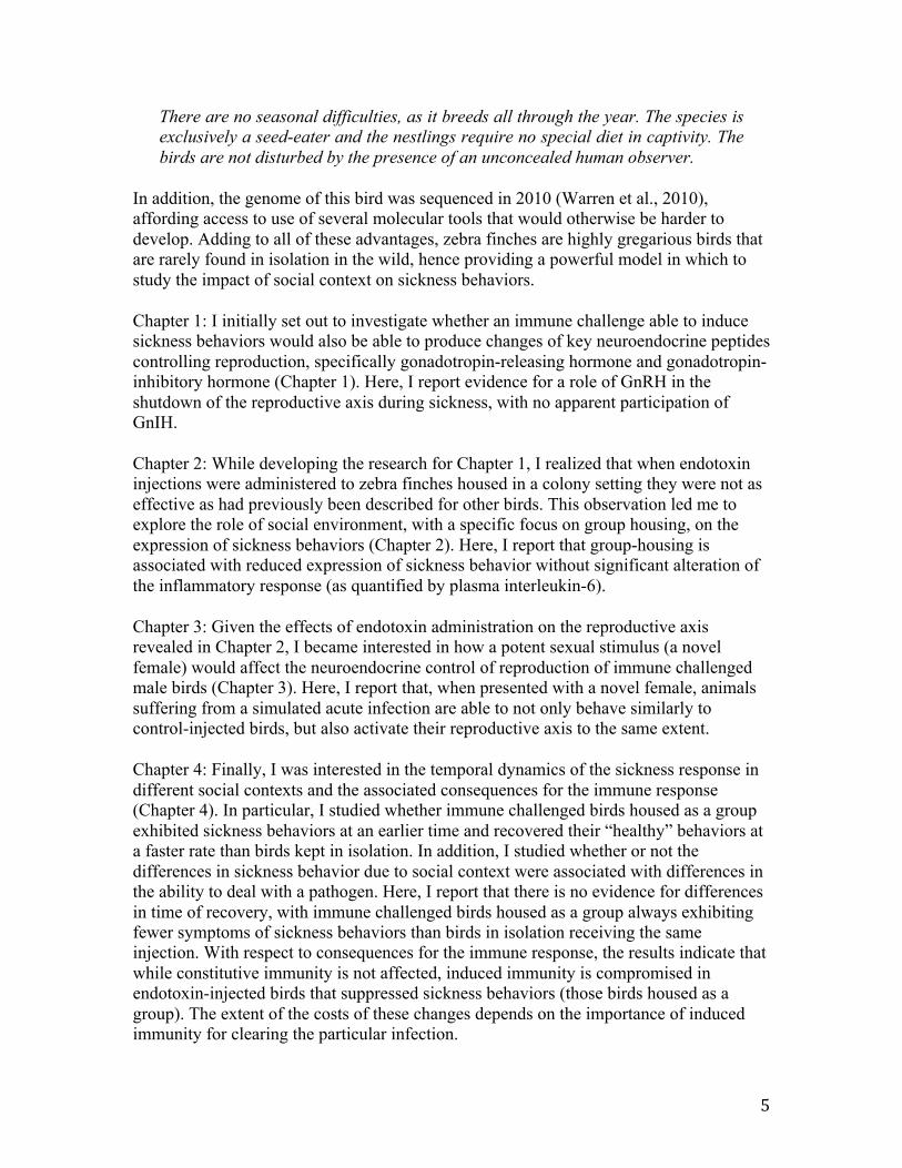
There are no seasonal difficulties, as it breeds all through the year. The species is exclusively a seed-eater and the nestlings require no special diet in captivity. The birds are not disturbed by the presence of an unconcealed human observer.
In addition, the genome of this bird was sequenced in 2010 (Warren et al., 2010), affording access to use of several molecular tools that would otherwise be harder to develop. Adding to all of these advantages, zebra finches are highly gregarious birds that are rarely found in isolation in the wild, hence providing a powerful model in which to study the impact of social context on sickness behaviors.
Chapter 1: I initially set out to investigate whether an immune challenge able to induce sickness behaviors would also be able to produce changes of key neuroendocrine peptides controlling reproduction, specifically gonadotropin-releasing hormone and gonadotropin-inhibitory hormone (Chapter 1). Here, I report evidence for a role of GnRH in the shutdown of the reproductive axis during sickness, with no apparent participation of GnIH.
Chapter 2: While developing the research for Chapter 1, I realized that when endotoxin injections were administered to zebra finches housed in a colony setting they were not as effective as had previously been described for other birds. This observation led me to explore the role of social environment, with a specific focus on group housing, on the expression of sickness behaviors (Chapter 2). Here, I report that group-housing is associated with reduced expression of sickness behavior without significant alteration of the inflammatory response (as quantified by plasma interleukin-6).
Chapter 3: Given the effects of endotoxin administration on the reproductive axis revealed in Chapter 2, I became interested in how a potent sexual stimulus (a novel female) would affect the neuroendocrine control of reproduction of immune challenged male birds (Chapter 3). Here, I report that, when presented with a novel female, animals suffering from a simulated acute infection are able to not only behave similarly to control-injected birds, but also activate their reproductive axis to the same extent.
Chapter 4: Finally, I was interested in the temporal dynamics of the sickness response in different social contexts and the associated consequences for the immune response (Chapter 4). In particular, I studied whether immune challenged birds housed as a group exhibited sickness behaviors at an earlier time and recovered their “healthy” behaviors at a faster rate than birds kept in isolation. In addition, I studied whether or not the differences in sickness behavior due to social context were associated with differences in the ability to deal with a pathogen. Here, I report that there is no evidence for differences in time of recovery, with immune challenged birds housed as a group always exhibiting fewer symptoms of sickness behaviors than birds in isolation receiving the same injection. With respect to consequences for the immune response, the results indicate that while constitutive immunity is not affected, induced immunity is compromised in endotoxin-injected birds that suppressed sickness behaviors (those birds housed as a group). The extent of the costs of these changes depends on the importance of induced immunity for clearing the particular infection.

6
Implications:
My research demonstrates the great plasticity of the sickness behavior response in face of social stimuli and the accompanying effects on the neuroendocrine control of reproduction and the immune system. The ability to modulate sickness according to the social context could prove adaptive in the sense that it should allow the animals to keep their social position in the group and keep mating opportunities open. At the same time, not giving the body the opportunity to fight the infection could have damaging effects on overall health. This creates an interesting trade-off: should animals invest in getting healthy or should they invest in appearing healthy? The findings provided herein are at the intersection of several fields (behavioral ecology, immunology, psychology, evolution, animal care) and should prove relevant in a world where infectious diseases cause a major burden in terms of lives lost and economic damage, by alerting for the ability of animals to modulate their sickness response. Since these findings demonstrate that animals can conceal sickness behavior in certain social circumstances, we should be aware of instances where we might not be able to identify sick animals. On the other hand, understanding how these effects are being mediated from an immunological and endocrine basis could possibly lead to tools to ameliorate sickness symptoms in captive animals and perhaps even in ourselves. Finally, social modulation of sickness behavior might provide a new paradigm for studying motivation underlying social behaviors (e.g. affiliation, parental care, etc).

7
References
Ashley, N.T., Wingfield, J.C., 2012. Sickness Behavior in Vertebrates: Allostasis, life history modulation and hormonal regulation. In: R.J. Nelson and G. Demas (Eds.), Ecoimmunology. Oxford University Press, London, pp. 45-91.
Aubert, A., Goodall, G., Dantzer, R., Gheusi, G., 1997. Differential effects of lipopolysaccharide on pup retrieving and nest building in lactating mice. Brain Behavior and Immunity 11, 107-118.
Avitsur, R., Sheridan, J.F., 2009. Neonatal stress modulates sickness behavior. Brain Behavior and Immunity 23, 977-85.
Avitsur, R., Yirmiya, R., 1999. The immunobiology of sexual behavior: Gender differences in the suppression of sexual activity during illness. Pharmacology Biochemistry and Behavior 64, 787-796.
Bouwman, K.M., Hawley, D.M., 2010. Sickness behaviour acting as an evolutionary trap? Male house finches preferentially feed near diseased conspecifics. Biology Letters 6, 462-465.
Cohn, D.W.H., de Sa-Rocha, L.C., 2006. Differential effects of lipopolysaccharide in the social behavior of dominant and submissive mice. Physiology & Behavior 87, 932-937.
Dantzer, R., 2004. Cytokine-induced sickness behaviour: a neuroimmune response to activation of innate immunity. European Journal of Pharmacology 500, 399-411.
Friedman, E.M., Reyes, T.M., Coe, C.L., 1996. Context-dependent behavioral effects of interleukin-1 in the rhesus monkey (Macaca mulatta). Psychoneuroendocrinology 21, 455-468.
Hart, B.L., 1988. Biological basis of the behavior of sick animals. Neuroscience and Biobehavioral Reviews 12, 123-137.
Hauber, M.E., Campbell, D.L.M., Woolley, S.M.N., 2010. The functional role and female perception of male song in Zebra Finches. Emu 110.
Hennessy, M.B., Deak, T., Schiml-Webb, P.A., Wilson, S.E., Greenlee, T.M., McCall, E., 2004. Responses of guinea pig pups during isolation in a novel environment may represent stress-induced sickness behaviors. Physiology & Behavior 81, 5-13.
Johnson, R.W., 2002. The concept of sickness behavior: a brief chronological account of four key discoveries. Veterinary Immunology and Immunopathology 87, 443-450.
Kent, S., Bluthe, R.M., Dantzer, R., Hardwick, A.J., Kelley, K.W., Rothwell, N.J., Vannice, J.L., 1992. Different receptor mechanisms mediate the pyrogenic and behavioral-effects of interleukin-1. Proceedings of the National Academy of Sciences of the United States of America 89, 9117-9120.

8
Miller, N.E., 1964. Some psychophysiological studies of motivation and of the behavioral-effects of illness. Bulletin of the British Psychological Society 17.
Morris, D., 1954. The reproductive behaviour of the zebra finch (Poephila guttata), with special reference to pseudofemale behaviour and displacement activities. Behaviour 6.
Owen-Ashley, N.T., Wingfield, J.C., 2006. Seasonal modulation of sickness behavior in free-living northwestern song sparrows (Melospiza melodia morphna). Journal of Experimental Biology 209, 3062-3070.
Sapolsky, R.M., 2005. The influence of social hierarchy on primate health. Science 308, 648-652.
Tuchscherer, M., Kanitz, E., Puppe, B., Tuchscherer, A., 2006. Early social isolation alters behavioral and physiological responses to an endotoxin challenge in piglets. Hormones and Behavior 50, 753-761.
Warren, W.C., Clayton, D.F., Ellegren, H., Arnold, A.P., Hillier, L.W., Kuenstner, A., Searle, S., White, S., Vilella, A.J., Fairley, S., Heger, A., Kong, L., Ponting, C.P., Jarvis, E.D., Mello, C.V., Minx, P., Lovell, P., Velho, T.A.F., Ferris, M., Balakrishnan, C.N., Sinha, S., Blatti, C., London, S.E., Li, Y., Lin, Y.-C., George, J., Sweedler, J., Southey, B., Gunaratne, P., Watson, M., Nam, K., Backstroem, N., Smeds, L., Nabholz, B., Itoh, Y., Whitney, O., Pfenning, A.R., Howard, J., Voelker, M., Skinner, B.M., Griffin, D.K., Ye, L., McLaren, W.M., Flicek, P., Quesada, V., Velasco, G., Lopez-Otin, C., Puente, X.S., Olender, T., Lancet, D., Smit, A.F.A., Hubley, R., Konkel, M.K., Walker, J.A., Batzer, M.A., Gu, W., Pollock, D.D., Chen, L., Cheng, Z., Eichler, E.E., Stapley, J., Slate, J., Ekblom, R., Birkhead, T., Burke, T., Burt, D., Scharff, C., Adam, I., Richard, H., Sultan, M., Soldatov, A., Lehrach, H., Edwards, S.V., Yang, S.-P., Li, X., Graves, T., Fulton, L., Nelson, J., Chinwalla, A., Hou, S., Mardis, E.R., Wilson, R.K., 2010. The genome of a songbird. Nature 464, 757-62.
Weil, Z.M., Bowers, S.L., Pyter, L.M., Nelson, R.J., 2006. Social interactions alter proinflammatory cytokine gene expression and behavior following endotoxin administration. Brain Behavior and Immunity 20, 72-79.
Weil, Z.M., Nelson, R.J., 2012. Sickness behaviors in vertebrates. In Demas, G.E. & Nelson, R.J. (Eds.), Ecoimmunology. Oxford University Press, London, pp.297-325.
Willette, A.A., Lubach, G.R., Coe, C.L., 2007. Environmental context differentially affects behavioral, leukocyte, cortisol, and interleukin-6 responses to low doses of endotoxin in the rhesus monkey. Brain Behavior and Immunity 21, 807-815.
Yirmiya, R., Avitsur, R., Donchin, O., Cohen, E., 1995. Interleukin-1 inhibits sexual-behavior in female but not in male-rats. Brain Behavior and Immunity 9, 220-233.
Zann R., 1996. The Zebra Finch: A Synthesis of Field and Laboratory Studies. Oxford University Press: Oxford, UK.

9
Chapter 1
Lipopolysaccharide injection induces rapid decrease of hypothalamic GnRH mRNA and peptide, but does not affect GnIH in zebra finches

10
Abstract
Lipopolysaccharide (LPS) is frequently used experimentally to mimic acute infection. Through activation of the host’s immune response, an LPS injection has profound effects on the adrenocortical response to stress and on behaviors including reduction in activity, water and food intake, and libido. These behavioral changes occurring during infection are collectively called “sickness behavior.” It is thought that adoption of sickness behavior reallocates energy from other fitness-enhancing activities, such as reproduction, for use in the immune response. Although the behavioral effects of LPS treatment are well-known, less information is available regarding the effects of LPS on the brain in terms of controlling reproductive behavior, specifically concerning a newly discovered neuropeptide, gonadotropin-inhibitory hormone (GnIH). This study investigated the effects of an LPS injection on the behavior and the hypothalamic neuropeptides controlling reproduction [GnIH and gonadotroping-releasing hormone (GnRH)] of zebra finches (Taeniopygia guttata). Overall, there was a decrease in activity in birds injected with LPS. The number of GnRH-immunoreactive neurons was significantly reduced in birds injected with LPS when compared to controls, while the number of GnIH-releasing neurons remained unchanged. At the level of gene expression, a similar pattern was found: there was reduced expression of GnRH mRNA in LPS-injected animals, whereas GnIH expression remained unchanged. Plasma testosterone did not change significantly in LPS-injected animals, nor did plasma corticosterone. Taken together, these results indicate a rapid (within 3h) inhibition of the reproductive axis during an immune challenge mimicking an infection, specifically acting on the GnRH system. The present study expands our knowledge on the interaction between the immune system and the reproductive system.

11
Introduction
Animals possess a finite amount of energy to distribute to their daily activities (such as foraging, growing, reproducing, immune function). Thus, if any given activity must be prioritized energetically, there will be less energy available for all others. For example, it has been hypothesized that animals experiencing an infection will save energy for the immune response by reducing their overall activity. These “sickness behaviors” include lethargy, apathy, anorexia (decreased food consumption), and adipsia (decreased fluid intake) (Hart 1988). Such responses often occur simultaneously with the suppression of other functions, e.g. reproduction, that are not essential for immediate individual survival (Yirmiya et al., 1995; Bonneaud et al., 2003; Owen-Ashley and Wingfield, 2006; Ashley and Wingfield, 2012).
In vertebrates, reproduction is regulated by the hypothalamic pituitary gonadal (HPG) axis. In males, the pulsatile release of the hypothalamic peptide gonadotropin-releasing hormone-I (GnRH-I) stimulates the pituitary gland, causing synthesis and release of the gonadotropins (luteinizing hormone (LH) and follicle stimulating hormone (FSH)). LH travels through the blood stream, ultimately causing testosterone synthesis and secretion from the testis. Testosterone has a multitude of behavioral effects in adult birds, including activation of sexual behavior, mate attraction and mate guarding (Wingfield et al., 1990; Hau 2007). Another neuropeptide, gonadotropin-inhibitory hormone (GnIH), acts within the hypothalamus, pituitary gland and gonads and has an overall suppressive effect on the HPG axis (for a review see Tsutsui et al., 2012). In addition to its neuroendocrine effects, GnIH has been linked to inhibition of reproductive behavior (birds: Bentley et al., 2006; mammals: Johnson et al., 2007). GnIH is also a potential mediator of energy balance (Johnson et al., 2007, Qi et al., 2009, Clarke et al., 2009, Tachibana et al., 2005). To our knowledge, GnIH has never been explored in the context of sickness behavior.
Lipopolysacharide (LPS), a component of gram-negative bacterial cell wall, is routinely administered experimentally to induce sickness behavior in animals, and it appears to disrupt gonadotropic functions in several mammals (for a review, see: Tomaszewska-Zaremba and Herman 2009). Most of the effects of endotoxin challenge on the reproductive system have been explored in females. For example, in ewes, exposure to an endotoxin causes: suppression of the pulsatile release of GnRH and LH secretion (Harris et al., 2000), inhibition of pituitary responsiveness to GnRH (Williams et al., 2001), disruption of the follicular phase (Battaglia et al., 2000), and disruption of cyclicity and induction of preterm labor (Schlafer et al., 1994). Also in female rats and monkeys, LH release is suppressed by endotoxin injection (rats: He et al., 2003; Iwasa et al., 2008; Watanobe and Hayakawa, 2003; and monkeys: Xiao et al., 2000). The work by He and colleagues (2003) suggests that the inhibitory effect of LPS on the HPG axis occurs upstream of the pituitary, because an intravenous injection of GnRH still induces LH release in LPS-injected female rats. Males are not well studied in the context of the effects of an LPS injection on the HPG axis. Nonetheless, castrate rats exhibit reduced levels of LH after receiving an injection or an implant of LPS (Refojo et al., 1998, Ebisui et al., 1992, Rivest and Rivier, 1993; Rivier, 1990). Only two studies report the effect of an LPS injection on LH levels in birds, but the pattern appears to be similar to what is

12
observed in mammals. Twenty-four hours after exposure to an endotoxin challenge, male and female white crowned-sparrows, Zonotrichia leucophrys, demonstrated low circulating LH levels (Owen-Ashley at al., 2006). Song sparrows (Melospiza melodia) have their LH levels reduced at 6h after an LPS injection, but LH levels are no different from control-injected birds at 22h post-injection (Adelman et al., 2010). To our knowledge, the brain neuropeptides controlling reproduction have never been explored in birds subjected to an endotoxin challenge.
Testosterone can have a suppressive effect on sickness behavior in male birds (Ashley et al., 2009). Hence, at least one component of the HPG axis interferes with the avian sickness response. Due to this intriguing relationship between testosterone and sickness behavior and our limited knowledge of how the immune system affects the reproductive system, especially in non-mammalian species, the present work was aimed at studying the effect of LPS administration on neuroendocrine components of the reproductive axis in male birds.
We predicted that males experiencing infections and exhibiting sickness behaviors would down-regulate their reproductive axis by increasing levels of GnIH mRNA and peptide, indicating an important role of GnIH in mediating the communication between immune and reproductive status. Additionally, we predicted that GnRH mRNA and peptide levels would be reduced, in similarity to what is found in mammalian females. We also predicted a decrease in circulating testosterone levels after LPS injection as a consequence. Because activation of the hypothalamic-pituitary-adrenal (HPA) axis is frequently seen upon an endotoxin challenge, an increase in plasma corticosterone was expected.
This study explores for the first time the effects of an endotoxin challenge on the main hypothalamic sites controlling reproductive function in male birds and specifically tries to generate new insights into the role GnIH might play during the course of an infection.

13
Methods
Animals and experimental design
The experiment was carried out in two phases (Phase I – April 2009 and Phase II – May 2011) at the University of California, Berkeley Field Station for the Study of Behavior, Ecology and Reproduction. All procedures were approved by and in compliance with the University of California Office of Lab Animal Care and federal regulations.
A colony of zebra finches (Taeniopygia guttata), including adult males, females and juveniles was housed in a 2.7 m by 2.5 m by 2.1 m indoor flight aviary. They were exposed to natural changes in day length, supplemented by artificial lighting at a light/dark schedule of 12L:12D. Food and water were provided ad libitum and consisted of German millet mixed with canary seed. All birds in our colony are uniquely color banded.
To facilitate individual identification within the colony, on the day prior to the experiment, twelve male zebra finches received randomized color markings on their chests using marker pens and were returned to the colony. The color used was the same for all (blue), with a different number of dots on the chest.
On the day of the experiment, the 12 male zebra finches were injected in the pectoral muscle with a sterile solution of either 100µL of LPS 0.3mg mL-1 (Sigma-Aldrich #L4005, Serotype 055:B5) or 100µL of 10mmol-1 phosphate buffer saline (pH 7.2). The region to be injected was sterilized with ethanol, which was allowed to dry before injecting the animal. The dose of LPS was ca. 2mg/kg of body weight. This dose is higher to what has been used previously in experiments with passerines. For example, Owen-Ashley et al. (2006) and Burness et al. (2010) used a dose of 1mg/kg of body weight in white-crowned sparrows and zebra finches, respectively. When we tested both this dose and 2mg/kg, the latter dose seemed to induce greater behavioral response (personal observation). Animals were randomly assigned to the injection treatment.
Behavior
Behavior was recorded two hours after the injection, by direct observation. Two observers that were naïve to the treatment stood outside of the aviary. Observers were instructed to count the number of hops, flights, calls and songs, within a five minute period. After the initial scoring, another five minutes were dedicated to observing the time the birds spent resting. Then, the observers moved on to the next bird. Thus, each bird was observed for a total of ten minutes and all birds were observed within the same sixty-minute period.
Approximately three hours after the injection, the birds were captured using butterfly nets, deeply anesthetized via isoflurane inhalation and decapitated. The brain was immediately removed and placed on dry ice and trunk blood was stored on regular ice. The blood was then centrifuged at 1500 g for ten minutes and the plasma portion was

14
placed into separate tubes. All tissues were maintained at -80oC until further analysis. Time from entering the aviary until euthanizing birds was on average 11.7 min (±S.E.M.: 2.6 min) for control-injected and 12.9 min (± S.E.M.: 2.8 min) for LPS-injected birds. This time is not significantly different between treatments (t(10)=0.702, P=0.499).
Immunohistochemistry (IHC)
Using a cryostat, 20µm coronal sections of the brains collected in Phase I were cut and every fifth slice was placed directly onto slides. A hydrophobic barrier was created around the slices on the slide, by the use of a PAP pen (Sigma-Aldrich # Z377821). The brain sections were then fixed using a 4% paraformaldehyde solution for one hour. The slides were then washed three times in phosphate buffered saline (PBS, 10mM, pH 7.2) and exposed to a 1% solution of hydrogen peroxide in PBS for thirty minutes. A new wash in PBS for five minutes was repeated three times, after which 2% normal goat serum (NGS) in 0.2% PBS-Triton (PBS-T) was added for one hour. Subsequently, GnRH primary antibody (HU60, gift from Dr. Henryk Urbanski, Portland, OR, USA) at a concentration of 1:5000 in 0.2% PBS-T was added and allowed to incubate at room temperature (r.t.) for one hour and subsequently for 48 hours at 4oC. The slides were then washed three times in 0.2% PBS-T, followed by incubation in 1:250 biotinylated goat anti-rabbit IgG (Vector Labs #BA-1000) in 0.2% PBS-T for one hour and an additional three washes in 0.2% PBS-T. After a one hour incubation in avidin-biotin complex (ABC, Vectastain Elite Kit, Vector Labs #PK6100), visualization was achieved by adding 0.03% 3,3’-diaminobenzine (DAB) for eight minutes.
The protocol for the labeling of GnIH (primary antiserum PAC 123, 124, Bentley, Berkeley, CA, USA, used at dilution 1:5000 in 0.2% PBS-T) was the same as for GnRH, with the difference that no r.t. incubation in primary antibody was done. Slides containing adjacent brain slices were used for each of these two antibodies. Successful use of these antibodies has been demonstrated previously in zebra finch brains using a similar protocol (Perfito et al., 2011).
Quantification of GnRH and GnIH immunoreactivity
Photographs were taken of the areas showing immunoreactivity using a Zeiss Axio Imager A1 microscope and AxioVision 4.5 software. Because GnRH-I and GnIH neurons occur in restricted areas of the brain (preoptic area and paraventricular nucleus, respectively), and because we ran the IHC on adjacent sections for each neuropeptide, we were easily able to count the number of GnRH-I-immunoreactive(-ir) neurons and the number of GnIH-ir neurons. The experimenter was blind to the injection treatment corresponding to the slides.
Gene expression
RNA isolation, purification and reverse-transcription
The brains collected in Phase II were cut using a cryostat and 20µm coronal sections were

15
either placed directly onto slides or collected into a tube containing 1mL PureZOL reagent (Bio-Rad Laboratories, Hercules, CA). In other words, for every section that was collected for histology, its adjacent slice was collected for RNA extraction. Two separate samples were collected from each brain: the first one consisted of the portion of the brain starting from when the tractus septomesencephalicus (TrSM) becomes visible until the disappearance of the anterior commissure (CoA); the second one consisted of the rest of the sections from that point until the disappearance of the optic tectum (TeO). Using this protocol, we avoided amplification of GnRH-II during the qRT-PCR procedure, which could be a confounding factor when analyzing GnRH-I data. We verified histologically that no GnRH-II cell bodies were detected in the part of the brain collected in the first sample. This was accomplished by immunolabeling slides using GnRH primary antibody, as described above and inspecting slides adjacent to the last sections collected in the first sample tube for the presence of GnRH cell bodies. The primary antibody used also binds GnRH-II peptide. Each brain sub-sample was homogenized and immediately stored at -80oC until extraction. Total RNA extraction was performed according to manufacturer’s instructions, with final dilution of RNA in 20µL of DEPC-treated water. Quantification of RNA was done via spectrophotometry (NanoDrop 2000). The RNA was then treated for any genomic DNA contamination (DNA free, Ambion) and 33 ng of RNA from each sample was reverse-transcribed to cDNA using iScript reverse transcriptase with 5X iScript reaction mix (Bio-Rad Laboratories, Hercules, CA). The cDNA was diluted 1:25, as this dilution was optimal for the genes being amplified.
Quantitative real-time PCR (qRT-PCR)
Primers for GnRH and GnIH were designed based on the published sequences for zebra finches (respectively, GenBank ID: NM_001142320.1 and GenBank ID: AB522971.1) and 18S (control gene) primers were designed based on the published rat sequence (GenBank ID: NR_046237.1). Primer sequences were: GnRH-F: ACTCCACAACCTCTCTCAGG; GnRH-R: CTCTGCTGCTCCTCCTCTAA; GnIH-F: CCCTGAGATTTGGAAGAGC; GnIH-R: CAGATTGACAGGCAGTGAC; 18S-F: CCATCCAATCGGTAGTAGCG; 18S-R: GTAACCCGTTGAACCCCATT. Amplification of primer-dimer was controlled for by running template-free controls. These samples always resulted in differences of at least 10 cycles of the Ct values compared to samples containing template. The qRT-PCR was performed in duplicate for each bird for each gene in 25µL reactions according to manufacturer’s instructions for 2x iQTM SyBR Green Supermix (Bio-Rad Laboratories, Hercules, CA). After checking for primer specificity by visual inspection of the melting curves, we used the RT-PCR Miner program (Zhao and Fernald 2005) for analysis of the raw fluorescent data. Gene expression was calculated using the following formula: 1/(1+E)Ct, where E is the average PCR efficiency and Ct is the cycle threshold. The reference gene used to normalize mRNA levels among samples was 18S, after verification that treatment did not affect its levels. Normalization was done for each individual by dividing expression values for the gene of interest by expression values of 18S.

16
RIA
Corticosterone
To measure plasma corticosterone concentrations, we followed the protocol of Wingfield et al. (1992). Plasma volume of samples was 20µL All samples were run in a single assay. The detection limit of the standard curve was 7.5pg, and intra-assay variation was 6.3%.
Testosterone
Testosterone assay was performed following Wingfield et al. (1991). Plasma volume of samples was 35 µL. The detection limit of the assay was 3.9pg. Intra-assay variation was 7.1%.
Statistical analysis
Statistical analyses were performed using JMP v.9. Data collected in Phase I of experiment consisted of behavior, immunohistochemistry data and corticosterone concentrations. Data collected during Phase II consisted of behavior, immunohistochemistry, gene expression and testosterone concentrations. Behaviors and counts of cell-ir from Phase I and Phase II were analyzed together, after verification that animals receiving the same injection did not differ between Phase I and II. The only behavior affected by Phase was preening (only in control-injected birds) and this behavior was thus removed from our analysis. Hence, for behaviors and cell-ir, N=12 for each treatment and for the rest of the parameters analyzed, N=6 for each treatment, except when noted in the text. Data that were not normally distributed and for which transformation did not resolve this issue were analyzed using non-parametric tests (Wilcoxon rank sums test). These include all behavioral data and the counts of ir-cell bodies. Gene expression data and hormone plasma concentrations were log-transformed prior to analysis and analyzed using Student’s t-test. For purposes of graphical representation of the gene expression results, the following procedure was used: after calculating the ratio relative to the housekeeping gene (normalization) we divided the mean for each treatment group by the mean of the control treatment. In this way, the control treatment is set at 1 (or 100% expression) and any variation on the LPS treatment is represented as a ratio of that. All other data are represented as means ± SEM. All tests were two-tailed and probability values of P<0.05 were considered to be statistically significant.
17
Results
Behavior
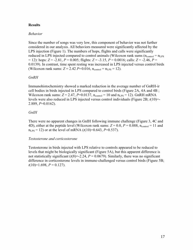
Since the number of songs was very low, this component of behavior was not further considered in our analysis. All behaviors measured were significantly affected by the LPS injection (Figure 1). The numbers of hops, flights and calls were significantly reduced in LPS injected compared to control animals (Wilcoxon rank sums (ncontrol = nLPS = 12): hops: Z = -2.81, P = 0.005; flights: Z = -3.15, P = 0.0016; calls: Z = -2.46, P = 0.0139). In contrast, time spent resting was increased in LPS injected versus control birds (Wilcoxon rank sums: Z = 2.42 P=0.016, ncontrol = nLPS = 12).
GnRH
Immunohistochemistry showed a marked reduction in the average number of GnRH-ir cell bodies in birds injected in LPS compared to control birds (Figure 2A, 4A and 4B ; Wilcoxon rank sums: Z = 2.47, P=0.0137, ncontrol = 10 and nLPS = 12). GnRH mRNA levels were also reduced in LPS injected versus control individuals (Figure 2B; t(10)=-2.889, P=0.0162).
GnIH
There were no apparent changes in GnIH following immune challenge (Figure 3, 4C and 4D), either at the peptide level (Wilcoxon rank sums: Z = 0.0, P = 0.888, ncontrol = 11 and nLPS = 12) or at the level of mRNA (t(10)=0.643, P=0.537).
Testosterone and corticosterone
Testosterone in birds injected with LPS relative to controls appeared to be reduced to levels that might be biologically significant (Figure 5A), but this apparent difference is not statistically significant (t(8)=-2.24, P = 0.0679). Similarly, there was no significant difference in corticosterone levels in immune-challenged versus control birds (Figure 5B; t(10)=1.698, P = 0.127).

18
Discussion
Treatment with LPS induced stereotypical sickness behaviors, with reduction in numbers of hops, flights and calls, and a simultaneous increase in the time spent resting. This finding is similar to that previously demonstrated in zebra finches (Burness et al., 2010), as well as in other bird species (house sparrow: Bonneaud et al., 2003, white-crowned sparrow: Owen-Ashley et al., 2006; chicken: Johnson et al., 1993).
As predicted, both gene expression and peptide data indicated that GnRH synthesis in the brain was down-regulated in response to LPS injection. These results corroborate previous studies showing down-regulation of GnRH mRNA levels in the brains of female mammals upon LPS challenge (rats: Nappi and Rivest (1997) and sheep: Herman and Tomaszewska-Zaremba (2010)). Our data provide the first assessment of the role of GnIH in the inhibition of the HPG axis in response to endotoxin challenge in birds. Because GnIH expression and protein content in cells were not affected by LPS injections, it appears that the effect of LPS might be somewhat specific to GnRH and the role of GnIH in mediating communication between the immune and reproductive system may be limited or absent. This finding is surprising, given the suggested role of GnIH in fine-tuning reproduction and its directly inhibitory effects upon the GnRH system in birds and mammals (reviewed in Kriegsfeld et al., 2010).
A decline in circulating testosterone would suggest that LPS injections reduced activity of the HPG axis as a whole. Our data showed no statistically significant differences in circulating testosterone at the time of sampling although inspection of the results (Figure 5A) suggests that LPS treatment may have exerted a biological effect on circulating testosterone. Several previous reports show an impact of LPS injection on LH. For example, LH release is suppressed by endotoxin injection in both female (He et al., 2003; Iwasa et al., 2008; Watanobe and Hayakawa, 2003) and castrate male rats (Refojo et al., 1998, Ebisui et al., 1992, Rivest and Rivier, 1993; Rivier, 1990). The two studies where LH concentrations were measured in birds receiving an LPS injection indicate that LH levels are reduced at 24h post-injection in white crowned-sparrows (Owen-Ashley at al., 2006) but not in song sparrows, where levels measured at 22h post-injection are not different from control (Adelman et al., 2010). In song sparrows, a difference between Control and LPS-injected birds is found only at 6h after the injection. Unfortunately, these two studies did not measure testosterone concentrations. The lack of statistical significance may be a result of the timing of sampling following LPS injection. We expected to see an increase in circulating corticosterone because immune challenges typically activate the HPA axis (reviewed in Beishuizen and Thijs 2003). Contrary to our prediction, there was no difference from control animals, even though corticosterone appears to be slightly increased in LPS-injected animals. Again, this might be a matter of the timing of sampling. However, in white-crowned sparrows, although the peak of the corticosterone response occurs at 1h after an LPS injection, at 3h after the injection corticosterone is still elevated (Owen-Ashley et al. 2006). Even at 6h, corticosterone in LPS-injected male white-crowned sparrows is higher than their control injected counterparts. Data from a different experiment in our lab employing male zebra finches indicates that at 5h after an LPS injection, there is no increase in corticosterone

19
(unpublished data). It is hard to draw comparisons between our results and those of Owen-Ashley et al. (2006), since we do not know how the corticosterone response dynamics might differ in a captive-bred species versus a wild caught species. It is also possible that the observed shutdown of the reproductive axis may be independent of any effect on corticosterone. Rivest and Rivier (1995) have previously suggested that the effects of IL-1 on the reproductive axis can occur independently of the activation of the HPA axis.
A wide range of environmental cues and endogenous factors influence the activity of the HPG axis. One of these stimuli is immune challenge, which is usually linked to suppression of this axis. Our study shows that an intramuscular injection of LPS is able to inhibit the HPG axis of male zebra finches at the level of the hypothalamus and, possibly, at the level of gonadal hormonal secretion. The suppressive effect of LPS on males has been explored primarily at the level of the anterior pituitary gland by quantification of gonadotropin release. For example, in both male rats and sheep, LPS administration led to reduced plasma LH (Refojo et al., 1998 and Coleman et al., 1993, respectively). In these animals, immune challenge disrupted the pulsatile release of GnRH, thereby affecting gonadotropin release and downstream processes. Even though alterations of circulating LH following LPS challenge have been observed in several species, our understanding of the hypothalamic components that affect these changes has been more limited. Nappi and Rivest (1997) used in situ hybridization to show that female rats have reduced GnRH transcripts in the hypothalamus during proestrus when injected with LPS. Herman and Tomaszewska-Zaremba (2010) showed a similar effect on anestrous ewes: an LPS challenge reduced the amount of GnRH mRNA in the preoptic area. This time, the researchers used real-time PCR for the quantification of gene expression. These results match our findings of decreased hypothalamic GnRH expression in male birds. Additionally, our study showed a decreased number of neurons immunoreactive for GnRH, indicating that GnRH peptide was also reduced.
GnIH has been an unexplored hypothalamic component of the reproductive axis in the context of sickness behavior. The actions of GnIH are inhibitory at several levels of the reproductive axis. In addition to its regulatory function in reproduction, GnIH has been suggested to play a role in the control of energy balance (Johnson et al., 2007, Qi et al., 2009, Clarke et al., 2009, Tachibana et al., 2005). As a result, we hypothesized that GnIH might also be involved in the response of the HPG axis to immune challenge. Specifically, we predicted that GnIH would rapidly respond to an immune challenge by being up-regulated and thereby inhibit the GnRH system. Contrary to our prediction, we saw no changes in GnIH during an immune challenge in male birds. GnIH levels were similar in sick and control birds, indicating a passive role for GnIH in the response to an infection. Thus there is likely to be an alternative route for the interaction between immune and reproductive systems.
The present work adds to the comparative understanding of how sickness affects reproduction by exploring the effects of an immune challenge on the reproductive axis of a songbird species. Overall, it appears that the reproductive axis responds to perceived infection very rapidly (within three hours post injection), and that, in this context, GnIH

20
does not influence the activity of the GnRH system. The exact mechanism(s) by which the immune system inhibits the GnRH system has yet to be elucidated.
Acknowledgements
This work was published in the Journal Hormones and Behavior (2012, vol. 62: 173-179) and co-authored by John C. Wingfield and George E. Bentley. I would like to thank the following sources of funding: Ministério para Ciência, Tecnologia e Ensino Superior (MCTES-Lisbon, Portugal) for financial support through doctoral grant [SFRH/BD/33251/2007] awarded to me and support from the National Science Foundation [0920753 and 0956338 to G.E.B.; and IOS-0750540 to J.C.W.]. Additionally, I am grateful for the support provided by the staff at the Field Station for the Study Behavior, Ecology and Reproduction, Sarah Fong for help with behavioral observations, and Tyrone Hayes and Eileen Lacey for comments on the manuscript.

21
References
Adelman, J.S., Bentley, G.E., Wingfield, J.C., Martin, L.B., Hau, M., 2010. Population differences in fever and sickness behaviors in a wild passerine: a role for cytokines. Journal of Experimental Biology 213, 4099-4109.
Ashley, N.T., Hays, Q.R., Bentley, G.E., Wingfield, J.C., 2009. Testosterone treatment diminishes sickness behavior in male songbirds. Hormones and Behavior 56, 169-176.
Ashley, N.T., Wingfield, J.C., 2012. Sickness Behavior in Vertebrates: Allostasis, life history modulation and hormonal regulation. In: R.J. Nelson and G. Demas (Eds.), Ecoimmunology. Oxford University Press, London, pp. 45-91.
Battaglia, D.F., Krasa, H.B., Padmanabhan, V., Viguie, C., Karsch, F.J., 2000. Endocrine alterations that underlie endotoxin-induced disruption of the follicular phase in ewes. Biology of Reproduction 62, 45-53.
Beishuizen, A., Thijs, L.G., 2003. Endotoxin and the hypothalamo-pituitary-adrenal (HPA) axis. Journal of Endotoxin Research 9, 3-24.
Bentley, G.E., Jensen, J.P., Kaur, G.J., Wacker, D.W., Tsutsui, K., Wingfield, J.C., 2006. Rapid inhibition of female sexual behavior by gonadotropin-inhibitory hormone (GnIH). Hormones and Behavior 49, 550-555.
Bonneaud, C., Mazuc, J., Gonzalez, G., Haussy, C., Chastel, O., Faivre, B., Sorci, G., 2003. Assessing the cost of mounting an immune response. American Naturalist 161, 367-379.
Burness, G., Armstrong, C., Fee, T., Tilman-Schindel, E., 2010. Is there an energetic-based trade-off between thermoregulation and the acute phase response in zebra finches? Journal of Experimental Biology 213, 1386-1394.
Clarke, I.J., Qi, Y., Sari, I.P., Smith, J.T., 2009. Evidence that RF-amide related peptides are inhibitors of reproduction in mammals. Frontiers in Neuroendocrinology 30, 371-378.
Coleman, E.S., Elsasser, T.H., Kemppainen, R.J., Coleman, D.A., Sartin, J.L., 1993. Effect of endotoxin on pituitary-hormone secretion in sheep. Neuroendocrinology 58, 111-122.
Ebisui, O., Fukata, J., Tominaga, T., Murakami, N., Kobayashi, H., Segawa, H., Muro, S., Naito, Y., Nakai, Y., Masui, Y., Nishida, T., Imura, H., 1992. Roles of interleukin-1-alpha and interleukin-1-beta in endotoxin-induced suppression of plasma gonadotropin-levels in rats. Endocrinology 130, 3307-3313.
Harris, T.G., Battaglia, D.F., Brown, M.E., Brown, M.B., Carlson, N.E., Viguie, C., Williams, C.Y., Karsch, F.J., 2000. Prostaglandins mediate the endotoxin-induced

22
suppression of pulsatile gonadotropin-releasing hormone and luteinizing hormone secretion in the ewe. Endocrinology 141, 1050-1058.
Hart, B.L., 1988. Biological basis of the behavior of sick animals. Neuroscience and Biobehavioral Reviews 12, 123-137.
Hau, M., 2007. Regulation of male traits by testosterone: implications for the evolution of vertebrate life histories. Bioessays 29, 133-44.
He, D., Sato, I., Kimura, F., Akema, T., 2003. Lipopolysaccharide inhibits luteinizing hormone release through interaction with opioid and excitatory amino acid inputs to gonadotropin-releasing hormone neurones in female rats: Possible evidence for a common mechanism involved in infection and immobilization stress. Journal of Neuroendocrinology 15, 559-563.
Herman, A.P., Tomaszewska-Zaremba, D., 2010. Effect of endotoxin on the expression of GnRH and GnRHR genes in the hypothalamus and anterior pituitary gland of anestrous ewes. Animal Reproduction Science 120, 105-111.
Iwasa, T., Matsuzaki, T., Murakami, M., Shimizu, F., Kuwahara, A., Yasui, T., Irahara, M., 2008. Decreased expression of kisspeptin mediates acute immune/inflammatory stress-induced suppression of gonadotropin secretion in female rat. Journal of Endocrinological Investigation 31, 656-659.
Johnson, R.W., Curtis, S.E., Dantzer, R., Bahr, J.M., Kelley, K.W., 1993. Sickness behavior in birds caused by peripheral or central injection of endotoxin. Physiology & Behavior 53, 343-348.
Kriegsfeld, L.J., Gibson, E.M., Williams, W.P., III, Zhao, S., Mason, A.O., Bentley, G.E., Tsutsui, K., 2010. The Roles of RFamide-Related Peptide-3 in Mammalian Reproductive Function and Behaviour. Journal of Neuroendocrinology 22, 692-700.
Nappi, R.E., Rivest, S., 1997. Effect of immune and metabolic challenges on the luteinizing hormone-releasing hormone neuronal system in cycling female rats: An evaluation at the transcriptional level. Endocrinology 138, 1374-1384.
Owen-Ashley, N.T., Turner, M., Hahn, T.P., Wingfield, J.C., 2006. Hormonal, behavioral, and thermoregulatory responses to bacterial lipopolysaccharide in captive and free-living white-crowned sparrows (Zonotrichia leucophrys gambelii). Hormones and Behavior 49, 15-29.
Owen-Ashley, N.T., Wingfield, J.C., 2006. Seasonal modulation of sickness behavior in free-living northwestern song sparrows (Melospiza melodia morphna). Journal of Experimental Biology 209, 3062-3070.
23
Perfito, N., Zann, R., Ubuka, T., Bentley, G., Hau, M., 2011. Potential roles for GNIH and GNRH-II in reproductive axis regulation of an opportunistically breeding songbird. General and Comparative Endocrinology 173, 20-26.
Qi, Y., Oldfield, B.J., Clarke, I.J., 2009. Projections of RFamide-related Peptide-3 Neurones in the Ovine Hypothalamus, with Special Reference to Regions Regulating Energy Balance and Reproduction. Journal of Neuroendocrinology 21, 690-697.
Refojo, D., Arias, P., Moguilevsky, J.A., Feleder, C., 1998. Effect of bacterial endotoxin on in vivo pulsatile gonadotropin secretion in adult male rats. Neuroendocrinology 67, 275-281.
Rivest, S., Rivier, C., 1993. Centrally injected interleukin-1-beta inhibits the hypothalamic lhrh secretion and circulating lh levels via prostaglandins in rats. Journal of Neuroendocrinology 5, 445-450.
Rivest, S., Rivier, C., 1995. The role of corticotropin-releasing factor and interleukin-i in the regulation of neurons controlling reproductive functions. Endocrine Reviews. 16, 177-199.
Rivier, C., 1990. Role of endotoxin and interleukin-1 in modulating ACTH, LH and sex steroid secretion. Advances in Experimental Medicine and Biology. 274, 295–301.
Schlafer, D.H., Yuh, B., Foley, G.L., Elssaser, T.H., Sadowsky, D., Nathanielsz, P.W., 1994. Effect of salmonella endotoxin administered to the pregnant sheep at 133-142 days gestation on fetal oxygenation, maternal and fetal adrenocorticotropic hormone and cortisol, and maternal plasma tumor-necrosis-factor-alpha concentrations. Biology of Reproduction 50, 1297-1302.
Tachibana, T., Sato, M., Takahashi, H., Ukena, K., Tsutsui, K., Furuse, M., 2005. Gonadotropin-inhibiting hormone stimulates feeding behavior in chicks. Brain Research 1050, 94-100.
Tomaszewska-Zaremba, D., Herman, A., 2009. The role of immunological system in the regulation of gonadoliberin and gonadotropin secretion. Reproductive Biology 9, 11-23.
Tsutsui, K., Ubuka, T., Bentley, G.E., Kriegsfeld, L.J., 2012. Gonadotropin-inhibitory hormone (GnIH): Discovery, progress and prospect. General and Comparative Endocrinology 177, 305-14.
Watanobe, H., Hayakawa, Y., 2003. Hypothalamic interleukin-1 beta and tumor necrosis factor-alpha, but not interleukin-6, mediate the endotoxin-induced suppression of the reproductive axis in rats. Endocrinology 144, 4868-4875.
24
Williams, C.Y., Harris, T.G., Battaglia, D.F., Viguie, C., Karsch, F.J., 2001. Endotoxin inhibits pituitary responsiveness to gonadotropin-releasing hormone. Endocrinology 142, 1915-1922.
Wingfield, J.C., Hegner, R.E., Dufty, A.M., Ball, G.F., 1990. The challenge hypothesis - theoretical implications for patterns of testosterone secretion, mating systems, and breeding strategies. American Naturalist 136, 829-846.
Wingfield, J.C., Hegner, R.E., Lewis, D.M., 1991. Circulating levels of luteinizing hormone and steroid hormones in relation to social status in the cooperatively breeding white-browed sparrow weaver, Plocepasser mahali. Journal of Zoology 225, 43-58.
Wingfield, J.C., Hegner, R.E., Lewis, D.M., 1992. Hormonal responses to removal of a breeding male in the cooperatively breeding white-browed sparrow weaver, Plocepasse mahali. Hormones and Behavior 26, 145-155.
Xiao, E., Xia-Zhang, L., Ferin, M., 2000. Inhibitory effects of endotoxin on LH secretion in the ovariectomized monkey are prevented by naloxone but not by an interleukin-1 receptor antagonist. Neuroimmunomodulation 7, 6-15.
Yirmiya, R., Avitsur, R., Donchin, O., Cohen, E., 1995. Interleukin-1 inhibits sexual-behavior in female but not in male-rats. Brain Behavior and Immunity 9, 220-233.
Zhao, S., Fernald, R.D., 2005. Comprehensive algorithm for quantitative real-time polymerase chain reaction. Journal of Computational Biology 12, 1047-1064.

25
Figure 1 – Mean number of hops (A), calls (B), flights (C), and time spent resting (D) recorded at 2h after an injection of either PBS (Control – white bar; n =12) or LPS (black bar; n =12). Bars represent mean ± S.E.M. Asterisks indicate significance at P<0.05.

26
Figure 1
A
C D
B *" *"
*" *"
0
5
10
15
20
25
30
Control LPS
# of
hop
s
0 1 2 3 4 5 6 7
Control LPS
# of
flig
hts
0
0.5
1
1.5
2
2.5
Control LPS
# of
cal
ls
0
50
100
150
200
250
300
Control LPS
Tim
e re
stin
g (s
ec)

27
Figure 2 – Mean number of GnRH-ir cell bodies (A) and fold change in GnRH-I expression (B) in brains of birds injected with PBS (Control – white bar) or LPS (black bar). In A, bars represent mean ± S.E.M. and n = 10 for Control and n = 12 for LPS injected birds. In B, bars represent the ratio of the means of each treatment over Control treatment ± S.E.M. and hence Control treatment is set at 1. In B, n = 6 for each treatment. Asterisks indicate significance at P<0.05.

28
Figure 2
A
B
*"
*"
0
10
20
30
40
50
60
Control LPS
# of
GnR
H-ir
cel
l bod
ies
0
0.2
0.4
0.6
0.8
1
1.2
1.4
1.6
Control LPS
Fold
cha
nge
in G
nRH
exp
ress
ion
rela
tive
to C
ontr
ol tr
eatm
ent
29
Figure 3 – Mean number of GnIH-ir cell bodies (A) and fold change in GnIH expression (B) in brains of birds injected with PBS (Control – white bar) or LPS (black bar). In A, bars represent mean ± S.E.M. and n = 11 for Control and n = 12 for LPS injected birds. In B, bars represent the ratio of the means of each treatment over Control treatment ± S.E.M. and hence Control treatment is set at 1. In B, n = 6 for each treatment.

30
Figure 3
A
B
0 20 40 60 80
100 120 140 160 180 200
Control LPS
# of
GnI
H-ir
cel
l bod
ies
0
0.2
0.4
0.6
0.8
1
1.2
1.4
Control LPS
Fold
cha
nge
in G
nIH
exp
ress
ion
rela
tive
to C
ontr
ol tr
eatm
ent

31
Figure 4 - Representative sections for GnRH-I (top row) and GnIH (bottom row) immunoreactivity in male zebra finches at 3h after either a saline (Control) injection (A and C) or an LPS injection (B and D). All images were taken at the same magnification.

32
Figure 4

33
Figure 5 – Plasma levels of testosterone (A) and corticosterone (B) of birds injected with PBS (Control – white bar) or LPS (black bar). Bars represent mean ± S.E.M. In A, n = 5 for Control and n = 6 for LPS injected. In B, n = 6 for each treatment.

34
Figure 5
A
B
0
0.5
1
1.5
2
2.5
3
3.5
4
4.5
Control LPS
Test
oste
rone
(ng/
mL)
0.0
5.0
10.0
15.0
20.0
25.0
30.0
35.0
Control LPS
Cor
ticos
tero
ne (n
g/m
L)

35
Chapter 2
Social context modulates sickness behavior

36
Abstract
Sickness behaviors constitute an array of symptoms exhibited by an animal during the course of an infection, including reduced activity, reduced food and water intake and reduced social interactions. It is hypothesized that these symptoms enable reallocation of finite energy resources to fight infection. In this way, by focusing energy on healing, available resources are being removed from other activities, potentially reducing adaptive opportunities, such as mating. Hence, to achieve increased reproductive success, animals might be able to adjust the expression of sickness behaviors to their environmental circumstances. While abiotic conditions such as temperature and season can modulate sickness behaviors, no studies in passerines have linked modulation of sickness behaviors to social settings. Here, it is demonstrated that social surroundings affect the extent to which animals exhibit symptoms of sickness. After an immune challenge, zebra finches kept in isolation markedly reduced activity, but those kept in a colony setting did not. The same trend is verified when looking at the time they spent resting. Additionally, a proinflammatory cytokine (interleukin-6) was quantified in plasma samples and all animals that had been immune challenged showed increased levels of this marker, showing that the physiological response was similar. Hence, birds in a social context were able to overcome the behavioral, but not physiological, symptoms usually associated with an inflammatory response. These findings suggest a trade-off between allowing the body to respond to an infection and taking advantage of being in a social situation.

37
Introduction
During an infection, most animals react by reducing their overall activities, food and water intake, and social interactions (Hart 1988; Kelley et al. 2003). This non-specific array of symptoms is collectively called sickness behavior and the main mediators of this response are the proinflammatory cytokines interleukin-1β (IL-1β), interleukin-6 (IL-6) and tumor necrosis factor-α (TNF-α) (Dantzer 2004). It has been postulated that adopting sickness behaviors could enhance the chances of recovery by relocating an animal’s resources into fighting the pathogen (Hart 1988). Evidence for this idea comes from experiments focusing on the study of fever, a component of the acute phase response that might accompany sickness behaviors: animals showing moderate fevers have lower morbidity and mortality (Kluger 1986).
Certain life-history and environmental circumstances may create instances in which suppressing sickness behaviors is adaptive, even if this might be detrimental to health in the short term. For example, when pups of lactating mice were exposed to life-threatening conditions (low ambient temperatures), dams that had received an lipopolysaccharide (LPS) injection and, in this way, induced to show sickness behaviors (as assessed by decreased food and water intake and loss in body weight) were able to partially suppress sickness and maintain nest-building behavior, whereas at a higher ambient temperature nest-building is abandoned (Aubert et al. 1997). At both ambient temperatures, pup retrieving is still present. Although an injection of IL-1 is able to induce sickness behaviors in male rats (reduced activity in an open field), it has no effect on their sexual behaviors (Yirmiya et al. 1995), suggesting that when given the opportunity to mate, these animals appear to suppress their behavioral symptoms. In song sparrows, the effect of an LPS injection is much less noticeable during the breeding season than during other parts of the year (Owen-Ashley and Wingfield 2006), revealing a tendency to overcome or mask sickness when life-history conditions present opportunities to increase fitness directly. Seasonal adjustments in immune function have been thoroughly explored in non-tropical mammals, where maintenance of host defense in the face of winter’s challenges becomes even more critical (reviewed in Nelson 2004). It is important to note that there is reduced survival in animals not allowed to develop certain components of the sickness response (reduced food intake: Murray and Murray 1979; or fever: Kluger 1986). Hence, in specific circumstances, animals may be investing in current reproductive success (e.g. mating opportunities or investment in offspring) at the cost of survival.
Withdrawal from social interactions is nowadays included in the definition of sickness behavior (Kelley et al. 2003) and there is extensive research on how sickness behavior impacts the social behavior of animals (e.g., Fishkin and Winslow 1997; Avitsur and Yirmiya 1999; Cohn and de Sa-Rocha 2006; Kavaliers et al. 2006; Eisenberger et al. 2010). In contrast, our understanding of how social context itself affects sickness behaviors, however, is still limited (but see Yee and Prendergast 2010). That is to say, could an individual’s social environment influence the extent to which sickness behaviors are expressed? In the case of gregarious species, expressing sickness behaviors could lead to loss of social position or mating opportunities and the associated fitness benefits. Thus

38
one might predict that, in highly social settings, where social dominance and/or mating opportunities are fluid and transient, sickness behaviors might be reduced, or masked, to allow for participation in adaptive opportunities, even though this might decrease life-span overall. To assess the effects of the social environment on sickness behavior, male zebra finches, Taeniopygia guttata (Vieillot 1817), were separated into two social treatments (isolation or group housing) and injected with lipopolyssacharide (LPS) or with a control solution of phosphate buffered saline. LPS is a component of the cell wall of gram-negative bacteria that acts as a potent stimulator of innate immunity in a wide range of eukaryotes (Alexander and Rietschel 2001). For that reason, it is commonly used to mimic infections experimentally, by exposing the test subjects to this substance. Differences in the behavioral response to LPS, specifically changes in activity, between social treatments were assessed and compared to a physiological marker of the acute phase response, plasma IL-6-like activity. This was carried out in order to verify whether changes in activity due to social environment would cause concurrent changes in IL-6 response, or if these two variables could be decoupled.

39
Materials and Methods
Two breeding colonies of zebra finches of 30 individuals each were maintained in 2.7 m by 2.5 m by 2.1 m aviaries at the Field Station for the Study of Behavior, Ecology and Reproduction, University of California, Berkeley. Birds were bred in these colonies and were between 180-270d of age. Sex ratio was approximately even. They were exposed to natural changes in day length, supplemented by artificial lighting at a light/dark schedule of 12L:12D, while fed German millet mixed with canary seed. The research was conducted for 4 weeks, in blocks of 4 days during the month of May 2010. On day 1 in the morning, 8 males were caught with butterfly nets, weighed with a Liberta scale (Escali, model PR100S, Burnsville, MN, USA), color marked on the chest with permanent marker to facilitate individual identification, and either put back in their original colony or transferred to a replicate aviary in isolation (visual; concerning the acoustics, the colony was still audible, but more faintly). On day 2, 4 trained volunteers collected behavioral data on the focal birds at 9:00 and at 14:00 hours (two birds per volunteer). To minimize disturbance of the birds, volunteers sat behind a glass wall with curtains. The behavioral collection consisted of focusing on one bird and one behavior at a time; therefore, volunteers first counted the number of hops for 3 minutes followed by calls, flights, songs and time resting each within sequential 3 minute periods. The volunteer would then do the same for a second bird, and then repeat the procedure for each bird, for a total of 30 minutes of observation per bird. At 15:00 hours, all the birds were captured, weighed and a blood sample (approximately 40µL) was collected from the wing vein into a capillary tube. Cloacal temperature was measured at this time by insertion of an 18-gauge Physitemp thermocouple probe into the bird’s cloaca (1cm deep) and assessed using a digital thermocouple thermometer. Due to a technical malfunction, insufficient data was collected for statistical analysis. On day 3, the procedure was repeated, except that at 10:00 hours half of the birds in each social treatment were injected with an LPS (Sigma-Aldrich #L4005, Serotype 055:B5) dose of 2 mg/kg and half with 100µL of a 10 mmol-1 phosphate buffer saline (LPS vehicle) and weighed. On day 4, the behavioral observation was performed at 10:00 hours, approximately 24h after injection. The behavior was checked at this time to verify whether the treatments were delaying recovery. All the birds were then captured, weighed, and the isolated birds were returned to their colonies. During weeks 1 and 2, 8 birds were assigned to each social treatment each week. On weeks 3 and 4 the same birds from weeks 1 and 2, respectively, were used and subjected to the same social treatment, but assigned to the opposite injection group. Hence, birds are their own controls for the injection effect within a social treatment. Sample size was N=8 in Group-Control and Group-LPS and N=7 in Isolated-Control and Isolated-LPS. The experimental timeline (Table 1) was decided based on previous work on passerines (Owen-Ashley et al. 2006; Burness et al. 2010). According to the same work, it has been found that recovery from an LPS injection (in terms of behavioral response, fever and corticosterone concentrations) usually occurs within 24 h of the injection. Although all males that were group-housed had the opportunity to mate, no mating attempts were observed within the timeframe the behavioral observations were taken.

40
Two time points of the experiment were analyzed: Day 3 PM, for the effect of social treatment on LPS effects; and Day 4 AM for the effect of treatment on recovery. Hops, flights and calls were summed to yield a measure of overall activity. Within each social treatment, each bird acted as it’s own control for the effect of injection and the order of the injection was randomized. Since the order in which the injection was administered (whether LPS or saline injection was given first) could impact the outcome of the experiment, a repeated-measures ANOVA with two among-subjects factors (social treatment and injection order) and one within-subjects factor (injection treatment) was used. The model included all biologically relevant interaction terms. Bird identity was treated as a random effect. To further explore the differences in activity, a Tukey HSD was used. Song number was not included in the analyses because it was too rare. Mass loss (mass at Day 1 – mass at Day 4) and time resting in seconds were analyzed in a similar fashion. All tests were two-tailed and statistical significance was set at P<0.05. Analyses were performed using JMP, version 9.
To obtain plasma corticosterone concentrations, a radioimmunoassay was used, following Wingfield et al. (1992). To 20µL of plasma, 180µL of distilled water was added. Each sample also received 2000cpm 3H-corticosterone and was then placed at 4°C overnight to achieve equilibrium. After adding 4mL of redistilled dichloromethane, the samples were vortexed and 2h later the nonpolar layer was collected into a new tube and dried using nitrogen gas at 45°C. 500µL of phosphate-buffered saline with 0.1% gelatin (pH 7.0) was then added to all tubes. 200µL aliquots of each sample were prepared in duplicate and 100µL placed in a vial to calculate percent recoveries post-extraction. To separate the unbound portion, 500µL of dextran-coated charcoal was added to all tubes and, after centrifugation at 2000rpm for 10min at 4oC, the supernatant was decanted into new vials. After adding 4.5mL of scintillation fluid to each vial, these were then placed in a Beckman scintillation counter, yielding the counts per minute of tritium. The detection limit of the standard curve was 7.5 pg. The intra-assay variation was 5.03%.
Corticosterone data were log10-transformed to better fit the assumption of normality. Only corticosterone from samples taken at 5h after the injection (Day 3PM) was used in the analysis. Data was analyzed by using a repeated-measures ANOVA with two among-subjects factors (social treatment and injection order) and one within-subjects factor (injection treatment), including all possible interaction terms. Bird identity was treated as a random effect. Sample size was N=5 in Isolation and N=8 in Group-housing.
In order to quantify the physiological immune response, plasma IL-6-like bioactivity was measured. IL-6 is one of the main cytokines having critical roles on the timing and on the extent of the sickness behavior response (Dantzer 2004; Elmquist et al. 1997; Kluger 1991). The refererence to IL-6-like bioactivity, not IL-6 concentration, results from the measurement being based on an in vitro cell-proliferation assay, rather than protein or mRNA quantification. Protein quantification (ELISA) has not yet been optimized for passerine cytokines and no study has yet linked mRNA cytokine expression to circulating protein levels or bioactivity in passerines. As such, the cell-proliferation assay described below was used in order to measure a functional signal of pro-inflammatory cytokine activity. IL-6-like bioactivity was assessed using the B9 cell proliferation assay of

41
Vanoers et al. (1988) as modified by Adelman et al. (2010). Briefly, cells from the B9 mouse B-cell hybridoma line, which are highly dependent upon IL-6 for proliferation, were grown in RPMI media (cat. no. 11875-085 Gibco, Invitrogen, Carslbad, CA, USA) with 5% fetal bovine serum (cat. no. SH3007002 Hyclone, Thermo Scientific, Waltham, MA, USA), 100IUml–1 penicillin/streptomycin (cat. no. SV30010, Hyclone), 50 µmol–1 2-mercaptoethanol (cat. no. M6250, Sigma-Aldrich) and 50 pgml–1 recombinant human IL-6 (cat. no. I1395, Sigma-Aldrich). Before the assay, cells were washed twice in RPMI without IL-6. Plasma samples were serially diluted in duplicate and placed in wells of a 96-well flat-bottom dish. 5,000 cells were added to each well, including a final concentration of polymyxin B (cat. no. P1004, Sigma-Aldrich) of 2.5 x 10⁻⁶ mol-1. After 72 hours at 37C, 25 µl of 5 mgml–1 Thiazolyl Blue tetrazolium bromide dye (MTT, cat. no. M5655, Sigma-Aldrich), was added to each well. After another four hour incubation, cells were treated with 50 ml per well of a mixture of 20% SDS, 37.75% N,N-dimethyl formamide, 2% glacial acetic acid, 2.5% 1 mol–1 HCl and 37.75% de-ionized water. Following overnight incubation at 37C, proliferation was assayed by reading absorbance on a Bio-Rad iMark plate reader (cat. no. 168-1135, Life Science Research, Hercules, CA, USA). Unlike in Adelman et al. (2010), final values were read as absorbance at 595 nm minus absorbance at 630nm to control for any differences in general opacity between wells.
Samples collected on day 3 PM (at 5h after injection) were used for analysis. Because the IL-6-like bioassay uses multiple dilutions of two identical aliquots taken from a single blood sample, and each bird contributed multiple blood samples, its analysis requires careful safeguards against non-independence. As such, a linear mixed model approach was used in R, version 2.11, which allowed for a nested random effects structure of aliquot within blood sample within bird (Pinheiro and Bates 2000; R Development Core Team 2010). Because overall IL-6-like bioactivity was low compared to other passerine species (Adelman et al. 2010, Hawley et al. 2012), consecutive serial dilutions were somewhat correlated with one another. To avoid such autocorrelation, only dilutions of 1:4 and 1:16 were used in the final analysis. The addition of an AR1 function to control for any remaining autocorrelation did not improve the model fit (likelihood ratio with and without AR1 function: 0.27, P = 0.61), so no such function was retained in the final model. Initial main effects in the model included LPS treatment, social treatment, plasma dilution, and all interactions. Interactions were removed from the model if their P-value was > 0.05, beginning with the 3-way interaction.

42
Results
The ANOVA on activity showed no effects of injection order (F=0.176, P=0.6827, df=1, 11) or social treatment (F=4.01, P=0.071, df=1, 11). The effect of injection was significant (F=13.40, P=0.0033, df=1, 12), as was the interaction between injection and social treatment (F=11.47, P=0.0054, df=1,12). Activity was reduced in isolated birds injected with LPS compared to isolated controls but did not differ amongst birds that were group-housed (Fig. 1a; Tukey HSD). Twenty-five hours after injection, birds in both social treatments returned to their baseline behavior levels (ANOVA: Injection treatment: F=0.31, P=0.59, df=1, 11; Social treatment: F=0.41, P=0.53, df=1, 10; Injection x Social: F=0.58, P=0.46, df=1, 10), showing no lasting impacts of LPS or social treatment on recovery. Also in this case, there were no effects on injection order on recovery (F=0.39, P=0.55, df=1, 10).
Birds subject to an LPS injection spent significantly more time resting (Fig. 1b; ANOVA: Injection effect: F=10.7, P=0.0066, df=1, 12). The social treatment had no effect on the response (F=0.15, P=0.71, df=1, 11) and neither did the order of injection (F=2.57, P=0.14, df=1, 11). There were no significant interactions. Differences between LPS-injected and control birds were driven primarily by birds in the isolated treatment.
Isolated birds lost significantly more mass (Fig. 2; ANOVA: Social treatment effect: F=8.59, P=0.015, df=1,10), regardless of the injection received (Injection treatment: F=0.018, P=0.89, df=1, 11). There was no effect of the order the injection treatment was administered (F=0.20, P=0.66, df=1, 10). There were no significant interactions.
Although at 5h after injection costicosterone concentrations seemed elevated in LPS-Isolated birds (mean ± standard error of the mean: 9.14ng/mL ± 2.17ng/mL) as compared to Control-Isolated birds (4.92ng/mL ± 2.49ng/mL), this was not found to be significant, since the model shows that corticosterone concentrations were not affected by either social or injection treatment at any of the time points analyzed (ANOVA, P>0.05; Table 2). The mean hormone concentrations for the group-housed birds were closer in value amongst injection treatments at 5h after the injections (Control-Group: 7.91ng/mL ± 1.94ng/mL; LPS-Group: 6.69ng/mL ± 1.67ng/mL).
IL-6-like bioactivity was increased in birds treated with LPS (Fig. 3, t=2.45, P=0.04, df=7). There was no significant effect of social setting on IL-6-like bioactivity (t=-0.12, P=0.90, df=7) or interaction between LPS treatment and social setting (t=-1.01, P=0.34, df=7).

43
Discussion
Typical sickness behaviors include decreased interest in social activities (Kelley et al. 2003) and it is well established that infection affects social behavior (Yirmiya et al. 1995; Aubert et al. 1997; Fishkin and Winslow 1997; Avitsur and Yirmiya 1999; Cohn and de Sa-Rocha 2006; Kavaliers et. 2006; Eisenberger et al. 2010). It is less well known how the social environment impacts the response to infection. The data presented here show that for a highly social species, zebra finch, social environment significantly impacts the extent to which animals exhibit sickness behavior.
Male zebra finches exhibited clear symptoms of sickness behaviors in isolation (reduced activity), but not in a colony setting following exposure to a simulated bacterial infection. In the colony setting, LPS-injected animals were just as active as controls, exhibiting no measurable signs of sickness behaviors – even though the IL-6 measurement indicated that all LPS-injected birds exhibited a physiological response to the immune challenge. On the other hand, LPS-injected animals in isolation were less active than isolated controls, and also less active than LPS-injected animals in the colony. These results, coupled with the plasma measures of IL-6-like bioactivity seem to indicate that although LPS is able to induce an immune response (increased IL-6-like bioactivity in animals injected with LPS) in any social condition, this is only translated into a behavioral response when the animals are in isolation. All animals lost mass during the experiment, but the amount of mass lost was higher in animals kept in isolation, regardless of the injection received. Taken together, these results indicate zebra finches might conceal sickness behaviors when in a social context.
Effect on behavior
The behavioral outcome observed in LPS-injected isolated males – overall reduced activity and increased resting time - is consistent with the definition of sickness behavior (Hart 1988) and with results obtained both in zebra finches (Burness et al. 2010), other birds (Owen-Ashley et al. 2006) and mammals (e.g. rats: Yirmiya et al. 1994; and pigs: Johnson and Vonborell 1994). In most of these studies, the experimental animals were kept in individual cages while being tested. When zebra finches were group-housed in the present study, LPS induced no behavioral deficit despite elevating IL-6, indicating a strong effect of the social environment on expression of sickness behavior. At first, the lack of reduction in activity within group-house animals could be interpreted as a flooring effect (i.e. no further reduction of activity would be possible). However, this explanation can likely be excluded since group-housed birds, although not as active as control-injected birds housed in isolation, were still exhibiting plenty of activity (Control-injected group housed: 38 ± 6; LPS-injected group housed: 31.6 ± 7.2; Fig. 1) and the LPS-injected animals in isolation demonstrate it is possible to reduce activities even further (8.3 ± 6.3; Fig. 1).
Effect on mass

44
All birds lost mass during this experiment, which we hypothesize could be due to manipulation stress (entering the aviaries, capturing the animals for weighing and blood sampling). Social treatment increased mass loss, with isolated animals losing more mass than group-housed subjects, regardless of LPS-injection. This response was attributed in the control-injected isolated birds to increased overall activity thus creating an energy deficit that could result in mass loss. Nonetheless, a potential effect of the social treatment on food intake cannot be excluded. In fact, changes in food intake might be especially important in contributing to the mass loss observed in the LPS-treated birds in isolation. Most other experiments utilizing birds as a model show an effect of LPS on mass loss (Owen-Ashley et al. 2006; Owen-Ashley and Wingfield 2006; Burness et al. 2010). It is hard to draw direct comparisons between this and other experiments in terms of mass loss, since the birds in the current experiment were kept in large flight aviaries, where their activities are not as constrained as in smaller cages, which might influence energy expenditure.
Corticosterone
No significant changes in corticosterone concentrations were detected after an injection with LPS. Previous studies both in mammals (Nakano et al 1987; Rivier et al. 1989; Klein and Nelson 1999) and in birds (Johnson et al. 1993; Owen-Ashley et al. 2006) have shown increases in corticosterone after an LPS injection.
Since the current experiment was performed in flight aviaries, it could have been detrimental to the outcome of the experiment to collect blood samples at several time points. Chasing birds in aviaries at several time points could affect the behavioral measurements in unpredictable ways. Hence, corticosterone was only measured at approximately 5h after the LPS injection. The potential increase of corticosterone by LPS, which according to what has been shown in white-crown sparrows and chickens (Johnson et al. 1993 and Owen-Ashley et al. 2006, respectively) should have peaked between 1-2h after the LPS injection, could have subsided at 5h after the injection. In previous research, male zebra finches showed increased plasma corticosterone concentrations at 3h after an LPS injection (Lopes et al.; unpublished data). In sum, the results do not indicate that differences in corticosterone are mediating the behavioral responses observed.
IL-6
IL-6-like bioactivity was increased in birds treated with LPS, though there was no effect of social setting on this increase. This result suggests that animals in both isolated and group settings mounted similar physiological responses to LPS treatment. IL-6-like activity is usually associated with the LPS treatment and the expression of sickness behaviors in mammalian and avian species (Leshchinsky and Klasing 2001; Kelley et al. 2003; Dantzer 2004; Adelman et al. 2010). The fact that LPS-treated birds in both social settings showed similar IL-6 induction suggests that social modulation of lethargy may be achieved independently from cytokine release (Adelman and Martin 2009). However, the possibility that other immunological responses to LPS differed between social treatments

45
cannot be ruled out. For instance, in rodents and chickens, although IL-6 and IL-1β both increase with LPS treatment, the correlation between and dynamics of these inflammatory cytokines can vary across individuals (Lenczowski et al. 1999; Leshchinsky and Klasing 2001). In rats, IL-1β has been shown to be elevated in isolated males, when compared to males living with two conspecific males (Yee and Prendergast 2010). Therefore, some differences in cytokine or other immunological responses could exist between social groups of zebra finches, but were not detected. Nonetheless, the similar IL-6-like activity between groups suggests that differences in sickness behaviors could involve modulation of physiological pathways other than pro-inflammatory cytokine release.
Conclusion
The results show that social environment affects the behavioral response to an immune challenge. Although the LPS injection did cause an increase in IL-6, indicating that there was a physiological response to the injection, this response was not different in LPS injected birds kept in isolation or in a group. Additionally, we did not detect differences in corticosterone concentrations due to either injection or social treatment. Hence, these results do not explain the mechanism underlying the effect of social context on sickness behavior. Nonetheless, the effect on behavior was dramatic, which might be an indication that masking sickness behavior when in the presence of conspecifics has some advantages. In the wild, the ability to perform “normally” in certain social contexts can provide direct fitness benefits, such as increased mating opportunities, functioning as motivation to suppress overt signs of illness. Simultaneously, the absence of sickness behaviors could become detrimental to health by diminishing chances of recovery. The balance between social interactions and inhibition of sickness behaviors uncovers a novel, unexplored trade-off providing exciting opportunity for further exploration.
Acknowledgements
This work was published in the jounral Behavioral Ecology and Sociobiology (2012, vol. 66: 1421-1428) and co-authored by James Adelman, John C. Wingfield and George E. Bentley. I would like to thank the following sources of funding: Ministério para Ciência, Tecnologia e Ensino Superior (MCTES-Lisbon, Portugal) for financial support through doctoral grant [SFRH/BD/33251/2007] awarded to me and support from the National Science Foundation [0920753 and 0956338 to G.E.B.; and IOS-0750540 to J.C.W.]. Additionally, I am grateful for the support provided by the staff at the Field Station for the Study Behavior, Ecology and Reproduction, Sean Liu, Eric Mendez, Alan Chen and Sarah Fong for hours of with observations, and Gregory Goldsmith, Nicole Perfito, Lance Kriegsfeld and Eileen Lacey for help with statistical analysis and comments on the manuscript.

46
References
Adelman, J.S., Bentley, G.E., Wingfield, J.C., Martin, L.B., Hau, M., 2010. Population differences in fever and sickness behaviors in a wild passerine: a role for cytokines. Journal of Experimental Biology 213, 4099-4109.
Adelman, J.S., Martin, L.B., 2009. Vertebrate sickness behaviors: Adaptive and integrated neuroendocrine immune responses. Integrative and Comparative Biology 49, 202-214.
Alexander, C., Rietschel, E.T., 2001. Bacterial lipopolysaccharides and innate immunity. Journal of Endotoxin Research 7, 167-202.
Aubert, A., Goodall, G., Dantzer, R., Gheusi, G., 1997. Differential effects of lipopolysaccharide on pup retrieving and nest building in lactating mice. Brain Behavior and Immunity 11, 107-118.
Avitsur, R., Yirmiya, R., 1999. The immunobiology of sexual behavior: Gender differences in the suppression of sexual activity during illness. Pharmacology Biochemistry and Behavior 64, 787-796.
Burness, G., Armstrong, C., Fee, T., Tilman-Schindel, E., 2010. Is there an energetic-based trade-off between thermoregulation and the acute phase response in zebra finches? Journal of Experimental Biology 213, 1386-1394.
Cohn, D.W.H., de Sa-Rocha, L.C., 2006. Differential effects of lipopolysaccharide in the social behavior of dominant and submissive mice. Physiology & Behavior 87, 932-937.
Dantzer, R., 2004. Cytokine-induced sickness behaviour: a neuroimmune response to activation of innate immunity. European Journal of Pharmacology 500, 399-411.
Eisenberger, N.I., Inagaki, T.K., Mashal, N.M., Irvin, M.R., 2010. Inflammation and social experience: An inflammatory challenge induces feelings of social disconnection in addition to depressed mood. Brain Behavior and Immunity 24, 558-563.
Elmquist, J.K., Scammell, T.E., Saper, C.B., 1997. Mechanisms of CNS response to systemic immune challenge: the febrile response. Trends in Neurosciences 20, 565-570.
Fishkin, R., Winslow, J., 1997. Endotoxin-induced reduction of social investigation by mice: interaction with amphetamine and anti-inflammatory drugs. Psychopharmacology 132, 335–341.
Hart, B.L., 1988. Biological basis of the behavior of sick animals. Neuroscience and Biobehavioral Reviews 12, 123-137.
Hawley, D.M., DuRant, S.E., Wilson, A.F., Adelman, J.S., Hopkins, W.A., 2012.
47
Additive metabolic costs of thermoregulation and pathogen infection. Functional Ecology 26, 701-710.
Johnson, R.W., Curtis, S.E., Dantzer, R., Kelley, K.W., 1993. Central and peripheral prostaglandins are involved in sickness behavior in birds. Physiology & Behavior 53, 127-131.
Johnson, R.W., Vonborell, E., 1994. Lipopolysaccharide-induced sickness behavior in pigs is inhibited by pretreatment with indomethacin. Journal of Animal Science 72, 309-314.
Kavaliers, M., Choleris, E., Agmo, A., Braun, W.J., Colwell, D.D., Muglia, L.J., Ogawa, S., Pfaff, D.W., 2006. Inadvertent social information and the avoidance of parasitized male mice: A role for oxytocin. Proceedings of the National Academy of Sciences of the United States of America 103, 4293–4298.
Kelley, K.W., Bluthe, R.M., Dantzer, R., Zhou, J.H., Shen, W.H., Johnson, R.W., Broussard, S.R., 2003. Cytokine-induced sickness behavior. Brain Behavior and Immunity 17, S112-S118.
Klein, S.L., Nelson, R.J., 1999. Activation of the immune-endocrine system with lipopolysaccharide reduces affiliative behaviors in voles. Behavioral Neuroscience 113, 1042-1048.
Kluger, M.J., 1986. Is fever beneficial. Yale Journal of Biology and Medicine 59, 89-95.
Kluger, M.J., 1991. Fever - role of pyrogens and cryogens. Physiological Reviews 71, 93-127.
Lenczowski, M.J.P., Bluthe, R.M., Roth, J., Rees, G.S., Rushforth, D.A., Van Dam, A.M., Tilders, F.J.H., Dantzer, R., Rothwell, N.J., Luheshi, G.N., 1999. Central administration of rat IL-6 induces HPA activation and fever but not sickness behavior in rats. American Journal of Physiology-Regulatory Integrative and Comparative Physiology 276, R652-R658.
Leshchinsky, T.V., Klasing, K.C., 2001. Divergence of the inflammatory response in two types of chickens. Developmental and Comparative Immunology 25, 629-638.
Murray, M.J., Murray, A.B., 1979. Anorexia of infection as a mechanism of host defense. American Journal of Clinical Nutrition 32, 593-596.
Nakano, K., Suzuki, S., Oh, C., 1987. Significance of increased secretion of glucocorticoids in mice and rats injected with bacterial endotoxin. Brain Behavior and Immunity 1, 159-172.
Nelson, R.J., 2004. Seasonal immune function and sickness responses. Trends in

48
Immunology 25, 187-192.
Owen-Ashley, N.T., Turner, M., Hahn, T.P., Wingfield, J.C., 2006. Hormonal, behavioral, and thermoregulatory responses to bacterial lipopolysaccharide in captive and free-living white-crowned sparrows (Zonotrichia leucophrys gambelii). Hormones and Behavior 49, 15-29.
Owen-Ashley, N.T., Wingfield, J.C., 2006. Seasonal modulation of sickness behavior in free-living northwestern song sparrows (Melospiza melodia morphna). Journal of Experimental Biology 209, 3062-3070.
Pinheiro, J.C., Bates, D.C., 2000. Mixed-effects models in S and S-PLUS. Springer, Berlin.
R Development Core Team, 2010. R: A language and environment for statistical computing. Vienna, Austria, R Foundation for Statistical Computing.
Rivier, C., Chizzonite, R., Vale, W., 1989. In the mouse, the activation of the hypothalamic-pituitary-adrenal axis by a lipopolysaccharide (endotoxin) is mediated through interleukin-1. Endocrinology 125, 2800-2805.
Vanoers, M.H.J., Vanderheyden, A., Aarden, L.A., 1988. Interleukin 6 (il-6) in serum and urine of renal-transplant recipients. Clinical and Experimental Immunology 71, 314-319.
Wingfield, J.C., Vleck, C.M., Moore, M.C., 1992. Seasonal-changes of the adrenocortical-response to stress in birds of the sonoran desert. Journal of Experimental Zoology 264, 419-428.
Yee, J.R., Prendergast, B.J., 2010. Sex-specific social regulation of inflammatory responses and sickness behaviors. Brain Behavior and Immunity 24, 942-951.
Yirmiya, R., Avitsur, R., Donchin, O., Cohen, E., 1995. Interleukin-1 inhibits sexual-behavior in female but not in male-rats. Brain Behavior and Immunity 9, 220-233.
Yirmiya, R., Rosen, H., Donchin, O., Ovadia, H., 1994. Behavioral-effects of lipopolysaccharide in rats - involvement of endogenous opioids. Brain Research 648, 80-86.
Zhao, S., Fernald, R.D., 2005. Comprehensive algorithm for quantitative real-time polymerase chain reaction. Journal of Computational Biology 12, 1047-1064.

49
Table 1 - Experimental timeline and type of data collected at each time point.
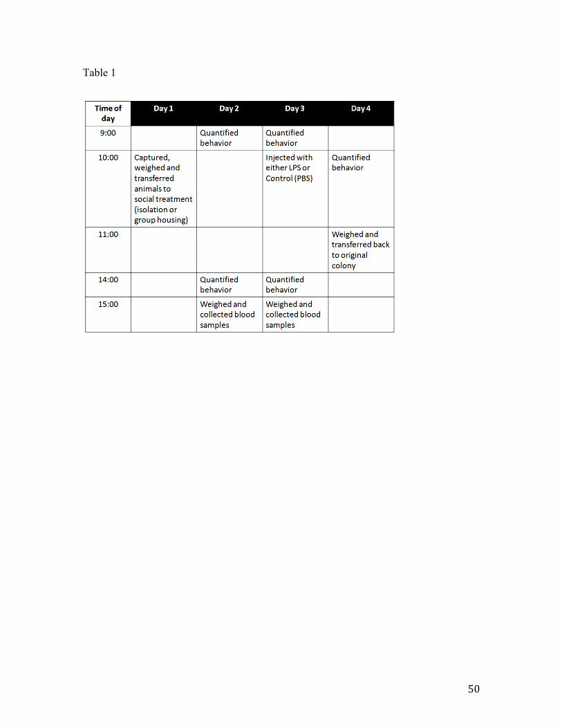
50
Table 1

51
Table 2 - Repeated-measures ANOVA table testing for the effect of two among-subjects factors (social treatment and injection order) and one within-subjects factor (injection treatment) on corticosterone concentrations at 5h after injection.

52
Table 2
Source DF Den. DF F ratio P-value
Social 1 9 0.2120 0.6561
Injection order 1 9 0.0611 0.8103
Social x Inj. order 1 9 1.7620 0.2171
Injection 1 10 0.1548 0.7023
Injection x Social 1 10 2.4689 0.1472
Injection x Inj. order 1 10 0.5085 0.4921
53
Figure 1 - Average number of activities (a) and time spent resting (b) 4h after injection of either a control solution (white bars) or an LPS solution (black bars). Bars represent means ± S.E.M.

54
Figure 1

55
Figure 2 - Mass lost during the course of the experiment. Bars represent means ± S.E.M.

56
Figure 2

57
Figure 3 - Plasma IL-6 bioactivity. Bars represent means ± S.E.M.

58
Figure 3

59
Chapter 3
Exposure to a novel female attenuates behavioral symptoms of infection and affects the hypothalamo-‐pituitary-‐gonadal axis

60
Abstract
Sickness behaviors are the behavioral alterations an animal suffers during the course of an infection, including reduced activity, food and water intake and social interactions. In addition, evidence exists for a shutdown of the reproductive axis during times of sickness. Adopting sickness behaviors is thought to add in overcoming the infection, by releasing energy from these activities into enhancement of the immune system. Nonetheless, sickness behaviors have been shown to be plastic, being reduced under certain environmental and social circumstances. Here, we wanted to test whether the presentation of a novel female to males suffering from a simulated infection could impact both the behavioral effects of sickness, as well as the effects on the reproductive axis. We demonstrate that the presence of a novel female can both diminish sickness behaviors and induce alterations of the reproductive axis within 30 minutes, with no associated changes in brain gene expression of proinflammatory cytokine involved in the regulation of sickness behaviors (interleukin-1β). In addition, we report evidence for an effect of social environment by itself in altering brain gene expression of IL-1β of control animals. The results of this experiment appear to indicate that the behavioral effects of IL-1β are context dependent. In sum, these findings indicate that males prioritize the opportunity to mate versus investment in recovery from an infection by expression of sickness behaviors.
61
Introduction
During the course of an infection, animals display a coordinated array of behavioral responses, collectively called “sickness behavior”. These behavioral changes include lethargy, anorexia, adipsia, and reduced interest in social interactions (Hart, 1988).
A common tool to simulate an infection in animals experimentally is injection of lipopolysacharide (LPS), a component of gram-negative bacteria cell walls. Peripheral LPS administration induces cytokine release into circulation, but it is the neural expression of these cytokines that eventually induces the behavioral response (Laye et al., 2000). The proinflammatory cytokines interleukin-1β (IL-1β), IL-6 and tumor necrosis factor α (TNFα) are the main mediators of this sickness response (Dantzer, 2004). Brain production of cytokines is thought to be induced by peripheral cytokines (Quan et al., 1998; Ek et al., 1998; Goehler et al., 1999).
LPS and interleukin injections have also been found to disrupt gonadotropic functions in mammals at all levels of the hypothalamic-pituitary-gonadal (HPG) axis, including: suppression of the pulsatile release of GnRH (Harris et al., 2000), prevention of the preovulatory LH surge (Rivier and Vale, 1990), suppression of LH release (Xiao et al., 2000; He et al., 2003; Watanobe and Hayakawa, 2003; Iwasa et al., 2008; Refojo et al., 1998, Ebisui et al., 1992, Rivest and Rivier, 1993; Rivier, 1990), alteration of pituitary responsiveness to GnRH (Williams et al., 2001) and suppression of testosterone production (Turnbull and Rivier, 1997). In birds, three studies indicate disruption of the HPG axis by LPS. In one study, male and female white crowned-sparrows, Zonotrichia leucophrys gambelii, exhibited low circulating LH levels 24h after LPS injection (Owen-Ashley at al., 2006). In the second, LH levels were reduced at 6h post-injection in song sparrows (Melospiza melodia), although no differences are found at 22h (Adelman et al., 2010). In a more recent study (Lopes et al. 2012a), male zebra finches (Taeniopygia guttata) showed reduction in GnRH mRNA and peptide expression in the brain, along with a tendency towards reduced circulating testosterone concentrations at 3h after an injection of LPS.
The adoption of sickness behavior is hypothesized to aid in recovery from infection by a reallocation of energy from activities that are not immediately critical for survival into the immune response (Hart, 1988; Kent et al., 1992). This hypothesis is in agreement with the evidence for disruption of the HPG axis following LPS injection. However, it is apparent that the sickness response might be plastic and affected by external factors and motivational variables. Miller (1964) demonstrated that the effect of LPS on conditioned bar pressing in rats was dependent on the stimulus to which bar pressing was linked. If bar pressing was maintained by an appetitive stimulus (such as food and water), LPS caused a decrease in frequency of bar pressing. In contrast, if bar pressing was used to avoid an aversive stimulus (such as escaping a rotating drum), LPS increased bar pressing (Miller 1964). While LPS injection was able to inhibit nest-building behavior in lactating mice injected at room temperature, the mice recovered this behavior once tested at 6oC, a temperature that would be life-threatening for their pups (Aubert et al. 1997). Further, while male mating behavior of rats is not changed by an injection of IL-1, female mating
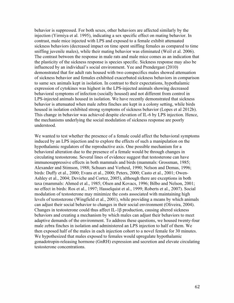
62
behavior is suppressed. For both sexes, other behaviors are affected similarly by the injection (Yirmiya et al. 1995), indicating a sex specific effect on mating behavior. In contrast, male mice injected with LPS and exposed to a female exhibit attenuated sickness behaviors (decreased impact on time spent sniffing females as compared to time sniffing juvenile males), while their mating behavior was eliminated (Weil et al. 2006). The contrast between the response in male rats and male mice comes as an indication that the plasticity of the sickness response is species specific. Sickness response may also be influenced by an individual’s social environment. Yee and Prendergast (2010) demonstrated that for adult rats housed with two conspecifics males showed attenuation of sickness behavior and females exhibited exacerbated sickness behaviors in comparison to same sex animals kept in isolation. In contrast to their expectations, hypothalamic expression of cytokines was highest in the LPS-injected animals showing decreased behavioral symptoms of infection (socially housed) and not different from control in LPS-injected animals housed in isolation. We have recently demonstrated that sickness behavior is attenuated when male zebra finches are kept in a colony setting, while birds housed in isolation exhibited strong symptoms of sickness behavior (Lopes et al 2012b). This change in behavior was achieved despite elevation of IL-6 by LPS injection. Hence, the mechanisms underlying the social modulation of sickness response are poorly understood.
We wanted to test whether the presence of a female could affect the behavioral symptoms induced by an LPS injection and to explore the effects of such a manipulation on the hypothalamic regulators of the reproductive axis. One possible mechanism for a behavioral alteration due to the presence of a female would be through changes in circulating testosterone. Several lines of evidence suggest that testosterone can have immunosuppressive effects in both mammals and birds (mammals: Grossman, 1985; Alexander and Stimson, 1988; Schuurs and Verheul, 1990; Nelson and Demas, 1996; birds: Duffy et al., 2000; Evans et al., 2000; Peters, 2000; Casto et al., 2001; Owen-Ashley et al., 2004; Deviche and Cortez, 2005), although there are exceptions in both taxa (mammals: Ahmed et al., 1985; Olsen and Kovacs, 1996; Bilbo and Nelson, 2001; no effect in birds: Ros et al., 1997; Hasselquist et al., 1999; Roberts et al., 2007). Social modulation of testosterone may minimize the costs associated with maintaining high levels of testosterone (Wingfield et al., 2001), while providing a means by which animals can adjust their social behavior to changes in their social environment (Oliveira, 2004). Changes in testosterone could thus affect IL-1β production, causing altered sickness behaviors and creating a mechanism by which males can adjust their behaviors to meet adaptive demands of the environment. To address these questions, we housed twenty-four male zebra finches in isolation and administered an LPS injection to half of them. We then exposed half of the males in each injection cohort to a novel female for 30 minutes. We hypothesized that males exposed to females would upregulate hypothalamic gonadotropin-releasing hormone (GnRH) expression and secretion and elevate circulating testosterone concentrations.

63
Methods
Animals and experimental set up
All the experiments were carried out at the University of California, Berkeley Field Station for the Study of Behavior, Ecology and Reproduction. All procedures were approved by the University of California Office of Lab Animal Care and were in compliance with federal regulations. The animals used in this experiment come from our captive colonies of zebra finches (Taeniopygia guttata). These colonies include adult males, females and juveniles, housed in a 2.7 m by 2.5 m by 2.1 m indoor flight aviary. They were exposed to natural changes in day length, supplemented by artificial lighting at a light/dark schedule of 12L:12D. Food and water were provided ad libitum and consisted of German millet mixed with canary seed.
The experiment was carried out in May 2011. A total of 24 male zebra finches were tested, employing a staggered design. Each day, 4 birds were tested and this was replicated for 6 consecutive days (24 birds). On the day previous to the experiment, 4 males were chosen randomly from the colony and placed individually in soundproof cages containing video cameras. At 9:00 on the day of the experiment, the males were injected in the pectoral muscle with a sterile solution of either 100µL of LPS 0.3mg mL-1 (Sigma-Aldrich #L4005, Serotype 055:B5) or 100µL of 10mmol-1 phosphate buffered saline (pH 7.2). The region to be injected was sterilized with ethanol, which was allowed to dry before injecting the animal. The dose of LPS was ca. 2mg/kg of body weight. At 12:00 (3h after the injection – Time 1), behavior was recorded for 30min. with the use of the video cameras. At 12:30 (3.5h after the injection – Time 2), unfamiliar females (no previous contact with the focal males) were introduced into the chambers (1 female per chamber) housing half of the males; as controls, no additional animals were introduced to the chamber housing the remaining. For ease of transferring of the females into the cages, social treatment was not randomized (i.e., the same cages always obtained a female). We do not believe this introduces any biases into the experiment, since all the cages were closed systems kept in the same exact conditions (soundproof and light and temperature controlled) and were placed side by side in the same room. Additionally, behaviors (hops and calls) at Time 1 were not different between cages containing animals that had received the same injection (t-test; p>0.05). Behavior was recorded for another 30min. At the end of the 30min, the males were deeply anesthetized via isoflurane inhalation and decapitated. The brain was immediately removed and placed on dry ice. Testes were removed, measured using a plastic caliper (SPI 2000) and placed on dry ice. Trunk blood was collected into a 2mL eppendorf tube and placed on regular ice, until centrifugation (1500 g for 10min at 4oC), after which the plasma portion was collected onto a separate tube. All tissues were kept at -80oC until further analysis. The females were returned to their original colonies.
Behavior
Behavior was scored from videotapes by two trained observers who were blind with respect to the experimental treatments and who had no knowledge of the overall purpose

64
of the experiment. The observers were instructed to count the number of hops, calls and songs displayed during each behavioral trial. Additionally, for the birds that received a female as a treatment during the second 30min. interval, they were instructed to count number of directed songs and copulation attempts (mounting). During the second 30min., the observers were instructed to ignore the first 5min of video, to give the animals time to settle down after the researcher’s hand had been in the cage. Thus each bird’s behavior was scored for 55 minutes of the available 60min. of video footage. For each group, N=6, except regarding number of calls. Due to camera failure to record sound (1 out of 4 cameras did not record sound), N=3 for each group that received a novel female (LPS-injected and Control-injected). The number of times birds that were observed singing was too low and this behavior was not considered further in our analyses.
To confirm that changes in behavior were due to the presence of a female and not just having any conspecific in the cage, we ran an additional control using the same experimental set-up: LPS injected birds (N = 6) were recorded at 3h post-injection in isolation for 30 min, followed by 30 min in the presence of a novel male, and the number of hops were counted.
Gene expression
To explore the impact of the treatments on the hypothalamic regulators of reproduction, we quantified mRNA production of GnRH and GnIH in the brain. Additionally, because these neuropeptides are now know to be produced in the testis, we decided to verify whether there would be an effect of treatment in local regulation of their expression. To obtain a measure of proinflammatory response we decided to quantify IL-1β. Because no direct methods for quantifying IL-1β are readily available for zebra finches, we cloned and quantified the expression of this gene in the brain. We also quantified expression of IL-1β in the brain of birds kept in a colony setting injected with saline (N=6) and collected at 3h after injection (collected as part of a different experiment – see Lopes et al. 2012a), jointly with the control isolated birds from the current experiment.
RNA isolation, purification and reverse-transcription
The brains were cut using a cryostat and 20µm coronal sections were alternatively placed directly onto slides or collected into a tube containing 1mL PureZOL reagent (Bio-Rad Laboratories, Hercules, CA). Two separate tubes of PureZOL were collected. The first consisted of the portion of the brain starting from where the tractus septomesencephalicus (TrSM) becomes visible and continuing to the disappearance of the anterior commissure (CoA); the second one consisted of the rest of the sections from that point until the disappearance of the optic tectum (TeO). Using this protocol, we avoided amplification of GnRH-II during the qRT-PCR procedure, which could be a confounding factor when analyzing GnRH-I data. We verified histologically that no GnRH-II cell bodies were detected in the part of the brain collected in the first sample. Each brain sub-sample was homogenized and stored at -80oC until extraction. The larger testis for each individual was similarly placed in PureZOL, homogenized and stored at 80oC until extraction. Total RNA extraction was performed according to manufacturer’s instructions, with final

65
dilution of RNA in 20µL of DEPC-treated water. Quantification of RNA was done via spectrophotometry (NanoDrop 2000). The RNA was then treated for any genomic DNA contamination (DNA free, Ambion) and 33 ng of RNA from each sample was reverse-transcribed to cDNA using iScript reverse transcriptase with 5X iScript reaction mix (Bio-Rad Laboratories, Hercules, CA). The cDNA was diluted 1:25, as this dilution was optimal for the genes being amplified.
Cloning IL-1β in zebra finches
Primers were developed based on the predicted IL-1β sequence for zebra finches (GenBank accession number: XM_002195564). PCR reactions were performed using 1 µl cDNA (100–200 ng µl–1) with 0.25 µl ExTaq (Takara), 5 µl 10X PCR buffer (Takara), 4 µl dNTPs (Takara) and 1 µl of each primer (forward and reverse) in a final volume of 50 µl. The PCR program consisted of 94°C for 3 min; 45 cycles of 94 °C for 30 s, annealing at 60 °C for 30 s and 72 °C for 1 min; followed by a final extension step of 72 °C for 3min. Product amplification was confirmed on a Tris-acetate and ETDA (TAE) agarose (1%) gel containing 0.05% ethidium bromide. The PCR products were then purified in using GE Kit Microspin columns (S-200HR), according to manufacture’s instructions. Cloning of PCR products was done using pGEM®-T Easy Vectors (Promega), following manufacture’s instructions. White bacterial colonies were picked and PCR amplified with 0.125 µl ExTaq (Takara), 2.5 µl 10X PCR buffer (Takara), 2 µl dNTPs (Takara) and 0.5 µl of each primer (forward and reverse) in a final volume of 25 µl. The PCR program consisted of 94°C for 10 min; 30 cycles of 94 °C for 30 s, annealing at 55 °C for 30 s and 72 °C for 1 min; followed by a final extension step of 72 °C for 3min. Appropriate products were chosen based on visualization on a Tris-acetate and ETDA (TAE) agarose (1%) gel containing 0.05% ethidium bromide, purified using ExoSap (BRAND) according to manufactures instructions. After quantification of the products (approximately 500 ng/µl of DNA was obtained for each product), products were sequenced at the UC Berkeley DNA Sequencing Facility (Berkeley, CA, USA) on an Applied Biosystems 3730 96-capillary DNA sequencer.
Quantitative real-time PCR (qRT-PCR)
Primers for GnRH and GnIH were designed based on the published sequences for zebra finches (respectively, GenBank ID: NM_001142320.1 and GenBank ID: AB522971.1) and 18S (control gene) primers were designed based on the published rat sequence (GenBank ID: NR_046237.1). Primers for IL-1β were designed based on the sequence we cloned (under submission to GenBank ID). Primer sequences were: GnRH-F: ACTCCACAACCTCTCTCAGG; GnRH-R: CTCTGCTGCTCCTCCTCTAA; GnIH-F: CCCTGAGATTTGGAAGAGC; GnIH-R: CAGATTGACAGGCAGTGAC; 18S-F: CCATCCAATCGGTAGTAGCG; 18S-R: GTAACCCGTTGAACCCCATT; IL-1β-F: TTATGGCCCCAACTGTCTGT; and IL-1β-R: TGCCAAGGTCACTATCAGCA Amplification of primer-dimer was controlled for by running template-free controls. These samples always resulted in differences of at least 10 cycles of the Ct values compared to samples containing template. The qRT-PCR was performed in duplicate for each bird for each gene in 25µL reactions according to manufacturer’s instructions for 2x

66
iQTM SyBR Green Supermix (Bio-Rad Laboratories, Hercules, CA). After checking for primer specificity by visual inspection of the melting curves, we used the RT-PCR Miner program (Zhao and Fernald 2005) for analysis of the raw fluorescent data. Gene expression was calculated using the following formula: 1/(1+E)Ct, where E is the average PCR efficiency and Ct is the cycle threshold. The reference gene used to normalize mRNA levels among samples was 18S, after verification that treatment did not affect its levels. Normalization was done for each individual by dividing expression values for the gene of interest by expression values of 18S.
Immunohistochemistry
In addition to measuring mRNA in the brain, we quantified the number of neurons that were immunoreactive to GnRH and GnIH in order to get a more dynamic picture of the impact of the treatments on the reproductive axis.
A hydrophobic barrier was created around the slices on the slide by the use of a PAP pen (Sigma-Aldrich # Z377821). The brain sections were then fixed using a 4% paraformaldehyde solution for one hour. The slides were washed three times in phosphate buffered saline (PBS, 10mM, pH 7.2) and exposed to a 1% solution of hydrogen peroxide in PBS for thirty minutes. A new wash in PBS for five minutes was repeated three times, after which 2% normal goat serum (NGS) in 0.2% PBS-Triton (PBS-T) was added for one hour. Subsequently, GnRH primary antibody (HU60, gift from Dr. Henryk Urbanski, Portland, OR, USA) at a concentration of 1:5000 in 0.2% PBS-T was added and allowed to incubate at room temperature (r.t.) for one hour and subsequently for 48 hours at 4oC. The slides were then washed three times in 0.2% PBS-T, followed by incubation in 1:250 biotinylated goat anti-rabbit IgG (Vector Labs #BA-1000) in 0.2% PBS-T for one hour and an additional three washes in 0.2% PBS-T. After a one hour incubation in avidin-biotin complex (ABC, Vectastain Elite Kit, Vector Labs #PK6100), visualization was achieved by adding 0.03% 3,3’-diaminobenzine (DAB) for eight minutes.
The protocol for the labeling of GnIH (primary antiserum PAC 123, 124, Bentley, Berkeley, CA, USA, used at dilution 1:5000 in 0.2% PBS-T) was the same as for GnRH, with the difference that no r.t. incubation in primary antibody was done. Slides containing adjacent brain slices were used for each of these two antibodies. Successful use of these antibodies has been demonstrated previously in zebra finch brains using a similar protocol (Perfito et al., 2011).
Quantification of GnRH and GnIH immunoreactivity
Photographs were taken of the areas showing immunoreactivity using a Zeiss Axio Imager A1 microscope and AxioVision 4.5 software. Because GnRH-I and GnIH neurons occur in restricted areas of the brain (preoptic area and paraventricular nucleus, respectively), and because we ran the IHC on adjacent sections for each neuropeptide, we were easily able to count the number of GnRH-I-immunoreactive(-ir) neurons and the number of GnIH-ir neurons.

67
Radioimmunoassay for testosterone
Testosterone constitutes the endpoint of the activation of the reproductive axis. As stated previously, testosterone might be an important mediator of the extent of the sickness behavior response.
Testosterone assays were performed following Wingfield et al. (1991). All samples were measured in a single assay. Plasma volume of samples was 35 µL. The detection limit of the assay was 3.9pg. Intra-assay variation was 7.1%.
Statistical analysis
Statistical analyses were performed using JMP v.9. Concerning the behavioral data, each time point was analyzed separately. The first time point served to verify that sickness behavior had been induced and was analyzed by use of a t-test with Welch’s correction, except for calls, which were analyzed using a Wilcoxon Rank Sums Test (non-normal distribution). Sample sizes for hopping are N=12 for LPS-injected and N=12 for Control-injected and for calling are N=9 for each of these groups. The second time point served to test for an effect of social treatment (presence of female) and an interaction of social treatment x injection and was analyzed using a two-way ANOVA, where factors were Social Treatment and Injection and Social Treatment x Injection interaction. At time 2, sample sizes are N=6 in each treatment, except for Calls, as noted in section 2.2, where N=3 for each of the groups (LPS and Control-injected) that received a novel female. Gene expression, ir-cell counts and testosterone data were similarly analyzed using a two-way ANOVA, with Social Treatment and Injection and Social Treatment*Injection interaction as factors. Behavioral data and GnRH gene expression data were log-transformed prior to analysis to better fit the assumption of normality and homogeneity of variance. Data are represented as means ± SEM. All tests were two-tailed and probability values of P<0.05 were considered to be statistically significant. To explore further some of our results, we decided to present effect size (Cohen’s d) for comparisons of two groups of particular interest. Effect size (ES) was calculated by dividing the difference between the means of the two groups being compared by the pooled standard deviation. An ES=0.2 was considered to indicate a small effect an ES=0.5 a moderate effect and an ES=0.8 a large effect (Cohen, 1992).

68
Results
Behavior
Time 1: LPS injection induced sickness behaviors
Overall, LPS injection reduced activity in birds, as can be seen in Fig.1. More specifically, there was a significant effect of injection on number of hops (t(1, 20) = 3.77, p= 0.0011) and number of calls (S=57, p=0.0108, NControl=NLPS=9).
Time 2: Addition of a novel female altered manifestation of sickness behavior
The activity of Control-Isolated animals was reduced over time (Fig.1 vs. Fig.2).
The presence of a novel female caused an significant increase in the number of times an individual was observed hopping (F(1,20) = 11.25, p= 0.0032) and calling (F(1,14) = 18.44, p= 0.0007) (Fig.2). No effects of injection or the interaction injection x presence of female were observed for any of the behaviors analyzed. LPS-injected birds kept in isolation hopped on average 3.04 times less then Control-injected birds (ES=0.59 – moderate effect), while the difference in hopping between Control and LPS-injected presented with a female is of only 1.47 times (ES=0.43 – small effect). A similar pattern is found for number of calls: in isolation, Control called 4.67 times more frequently than LPS-injected animals (ES=0.69 – moderate effect); with female, Control called on average 1.41 times more than LPS-injected (ES=0.25 – small effect).
Animals that received females were observed displaying directed singing (3 Control and 3 LPS-injected birds) and copulation attempts (1 Control and 1 LPS-injected bird) regardless of the injection. We did not perform statistical analysis on these behaviors given the reduced sample size.
The behavior of LPS injected males in isolation was not different from that of the same males when presented with the novel male (paired t-test: t(5) = -1.009, p= 0.359).
Reproductive effects at hypothalamic level
The number of GnRH-ir cell bodies in the hypothalamus was not affected by injection (F(1,20) = 2.08, p= 0.167) or presence of a novel female (F(1,20) = 0.73, p= 0.406; Fig.3A). There was no significant interaction between injection and presence of female (F(1,20) = 2.65, p= 0.122). Regarding GnRH mRNA expression, no statistical significant effects were found for injection, presence of female or their interaction (p>0.05).
No statistical effect of injection on number of GnIH-ir cell bodies was found for LPS injected birds (F(1,20) = 2.21, p= 0.163; Fig. 3C). At the level of mRNA (Fig.3D), the presence of a novel female increased GnIH mRNA expression (F(1,20) = 4.61, p= 0.045).

69
Reproductive effects at testicular level
Testicular GnRH expression (Fig.4A) increased when animals were in the presence of a novel female (Fig.4) (F(1,20) = 4.60, p= 0.044). Testicular GnIH expression (Fig.4B) was not affected by either LPS or presence of female (p>0.05).
Testosterone concentrations were the highest in Control isolated animals (Fig.4C). When compared, a large effect was found both on testosterone concentrations of LPS vs. Control injected animals in isolation (ES=1.16) and on Control isolated vs. Control with female (ES=1.27). Neither presence of a novel female or injection affected testosterone concentrations, but a nearly significant interaction existed between the two (F(1,20) = 3.97, p= 0.060).
Immune effects
Brain mRNA levels of IL-1β were the lowest in Control-Isolated individuals (Fig.5). Injection had a borderline statistically significant effect on IL-1β expression in the brain (Fig.5; F(1,20) = 4.19, p= 0.0546), increasing it. Presence of a novel female had no effect on IL-1β (F(1,20) = 3.07, p= 0.0958). To further explore these nearly significant relationships, we calculated effect size to compare the magnitude of change in expression induced by LPS in isolation and in the presence of a female. In isolation, ES=1.12 (large effect) and in the presence of a female, ES=0.70 (moderate effect).
When we quantified mRNA levels of IL-1β in animals that were control-injected and kept in isolation simultaneous to animals that were control-injected and kept in a colony setting, we verified that birds kept in isolation had significantly lower expression of IL-1β expression than birds kept in a colony (t(8.5)=-3.44, p=0.008; Fig.5B).

70
Discussion
This study is the first to explore the effects of the presence of a female on the behavioral, neuroendocrine and testosterone profile of male birds subjected to an endotoxin challenge. The presence of a novel female induced both behavioral and physiological changes in birds that had received an endotoxin injection. Importantly, these changes occurred within only 30min of being in the presence of the novel female.
Isolated males that received an LPS injection showed drastic reductions in the number of hops and number of calls compared to birds injected with a saline control. When LPS-injected males were given a female they had never encountered previously, their behaviors were indistinguishable from Control-injected males in the same social condition (presence of a novel female).
Activation of the HPG axis was found both at the level of the brain and, most significantly, the testis, through enhanced expression of GnRH mRNA. Contrary to our predictions, this was not translated in terms of increased testosterone concentration at the time point analyzed. Curiously, LPS-injected males when in the presence of a female had the highest levels of brain IL-1β gene expression. Control-injected males also showed IL-1β levels comparable to Isolated LPS-injected males. This is surprising, since IL-1β has been extensively documented as having a primary role in the expression of sickness behaviors. It appears that in zebra finches social environment alone is able to induce changes of IL-1β expression.
Effects on behavior
Stereotypical sickness behaviors were observed at 3h after LPS injection, as quantified by a reduction in all activities measured. With continued time in isolation, Control-injected males further decreased their activity, although not to the same extent as LPS-injected individuals. This finding makes it somewhat difficult to interpret the effects of social environment on sickness response statistically. We had predicted that if addition of a novel female caused an effect on LPS-injected animals, we would find an interaction between social treatment and injection. Given the increased lethargy of control-injected isolated animals, we would now expect a main effect of social treatment to indicate that the novel females affect the behavior of LPS-injected animals. Graphically, it is easier to understand the effects observed: while LPS-injected animals kept in isolation did not alter their behaviors from Time 1 to Time 2, LPS-injected animals that received a female increased in activity. Thus, the presence of a novel female attenuated the behavioral symptoms of sickness in LPS-injected animals, to the point that these animals were behaviorally indistinguishable from Control-Injected animals in the same social setting. Changes in behavior due to social conditions have been previously observed in zebra finches and other animals. These effects do not appear to be linked to having any conspecific in the cage, nor to the novelty of that cage mate, since the behavior of LPS-injected males kept in isolation was not different from their own behavior when in the presence of a novel male.

71
Effects on the HPG axis
While brain GnRH expression appears slightly increased in males exposed to a female, this effect was more strongly verifiable at the level of the testis. The significance of this response at the testicular level is not entirely clear, since the effects of GnRH at this level are not yet fully understood and both inhibitory and stimulatory effects have been found (McGuire and Bentley 2010). Little is known about its general effects in birds, with one experiment demonstrating that in isolated granulosa cells from chicken (Gallus domesticus), GnRH induces an increase in LH-stimulated progesterone production (Hertelendy 1982). Even though the functional consequences of increased expression of testicular GnRH is unknown in birds, it is relevant to realize that the testes are able to respond rapidly to a social cue (presence of a female). Although social effects on testis size have been previously shown in birds (for example, Flickinger 1966), to our knowledge, ours are the first results on rapid (within 30min) changes in GnRH expression at the testicular level. On the other hand, GnIH appears to respond faster at the level of the brain, with no changes in expression being found on the testis.
The brain immunohistochemistry results coupled with the gene expression results may contribute to our understanding of the dynamics of what is happening. Although it would be likely to see changes in gene expression within 30min, if would be unlikely to see de novo protein production within this timeframe. A change in number of ir-cells within this time would mean release of the neuropeptide. On the other hand, changes in cell-ir number within 3h could either be related to de novo neuropeptide synthesis, degradation or neuropeptide release from the cells. Previous results using zebra finches have shown that the number of GnRH-ir cells is reduced in birds injected with LPS at 3h after an injection (Lopes et al 2012a), with a trend for reduction in testosterone concentration. The current results seem to corroborate these findings: when birds were kept in isolation and injected with LPS, both a reduction in number of GnRH-ir cells and T was verified. However, GnRH expression was not different between LPS and Control-injected birds in isolation. Jointly, these results appear to indicate an inhibition of the HPG axis in LPS-isolated birds, but a readiness to respond to environmental alterations in Control-isolated birds. In birds that received a novel female, increase in GnRH gene expression was not coupled with increase in number of cells showing GnRH-ir. These results could be interpreted as an activation of the axis (through increased mRNA expression), and neuropeptide release in Control –injected animals presented with a female. In what regards LPS-injected males in this social condition, the interpretation becomes harder, since we would expect them to either have a similar GnRH cell-ir profile as LPS-injected animals kept in isolation or lower number of GnRH-ir cells, indicating GnRH release. An increase in number of GnIH-ir cells is only apparent in LPS-injected birds, regardless of social treatment. This result fits with previous observations that number of GnIH-ir cells is increased by LPS injection. High levels of GnIH neuropeptide in the hypothalamus could mean a preparedness to “pause” reproduction (Calisi et al 2012), which would make sense during infectious states.
Given that several studies suggest that the presence of a female can induce an elevation of testosterone concentrations within a short period of time (Feder et al. 1977; Dufty and
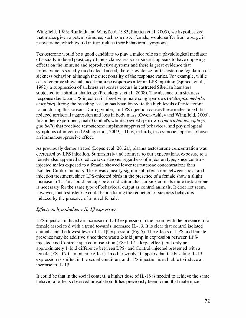
72
Wingfield, 1986; Runfeldt and Wingfield, 1985; Pinxten et al. 2003), we hypothesized that males given a potent stimulus, such as a novel female, would suffer from a surge in testosterone, which would in turn reduce their behavioral symptoms.
Testosterone would be a good candidate to play a major role as a physiological mediator of socially induced plasticity of the sickness response since it appears to have opposing effects on the immune and reproductive systems and there is great evidence that testosterone is socially modulated. Indeed, there is evidence for testosterone regulation of sickness behavior, although the directionality of the response varies. For example, while castrated mice show enhanced immune responses after an LPS injection (Spinedi et al., 1992), a suppression of sickness responses occurs in castrated Siberian hamsters subjected to a similar challenge (Prendergast et al., 2008). The absence of a sickness response due to an LPS injection in free-living male song sparrows (Melospiza melodia morphna) during the breeding season has been linked to the high levels of testosterone found during this season. During winter, an LPS injection causes these males to exhibit reduced territorial aggression and loss in body mass (Owen-Ashley and Wingfield, 2006). In another experiment, male Gambel's white-crowned sparrow (Zonotrichia leucophrys gambelii) that received testosterone implants suppressed behavioral and physiological symptoms of infection (Ashley et al., 2009). Thus, in birds, testosterone appears to have an immunosuppressive effect.
As previously demonstrated (Lopes et al. 2012a), plasma testosterone concentration was decreased by LPS injection. Surprisingly and contrary to our expectations, exposure to a female also appeared to reduce testosterone, regardless of injection type, since control-injected males exposed to a female showed lower testosterone concentrations than Isolated Control animals. There was a nearly significant interaction between social and injection treatment, since LPS-injected birds in the presence of a female show a slight increase in T. This could perhaps be an indication that for sick animals more testosterone is necessary for the same type of behavioral output as control animals. It does not seem, however, that testosterone could be mediating the reduction of sickness behaviors induced by the presence of a novel female.
Effects on hypothalamic IL-1β expression
LPS injection induced an increase in IL-1β expression in the brain, with the presence of a female associated with a trend towards increased IL-1β. It is clear that control isolated animals had the lowest level of IL-1β expression (Fig.5). The effects of LPS and female presence may be additive since there was a 2-fold jump in expression between LPS-injected and Control-injected in isolation (ES=1.12 – large effect), but only an approximately 1-fold difference between LPS- and Control-injected presented with a female (ES=0.70 – moderate effect). In other words, it appears that the baseline IL-1β expression is shifted in the social condition, and LPS injection is still able to induce an increase in IL-1β.
It could be that in the social context, a higher dose of IL-1β is needed to achieve the same behavioral effects observed in isolation. It has previously been found that male mice

73
injected with LPS and placed in the presence of a female have their IL-1 gene expression increased relative to mice in isolation (Weil et al., 2006). In this experiment, however, no changes in IL-1 were found in control-injected males presented with a female. It might be that additional cytokines would help explain the effects observed in our experiment. For example, TNFα and IL-6 are the other two major cytokines involved in the sickness response. However, Weil et al. (2006) found TNFα to be altered by treatment in a similar way as IL-1. In a different experiment, housing with conspecifics was itself sufficient to cause an LPS-induced increase in TNFα concentrations relative to isolation in male rats, as measured by an EIA (Yee and Prendergast, 2010). However, the animals with the most robust sickness responses (LPS isolated rats) were the ones with the lowest levels of TNFα. In our experiment, isolated males follow the expected pattern: an LPS injection increased IL-1 expression while reducing activity. On the other hand, when males were exposed to females, IL-1 expression was elevated, regardless of injection and LPS-injected males became behaviorally unresponsive to the effects of injection (and IL-1). It has been suggested previously that exposure to a female might be able to alter behavioral responsiveness to cytokines (Weil et al, 2006). Also, it might be that additional factors affect expression of IL-1, such as stress. De Miguel et al. (2011), showed that social stress increases corticosterone, adrenaline and IL-1 in mice and that within the group of stressed animals, the most active mice are the ones with the highest IL-1.
Life-history trade-off theory predicts that, given limited resources, animals will invest towards maximization of reproductive success. A simulated acute infection may indicate to the host that its residual reproductive value is low. This in turn may lead the host to invest in current reproductive or effort in case the opportunity presents itself. The present work favors the terminal investment hypothesis as an explanation for the modulation of sickness behaviors (Adelman and Martin 2009): males kept in isolation and injected with an endotoxin invested heavily in the expression of sickness behaviors and shut down their reproductive system, while males that received a female invested in current reproductive success by activating their reproductive axis and attempting to mate, while not allocating time to exhibiting sickness behaviors. In this case, reduction in sickness behaviors may be a way to shift time and energy into reproductive opportunities. The baseline (control-injected) level of the main proinflammatory cytokine involved in modulating sickness behavior was increased by sole presence of a female. Evolutionarily, it would make sense for isolated animals to downregulate the immune response (therefore reducing circulating levels of proinflamatory cytokines), since they are at no risk of contracting an infection from conspecifics. In fact, to test for this hypothesis, we ran a RT-PCR for quantification of expression of IL-1 in the brain of birds kept in a colony setting injected with saline (N=6) and collected at 3h after injection (collected as part of a different experiment – see Lopes et al. 2012a), jointly with the control isolated birds from the current experiment. Indeed, isolation alone significantly reduces IL-1β expression (Fig.5B). Together, these results suggest that there is social modulation of interleukin expression and that the behavioral effects induced by this cytokine are context dependent. These results support findings by Larson et al (2002), where they showed that the behavioral effects of administration of IL-1 to mice was dependent on the level of motivation and on the cost of the behavioral response. More specifically, in food restricted mice (higher motivation), IL-1β administration induced a lower decrease in milk intake than it did on free-feeding

74
mice (low motivation). In the case of our experiment, similar brain levels of IL-1β expression were associated with sickness behaviors only in birds kept in isolation and not in birds in the presence of a female. It is interesting to note that zebra finches are gregarious in the wild, being most frequently found in flocks (Zann, 1996). It might be that gregarious species have naturally higher base levels of IL-1 than solitary species. It would be interesting to quantify the baseline levels of IL-1 in species that are solitary and verify whether these are changed by the presence of conspecifics.
Conclusion
In summary, while LPS injection of isolated males reduced overall activity and downregulated the HPG axis with a concomitant increase in brain IL-1 expression, the presence of a novel female abolished all of these effects except IL-1 expression, which remained elevated. IL-1 expression was similarly elevated in control males exposed to a female. Taken together these results indicate that the presence of a novel female can attenuate sickness behavior and the impact on the HPG axis activity without significantly altering the inflammatory response. The results of this experiment demonstrate that the effects of IL-1β are context dependent. Further research is necessary to investigate how this effect arises and to identify other factors involved in mediating changes in IL-1 and its impact on behavior.
Acknowledgements
I would like to thank Ministério para Ciência, Tecnologia e Ensino Superior (MCTES-Lisbon, Portugal) for financial support through doctoral grant [SFRH/BD/33251/2007] awarded to me. Additionally, John Wingfield and Paulina Gonzalez provided help with the hormonal assays and Hilary Chan and Sophie Demathieu helped with immunohistochemistry work. I am also grateful for the support provided by the staff at the Field Station for the Study Behavior, Ecology and Reproduction, and Gregory Goldsmith and Eileen Lacey for help with statistical analysis and comments on the manuscript.

75
References
Adelman, J.S., Bentley, G.E., Wingfield, J.C., Martin, L.B., Hau, M., 2010. Population differences in fever and sickness behaviors in a wild passerine: a role for cytokines. Journal of Experimental Biology 213, 4099-4109.
Adelman, J.S., Martin, L.B., 2009. Vertebrate sickness behaviors: Adaptive and integrated neuroendocrine immune responses. Integrative and Comparative Biology 49, 202-214.
Ahmed, S.A., Dauphinee, M.J., Talal, N., 1985. Effects of short-term administration of sex-hormones on normal and autoimmune mice. Journal of Immunology 134, 204-210.
Alexander, J., Stimson, W.H., 1988. Sex-hormones and the course of parasitic infection. Parasitology Today 4, 189-193.
Ashley, N.T., Hays, Q.R., Bentley, G.E., Wingfield, J.C., 2009. Testosterone treatment diminishes sickness behavior in male songbirds. Hormones and Behavior 56, 169-176.
Aubert, A., Goodall, G., Dantzer, R., Gheusi, G., 1997. Differential effects of lipopolysaccharide on pup retrieving and nest building in lactating mice. Brain Behavior and Immunity 11, 107-118.
Bilbo, S.D., Nelson, R.J., 2001. Sex steroid hormones enhance immune function in male and female Siberian hamsters. American Journal of Physiology-Regulatory Integrative and Comparative Physiology 280, R207-R213.
Calisi, R.M., Diaz-Munoz, S.L., Wingfield, J.C., Bentley, G.E., 2011. Social and breeding status are associated with the expression of GnIH. Genes Brain and Behavior 10, 557-564.
Casto, J.M., Nolan, V., Ketterson, E.D., 2001. Steroid hormones and immune function: Experimental studies in wild and captive dark-eyed juncos (Junco hyemalis). American Naturalist 157, 408 -420.
Cohen, J., 1992. A power primer. Psychological Bulletin 112, 155-159.
Dantzer, R., 2004. Cytokine-induced sickness behaviour: a neuroimmune response to activation of innate immunity. European Journal of Pharmacology 500, 399-411.
De Miguel, Z., Vegas, O., Garmendia, L., Arregi, A., Beitia, G., Azpiroz, A., 2011. Behavioral coping strategies in response to social stress are associated with distinct neuroendocrine, monoaminergic and immune response profiles in mice. Behavioural Brain Research 225, 554-561.

76
Deviche, P., Cortez, L., 2005. Androgen control of immunocompetence in the male house finch, Carpodacus mexicanus Muller. Journal of Experimental Biology 208, 1287-1295.
Duffy, D.L., Bentley, G.E., Drazen, D.L., Ball, G.F., 2000. Effects of testosterone on cell-mediated and humoral immunity in non-breeding adult European starlings. Behavioral Ecology 11, 654–662.
Dufty, A.M., Wingfield, J.C., 1986. The influence of social cues on the reproductive endocrinology of male brown-headed cowbirds - field and laboratory studies. Hormones and Behavior 20, 222-234.
Ebisui, O., Fukata, J., Tominaga, T., Murakami, N., Kobayashi, H., Segawa, H., Muro, S., Naito, Y., Nakai, Y., Masui, Y., Nishida, T., Imura, H., 1992. Roles of interleukin-1-alpha and interleukin-1-beta in endotoxin-induced suppression of plasma gonadotropin-levels in rats. Endocrinology 130, 3307-3313.
Ek, M., Kurosawa, M., Lundeberg, T., Ericsson, A., 1998. Activation of vagal afferents after intravenous injection of interleukin-1 beta: Role of endogenous prostaglandins. Journal of Neuroscience 18, 9471-9479.
Evans, M.R., Goldsmith, A.R., Norris, S.R.A., 2000. The effects of testosterone on antibody production and plumage coloration in male house sparrows (Passer domesticus). Behavioral Ecology and Sociobiology 47, 156-163.
Feder, H.H., Storey, A., Goodwin, D., Reboulleau, C., Silver, R., 1977. Testosterone and 5alpha-dihydrotestosterone levels in peripheral plasma of male and female ring doves (Streptopelia risoria) during reproductive-cycle. Biology of Reproduction 16, 666-677.
Fishkin, R.J., Winslow, J.T., 1997. Endotoxin-induced reduction of social investigation by mice: interaction with amphetamine and anti-inflammatory drugs. Psychopharmacology 132, 335-341.
Flicking.Gl, 1966. Response of testes to social interaction among grouped chickens. General and Comparative Endocrinology 6, 89-98.
Goehler, L.E., Gaykema, R.P.A., Nguyen, K.T., Lee, J.E., Tilders, F.J.H., Maier, S.F., Watkins, L.R., 1999. Interleukin-1 beta in immune cells of the abdominal vagus nerve: a link between the immune and nervous systems? Journal of Neuroscience 19, 2799-2806.
Grossman, C.J., 1985. Interactions between the gonadal-steroids and the immune system. Science 227, 257-261.
Harris, T.G., Battaglia, D.F., Brown, M.E., Brown, M.B., Carlson, N.E., Viguie, C., Williams, C.Y., Karsch, F.J., 2000. Prostaglandins mediate the endotoxin-induced suppression of pulsatile gonadotropin-releasing hormone and luteinizing hormone secretion in the ewe. Endocrinology 141, 1050-1058.

77
Hart, B.L., 1988. Biological basis of the behavior of sick animals. Neuroscience and Biobehavioral Reviews 12, 123-137.
Hasselquist, D., Marsh, J.A., Sherman, P.W., Wingfield, J.C., 1999. Is avian humoral immunocompetence suppressed by testosterone? Behavioral Ecology and Sociobiology 45, 167-175.
He, D., Sato, I., Kimura, F., Akema, T., 2003. Lipopolysaccharide inhibits luteinizing hormone release through interaction with opioid and excitatory amino acid inputs to gonadotropin-releasing hormone neurones in female rats: Possible evidence for a common mechanism involved in infection and immobilization stress. Journal of Neuroendocrinology 15, 559-563.
Hertelendy, F., Lintner, F., Asem, E.K., Raab, B., 1982. Synergistic effect of gonadotropin-releasing hormone on lh-stimulated progesterone production in granulosa-cells of the domestic-fowl (Gallus domesticus). General and Comparative Endocrinology 48, 117-122.
Iwasa, T., Matsuzaki, T., Murakami, M., Shimizu, F., Kuwahara, A., Yasui, T., Irahara, M., 2008. Decreased expression of kisspeptin mediates acute immune/inflammatory stress-induced suppression of gonadotropin secretion in female rat. Journal of Endocrinological Investigation 31, 656-659.
Kent, S., Bluthe, R.M., Dantzer, R., Hardwick, A.J., Kelley, K.W., Rothwell, N.J., Vannice, J.L., 1992. Different receptor mechanisms mediate the pyrogenic and behavioral-effects of interleukin-1. Proceedings of the National Academy of Sciences of the United States of America 89, 9117-9120.
Larson, S.J., Romanoff, R.L., Dunn, A.J., Glowa, J.R., 2002. Effects of interleukin-1 beta on food-maintained behavior in the mouse. Brain Behavior and Immunity 16, 398-410.
Laye, S., Gheusi, G., Cremona, S., Combe, C., Kelley, K., Dantzer, R., Parnet, P., 2000. Endogenous brain IL-1 mediates LPS-induced anorexia and hypothalamic cytokine expression. American Journal of Physiology-Regulatory Integrative and Comparative Physiology 279, R93-R98.
Lopes, P.C., Adelman, J., Wingfield, J.C., Bentley, G.E., 2012a. Social context modulates sickness behavior. Behavioral Ecology and Sociobiology 66, 1421-1428.
Lopes, P.C., Wingfield, J.C., Bentley, G.E., 2012b. Lipopolysaccharide injection induces rapid decrease of hypothalamic GnRH mRNA and peptide, but does not affect GnIH in zebra finches. Hormones and Behavior 62, 173–179.
Miller, N.E., 1964. Some psychophysiological studies of motivation and of the behavioral-effects of illness. Bulletin of the British Psychological Society 17.

78
Nelson, R.J., Demas, G.E., 1996. Seasonal changes in immune function. Quarterly Review of Biology 71, 511-548.
Oliveira, R.F., 2004. Social modulation of androgens in vertebrates: Mechanisms and function. Advances in the Study of Behavior, Vol 34 34, 165–239.
Olsen, N.J., Kovacs, W.J., 1996. Gonadal steroids and immunity. Endocrine Reviews 17, 369-384.
Owen-Ashley, N.T., Hasselquist, D., Wingfield, J.C., 2004. Androgens and the immunocompetence handicap hypothesis: Unraveling direct and indirect pathways of immunosuppression in song sparrows. American Naturalist 164, 490-505.
Owen-Ashley, N.T., Turner, M., Hahn, T.P., Wingfield, J.C., 2006. Hormonal, behavioral, and thermoregulatory responses to bacterial lipopolysaccharide in captive and free-living white-crowned sparrows (Zonotrichia leucophrys gambelii). Hormones and Behavior 49, 15-29.
Owen-Ashley, N.T., Wingfield, J.C., 2006. Seasonal modulation of sickness behavior in free-living northwestern song sparrows (Melospiza melodia morphna). Journal of Experimental Biology 209, 3062-3070.
Peters, A., 2000. Testosterone treatment is immunosuppressive in superb fairy-wrens, yet free-living males with high testosterone are more immunocompetent. Proceedings of the Royal Society B-Biological Sciences 267, 883-889.
Pinxten, R., de Ridder, E., Eens, M., 2003. Female presence affects male behavior and testosterone levels in the European starling (Sturnus vulgaris). Hormones and Behavior 44, 103-109.
Prendergast, B.J., Baillie, S.R., Dhabhar, F.S., 2008. Gonadal hormone-dependent and -independent regulation of immune function by photoperiod in Siberian hamsters. American Journal of Physiology-Regulatory Integrative and Comparative Physiology 294, R384-R392.
Quan, N., Whiteside, M., Herkenham, M., 1998. Time course and localization patterns of interleukin-1 beta messenger RNA expression in brain and pituitary after peripheral administration of lipopolysaccharide. Neuroscience 83, 281-293.
Rivest, S., Rivier, C., 1993. Centrally injected interleukin-1-beta inhibits the hypothalamic lhrh secretion and circulating lh levels via prostaglandins in rats. Journal of Neuroendocrinology 5, 445-450.
Rivier, C., Vale, W., 1990. Cytokines act within the brain to inhibit luteinizing-hormone secretion and ovulation in the rat. Endocrinology 127, 849-856.

79
Roberts, M.L., Buchanan, K.L., Hasselquist, D., Evans, M.R., 2007. Effects of testosterone and corticosterone on immunocompetence in the zebra finch. Hormones and Behavior 51, 126-134.
Ros, A.F.H., Groothuis, T.G.G., Apanius, V., 1997. The relation among gonadal steroids, immunocompetence, body mass, and behavior in young black-headed gulls (Larus ridibundus). American Naturalist 150, 201-219.
Runfeldt, S., Wingfield, J.C., 1985. Experimentally prolonged sexual-activity in female sparrows delays termination of reproductive activity in their untreated mates. Animal Behaviour 33, 403-410.
Schuurs, A., Verheul, H.A.M., 1990. Effects of gender and sex steroids on the immune-response. Journal of Steroid Biochemistry and Molecular Biology 35, 157-172.
Spinedi, E., Suescun, M.O., Hadid, R., Daneva, T., Gaillard, R.C., 1992. Effects of gonadectomy and sex-hormone therapy on the endotoxin-stimulated hypothalamo-pituitary-adrenal axis - evidence for a neuroendocrine-immunological sexual dimorphism. Endocrinology 131, 2430-2436.
Turnbull, A.V., Rivier, C., 1997. Inhibition of gonadotropin-induced testosterone secretion by the intracerebroventricular injection of interleukin-1 beta in the male rat. Endocrinology 138, 1008-1013.
Watanobe, H., Hayakawa, Y., 2003. Hypothalamic interleukin-1 beta and tumor necrosis factor-alpha, but not interleukin-6, mediate the endotoxin-induced suppression of the reproductive axis in rats. Endocrinology 144, 4868-4875.
Weil, Z.M., Bowers, S.L., Pyter, L.M., Nelson, R.J., 2006. Social interactions alter proinflammatory cytokine gene expression and behavior following endotoxin administration. Brain Behavior and Immunity 20, 72-79.
Williams, C.Y., Harris, T.G., Battaglia, D.F., Viguie, C., Karsch, F.J., 2001. Endotoxin inhibits pituitary responsiveness to gonadotropin-releasing hormone. Endocrinology 142, 1915-1922.
Wingfield, J.C., Hegner, R.E., Lewis, D.M., 1991. Circulating levels of luteinizing hormone and steroid hormones in relation to social status in the cooperatively breeding white-browed sparrow weaver, Plocepasser mahali. Journal of Zoology 225, 43-58.
Wingfield, J.C., Lynn, S.E., Soma, K.K., 2001. Avoiding the 'costs' of testosterone: Ecological bases of hormone-behavior interactions. Brain Behavior and Evolution 57, 239-251.

80
Xiao, E., Xia-Zhang, L., Ferin, M., 2000. Inhibitory effects of endotoxin on LH secretion in the ovariectomized monkey are prevented by naloxone but not by an interleukin-1 receptor antagonist. Neuroimmunomodulation 7, 6-15.
Yee, J.R., Prendergast, B.J., 2010. Sex-specific social regulation of inflammatory responses and sickness behaviors. Brain Behavior and Immunity 24, 942-951.
Yirmiya, R., Avitsur, R., Donchin, O., Cohen, E., 1995. Interleukin-1 inhibits sexual-behavior in female but not in male-rats. Brain Behavior and Immunity 9, 220-233.
Zann, R., 1996. ‘The Zebra Finch: A Synthesis of Field and Laboratory Studies.’ Oxford University Press: Oxford, UK.

81
Figure 1 - Effect of LPS injection on number of hops (A) and calls (B) by male birds housed in isolation. Measurements were made 3h post-injection. Bars represent mean ± S.E.M.

82
Figure 1
0"
50"
100"
150"
200"
250"
300"
350"
CONTROL" LPS"
#"of"hop
s"
0"
20"
40"
60"
80"
100"
120"
CONTROL" LPS"
#"of"calls"
0"
50"
100"
150"
200"
250"
CONTROL" LPS" CONTROL"+" LPS"+"
ISOLATED" ISOLATED" FEMALE" FEMALE"
#"of"hop
s"
0"
20"
40"
60"
80"
100"
120"
CONTROL" LPS" CONTROL"+" LPS"+"
ISOLATED" ISOLATED" FEMALE" FEMALE"
#"of"calls"
A B"
A B"

83
Figure 2 - Effect of LPS injection on number of hops (A) and calls (B) by male birds housed either in isolation or exposed to a novel female for 30 minutes. Measurements were made at 3:30h post-injection. Bars represent mean ± S.E.M.

84
Figure 2
0"
50"
100"
150"
200"
250"
300"
350"
CONTROL" LPS"
#"of"hop
s"
0"
20"
40"
60"
80"
100"
120"
CONTROL" LPS"
#"of"calls"
0"
50"
100"
150"
200"
250"
CONTROL" LPS" CONTROL"+" LPS"+"
ISOLATED" ISOLATED" FEMALE" FEMALE"
#"of"hop
s"
0"
20"
40"
60"
80"
100"
120"
CONTROL" LPS" CONTROL"+" LPS"+"
ISOLATED" ISOLATED" FEMALE" FEMALE"
#"of"calls"
A B"
A B"

85
Figure 3 - Effect of LPS injection on number of GnRH-immunoreactive cell bodies (A), brain GnRH mRNA expression (B), GnIH-immunoreactive cell bodies (C) and brain GnIH mRNA expression (D) in birds housed either in isolation or exposed to a novel female for 30 minutes. Measurements were made 4h post-injection. In A and C, bars represent mean ± S.E.M. In B and D, bars represent the ratio of the means of each treatment over Control isolated treatment ± S.E.M. and hence Control isolated treatment is set at 1.

86
Figure 3
0.0"
0.5"
1.0"
1.5"
2.0"
2.5"
CONTROL" LPS" CONTROL"+" LPS"+"
ISOLATED" ISOLATED" FEMALE" FEMALE"
Brain"Gn
RH"re
la3v
e"gene
"expression"
0.0"
0.5"
1.0"
1.5"
2.0"
2.5"
3.0"
CONTROL" LPS" CONTROL"+" LPS"+"
ISOLATED" ISOLATED" FEMALE" FEMALE"
Brain"Gn
IH"re
la3v
e"gene
"expression"
A B"
0"
5"
10"
15"
20"
25"
CONTROL" LPS" CONTROL"+" LPS"+"
ISOLATED" ISOLATED" FEMALE" FEMALE"
#"of"GnIH8ir"cell"bo
dies"
0"5"
10"15"20"25"30"35"40"45"
CONTROL" LPS" CONTROL"+" LPS"+"
ISOLATED" ISOLATED" FEMALE" FEMALE"
#"of"GnR
H8ir"cell"bo
dies"
C D"

87
Figure 4 - Effect of LPS injection on testicular GnRH (A) and GnIH (B) expression, and on circulating testosterone concentrations (C) in birds either housed in isolation or exposed to a novel female for 30 minutes. Measurements were made 4h post-injection. In A and B, bars represent the ratio of the means of each treatment over Control isolated treatment ± S.E.M. and hence Control isolated treatment is set at 1. In C, bars represent mean ± S.E.M.

88
Figure 4
0.0"
0.5"
1.0"
1.5"
2.0"
2.5"
3.0"
CONTROL" LPS" CONTROL"+" LPS"+"
ISOLATED" ISOLATED" FEMALE" FEMALE"
Tes3s"G
nRH"rela3v
e"gene
"expression"
0.0"
0.2"
0.4"
0.6"
0.8"
1.0"
1.2"
1.4"
CONTROL" LPS" CONTROL"+" LPS"+"
ISOLATED" ISOLATED" FEMALE" FEMALE"
Tes3s"G
nIH"rela3v
e"gene
"expression"
0"
0.5"
1"
1.5"
2"
2.5"
CONTROL" LPS" CONTROL"+" LPS"+"
ISOLATED" ISOLATED" FEMALE" FEMALE"
Testosteorne
"(ng/mL)"
A B"
C
0.0"
0.5"
1.0"
1.5"
2.0"
2.5"
3.0"
CONTROL" LPS" CONTROL"+" LPS"+"
ISOLATED" ISOLATED" FEMALE" FEMALE"
Tes3s"G
nRH"rela3v
e"gene
"expression"
0.0"
0.2"
0.4"
0.6"
0.8"
1.0"
1.2"
1.4"
CONTROL" LPS" CONTROL"+" LPS"+"
ISOLATED" ISOLATED" FEMALE" FEMALE"
Tes3s"G
nIH"rela3v
e"gene
"expression"
0"
0.5"
1"
1.5"
2"
2.5"
CONTROL" LPS" CONTROL"+" LPS"+"
ISOLATED" ISOLATED" FEMALE" FEMALE"
Testosteorne
"(ng/mL)"
A B"
C

89
Figure 5 - Effect of LPS injection on brain IL-1B gene expression in birds either housed in isolation or exposed to a novel female for 30 minutes (A) as well as in birds housed in in isolation or in a colony-setting (B). Measurements were made 4h post-injection. Bars represent the ratio of the means of each treatment over Control isolated treatment ± S.E.M. and hence Control isolated treatment is set at 1.

90
Figure 5
0.0"
0.5"
1.0"
1.5"
2.0"
2.5"
3.0"
3.5"
4.0"
CONTROL" LPS" CONTROL"+" LPS"+"
ISOLATED" ISOLATED" FEMALE" FEMALE"
Brain"IL81β""re
la3v
e"gene
"expression"
A B
0"
0.5"
1"
1.5"
2"
2.5"
3"
3.5"
CONTROL" CONTROL"
ISOLATED" GROUP"
Brain"IL81β"rela3v
e"gene
"expression"""
91
Chapter 4
Temporal dynamics of social modulation of sickness behaviors and associated impacts on immune defenses
92
Abstract
While suffering from an infection, animals experience both behavioral and physiological alterations that potentiate the immune system in fighting the pathogen. To the behavioral component of this response we call “sickness behaviors” and these are characterized by overall reductions in activity. A growing number of reports are unveiling the great amount of plasticity of these sickness behaviors, which can be partially overcome in response to, for example, mates, intruders and parental duties. Here, we tested whether this plasticity is temporal, with animals experiencing sickness behaviors at an earlier stage of infection and later reducing them to accommodate for adaptive opportunities. Since it is hypothesized that adopting sickness behaviors frees up resources that can be used in mounting an immune response, we also tested whether diminished time spent exhibiting sickness behaviors could be associated with reduction of immunity. While we found no evidence for time-induced changes in the modulation of sickness behaviors, a reduction in the amount of sickness behaviors expressed by animals exposed to an immune challenge was associated with a lessened ability of the immune system to mount a response. These findings indicate that these animals are facing potential costs when reducing expression of sickness behaviors. The extent of these costs will be dependent on how relevant for fighting the infection these components of immunity are.

93
Introduction
During the onset of an infection, vertebrates suffer from fever, inflammation, hormonal changes, alteration of liver release of acute phase proteins and behavioral modifications (Andus et al. 1991; Baumann and Gauldie, 1994), and collectively these represent the generalized acute phase response (APR). The behavioral component of this response includes reduced activity, reduced food and water intake, and withdrawal from social activities, and is collectively known as “sickness behavior” (Johnson 2002). While historically viewed as maladaptive, sickness behavior is more recently hypothesized to be a highly organized, adaptive host strategy to fight the infection by redirecting energy from other activities into immune defenses and fever (Hart, 1988).
A growing number of reports demonstrate that the sickness behavior response appears to be plastic, with potential for social and environmental stimuli to affect its expression. For example, alteration of territorial aggression after lipopolysaccharide (LPS) treatment in male song sparrows (Melospiza melodia morphna) is dependent on season: during the early breeding season when territorial defense is critical, little reduction of territorial aggression occurs, in contrast with what happens in the non-breeding season when this behavior is greatly reduced (Owen-Ashley and Wingfield 2006). In a different experiment, alteration of parental behaviors due to LPS was modulated by potential damage to offspring of female mice: while at ambient temperature LPS-treated females significantly reduced pup retrieval and nest building, at low temperatures these behaviors were maintained, to prevent young from being exposed to cold temperatures (Aubert et al 1997).
The effects on an induced acute infection using LPS generally last for less than 24h. The exact time at which LPS stops inducing changes in behavior is not known in birds since it hasn’t been assessed for more than 12h after an injection, but findings in zebra finches indicate that at 8h after an injection it is still possible to find changes in the amount of hopping (Burness et al, 2010). According to the same study, the peak of the effect on body temperature seems to occur at approximately 5h after injection. A study by Owen-Ashley and colleagues (2006) in white crown sparrows indicates that a peak in corticosterone secretion is obtained at 1h after injection, which starts to subside more dramatically at around 6h and with no differences found at 24h. Within the timespan in which LPS-induced sickness behaviors are known to occur, some temporal flexibility exists to accommodate other needs, such as sexual behavior and offspring care, at the cost of suppressed sickness behaviors. However, we don’t know if animals that suppress sickness behaviors are acting in this way transiently or whether the suppression of these behaviors is permanent during the course of the simulated infection. Additionally, since responding to an infection is linked to increased survival (Kluger et al., 1975; Kluger and Vaughn, 1978; Hart, 1988), we would expect some costs of reducing sickness behaviors. The physiological consequences of suppressing expression of sickness behaviors have received little attention.
We have recently discovered that social environment can affect the extent to which birds show sickness behaviors (Lopes et al 2012a). While at 4h after an LPS injection the

94
activity of zebra finches was reduced in animals kept in isolation, animals that were housed in colonies suffered no significant alterations of behavior (Lopes et al 2012a). However, in both social treatments, animals exhibited elevated levels of interleukin-6 (IL-6), a proinflammatory cytokine, indicating that part of the acute phase response was taking place. In contrast, in a different experiment, zebra finches receiving an LPS injection and maintained in their colonies showed reduced activity when compared to control-injected animals at 2h after injection (Lopes et al 2012b). Taken together, these results suggest that there could be an effect of time since injection in modulating the extent to which birds in a group exhibit sickness behaviors. Hence, we decided to use these two time points (2h and 4h after an LPS injection) to test whether social modulation of sickness behavior varies with time. Thus, in contrast with Lopes et al (2012a), in which behaviors were quantified at one time point only (4h), here we add an additional time point (2h) to try to infer whether sickness behaviors are changing over time. We predicted that while the behaviors of zebra finches housed in isolation and in group would be similarly reduced at 2h after an LPS injection, at 4h post-injection birds in a group would be more active. Also, we wanted to determine whether there was an impact on the animal’s immune response associated with the suppression of sickness behaviors in the short term. To assess this, we measured both constitutive and induced components of the innate immune system. We additionally predicted that birds injected with LPS and housed in group would show decreased ability to kill bacteria, lower haptoglobin-like activity, reduced ability to mount a fever response, and different amount of mass lost, as compared to LPS injected birds housed in isolation. Since the expression of sickness behaviors is linked to increased production of pro-inflammatory cytokines in the brain and these are, in turn, induced by peripheral cytokine production (Dantzer et al, 2006), we anticipated differences in circulating expression of these cytokines between animals showing different degrees of sickness behaviors. To assess this, we quantified the expression of IL-1β, a pro-inflammatory cytokine, in the blood.
95
Methods
Animals
The animals used as part of this experiment belonged to our two captive colonies of zebra finches (Taeniopygia guttata), which include adult males, females and juveniles. These colonies were kept in 2.7 m by 2.5 m by 2.1 m indoor flight aviaries, where birds were exposed to natural changes in day length, supplemented by artificial lighting at a light/dark schedule of 12L:12D. Food and water were provided ad libitum and consisted of German millet mixed with canary seed. All birds in our colony are uniquely color banded. All the experiments were carried out at the University of California, Berkeley Field Station for the Study of Behavior, Ecology and Reproduction. All procedures were approved by the University of California Berkeley Office of Lab Animal Care and were in compliance with federal regulations.
Behavioral recording device
The main goal of this experiment was to test for the effect of time on the social modulation of sickness behaviors of birds kept in flight aviaries, where they can exhibit their normal range of activities. In order to achieve this, we developed miniaturized devices (from here on now named backpacks) that could be attached onto the birds, and that could record their activity continuously over a range of time. The device measured vertical acceleration 40 times per second. The standard deviation of this acceleration per second was then compared to standard deviations that were empirically determined (by direct observation of the behavior of birds carrying active backpacks) and separated into 3 categories: resting (<250), hopping (from 250 to 5000; this category encompasses more activities other than hopping, such as preening, but was named hopping for easiness throughout the text) and flying (>5000). The amount of seconds at each category was added up and provided the total amount of seconds per activity. The device recorded for approximately 30 min (exactly 1832 seconds) intervals at every hour.
Experimental set up
On Day 1 of the experiment, 10 male birds were caught (5 from each aviary) at approximately 15:00, weighed with a Liberta scale (Escali, model PR100S, Burnsville, MN, USA) and given a dummy backpack. The dummy backpack consisted of a piece of clay with an identical shape and mass to the real backpack and was employed to acclimate the birds to carrying the actual backpack weight. The backpacks were attached to the birds using a leg-loop harness, following the design of Naef-Daenzer (2007). Half of these birds were placed in a cage in an isolated room (visual isolation; acoustic isolation from the colony, but not from other birds in isolation) and the other half was placed in cages in groups back in their colony room. The group cages consisted of either 2 or 3 test males. On Day 2, all birds were caught at approximately 09:30 and received an injection of either 100µL of LPS 0.3mg mL-1 (Sigma-Aldrich #L4005, Serotype 055:B5) in phosphate buffer saline (PBS; pH 7.2) or 100µL of 10mmol L-1 PBS in the pectoral muscle. The region to be injected was cleaned with ethanol, which was allowed to dry

96
before injecting the animal. The dose of LPS was ca. 2mg/kg of body weight, which has been shown to successfully induce sickness behaviors in zebra finches in other experiments by our group (for example, Lopes et al 2012a and b). The dummy backpack was removed and substituted with an active backpack and the birds were released into the flight aviaries in which they had been kept in cages overnight (in isolation or with the colony). The receiver system was turned on once all the birds had been injected at approximately 10:00 and the behavioral recording started at 12:00 (approximately 2h after injection). At 15:00, all birds were recaptured and three blood samples were collected from the wing vein: one consisted of a 5uL sample that was placed in an empty tube, to be immediately frozen; one consisted of a 5uL sample that was placed in a tube containing 1mL of PureZOL reagent, which was kept on ice, until homogenization; the last one consisted of approximately 40uL, which was kept on ice until centrifugation. The backpacks were removed and the birds were weighed and returned to their original colonies. On this day, a new group of 10 birds was caught and the procedure of Day 1 was repeated. In total, 38 birds were tested as part of this experiment.
Immune changes
To assess changes to immune defenses, we opted to look at components of the innate immune system (Diagram 1). The innate immune system produces constitutive immune defenses, which survey the body and act as a first level of defense, such as macrophages, which kill invaders through phagocytosis, and complement, which can directly lyse pathogens through formation of a transmembrane pore, but mostly functions by enhancing antibody and phagocytosis clearance of the pathogen (Janeway, 2005). To assess this aspect of the innate immunity, we employed a bacterial killing assay. Upon infection, macrophages (and other cells) are stimulated to release proinflammatory cytokines (Janeway, 2005). The main cytokines involved in sickness behaviors are IL-1, IL-6 and TNFα (Dantzer et al., 2006). These cytokines further activate the immune system, by promoting growth, differentiation and activation of other immune cells, such as T- and B-cells and macrophages, inducing fever response (IL-1β) and by stimulating production of acute phase proteins (IL-6), such as haptoglobin (Janeway, 2005). Hence, to further explore the extent to which the immune system was being stimulated, we assessed levels of IL-1β, haptoblobin and changes in temperature.
Body temperature changes
During the course of an infection, vertebrates frequently develop a fever response (Baumann and Gauldie, 1994). In small passerines, this is more frequently translated into hypothermia (Owen- Ashley and Wingfield, 2007). To assess the degree of hypothermia experienced by our birds, body temperature was measured at two time points during Day 2: at 09:30 (at the time of injection) and 15:00 (5h post-injection). This was achieved by insertion of an 18-gauge Physitemp thermocouple probe into the bird’s cloaca (1cm deep) and assessing the temperature using a digital thermocouple thermometer. Body temperature change was calculated as Temp.PM-Temp.AM.
Gene expression

97
The proinflammatory cytokine IL-1β is key for the development of sickness behavior (Kent et al, 1992). An LPS injection is able to strongly induce production of this and other important cytokines (such as interleukin-6 and tumor-necrosis factor-α) both at the periphery and in the brain (Gatti and Bartfai, 1993; Laye et al., 1994 and van Dam et al., 1998). To assess inflammatory response, and because no traditional methods are available for detection of IL-1β in the circulation of zebra finches, we quantified production of this cytokine in the blood by real-time PCR with specific primers.
RNA isolation, purification and reverse-transcription
The 5µL blood sample collected into a tube containing 1mL PureZOL reagent (Bio-Rad Laboratories, Hercules, CA) was used for RNA extraction. This sample was homogenized and stored at -80oC until extraction. Total RNA extraction was performed according to manufacturer’s instructions, with final dilution of RNA in 20µL of DEPC-treated water. Quantification of RNA was done via spectrophotometry (NanoDrop 2000). The RNA was then treated for any genomic DNA contamination (DNA free, Ambion) and 33 ng of RNA from each sample was reverse-transcribed to cDNA using iScript reverse transcriptase with 5X iScript reaction mix (Bio-Rad Laboratories, Hercules, CA). The cDNA was diluted 1:25, as this dilution was optimal for the genes being amplified.
Quantitative real-time PCR (qRT-PCR)
Primers for IL-1 β were designed based on the sequence we cloned (under submission to GenBank; see Chapter 3) and 18S (control gene) primers were designed based on the published rat sequence (GenBank ID: NR_046237.1). Primer sequences were: GnRH-F: ACTCCACAACCTCTCTCAGG; GnRH-R: CTCTGCTGCTCCTCCTCTAA; GnIH-F: CCCTGAGATTTGGAAGAGC; GnIH-R: CAGATTGACAGGCAGTGAC; 18S-F: CCATCCAATCGGTAGTAGCG; 18S-R: GTAACCCGTTGAACCCCATT; IL-1β-F: TTATGGCCCCAACTGTCTGT; and IL-1β-R: TGCCAAGGTCACTATCAGCA. Gene expression data for IL-1 β were obtained following the procedure described in Chapter 3.
Bacterial killing assay
The bacterial killing assay constitutes an integrative in vitro quantification of a combination of several mechanisms of the immune system, including phagocytic activities of leukocytes and microbicidal activities of humoral proteins (e.g., Tieleman et al, 2005; Matson et al 2006; Millet et al 2007). Bacterial killing ability was quantified following the method described by Millet et al. (2007) for whole blood, using Escherichia coli (ATCC#8739) supplied as 107 organisms per lyophilized pellet (Epower Assayed Microorganism Preparation; Microbiologics Inc., Saint Cloud, MN). Briefly, we diluted 5 µL of whole blood into tubes containing 100 µL of a broth consisting of CO2 independent media and 4mM L-Glutamine. A 20 µL aliquot containing approximately 500 colony forming units of the E. coli was added to the diluted blood and the mixture was vortexed and incubated for 30 minutes at 41oC. Samples were again vortexed. Aliquots of 45 µL were pipetted on to tryptic soy agar plates and spread uniformly over the surface of the agar. The samples were plated in duplicate. Additionally, 3 control
98
tubes containing only bacteria and broth were incubated in the same way as the sample tubes and plated at the same time. Any plate for which a bacterial mat was formed was excluded from the analysis. The proportion of colonies killed was calculated as: [µ (number of colonies on control plates) – µ(number of colonies on experimental plates)]/µ(number of colonies on control plates).
Haptoglobin-like activity
Haptoglobin prevents oxidative stress by binding free hemoglobin in the blood, which is commonly released during the course of an infection due to hemolysis (Kristiansen et al 2001). Haptoglobin-like activity was measured with the Tridelta kit (TP801; Kildare, Ireland), following the suggested protocol with minor modifications according to Martin et al. (2010). All samples were measured in a single assay. Intra-assay variation was 5.96%.
Statistical analysis
Statistical analyses were performed using JMP v.10 and graphs were prepared in R (R Development Core Team, 2011). For purposes of analysis and graphical representation, behavioral data were converted into the proportion of seconds performing an activity out of all seconds available at each time point (1832 sec). Data that were not normally distributed were transformed prior to analysis. These included hopping and resting data (arcsine transformation), IL-1 expression (log10 transformation) and haptoglobin-like activity (square root transformation). Dependent variables that were sampled repeatedly on Day 2 were first compared between social condition and injection treatment using a repeated-measures MANOVA. When the factor time was non-significant, the time point closer to blood sampling (4h post injection) was analyzed using a factorial ANOVA and planned contrasts were used to compare the two subgroups of interest (LPS-isolated versus LPS-Group). All other dependent variables were compared using the factorial ANOVA and planned contrasts, except for IL-1 gene expression data, which was only collected for LPS injected animals. These were analyzed by t-tests assuming unequal variance. For purposes of graphical representation of the gene expression results, the following procedure was used: after calculating the ratio relative to the housekeeping gene (normalization) we divided the mean for each treatment group by the mean of the control treatment. In this way, the control treatment is set at 1 (or 100% expression) and any variation on the LPS treatment is represented as a ratio of that. All other data are represented as means ± SEM. Tests for which we had no a priori directionality assumptions are two-tailed, including IL-1 and mass loss data. All others are one-tailed and probability values of P<0.05 were considered to be statistically significant.

99
Results
Behavior
The proportion of time performing each behavior was significantly affected by injection. Resting was increased after LPS injection (Fig1; F=9.4053, P=0.0059, df=1, 21) and hopping and flying were decreased (Fig.1; Hopping: F=8.5474, P=0.0081, df=1, 21; Flying: F=15.6618, P=0.0007, df=1, 21). Social treatment significantly affected resting (F=6.0143, P=0.0230, df=1, 21), with isolated animals resting more than group-housed animals (Fig.1). Birds housed in isolation spent significantly less time spent hopping than group-housed animals (Fig.1; F=7.1087, P=0.0145, df=1, 21). In contrast, social treatment did not affect time flying (F=3.6304, P=0.0705, df=1, 21). No significant interactions of social by injection treatment were found for any behavior (P>0.05). Additionally, time since injection did not significantly affect the behaviors (Fig.1; P>0.05). We utilized planned contrasts based on a factorial ANOVA to test for the hypothesis that at 4h post-injection time hopping and flying were higher in LPS injected birds kept as a group than LPS injected birds kept in isolation. At this time (Fig. 1), there is a significant effect of social housing on hopping (one-tailed ttest, p=0.043), but no effect on flying (one tailed t-test, p=0.356). We hypothesized an inverse effect on resting: LPS injected birds in a group would spend less time resting than LPS injected in isolation and, once again, found a significant effect (one-tailed ttest, p=0.0455).
Figure 2 represents the percent of time allocated to each behavior for LPS-injected birds at the two time points analyzed. The greatest percentage of time was allocated to resting, followed by hopping and then flying, regardless of social treatment.
Interleukin-1 expression
Circulating IL-1 expression was quantified only in LPS injected birds and it is significantly affected by social treatment (t=-2.39, P=0.0415, df=8.65), with animals in group having higher expression of this interleukin (Fig. 3).
Haptoglobin-like activity
No overall effects of social treatment, injection treatment or an interaction between the two was found for haptoglobin levels (Fig. 4; ANOVA, p>0.05). Planned contrasts revealed that haptoglobin-like activity in LPS injected birds is higher in isolated than group-housed birds (one-tailed t-test, P=0.0177).
Changes in body temperature
No overall significant effects of social treatment, injection and interaction between the two were found for body temperature (Fig. 5; ANOVA, p>0.05). Planned contrasts revealed significantly greater body temperature changes in LPS-injected birds kept in isolation as compared to LPS-injected birds kept in group (one-tailed t-test, p=0.01845).

100
Mass loss
Isolation significantly increased the amount of mass lost (Fig. 6; ANOVA, t=2.41, p=0.0216), with no significant effect of injection or interaction between the two (p>0.05). Planned contrasts of the LPS injected animals indicates no significant differences in terms of amount of mass lost due to social environment (two-tailed t=1.8873, p=0.0691).
Bacterial killing assay
Bacterial killing capacity was significantly affected by injection (ANOVA, F=7.2574, p=0.0110), with LPS injection increasing it, independently of housing conditions (Fig. 7). Planned contrasts show that in LPS injected birds the capacity to kill bacteria is not reduced when animals are housed in group (one-tailed t-test, t=-0.564, P=0.2883).

101
Discussion
Social context affects the extent to which birds in captivity exhibit sickness behaviors measured at 4h after an LPS injection (Lopes et al. 2012a), with LPS-injected birds that were group housed not differing from Control-injected birds housed in group. However, social context failed to induce a similar reduction in sickness behaviors in group-housed birds at 2h post LPS-injection (Lopes et al., 2012b). We wanted to follow up on these results and test whether group-housed birds showed different temporal dynamics of the behavioral response to LPS injection from birds kept in isolation, while assessing changes in immunity associated with suppressed sickness behaviors. Specifically, we hypothesized that birds injected with LPS and kept in a group would have, when compared to birds in isolation: lessened capacity to kill bacteria, lessened capacity to elevate haptoglobin in response to simulated infection, different levels of circulating IL-1 expression and different amount of mass lost.
Overall, isolation and LPS injection reduced time spent hopping and increased time spent resting, while flying was only affected by injection. Birds injected with LPS allocated on average different proportions of time to resting and hopping depending on whether they were kept housed with their colony or housed in isolation. However, these differences were only slight, with on average 5% more time being allocated from resting into hopping in LPS-injected animals housed in group. Contrary to our expectations, we found that expression of sickness behaviors does not vary with time after an injection of LPS (Fig1). In our previous experiment, we found no differences between LPS vs Control injected birds housed in group at 4h post-injection (Lopes et al, 2012a). We attribute the contrast between the current and previous experimental results (Lopes et al., 2012a) to the method of behavioral data collection. In our previous experiment, behavioral data was collected by direct observation of the birds (counts of number of activities), while in the current experiment the animals were fitted with a backpack that was able to log the activity (time of activity). Also, in our previous experimental design animals were their own controls for the effect of injection. In other words, animals were injected twice with opposite injections and differences in behavior were analyzed. That design might reduce variability due to personality traits, allowing for easier detection of an effect. Nonetheless, differences in social context were associated with changes in circulating expression levels of IL-1 in LPS injected birds, with group-house birds having higher expression of this cytokine. IL-1 is one of the main mediators of the sickness behavior response and injection of IL-1 itself is able to induce sickness behaviors (Dantzer 2004). These results are in agreement with a previous study by our group where males injected with LPS and kept in the presence of a female for 30 min had higher levels of IL-1 expression in the brain than isolated animals that had been LPS-injected (Chapter 3). Once again, it is surprising to verify that the behavioral response is decoupled from the level of IL-1 expression. In other words, the increase in IL-1 caused by LPS injection should be associated with decreased activity, and here we find that the more active LPS-injected birds also have higher IL-1 expression. This might be an indicator that alterations at the level of the signal transduction pathway of IL-1β are occurring, which deserves further investigation.

102
In keeping with our predictions, haptoglobin-like activity was reduced in LPS injected birds maintained in group as compared to LPS injected birds that were kept in isolation. During the course of an acute infection, red blood cells can undergo hemolysis, releasing hemoglobin. Because of the oxidative and toxic properties of the iron-containing heme in hemoglobin, hemoglobin needs to be rapidly removed from circulation to prevent cell damage caused by iron-derived reactive oxygen species. Haptoglobin’s function is to bind free hemoglobin in the blood. In this way, haptoglobin helps prevent oxidative stress induced by free heme and iron (Kristiansen et al 2001). Haptoglobin levels in circulation are generally low, but are increased during an inflammatory response. Simulated immune challenges, such as LPS injections, can significantly increase the plasma concentration of haptoblobin in birds (Millet et al 2007). The failure of the LPS injected animals housed in group to elevate their haptoglobin levels in response to injection is an indirect indication that, in the presence of an actual infection, these birds would be exposed to increased oxidative stress. Oxidative stress has been shown to impact reproductive performance, cellular senescence and aging and a reduced ability to deal with this type of stress might have long term fitness consequences (Beckman and Ames, 1998; Hulbert et al, 2007). Additionally, and as predicted, LPS-injected birds in isolation suffered from a greater degree of hypothermia than birds receiving the same injection and kept in group. Hypothermia is a common response to LPS in small passerines (Owen- Ashley and Wingfield, 2007), including zebra finches (Burness et al 2010). It is suggested that since passerines normally have a high body temperature, further elevations of temperature due to LPS might not be possible (Owen-Ashley and Wingfield, 2007). The ability to mount a fever response has been linked to increased survival in other species (iguanas: Kluger et al, 1975; rabbits: Kluger and Vaughn, 1978). Shifting body temperature during the course of an infection is thought to aid in the ability to respond to the infection by shifting the optimal temperature for pathogenic parasite and virus growth (Kluger, 1979). Hence, a decreased ability in shifting body temperature can lead to decreased capacity to slow down the infection, translating into costs for the immune challenged animals housed in group.
Animals kept in isolation lost more mass than animals kept in group. This is in accordance with our previous results (Lopes et al 2012a). These results could be an indication that isolation alone induces increased anorexia and adipsia, since the sole changes in activity would have caused a different effect (i.e. the more active birds should have lost more mass due to higher energy expenditure). This effect could additionally be due to the amount of energy necessary for a shift in body temperature (hypothermia) experienced to a greater extent by LPS injected birds in isolation. However, control-injected isolated birds did not experience this response (Fig. 5), which makes this suggestion less likely. Also, an effect of increased resting metabolic rate on the mass loss cannot be excluded (Martin et al 2003).
The bacterial killing assay provides an index of the blood’s general ability to limit microbial infection. Ability to kill bacteria was increased by LPS injection but, contrary to our expectation, social context did not affect the ability to kill bacteria in the LPS injected birds. An LPS injection is expected to increase bacterial killing capacity, by eliciting an innate immune response, including production of pro-inflammatory

103
cytokines, such as IL-1, which in turn stimulate production of acute phase proteins, such as haptoglobin (Sijben et al, 2003; Bliss et al, 2005; Leshchinsky and Klasing, 2001).
While LPS induced increased IL-1 production in animals housed in group, haptoglobin was not elevated by LPS in these animals, as compared to LPS-injected birds in isolation. Hence, while it appears that different components of the immune response might be differentially affected during a reduction of sickness behaviors, such as the one experienced by the animals in group, the overall effect on the ability to kill bacteria was not changed. However, it is important to note that the ability to shift body temperature was also affected by social housing in the LPS injected birds, with birds in isolation reducing their body temperature to an average of 41.3oC, while birds in group reduced it to 42.15oC. To our knowledge, no data on birds exist for the effect of body temperature in enhancing/diminishing immunological defenses, but Kluger (1991) suggests that a change in body temperature can enhance immunological defenses through temperature-dependent and independent mechanisms, and in this way benefit the hosts ability to fight the infection. If this was found to be true for birds, it could explain the lack of differences in the bacterial killing ability in the two social treatments after LPS injection. Since the assay is conducted in vitro, the temperature at which the blood is allowed to kill bacteria (41oC) is the same for the two treatments. If body temperature in any way impacts the effectiveness of the immune response in birds, we would expect to find differences. This could be empirically tested.
The types of immune defenses quantified in this study fall in to two main categories of the innate immune system: constitutive (complement and phagocytosis, quantified by whole blood bacterial killing) and induced (IL-1, haptoglobin, fever and sickness behaviors). The cost of constitutive defenses is thought to be low as compared to induced defenses, which might be highly costly for the host in terms of nutrients and energy required (Klasing and Leshchinsky, 1999). Our results suggest that LPS injected birds kept in a group might have traded off the cost of activating induced immunity by the ability to suppress sickness behaviors, with no alteration of the constitutive immunity. Hence, the cost of suppressing sickness behaviors will depend on the extent to which innate immunity is relevant during an infection. As we have discussed above, these costs could be translated in increased somatic damage, due to a reduced ability to react to oxidative stress. In sum, our results indicate that social modulation of sickness behaviors is linked to a reduced ability of activating the induced components of the immune system and, thus, to the potential costs associated with it. How the body communicates social context to the immune system remains an open challenge for future work.
Acknowledgements
I would like to thank Ministério para Ciência, Tecnologia e Ensino Superior (MCTES-Lisbon, Portugal) for financial support through doctoral grant [SFRH/BD/33251/2007] awarded to me. Additionally, Dwight Springthorpe built the backpacks for this work. I am also grateful for the support provided by the staff at the Field Station for the Study Behavior, Ecology and Reproduction, and Gregory Goldsmith and Eileen Lacey for help with statistical analysis and comments on the manuscript.

104
References
Andus, T., Bauer, J., Gerok, W., 1991. Effects of cytokines on the liver. Hepatology 13, 264-375.
Aubert, A., Goodall, G., Dantzer, R., Gheusi, G., 1997. Differential effects of lipopolysaccharide on pup retrieving and nest building in lactating mice. Brain Behavior and Immunity 11, 107-118.
Baumann, H., Gauldie, J., 1994. The acute-phase response. Immunology Today 15, 74-80.
Beckman, K.B., Ames, B.N., 1998. The free radical theory of aging matures. Physiological Reviews 78, 547-81.
Bliss, T.W., Dohms, J.E., Emara, M.G., Keeler, C.L., 2005. Gene expression profiling of avian macrophage activation. Veterinary Immunology and Immunopathology 105, 289-299.
Burness, G., Armstrong, C., Fee, T., Tilman-Schindel, E., 2010. Is there an energetic-based trade-off between thermoregulation and the acute phase response in zebra finches? Journal of Experimental Biology 213, 1386-1394.
Dantzer, R., 2004. Cytokine-induced sickness behaviour: a neuroimmune response to activation of innate immunity. European Journal of Pharmacology 500, 399-411.
Dantzer, R., 2006. Cytokine, sickness behavior, and depression. Neurologic Clinics 24, 247-264.
Gatti, S., Bartfai, T., 1993. Induction of tumor-necrosis-factor-alpha messenger-rna in the brain after peripheral endotoxin treatment - comparison with interleukin-1 family and interleukin-6. Brain Research 624, 291-294.
Hart, B.L., 1988. Biological basis of the behavior of sick animals. Neuroscience and Biobehavioral Reviews 12, 123-137.
Hulbert, A.J., Pamplona, R., Buffenstein, R., Buttemer, W.A., 2007. Life and death: Metabolic rate, membrane composition, and life span of animals. Physiological Reviews 87, 1175–1213.
Janeway, C., Travers, P., Walport, M., 2005. Immunobiology: The Immune System in Health and Disease Garland Science, New York, USA.
Johnson, R.W., 2002. The concept of sickness behavior: a brief chronological account of four key discoveries. Veterinary Immunology and Immunopathology 87, 443-450.

105
Kent, S., Bluthe, R.M., Dantzer, R., Hardwick, A.J., Kelley, K.W., Rothwell, N.J., Vannice, J.L., 1992. Different receptor mechanisms mediate the pyrogenic and behavioral-effects of interleukin-1. Proceedings of the National Academy of Sciences of the United States of America 89, 9117-9120.
Klasing, K.C., Leshchinsky, T.V., 1999. Functions, costs, and benefits of the immune system during development and growth. Ostrich, 2817–2832.
Kluger, M.J., 1979. Fever: Its Biology, Evolution, and Function. Princeton University Press: Princeton, N.J., USA.
Kluger, M.J., 1991. Fever - role of pyrogens and cryogens. Physiological Reviews 71, 93-127.
Kluger, M.J., Ringler, D.H., Anver, M.R., 1975. Fever and survival. Science 188, 166-168.
Kluger, M.J., Vaughn, L.K., 1978. Fever and survival in rabbits infected with pasteurella-multocida. Journal of Physiology 282, 243-251.
Kristiansen, M., Graversen, J.H., Jacobsen, C., Sonne, O., Hoffman, H.J., Law, S.K.A., Moestrup, S.K., 2001. Identification of the haemoglobin scavenger receptor. Nature 409, 198-201.
Laye, S., Parnet, P., Goujon, E., Dantzer, R., 1994. Peripheral administration of lipopolysaccharide induces the expression of cytokine transcripts in the brain and pituitary of mice. Molecular Brain Research 27, 157-162.
Leshchinsky, T.V., Klasing, K.C., 2001. Divergence of the inflammatory response in two types of chickens. Developmental and Comparative Immunology 25, 629-638.
Lopes, P.C., Adelman, J., Wingfield, J.C., Bentley, G.E., 2012a. Social context modulates sickness behavior. Behavioral Ecology and Sociobiology 66, 1421-1428.
Lopes, P.C., Wingfield, J.C., Bentley, G.E., 2012b. Lipopolysaccharide injection induces rapid decrease of hypothalamic GnRH mRNA and peptide, but does not affect GnIH in zebra finches. Hormones and Behavior 62, 173–179.
Martin, L.B., Alam, J.L., Imboma, T., Liebl, A.L., 2010. Variation in inflammation as a correlate of range expansion in Kenyan house sparrows. Oecologia 164, 339-347.
Martin, L.B., Scheuerlein, A., Wikelski, M., 2003. Immune activity elevates energy expenditure of house sparrows: a link between direct and indirect costs? Proceedings of the Royal Society B-Biological Sciences 270, 153-158.

106
Matson, K.D., 2006. Are there differences in immune function between continental and insular birds? Proceedings of the Royal Society B-Biological Sciences 273, 2267–2274.
Millet, S., Bennett, J., Lee, K.A., Hau, M., Klasing, K.C., 2007. Quantifying and comparing constitutive immunity across avian species. Developmental and Comparative Immunology 31, 188-201.
Naef-Daenzer, B., 2007. An allometric function to fit leg-loop harnesses to terrestrial birds. Journal of Avian Biology 38, 404-407.
Owen-Ashley, N.T., Turner, M., Hahn, T.P., Wingfield, J.C., 2006. Hormonal, behavioral, and thermoregulatory responses to bacterial lipopolysaccharide in captive and free-living white-crowned sparrows (Zonotrichia leucophrys gambelii). Hormones and Behavior 49, 15-29.
Owen-Ashley, N.T., Wingfield, J.C., 2006. Seasonal modulation of sickness behavior in free-living northwestern song sparrows (Melospiza melodia morphna). Journal of Experimental Biology 209, 3062-3070.
Owen-Ashley, N.T., Wingfield, J.C., 2007. Acute phase responses of passerine birds: characterization and seasonal variation. Journal of Ornithology 148, 583-591.
Sijben, J.W.C., Klasing, K.C., Schrama, J.W., Parmentier, H.K., van der Poel, J.J., Savelkoul, H.F.J., Kaiser, P., 2003. Early in vivo cytokine genes expression in chickens after challenge with Salmonella typhimurium lipopolysaccharide and modulation by dietary n-3 polyunsaturated fatty acids. Developmental and Comparative Immunology 27, 611-619.
Tieleman, B.I., Williams, J.B., Ricklefs, R.E., Klasing, K.C., 2005. Constitutive innate immunity is a component of the pace-of-life syndrome in tropical birds. Proceedings of the Royal Society B-Biological Sciences 272, 1715-1720.
Tracey, K.J., 2010. Understanding immunity requires more than immunology. Nature Immunology 11, 561-564.
Van Dam, A.M., Poole, S., Schultzberg, M., Zavala, F., Tilders, F.J.H., 1998. Effects of peripheral administration of LPS on the expression of immunoreactive interleukin-1 alpha, beta, and receptor antagonist in rat brain. Neuroimmunomodulation: Molecular Aspects, Integrative Systems, and Clinical Advances 840, 128-138.

107
Diagram 1 – Schematic representation of the innate immune system components measured as part of this experiment (modified from Tracey, 2010). Elements in bold font were the ones quantified as part of this study.

108
Diagram 1
Bacteria Macrophage
Complement
Cytokine release
IL-1
IL-6
Infection Constitutive immunity Induced immunity
Acute phase protein production
Fever
Sickness behaviors
Phagocytosis Cell lysis

109
Figure 1 – Average portion of time resting (A), hopping (B) and flying (C), at 2h (dark gray bars) and 4h (light gray bars) after an injection of either a control or LPS solution. Bars represent means ± SEM. Sample sizes were Group-Control=4, Isolated-Control=7, Group-LPS=6, Isolated-LPS=6. The ANOVA indicated significant main effect for injection for all behaviors, and significant main effect for social treatment for resting (A) and hopping (B). Asterisks on the indicate results of planned comparisons at P<0.05.

110
Figure 1
Group Isolated Group Isolated
Pro
porti
on o
f tim
e re
stin
g (s
ec)
0.70
0.75
0.80
0.85
0.90
0.95
1.002 hr4 hr
Control LPSGroup Isolated Group Isolated
Pro
porti
on o
f tim
e re
stin
g (s
ec)
0.70
0.75
0.80
0.85
0.90
0.95
1.002 hr4 hr
Control LPS
*A
Group Isolated Group Isolated
Pro
porti
on o
f tim
e ho
ppin
g (s
ec)
0.00
0.05
0.10
0.15
0.20
0.25
0.302 hr4 hr
Control LPS
*
B
Group Isolated Group Isolated
Pro
porti
on o
f tim
e fly
ing
(sec
)
0.000
0.005
0.010
0.015
0.0202 hr4 hr
Control LPS
C
111
Figure 2 – Percent of time spent performing each behavior at 4h of LPS injection in group and isolated animals.

112
Figure 2

113
Figure 3 – Fold change in expression of IL-1β in blood collected at 5h after an LPS injection. The circles represent the ratio of the means of each treatment over isolation treatment t± S.E.M. Sample sizes were Group=9 and Isolated=9. Asterisk indicates significance at P<0.05.

114
Figure 3
0"
0.5"
1"
1.5"
2"
2.5"
Group" Isolated"
LPS"
Fold%cha
nge%in%IL/1β%expression
%
*

115
Figure 4 – Haptoglobin-like activity of plasma collected at 5h after an injection of either a control or LPS solution, represented as means ± SEM. Sample sizes were Group-Control=9, Isolated-Control=8, Group-LPS=11, Isolated-LPS=10. The ANOVA indicated no main effects significant main effects. Asterisk indicates results of planned comparisons at P<0.05.
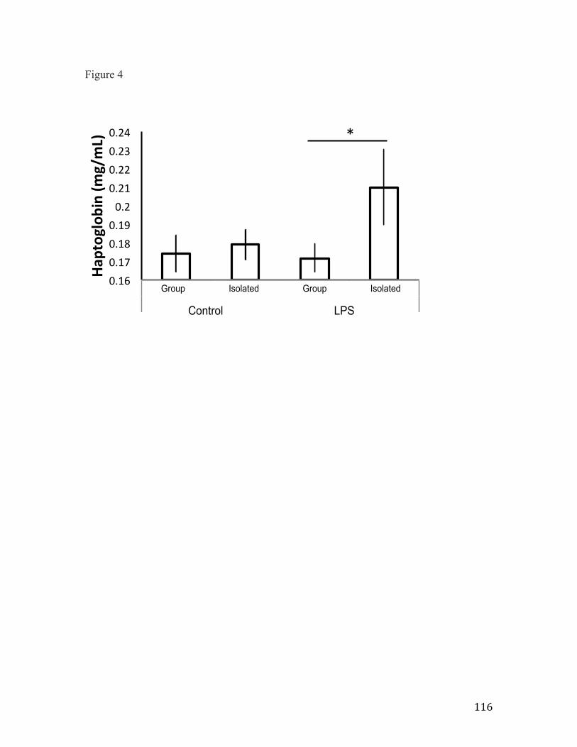
116
Figure 4
Group& Isolated& Group& Isolated&
CONTROL& LPS&
2h& 0.17407514& 0.179024369& 0.171685202& 0.210051118&
0.16&0.17&0.18&0.19&0.2&0.21&0.22&0.23&0.24&
Haptoglobin+(m
g/mL)+
Group Isolated Group Isolated
Pro
port
ion
of ti
me
hopp
ing
(sec
)
0.00
0.05
0.10
0.15
0.20
0.25
0.302 hr4 hr
Control LPS
*

117
Figure 5 – Mean change in body temperature (Tpost-injection -Tat injection time), represented as means ± SEM. Higher positive values indicate higher degree of hypothermia. Sample sizes were Group-Control=8, Isolated-Control=5, Group-LPS=7, Isolated-LPS=6. The ANOVA indicated no main effects significant main effects. Asterisk indicates results of planned comparisons at P<0.05.

118
Figure 5
Group& Isolated& Group& Isolated&
CONTROL& LPS&
2h& 0.6625& 1.16& 1.085714286& @1&
@2&@1.5&@1&
@0.5&0&
0.5&1&
1.5&2&
Delta
&bod
y&temp.&(oC)&
Group Isolated Group Isolated
Pro
port
ion
of ti
me
hopp
ing
(sec
)
0.00
0.05
0.10
0.15
0.20
0.25
0.302 hr4 hr
Control LPS
*

119
Figure 6 – Mass lost during the course of the experiment (Day1 – Day2), represented as means ± SEM. Sample sizes were Group-Control=10, Isolated-Control=8, Group-LPS=10, Isolated-LPS=10. The ANOVA indicated a significant main effect for social treatment. Asterisk indicates results of planned comparisons at P<0.05.

120
Figure 6
Group& Isolated& Group& Isolated&
CONTROL& LPS&
2h& 80.404& 80.73125& 80.598& 80.974&
81.2&
81&
80.8&
80.6&
80.4&
80.2&
0&Mass$loss$(g)$
Group Isolated Group Isolated
Pro
port
ion
of ti
me
hopp
ing
(sec
)
0.00
0.05
0.10
0.15
0.20
0.25
0.302 hr4 hr
Control LPS
*

121
Figure 7 – Bacterial killing ability of whole blood expressed as % colonies killed, represented as means ± SEM. Sample sizes were Group-Control=10, Isolated-Control=8, Group-LPS=9, Isolated-LPS=10. Asterisk indicates significance results at P<0.05.

122
Figure 7
Group& Isolated& Group& Isolated&
CONTROL& LPS&
2h& 50.51851852& 57.12962963& 71.6872428& 77.77777778&
30&
40&
50&
60&
70&
80&
90&
%"colon
ies"k
illed
"
Group Isolated Group Isolated
Pro
port
ion
of ti
me
hopp
ing
(sec
)
0.00
0.05
0.10
0.15
0.20
0.25
0.302 hr4 hr
Control LPS
*