Embed Size (px)
Citation preview
Livestock Health, Management and Production › High Impact Diseases › Vector-borne Diseases › African Horse Sickness ›
1 | P a g e
African Horse Sickness Author: Dr Melvyn Quan
Adapted from: Coetzer, J.A.W and Guthrie, A.J. 2004. African horse sickness, in Infectious diseases of livestock, edited by J.A.W. Coetzer & R.C. Tustin. Oxford University Press, Cape Town, 2: 1231-1264
Licensed under a Creative Commons Attribution license.
TABLE OF CONTENTS
Table of contents........................................................................................................... 1
Introduction ................................................................................................................... 2
Epidemiology ................................................................................................................. 2
Pathogenesis ................................................................................................................. 5
Diagnosis and differential diagnosis ........................................................................... 7
Clinical signs and pathology .................................................................................................. 7
“Dunkop” or pulmonary form ......................................................................................... 7
“Dikkop” or cardiac form ............................................................................................... 9
Mixed form ..................................................................................................................13
Horse sickness fever ...................................................................................................13
Laboratory confirmation ........................................................................................................14
Differential diagnosis ............................................................................................................14
Control / Prevention .................................................................................................... 15
Marketing and trade / Socio-economics .................................................................... 17
Important outbreaks .................................................................................................... 18
FAQ ............................................................................................................................... 19
References ................................................................................................................... 21
Livestock Health, Management and Production › High Impact Diseases › Vector-borne Diseases › African Horse Sickness ›
2 | P a g e
INTRODUCTION
African horse sickness (AHS) is a peracute, acute, subacute or mild infectious but non-contagious disease of equids caused by African horse sickness virus (AHSV). The virus is classified in the genus Orbivirus of the family Reoviridae, of which there are nine serotypes, all transmitted by Culicoides midges. AHSV is similar in morphology and shares many properties with other orbiviruses, such as equine encephalosis virus, bluetongue virus and epizootic haemorrhagic disease virus. The virus is closely associated with erythrocytes. Of the nine serotypes of AHSV, there is cross-neutralization between serotypes 1 and 2, 3 and 7, 5 and 8 and 6 and 9.
AHS is a World Organisation for Animal Health (OIE) - listed disease and manifests pyrexia and clinical signs compatible with impaired respiratory and circulatory functions, characterized by oedema of subcutaneous and intermuscular tissues and of the lungs, transudation into the body cavities and haemorrhages on serosal surfaces. The mortality rate in horses, the most susceptible species, may be as high as 95 per cent, while mules are less susceptible and donkeys with rare exceptions only develop inapparent infections.
Although mortality in horses as a result of AHS occurs every year in South Africa, major epidemics prior to 1953 seem to have occurred at intervals of roughly 20 to 30 years. The timing of these epizootics has been associated with the warm (El Niño) phase of the El Niño/Southern Oscillation (ENSO) phenomenon. Severe losses were reported in 1780, 1801, 1839, 1855, 1862, 1891, 1914, 1918, 1923, 1940, 1946 and 1953.
EPIDEMIOLOGY
AHS is endemic to sub-Saharan Africa, with recent reports of outbreaks in southern Africa, Ethiopia Ghana, Nigeria and Senegal. Occasional outbreaks have, in the past, been reported in northern Africa, the Iberian Peninsula and the Middle East (Figure 1). Its intrusion into North Africa and countries around the Mediterranean Sea and Asia is impeded by the Sahara desert. Apparently AHS does not occur in Madagascar or Mauritius.

Livestock Health, Management and Production › High Impact Diseases › Vector-borne Diseases › African Horse Sickness ›
3 | P a g e
Fig. 1: Worldwide distribution of African horse sickness
In South Africa, AHS appears to be endemic in the low-lying, eastern, summer-rainfall areas of the country. Although the inland plateau “Highveld” regions and most of the three Cape Provinces are usually free of the disease, the infection has sometimes extended into these areas and caused serious losses. According to Du Toit, heavy losses were reported even at Belfast, a town situated at almost the highest altitude (about 2 100 metres above sea level) in Mpumalanga, during the severe epidemic of 1923. Outbreaks of AHS in the Cape Peninsula of the Western Cape Province have only been reported in 1967, 1990, 1999, 2001, 2004, 2006 and 2011, all associated with the importation of infected horses.
Heavy rains followed by warm, dry spells favour the occurrence of epidemics. In South Africa, the first cases of AHS usually occur in the beginning of February, but the most serious outbreaks are commonly encountered in March and April. Following the first frosts, which are usually experienced towards the end of April or in May, the disease disappears abruptly. However, in the Lowveld region, where frost is less common, cases of the disease may continue into May and June.
During outbreaks of AHS in endemic areas, different virus serotypes may be active simultaneously within an area, but one serotype usually dominates during a particular season, followed the next year by the dominance of another serotype. While all serotypes are detected with varying frequencies in southern Africa, the most common circulating serotypes currently in northern Africa are serotypes 2, 4 and 9. Serotypes 3, 4 and 9 have been recorded outside Africa.
Livestock Health, Management and Production › High Impact Diseases › Vector-borne Diseases › African Horse Sickness ›
4 | P a g e
AHS affects primarily equine species. Horses are the species most susceptible to the disease (mortality rate between 70 to 95 per cent) and mules less so (mortality rate of 50 to 70 per cent). Donkeys and zebras are very resistant, with most infections subclinical. Horses of all breeds are in general equally susceptible to AHS but variation in susceptibility to the same virus of individual horses has been reported. There are horses in North and West Africa which descended from animals present there since 2 000 BC, and which have apparently acquired natural resistance to AHS. Theiler was unable to produce fatal disease in donkeys inoculated with large quantities of virulent blood; at most a mild fever reaction was produced. Donkeys in the Middle East, however, appear to be far more susceptible to disease (the mortality rate may reach 10 per cent) than donkeys in Africa. Zebras are highly resistant to the disease and only show a mild fever following experimental infection.
Foals born to immune mares acquire passive immunity by the ingestion of colostrum soon after birth. This immunity progressively declines as a result of catabolism of the immunoglobulins until it is completely lost after about four to six months. Foals born to fully susceptible mares are just as susceptible as are previously unexposed adult horses.
In addition to equids, dogs are the only other species that contract a highly fatal form of the disease after infection with AHSV. All documented natural cases in dogs have resulted from the ingestion of infected horse meat, but experimentally they are also susceptible to infection by inoculation of virus by various routes. AHSV serotype 9 has been isolated from the blood of stray dogs in Egypt, and antibodies to AHSV have been detected in the sera of dogs in India and South Africa. The role dogs play in the epidemiology of AHSV, if any, is unknown.
Besides early reports, little evidence of antibody to AHSV has been detected in domestic or wild ruminants with the possible exception of camels. Pigs, cats and monkeys are refractory to infection with AHSV.
Amongst wildlife, antibodies to AHSV have only been found in zebras, African elephants (Loxondonta africana) and black and white rhinoceroses (Diceros bicornis and Ceratotherium simium). However, the possibility that elephants are reservoir hosts of AHSV is questioned as a 100 elephants tested in an endemic AHS area in South Africa had high complement fixing titres to both bluetongue and AHS viruses, but no neutralizing antibodies to these viruses, indicating that the sera of elephants reacts non-specifically in the complement fixation test. In contrast, in a similar study in Kenya, elephants were shown to have neutralizing antibody to AHSV.
AHS is not contagious. AHSV is transmitted biologically by Culicoides spp., of which C. imicola and C. bolitinos have been shown to play an important role in Africa. Culicoides variipennis, a midge prevalent in the USA but not present in southern Africa, can transmit the virus to chicken embryos in the lab. In Europe, C. dewulfi, C. obsoletus sensu strictu, C. scoticus, C. pulicaris, and C. chiopterus have been implicated as vectors of the related bluetongue virus. Culicoides midges, in general, breed in damp soil

Livestock Health, Management and Production › High Impact Diseases › Vector-borne Diseases › African Horse Sickness ›
5 | P a g e
rich in organic matter, however C. bolitinos breeds in bovine dung, and it therefore not as dependant on annual rainfall and soil-type.
Adult midges become infected by taking blood meals from viraemic animals. Virogenesis in the midge is approximately eight days after which AHSV localises in the salivary glands and is transmitted to the next host when the midge takes its next blood meal. Both infection rate and rate of virogenesis of AHSV within midges are temperature dependent: the lower the temperature, the lower the infection rate and longer the virogenesis. Transovarial transmission and overwintering of AHSV in Culicoides spp. has not been demonstrated.
Free-ranging animals become infected with AHSV during the period between sunset and sunrise when midges are most active. The virus spreads by movement of either infected vertebrate hosts which then infect vectors in the “new” region, or vectors. AHSV can be distributed over great distances if equids incubating AHS are translocated by land, sea or air. During their lifetimes midges remain generally within a radius of a few kilometres of the site where they breed, but it has been postulated that they can be borne for many hundreds of kilometres in air currents. It has been suggested that infected midges transported by wind were responsible for the spread of the disease over the sea from Senegal to the Cape Verde Islands in 1943, from Turkey to Cyprus in 1960 and from Morocco to Spain in 1966.
It has been shown that a continuous transmission cycle of AHSV between Culicoides midges and zebras exists in the Kruger National Park in South Africa. Under such circumstances a sufficiently large zebra population can act as a reservoir for virus. It has also been suggested that donkeys may play a similar role in parts of Africa where there are large donkey populations. In view of the high mortality rate in horses, this species is regarded as an accidental or indicator host. Horses that have recovered from the disease do not remain carriers of the virus, which explains the failure of the disease to become permanently established outside tropical and subtropical Africa, despite the occurrence of the many outbreaks that have occurred outside endemic areas.
PATHOGENESIS
The outcome of infection in horses, including the incubation period and severity of disease, depends largely on the virulence of the virus and susceptibility of the animal. In experimentally infected cases, the incubation period of AHS varies between five and seven days, but it may be as short as two days and is rarely longer than ten days. After infection, initial multiplication of virus occurs in the regional lymph nodes and is followed by a primary viraemia with subsequent infection of target organs, namely, the lungs and lymphoid tissues throughout the body. Virus multiplication at these sites gives rise to a secondary viraemia of variable duration; in horses it is generally not higher than 105 TCID50/ml and lasts four to eight days, but does not exceed 21 days, whereas in donkeys and zebras the levels of viraemia are lower but
Livestock Health, Management and Production › High Impact Diseases › Vector-borne Diseases › African Horse Sickness ›
6 | P a g e
they may last as long as four weeks. In zebras viraemia has been reported to occur in the presence of circulating antibodies. AHSV is closely associated with the erythrocytes in the blood.
Effusions into body cavities and oedematous changes of various tissues (particularly of the lungs), as well as serosal and visceral haemorrhages, are often evident in fatal cases of AHS and indicate endothelial cell damage. Although no significant ultrastructural changes or evidence of viral replication could be detected in endothelial cells in the lungs in one study, in another the presence of virus and ultrastructural changes in, and separation of, endothelial cells in the lungs were found.
The factors determining the course and severity of infection are not fully understood. Theiler described four forms of AHS in horses, and these are still useful in categorizing the different effects AHSV may have on equids. These are, in order of decreasing severity: the peracute, “pulmonary” or “dunkop” (“thin head”) form, i.e. cases in which subcutaneous swelling of the head is absent); the acute or “mixed” form; the subacute, oedematous, “cardiac” or “dikkop” (“thick” or “swollen head”) form; and the horse sickness fever form. It has been shown that small plaque variants produce severe clinical reaction, while large plaque variants of AHSV produce no reactions or only mild ones. There is some evidence that serial passage of AHSV in horses using virus that has originated from the lung tissue of each successive animal consistently produces the peracute disease with marked pulmonary involvement, whereas passage using spleen homogenates from the same animal results in a progressively milder disease.
Fully susceptible horses, such as foals that have lost their colostral immunity or horses that have never been exposed to the AHSV, usually develop the “dunkop” form of AHS. Exercise during the febrile stage of the disease may also precipitate the “dunkop” form. Theiler claimed that the “mixed” form is rare and that many such cases arise from dual infections with various AHS strains. Subsequent observations indicated that the rigid distinction between different forms of AHS is not fully justifiable and that most cases of AHS are of the “mixed” form. Although more than one serotype may be active during an outbreak, isolation of more than one serotype from a naturally infected animal has never been recorded.
Pulmonary alveolar flooding by oedematous fluid would seem to occur terminally in most fatal cases although once it is initiated it may develop very rapidly, particularly in “dunkop” cases. The histological demonstration of an abundance of fibrin in the pulmonary alveoli of horses that have died of the “dunkop” form indicates that the oedema fluid has a high protein content, compatible with that of a permeability oedema. This oedema results in respiratory distress, and ultimately, asphyxia.
Livestock Health, Management and Production › High Impact Diseases › Vector-borne Diseases › African Horse Sickness ›
7 | P a g e
DIAGNOSIS AND DIFFERENTIAL DIAGNOSIS
Clinical signs and pathology
Four clinical forms of AHS have been described – the “dunkop” or pulmonary, “dikkop” or cardiac, mixed and horse sickness fever forms.
“Dunkop” or pulmonary form
This form of the disease occurs commonly when AHSV infects fully susceptible horses, notably foals that have lost their passively acquired immunity. It is the form usually seen in dogs.
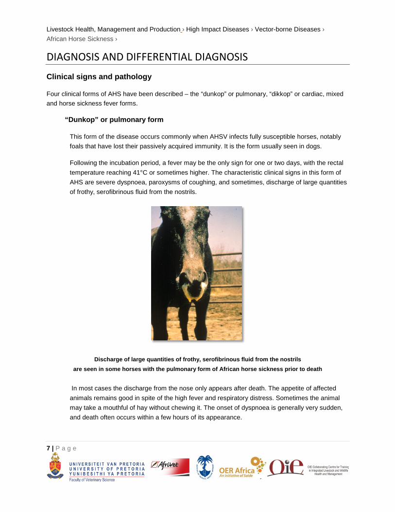
Following the incubation period, a fever may be the only sign for one or two days, with the rectal temperature reaching 41°C or sometimes higher. The characteristic clinical signs in this form of AHS are severe dyspnoea, paroxysms of coughing, and sometimes, discharge of large quantities of frothy, serofibrinous fluid from the nostrils.
Discharge of large quantities of frothy, serofibrinous fluid from the nostrils are seen in some horses with the pulmonary form of African horse sickness prior to death
In most cases the discharge from the nose only appears after death. The appetite of affected animals remains good in spite of the high fever and respiratory distress. Sometimes the animal may take a mouthful of hay without chewing it. The onset of dyspnoea is generally very sudden, and death often occurs within a few hours of its appearance.
Livestock Health, Management and Production › High Impact Diseases › Vector-borne Diseases › African Horse Sickness ›
8 | P a g e
The prognosis for horses suffering from the “dunkop” form is extremely grave; less than five per cent recover. In recovering horses the fever gradually subsides but the breathing remains laboured for some days.
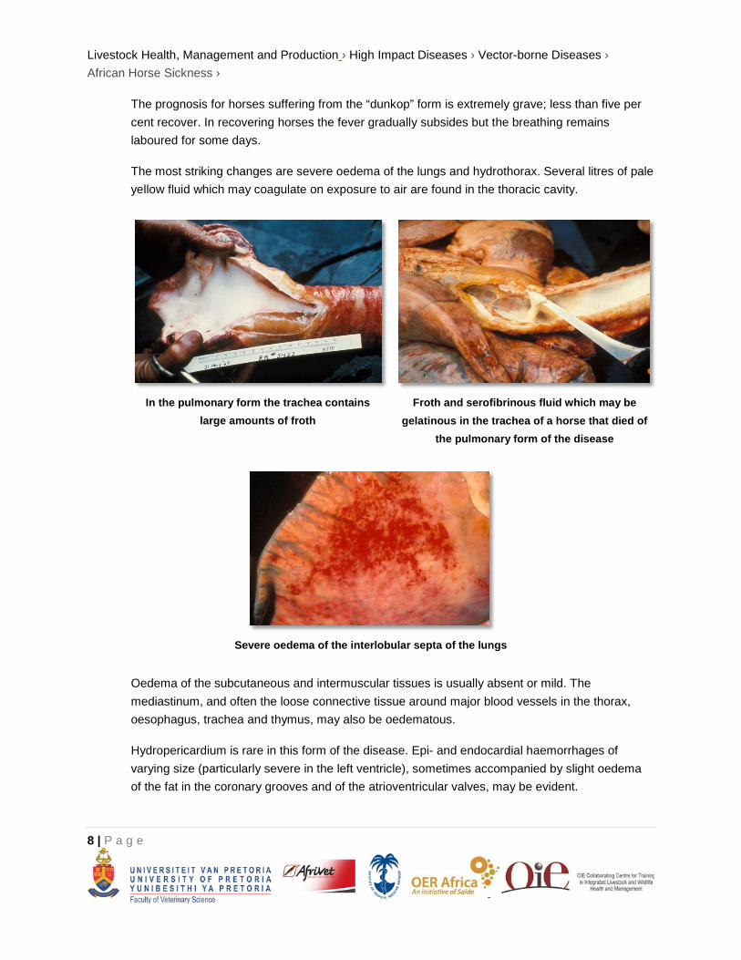
The most striking changes are severe oedema of the lungs and hydrothorax. Several litres of pale yellow fluid which may coagulate on exposure to air are found in the thoracic cavity.
In the pulmonary form the trachea contains
large amounts of froth
Froth and serofibrinous fluid which may be
gelatinous in the trachea of a horse that died of the pulmonary form of the disease
Severe oedema of the interlobular septa of the lungs
Oedema of the subcutaneous and intermuscular tissues is usually absent or mild. The mediastinum, and often the loose connective tissue around major blood vessels in the thorax, oesophagus, trachea and thymus, may also be oedematous.
Hydropericardium is rare in this form of the disease. Epi- and endocardial haemorrhages of varying size (particularly severe in the left ventricle), sometimes accompanied by slight oedema of the fat in the coronary grooves and of the atrioventricular valves, may be evident.

Livestock Health, Management and Production › High Impact Diseases › Vector-borne Diseases › African Horse Sickness ›
9 | P a g e
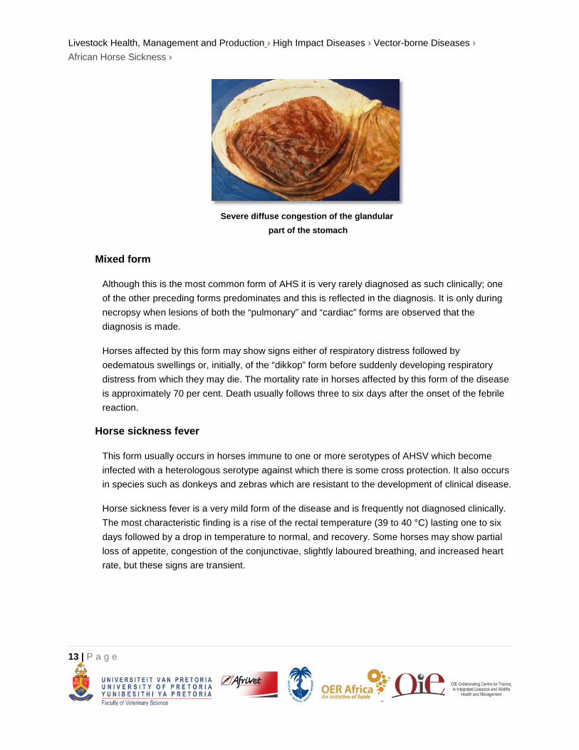
Marked diffuse congestion of the mucosa of the glandular part of the stomach is a consistent finding. This may be accompanied by patchy congestion and petechiation of the serosa and, sometimes, of the mucosa of the intestine. The liver may be slightly enlarged and congested, and its lobulation slightly more distinct than normal. There is usually some degree of ascites.
“Dikkop” or cardiac form
This form of AHS is characterized by subcutaneous oedema of the head and neck, and particularly the supraorbital fossae.
Facial swelling and oedema of the
supraorbital fossae
The oedema usually appears late in the course of the disease, but if it appears early, the condition is more serious, more acute and with a higher rate of mortality. In severe cases, there may be oedema of the eyelids, lips, cheeks, tongue, intermandibular space, and sometimes also the neck, chest and shoulders, but usually not the lower parts of the legs (i.e. below the elbow or stifle joints).

Livestock Health, Management and Production › High Impact Diseases › Vector-borne Diseases › African Horse Sickness ›
10 | P a g e
Oedema of the supraorbital
fossa and eye lids
Severe oedema of the eyelids in a horse suffering from African horse sickness
As the oedema worsens, dyspnoea and cyanosis may supervene.
Fever peaks at a later stage in the course of the disease than in the “dunkop” form, and may remain high for three to six days before declining. Some animals will only develop a very mild fever. Some animals may lie down repeatedly or are restless when standing, and frequently paw the ground with their front feet as a result of severe colic. Interference or difficulty in swallowing as a result of paralysis of the oesophagus may be a complication, particularly in those cases where there is severe oedema of the head.
The “dikkop” form of AHS is always more protracted and milder than the “dunkop” form, with a mortality rate of about 50 per cent. Death usually occurs within four to eight days of the onset of the febrile reaction. Unfavourable prognostic signs are petechiae in the mucosa of the conjunctivae and mouth, on the ventral aspect of the tongue. These, if they occur, are evident shortly before death.
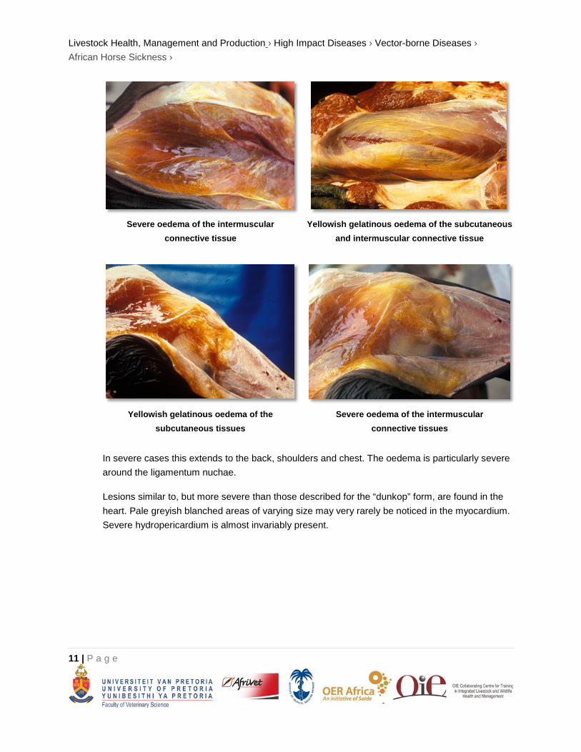
The most characteristic pathological change is yellowish gelatinous oedema of the subcutaneous and intermuscular connective tissues of the head and neck.
Livestock Health, Management and Production › High Impact Diseases › Vector-borne Diseases › African Horse Sickness ›
11 | P a g e
Severe oedema of the intermuscular
connective tissue
Yellowish gelatinous oedema of the subcutaneous
and intermuscular connective tissue
Yellowish gelatinous oedema of the
subcutaneous tissues
Severe oedema of the intermuscular
connective tissues
In severe cases this extends to the back, shoulders and chest. The oedema is particularly severe around the ligamentum nuchae.
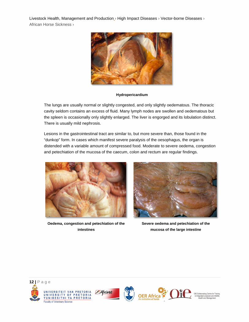
Lesions similar to, but more severe than those described for the “dunkop” form, are found in the heart. Pale greyish blanched areas of varying size may very rarely be noticed in the myocardium. Severe hydropericardium is almost invariably present.
Livestock Health, Management and Production › High Impact Diseases › Vector-borne Diseases › African Horse Sickness ›
12 | P a g e
Hydropericardium
The lungs are usually normal or slightly congested, and only slightly oedematous. The thoracic cavity seldom contains an excess of fluid. Many lymph nodes are swollen and oedematous but the spleen is occasionally only slightly enlarged. The liver is engorged and its lobulation distinct. There is usually mild nephrosis.
Lesions in the gastrointestinal tract are similar to, but more severe than, those found in the “dunkop” form. In cases which manifest severe paralysis of the oesophagus, the organ is distended with a variable amount of compressed food. Moderate to severe oedema, congestion and petechiation of the mucosa of the caecum, colon and rectum are regular findings.
Oedema, congestion and petechiation of the
intestines
Severe oedema and petechiation of the
mucosa of the large intestine
Livestock Health, Management and Production › High Impact Diseases › Vector-borne Diseases › African Horse Sickness ›
13 | P a g e
Severe diffuse congestion of the glandular
part of the stomach
Mixed form
Although this is the most common form of AHS it is very rarely diagnosed as such clinically; one of the other preceding forms predominates and this is reflected in the diagnosis. It is only during necropsy when lesions of both the “pulmonary” and “cardiac” forms are observed that the diagnosis is made.
Horses affected by this form may show signs either of respiratory distress followed by oedematous swellings or, initially, of the “dikkop” form before suddenly developing respiratory distress from which they may die. The mortality rate in horses affected by this form of the disease is approximately 70 per cent. Death usually follows three to six days after the onset of the febrile reaction.
Horse sickness fever
This form usually occurs in horses immune to one or more serotypes of AHSV which become infected with a heterologous serotype against which there is some cross protection. It also occurs in species such as donkeys and zebras which are resistant to the development of clinical disease.
Horse sickness fever is a very mild form of the disease and is frequently not diagnosed clinically. The most characteristic finding is a rise of the rectal temperature (39 to 40 °C) lasting one to six days followed by a drop in temperature to normal, and recovery. Some horses may show partial loss of appetite, congestion of the conjunctivae, slightly laboured breathing, and increased heart rate, but these signs are transient.

Livestock Health, Management and Production › High Impact Diseases › Vector-borne Diseases › African Horse Sickness ›
14 | P a g e
Laboratory confirmation
The epidemiology, clinical signs and gross lesions of AHS are often sufficiently specific to allow a provisional diagnosis of the disease to be made. To confirm the diagnosis, the virus should be isolated from blood collected, preferably in heparin-containing tubes, during the febrile stage of the disease or from specimens of the lungs, spleen and lymph nodes collected at necropsy and kept at 4 °C. In fatal cases high levels of infectivity are found in the lungs, spleen and lymph nodes. Virus isolation may be achieved by the use of a variety of cell cultures (e.g. BHK-21, VERO, monkey stable), by intracerebral inoculation of suckling mice or by intravenous inoculation of embryonated hens' eggs. When heparin is not available, blood collected in chelating agents such as Na-EDTA should be diluted five to ten fold to prevent detachment of the monolayer of cell cultures.
Virus isolates are identified by group-specific tests such as complement fixation (CF), agar gel immunodiffusion (AGID), direct and indirect immunofluorescence (IFA) or enzyme-linked immunosorbent assay (ELISA). Serotyping of AHS virus isolates is performed using virus neutralization (VN) tests employing type-specific antisera. Virus neutralization tests may be conducted in mice or various cell cultures e.g. plaque neutralization. Antibody to AHSV can be detected by utilizing CF, AGID, IFA, VN and ELISA tests.
In horses that have recovered from the disease, high CF antibody titres indicate infection with the virus within the previous few months. Antibodies detectable by VN tests and ELISA persist for a number of years.
A number of conventional reverse transcription polymerase chain reaction (RT-PCR) assays and real-time reverse transcription PCR assays have been developed for the detection of AHSV and to differentiate between the serotypes of AHSV. Genomic probes can also be developed and applied for in situ hybridization in tissues. Immunohistochemical staining methods have also been used successfully to determine the localization of AHS antigen within various tissues. The advantages of these new approaches are that they have the potential to be rapid, sensitive and versatile, and they may supplement or replace some of the older conventional methods. Furthermore, they can be applied to specimens from clinical cases that do not contain live virus. Immunohistochemical staining can be applied to tissues that have been preserved in 10% buffered formalin, which is an advantage if it is not possible for the samples to reach the laboratory rapidly.
Differential diagnosis
It is not possible to differentiate the AHS fever form of the disease from the early febrile stages of other forms of AHS, or from the early febrile stages of many other equine infectious diseases.
The clinical signs and lesions of AHS may be confused with those of equine encephalosis. As many of the epidemiological features of the two diseases are similar, they may occur simultaneously in South Africa and possibly also in other regions in southern Africa. Horses manifesting swelling of the eyelids,

Livestock Health, Management and Production › High Impact Diseases › Vector-borne Diseases › African Horse Sickness ›
15 | P a g e
supraorbital fossae or of the entire head as a result of equine encephalosis cannot be differentiated clinically from the “dikkop” form of AHS. However, the mortality rate of AHS is much higher than is that of equine encephalosis. Virus isolation and identification or serological tests on paired serum samples are essential to confirm either diagnosis.
Although the lesions of AHS at necropsy are fairly characteristic, the disseminated small haemorrhages and the oedema found in some parts of the body that are associated with the “dikkop” form of AHS may be very similar to those found in cases of purpura haemorrhagica and equine viral arteritis. In horses affected by these conditions, subcutaneous oedema often occurs ventral to the elbow and stifle joints, as well as in other locations, while in those suffering from AHS it does not occur below these joints. In purpura haemorrhagica, oedema and haemorrhages tend to be more numerous, widespread and severe than in AHS.
The early stages of piroplasmosis, when Babesia/Theileria parasites may be difficult to demonstrate in blood smears, are occasionally confused with AHS. AHS may also be complicated by piroplasmosis, and in such cases ventral oedema may be severe.
CONTROL / PREVENTION
Apart from supportive treatment, there is no specific therapy for AHS. Affected animals should be well nursed, fed and rested, as the slightest exertion may result in death as a result of cardiac failure. After recovery they should be rested for at least four weeks before being returned to light work. As piroplasmosis may be a complication of AHS, blood smears as well as the rectal temperature (to detect a secondary febrile reaction) should be taken regularly and, if the smears are found to be positive, animals should be treated appropriately.
As all AHSV serotypes are present in South Africa and in most parts of sub-Saharan Africa, the use of a polyvalent vaccine is necessary to protect horses in these areas. Several methods of immunization were attempted prior to 1930 with varying success. Since the demonstration in the early 1930s that AHSV can be attenuated by serial intracerebral passage in mice, the immunization of horses against the disease has been greatly simplified and improved. By exploiting these findings Alexander, Neitz and du Toit showed in 1936 that an effective attenuated vaccine could be produced. Today, large plaque variants of AHSV are selected as candidate vaccine strains.
In endemic areas and in regions where AHS occurs almost every year, viz. most parts of Africa south of the Sahara, annual vaccination of horses is a very practical means of control. Although prophylactic immunization against AHS is an efficient method of preventing serious losses, it cannot be relied upon fully to protect horses against infection or disease. However, the majority of horses that have received three or more courses of immunization are usually well protected against the disease. In South Africa,
Livestock Health, Management and Production › High Impact Diseases › Vector-borne Diseases › African Horse Sickness ›
16 | P a g e
annual immunization with a live polyvalent attenuated vaccine in late winter or early summer (September to November), which is some time before the peak AHS season, is advocated and allows immunized animals to respond adequately to the vaccine before possibly being challenged by natural exposure. Onderstepoort Biological Products currently produces a polyvalent vaccine containing attenuated strains prepared in two components one of which is trivalent (serotypes 1, 3 and 4) and the other of which is quadrivalent (serotypes 2, 6, 7 and 8). A course of immunization consists of the administration of these component vaccines three to four weeks apart. Serotypes 5 and 9 are not included in the vaccines as serotype 8 and 6, respectively, afford adequate cross protection.
Generally, immunization has no, or only limited, side effects. A slight temperature response may ensue between five and 13 days after inoculation as a result of a low-level virus replication in immunized animals. Horses should not be excessively exerted for about three weeks after the first course of immunization.
Immunity to either live viscerotropic or attenuated AHSV strains is solid against the homologous virus and probably lasts indefinitely. Cross immunity between serotypes may be enhanced by repeated inoculation of the same virus. There is evidence that animals that recover from infection with a field virus acquire a broader cross immunity to the other serotypes than that which is obtained from immunization. The simultaneous inoculation of several serotypes of attenuated AHS in horses usually results in the production of antibody against each serotype, although the response of individual horses may vary, and in some animals, antibody against one or more of the serotypes inoculated may not be detectable by VN tests. This is possibly because of interference between viruses in the polyvalent vaccine or over attenuation of vaccine strains. For these reasons annual vaccination of horses with the polyvalent vaccine is advocated in high risk areas in Africa in order to ensure the production of the widest possible polyvalent immunity to the different serotypes. During outbreaks of AHS in Spain in the late 1980s about ten per cent of animals (horses, mules and donkeys) immunized for the first time with an attenuated monovalent AHSV serotype 4 vaccine failed to seroconvert.
In most instances the levels of antibodies acquired by foals from colostrum correlate well with the levels of antibodies in the sera of their dams and determine the duration of their passive immunity. Because of the passive immunity acquired by foals born to immune mares, it is generally recommended that foals should not be immunized before they are six months of age. However, in foals which acquire low levels of antibody to one or more AHSV serotypes via the colostrum, neutralizing antibody to individual serotypes may decline to undetectable levels two to four months after birth suggesting that foals could become susceptible to infection well before the age of six months which is the age commonly recommended for initial vaccination. As a result of the restricted breeding season of Thoroughbreds, most foals in southern Africa are three to five months of age by late February when possible challenge with AHSV can be expected to be highest. It is therefore customary in Zimbabwe to immunize foals in January and again in September. Animals should be vaccinated every six months in their first and second years of life and thereafter annually.
Livestock Health, Management and Production › High Impact Diseases › Vector-borne Diseases › African Horse Sickness ›
17 | P a g e
Recombinant vaccines have been developed and in some cases shown to be effective vaccines. None are commercially available.
Infection of susceptible horses can be prevented to a large degree by stabling them some hours before sunset and letting them out a few hours after sunrise, as most Culicoides spp. are nocturnal and are not inclined to enter buildings at night. Thus, horses in urban and peri-urban locations which are stabled at night are less likely to become infected. The application of insect repellents and the use of insecticides on animals' coats will also discourage Culicoides from feeding on them.
MARKETING AND TRADE / SOCIO-ECONOMICS
Before the advent of mechanized transport the horse played a very important role in transportation, was vital in military operations and was used for draught power in agriculture and mining. Heavy losses as a result of AHS were therefore very disruptive and experienced very negatively. Although horses are nowadays seldom used for these purposes, many have considerable monetary value as performance horses or animals used for other forms of recreation.
Up to the late 1950s, it was thought that South Africa had exported (by sea) close to 350 000 horses to other parts of the world, largely in support of the war effort during the first and second World Wars. The outbreak of AHS in the Middle East in 1959 raised global fears of AHS and an embargo on the movement of horses out of Africa was put in place for the next four decades. The exception was the USA which accepted horses from Africa on the basis of a 60-day post arrival vector-proof quarantine.
Following a combined initiative of the broader equestrian industry, scientific and veterinary sectors, the European Union (EU) ratified the South African Horse Export Protocol in 1997 and South Africa has since exported close to 1000 horses from Kenilworth Quarantine Station in the AHS-Free Zone in Cape Town worth an estimated R250 million per annum. Since then, there have been five outbreaks of AHS in the AHS surveillance zone. Every time this has led to the temporary suspension of imports from South Africa to the EU. Consequently, the Directorate of Animal Health of the Department of Agriculture, Forestry and Fisheries (DAFF) formally applied to a number of South Africa’s trading partners to consider a protocol that would allow direct imports from Cape Town during the low-vector season. The application is based on the 2008 version of the OIE AHS Code with the offer of equivalence which is summarized as follows:
• Export during the low risk time of the year (80 days after last case).
• Export only from the AHS-Free Zone.
• Continuous residence in vector-proof quarantine station.
• Limited exercise under vector protection.
Livestock Health, Management and Production › High Impact Diseases › Vector-borne Diseases › African Horse Sickness ›
18 | P a g e
• Additional testing, including real-time RT-PCR (in process of OIE validation).
Revisions to the 2008 version of the OIE AHS code will address the risk posed by AHS to importing countries. Changes that will, most likely, be adopted at the 2012 OIE General Assembly will introduce the following principles:
• Official OIE recognition of AHS.
• South Africa to define own control measures as accepted by the OIE.
• Pre-export quarantine to be significantly reduced with testing (RT- PCR).
• If an outbreak occurred in the AHS Controlled Area (Containment Zone), it will be possible to resume exporting 80 days after the last case of AHS was confirmed.
IMPORTANT OUTBREAKS
In Egypt the disease occurred in 1928, 1943, 1953, 1958 and 1971 (all outbreaks originated in the areas of Aswan and Qena Provinces, and the international boundaries between Egypt and Sudan), in Yemen in 1930 and in the then Palestine, Syria, Lebanon and Jordan in 1944.
During the summer of 1959, AHS caused by serotype 9 occurred in the south eastern regions of Iran. This was followed by outbreaks during the spring of 1960 in most of the Persian Gulf area (Cyprus, Iraq, Syria, Lebanon, Jordan) as well as in Afghanistan, Pakistan, India and Turkey. Between 1959 and 1961 this region lost more than 300 000 equids.
In 1965 the disease occurred in Libya, Tunisia, Algeria and Morocco and also spread across the Strait of Gibraltar into Spain in 1966. Fortunately it occurred at the end of the summer and the outbreak in Spain was quickly terminated by application of strict control measures.
Between 1987 and 1990 outbreaks of AHS occurred again in Spain; the original source of infection was suspected to be ten zebras (Equus burchelli) imported from Namibia. AHS was also diagnosed in southern Portugal in 1989 and northern Morocco between 1989 and 1991; these outbreaks are thought to be extensions of outbreaks that occurred in August of the same year in southern Spain.
In 1989 a minor outbreak of AHS, caused by serotype 9, was reported in Saudi Arabia after an absence of 30 years. In 1997 outbreaks of AHS were reported in Saudi Arabia and Yemen.
AHS is endemic to southern Africa. An outbreak of 1855 is still considered to have been the largest in the region; nearly 70 000 horses, making up more than 40 per cent of the entire horse population of the Cape of Good Hope, died. In northern Africa, AHS is reported regularly in Ethiopia and Senegal and on

Livestock Health, Management and Production › High Impact Diseases › Vector-borne Diseases › African Horse Sickness ›
19 | P a g e
occasion from Eritrea, Burkina Faso, Nigeria and The Gambia. It has been shown recently that a live-attenuated vaccine-derived serotype 9 strain of AHSV is circulating in The Gambia. Outbreaks of AHS have only been reported once in Côte d’Ivoire (1997) and on the Cape Verde islands (1999).
FAQ
1. Which samples should be submitted for AHS diagnosis?
From a live animal, collect blood samples in a yellow (or red), purple and green-top tube (no additive, EDTA and heparin respectively). From a dead animal, collect heart blood or blood clots and/or 3x3x3 cm blocks of spleen and lung tissue in clean containers, for virus isolation. Collect two to three 1x1x1 cm blocks of heart (in particular a piece of the left ventricle through the papillary muscle, and the interventricular septum) spleen (any area) and lung (especially congested and oedematous areas) tissue in containers with 10% buffered formalin, for immunohistochemistry testing. If a post-mortem examination is not performed, collect jugular blood and spleen tissue through an incision in the abdominal wall.
2. What is a CT value and does a positive PCR assay confirm that my horse has AHS?
The polymerase chain reaction (PCR) amplifies a target region of DNA in a sample (in this case AHSV nucleic material) by cycling the sample, to which reagents and enzymes have been added, through a range of temperatures. One cycle consists of raising the temperature of the sample to 95°C (to denature the DNA and separate the complementary strands of DNA), dropping the temperature to 55-60°C (to allow primers complementary to the target DNA sequence to attach), and then raising the temperature to 72°C (to allow the DNA polymerase to amplify the sequence). This sequence of temperature changes is repeated 35 to 40 times and with each cycle, the target DNA is doubled (assuming perfect efficiency). If one copy of the target DNA is present in the sample before PCR, after 40 PCR cycles, the target DNA will have been amplified to 1×10¹² copies. Amplification of the target sequence is detected by measuring the fluorescence in a sample using a fluorescent DNA probe. The cycle number where the fluorescence in the sample reaches a threshold set by the user is called the “cycle threshold” (CT) (Figure 2). The higher the amount of target DNA (AHSV) in the sample before amplification by PCR, the lower the CT value. CT values of infected horses showing clinical signs of AHS are typically in the range of 20-30. If the CT value is high (> 30), it indicates that either the horse has AHS, but the sample was taken either early or late in the course of the infection; the horse was recently vaccinated (the PCR does not distinguish vaccinated from infected animals); or a false positive result was obtained from the laboratory. A positive PCR result does not necessarily mean the horse has AHS.

Livestock Health, Management and Production › High Impact Diseases › Vector-borne Diseases › African Horse Sickness ›
20 | P a g e
PCR results should always be evaluated together with clinical and epidemiological findings. The PCR detects AHSV nucleic material but does not confirm the infectivity of the virus. After a horse is infected with AHSV, infectious virus can be recovered for up to 21 days, but the horse will remain PCR-positive for up at three months.
3. My horse has been vaccinated every year with the AHS vaccine yet still got AHS.
The multivalent, modified-live vaccine does not guarantee protection of horses against AHS. It will decrease the probability of a horse getting the disease and if a horse does get the disease, the clinical signs in some horses will be milder than if the horse was not vaccinated. In rare cases, horses vaccinated several times may still succumb to the disease.
4. Can my horse be worked after vaccination?
Horses receiving their first vaccination should not be exercised, or only minimally, during the six week vaccination period, because it may place undue stress on the heart. Horses vaccinated previously can be worked normally during the vaccination period provided that no temperature reaction to the vaccine is seen.
5. Neighbours have moved zebras onto their properties and now my horses have AHS.
Zebra are often implicated as the cause of outbreaks of AHS. The role they play in the epidemiology of AHS and in the spread of the virus needs clarification. The duration of viraemia in an infected zebra is longer than in horses. In an experimental study, low levels of infectious AHSV were isolated intermittently from a zebra for up to 40 days. In horses viraemia does not exceed 21 days. What is clear is that a carrier state in zebra has never been proven. Zebra may be infected with AHSV, but then develop antibodies to the virus and are therefore not infectious to midges once the infection is cleared from the circulation. As most zebra occur in areas where AHS is endemic, adults will have antibodies to most serotypes by 12 months of age. The only susceptible zebra are between 6 months (when maternally-derived immunity declines) and 12 months of age. On the other hand, horses are totally susceptible to AHS in the absence of vaccination and with vaccination it takes about three years before they are considered “fully protected”. Zebra are not important reservoirs of AHSV and do not play an important role in overwintering of the virus. In 1987, zebra imported into Spain caused an outbreak of AHS, yet the disease persisted for four years, in an area where no zebra existed. Other examples are AHS outbreaks in the Middle East in the 1960s and a recent outbreak of bluetongue virus, a related orbivirus,

Livestock Health, Management and Production › High Impact Diseases › Vector-borne Diseases › African Horse Sickness ›
21 | P a g e
which overwintered in northern Europe. Although the period of viraemia in a zebra is longer than in a horse, due to the greater susceptibility of horses to AHS and the more frequent movement of horses around the country, horses probably play a far greater role in the spread and transmission of AHS than zebra do. Outbreak data show that AHS outbreaks start in areas of high horse density, such as Gauteng, the Natal Midlands and the Eastern Cape, in areas where zebras are not necessarily common.
6. A horse in the yard has AHS and I’m worried that it will infect the other horses
AHS is not contagious. A horse can only get the disease from a bite from an infected midge.
7. I want to move my horse into the AHS controlled area – what do I need to do?
1. All registered horses in the Republic of South Africa destined to enter the AHS Control Area must be vaccinated by a veterinarian or a specifically authorized animal health technician (AHT) in the employment of the provincial veterinary services under direct supervision of the state veterinarian concerned.
2. Vaccination must be done annually with AHS I and AHS II vaccine.
3. There must be a minimum of 3 weeks between I and II and the horse may not move into the AHS Control Area less than 60 days after the second vaccination.
4. All horses must be registered and identified by means of a passport. If not a competing horse, then a certificate of identification, acceptable to the state veterinarian (Boland magisterial district).
REFERENCES
1. Bayley, T. B. (1856). Notes on the Horse-sickness at the Cape of Good Hope, in 1854-55. Cape Town: Saul Solomon & Co.
2. Coetzer, J. A.W. & Guthrie, A. J. (2004). African horse sickness. In Infectious Diseases of Livestock, 2nd edn, pp. 1231-1246. Edited by J. A.W. Coetzer & R. C. Tustin. Cape Town: Oxford University Press Southern Africa.
3. Erasmus, B. J. (1963). Cultivation of horsesickness virus in tissue culture. Nature 200:716., 716.
4. Guthrie, A. J., Quan, M., Lourens, C. W., Audonnet, J. C., Minke, J. M., Yao, J., He, L., Nordgren, R., Gardner, I. A. & MacLachlan, N. J. (2009). Protective immunization of horses with a
Livestock Health, Management and Production › High Impact Diseases › Vector-borne Diseases › African Horse Sickness ›
22 | P a g e
recombinant canarypox virus vectored vaccine co-expressing genes encoding the outer capsid proteins of African horse sickness virus. Vaccine 27, 4434-4438.
5. Hamblin, C., Mertens, P. P. C., Mellor, P. S., Burroughs, J. N. & Crowther, J. R. (1991). A serogroup specific enzyme-linked immunosorbent assay for the detection and identification of African horse sickness viruses. J Virol Methods 31, 285-292.
6. Mellor, P. S. & Hamblin, C. (2004). African horse sickness. Vet Res 35, 445-466.
7. Quan, M., Lourens, C. W., MacLachlan, N. J., Gardner, I. A. & Guthrie, A. J. (2010). Development and optimisation of a duplex real-time reverse transcription quantitative PCR assay targeting the VP7 and NS2 genes of African horse sickness virus. J Virol Methods 167, 45-52.
8. Roy, P., Mertens, P. P. & Casal, I. (1994). African horse sickness virus structure. Comp Immunol Microbiol Infect Dis 17, 243-273. Theiler, A. (1921). African horse sickness (pestis equorum). Sci Bull 19, 1-29.
9. Wilson, A., Mellor, P. S., Szmaragd, C. & Mertens, P. P. (2009). Adaptive strategies of African horse sickness virus to facilitate vector transmission. Vet Res 40, 16.
Websites
World Organisation for Animal Health - www.oie.int African Horse Sickness Trust - www.africanhorsesickness.co.za