Embed Size (px)
Citation preview
HAL Id: pasteur-00874385https://hal-pasteur.archives-ouvertes.fr/pasteur-00874385
Submitted on 18 Oct 2013
HAL is a multi-disciplinary open accessarchive for the deposit and dissemination of sci-entific research documents, whether they are pub-lished or not. The documents may come fromteaching and research institutions in France orabroad, or from public or private research centers.
L’archive ouverte pluridisciplinaire HAL, estdestinée au dépôt et à la diffusion de documentsscientifiques de niveau recherche, publiés ou non,émanant des établissements d’enseignement et derecherche français ou étrangers, des laboratoirespublics ou privés.
Small misfolded Tau species are internalized via bulkendocytosis and anterogradely and retrogradely
transported in neurons.Jessica W Wu, Mathieu Herman, Li Liu, Sabrina Simoes, Christopher M
Acker, Helen Figueroa, Joshua I Steinberg, Martin Margittai, Rakez Kayed,Chiara Zurzolo, et al.
To cite this version:Jessica W Wu, Mathieu Herman, Li Liu, Sabrina Simoes, Christopher M Acker, et al.. Small misfoldedTau species are internalized via bulk endocytosis and anterogradely and retrogradely transported inneurons.. Journal of Biological Chemistry, American Society for Biochemistry and Molecular Biology,2013, 288 (3), pp.1856-70. �10.1074/jbc.M112.394528�. �pasteur-00874385�
Gilbert Di Paolo and Karen E. DuffMargittai, Rakez Kayed, Chiara Zurzolo, Figueroa, Joshua I. Steinberg, MartinSabrina Simoes, Christopher M. Acker, Helen Jessica W. Wu, Mathieu Herman, Li Liu, Transported in NeuronsAnterogradely and RetrogradelyInternalized via Bulk Endocytosis and Small Misfolded Tau Species AreMolecular Bases of Disease:
doi: 10.1074/jbc.M112.394528 originally published online November 27, 20122013, 288:1856-1870.J. Biol. Chem.
10.1074/jbc.M112.394528Access the most updated version of this article at doi:
.JBC Affinity SitesFind articles, minireviews, Reflections and Classics on similar topics on the
Alerts:
When a correction for this article is posted•
When this article is cited•
to choose from all of JBC's e-mail alertsClick here
http://www.jbc.org/content/288/3/1856.full.html#ref-list-1
This article cites 76 references, 25 of which can be accessed free at
at INSTITUT PASTEUR MEDIATHEQUE on October 17, 2013http://www.jbc.org/Downloaded from at INSTITUT PASTEUR MEDIATHEQUE on October 17, 2013http://www.jbc.org/Downloaded from at INSTITUT PASTEUR MEDIATHEQUE on October 17, 2013http://www.jbc.org/Downloaded from at INSTITUT PASTEUR MEDIATHEQUE on October 17, 2013http://www.jbc.org/Downloaded from at INSTITUT PASTEUR MEDIATHEQUE on October 17, 2013http://www.jbc.org/Downloaded from at INSTITUT PASTEUR MEDIATHEQUE on October 17, 2013http://www.jbc.org/Downloaded from at INSTITUT PASTEUR MEDIATHEQUE on October 17, 2013http://www.jbc.org/Downloaded from at INSTITUT PASTEUR MEDIATHEQUE on October 17, 2013http://www.jbc.org/Downloaded from at INSTITUT PASTEUR MEDIATHEQUE on October 17, 2013http://www.jbc.org/Downloaded from at INSTITUT PASTEUR MEDIATHEQUE on October 17, 2013http://www.jbc.org/Downloaded from at INSTITUT PASTEUR MEDIATHEQUE on October 17, 2013http://www.jbc.org/Downloaded from at INSTITUT PASTEUR MEDIATHEQUE on October 17, 2013http://www.jbc.org/Downloaded from at INSTITUT PASTEUR MEDIATHEQUE on October 17, 2013http://www.jbc.org/Downloaded from at INSTITUT PASTEUR MEDIATHEQUE on October 17, 2013http://www.jbc.org/Downloaded from at INSTITUT PASTEUR MEDIATHEQUE on October 17, 2013http://www.jbc.org/Downloaded from at INSTITUT PASTEUR MEDIATHEQUE on October 17, 2013http://www.jbc.org/Downloaded from
Small Misfolded Tau Species Are Internalized via BulkEndocytosis and Anterogradely and RetrogradelyTransported in Neurons*
Received for publication, June 22, 2012, and in revised form, November 21, 2012 Published, JBC Papers in Press, November 27, 2012, DOI 10.1074/jbc.M112.394528
Jessica W. Wu‡, Mathieu Herman‡, Li Liu‡, Sabrina Simoes‡, Christopher M. Acker§, Helen Figueroa‡,Joshua I. Steinberg‡, Martin Margittai¶, Rakez Kayed�, Chiara Zurzolo**, Gilbert Di Paolo‡, and Karen E. Duff‡,‡‡1
From the ‡Department of Pathology and Cell Biology, Taub Institute for Alzheimer’s Disease Research, Columbia University, NewYork, New York 10032, the §Department of Pathology, Albert Einstein College of Medicine, Bronx, New York 10461, the¶Department of Chemistry and Biochemistry, University of Denver, Denver, Colorado 80208, the �Department of Neurology, TheGeorge P. and Cynthia Woods Mitchell Center for Neurodegenerative Diseases, University of Texas Medical Branch, Galveston,Texas 77555, the **Unité de traffic membranaire et pathogenèse, Institut Pasteur Paris, Paris, France, and the ‡‡Department ofPsychiatry, New York State Psychiatric Institute, New York, New York 10032
Background: Exogenous, misfolded Tau can be internalized, but details of the mechanism are unknown.
Results: Small misfolded Tau species are internalized through endocytosis, anterogradely and retrogradely transported.
Conclusion: Tau uptake is dependent on conformation and size of aggregates, and regulated through endocytosis.
Significance: Understanding the mechanism by which pathological Tau is internalized provides a foundation for therapeutic
approaches targeting uptake and propagation of tauopathy.
The accumulation of Tau into aggregates is associated with
key pathological events in frontotemporal lobe degeneration
(FTD-Tau) and Alzheimer disease (AD). Recent data have
shown that misfolded Tau can be internalized by cells in vitro
(Frost, B., Jacks, R. L., and Diamond, M. I. (2009) J. Biol. Chem.
284, 12845–12852) and propagate pathology in vivo (Clavagu-
era, F., Bolmont, T., Crowther, R. A., Abramowski, D., Frank, S.,
Probst, A., Fraser, G., Stalder, A. K., Beibel, M., Staufenbiel, M.,
Jucker,M.,Goedert,M., andTolnay,M. (2009)Nat.Cell Biol.11,
909–913; Lasagna-Reeves, C. A., Castillo-Carranza, D. L., Sen-
gupta, U., Guerrero-Munoz,M. J., Kiritoshi, T., Neugebauer, V.,
Jackson, G. R., and Kayed, R. (2012) Sci. Rep. 2, 700). Here we
show that recombinant Taumisfolds into lowmolecular weight
(LMW) aggregates prior to assembly into fibrils, and both extra-
cellular LMW Tau aggregates and short fibrils, but not mono-
mers, long fibrils, nor long filaments purified from brain extract
are taken up by neurons. Remarkably, misfolded Tau can be
internalized at the somatodendritic compartment, or the axon
terminals and it can be transported anterogradely, retrogradely,
and can enhance tauopathy in vivo. The internalized Tau aggre-
gates co-localize with dextran, a bulk-endocytosis marker, and
with the endolysosomal compartments. Our findings demon-
strate that exogenous Tau can be taken up by cells, uptake
depends on both the conformation and size of the Tau aggre-
gates and once inside cells, Tau can be transported. These data
provide support for observations that tauopathy can spread
trans-synaptically in vivo, via cell-to-cell transfer.
Neurofibrillary tangles (NFTs)2 composed of conformation-ally abnormal Tau are one of the key neuropathological hall-marks of tauopathies such as FTD-Tau and AD, but they alsooccur in various forms in numerous other degenerative diseases(4). Autosomal dominantmutations occur in theMAPT gene ofpatients with FTD-Tau, establishing a direct causal role forabnormal Tau in the primary tauopathies (5–9). Althoughmutations that cause AD have not been identified in theMAPT
gene, inheritance of one of the Tau haplotypes, MAPT1c, isassociated with increased risk of disease (10).One of the most notable and intriguing aspects of Tau
pathology in AD is the anatomically defined temporal and spa-tial spread of NTFs through the brain from a region of initialvulnerability. Studies of human post-mortem brain tissue haveshown that NFTs initially form in the somatodendritic com-partment of neurons located in the trans-entorhinal cortex(EC) (11). With time, NFTs are found in greater abundancewithin the entorhinal cortex but they also start to accumulate inthe hippocampal subfields and limbic areas, followed by theneocortex (11). The appearance of pathology in limbic and neo-cortical association areas correlates with cognitive decline, andit is the density and regional distribution of NFTs, rather thanplaques that most closely correlates with cognitive decline inAD. Mapping the anatomical distribution of tangles in post-mortem brain tissue from patients at different stages of ADsuggests that affected areas are anatomically connected, andthat the pathology may spread from region to region trans-synaptically, in both an anterograde and retrograde direction
* This work was supported, in whole or in part, by National Institutes of HealthGrants NS074874 (to K. D.), NS076619 (to M. M.), and NS056049 (to G. D. P.)from the NINDS and an American Health Assistance Foundation AHAF fel-lowship (to J. W. W.).
1 To whom correspondence should be addressed: Columbia University Med-ical Center, Taub Institute for Alzheimer’s Disease Research, Department ofPathology, P&S 12-461, 630 West 168th St., New York, NY 10032. Tel.: 212-305-8970; Fax: 212-342-0119; E-mail: [email protected].
2 The abbreviations used are: NFTs, neurofibrillary tangles; AD, Alzheimer dis-ease; Fs, fibrils; LMW, low molecular weight; MTBR, microtubule-bindingregion of Tau; MF, microfluidic, A�, amyloid-�; poly(Q), polyglutamine;Sup35, the yeast prion protein; PAG, protein A coupled to gold; Lys, lysosomes;Lamp1, lysosomal-associated membrane protein 1; SF, short filament; BisTris,2-[bis(2-hydroxyethyl)amino]-2-(hydroxymethyl)propane-1,3-diol; EC, entorhinalcortex.
THE JOURNAL OF BIOLOGICAL CHEMISTRY VOL. 288, NO. 3, pp. 1856 –1870, January 18, 2013© 2013 by The American Society for Biochemistry and Molecular Biology, Inc. Published in the U.S.A.
1856 JOURNAL OF BIOLOGICAL CHEMISTRY VOLUME 288 • NUMBER 3 • JANUARY 18, 2013
(11, 12). This idea was recently tested through the creation oftransgenic mice that express a pathological Tau transgene pre-dominantly in the entorhinal cortex (13, 14). Tracking the spa-tial and temporal time course of pathology development in neu-roanatomically connected cells demonstrated that there wasanterograde spread of pathology out from the entorhinal cortexto hippocampal subfields. Furthermore, the observation ofhuman Tau protein in cells that did not express the human Tautransgene suggested that Tau can transfer transneuronally,including across a synapse. These data supported an earlierstudy showing that filamentous Tau from mouse brain extractinjected into a transgenic mouse with very mild tauopathycould induce the formation of fibrils from endogenously pro-duced Tau, and that mature tangles would form both locally,and at anatomically connected sites distant to the injection site(2).Trans-cellular spread of proteins has been reported for pri-
ons, �-synuclein, and Tau (15–20). In vitro studies have shownthat protein aggregates may spread between cells via physicalconnections such as tunneling nanotubes as proposed for prionaggregates (20, 21), or alternatively they may be released viaexosomes (22, 23) and internalized by neighboring cells asshown for superoxide dismutase-1 (24), �-synuclein (17, 25,26), and polyglutamine aggregates (27). An alternative that isespecially relevant for Tau is that aggregates may be releasedinto the extracellular space following degeneration of cellularcompartments. The observation of “ghost tangles” in the ADbrain that represent tangles remaining in the parenchyma afterthe affected cell has degenerated could be a source of suchaggregates. Additionally, the observation of Tau in ISF and CSFin mouse models (28) or humans with tauopathy (23) furthersuggests that Tau can be released from cells. Recent in vitro
studies support the idea of release and internalization of Tau asfibrillar aggregates formed from a highly aggregable region ofTau, the microtubule-binding region (MTBR). Tau can bereleased from human embryonic kidney (HEK), murine neuralprogenitor cells (C17.2), and can be internalized by neighboringcells (1, 18).Several unresolved questions of relevance to the in vivo
observations of propagation of tauopathy between neuroanat-omically connected cells remain, including whether primaryneurons can internalize physiologically relevant Tau aggre-gates, which cellular compartments can internalize Tau, andwhether uptake and transport can occur in an anterograde orretrograde direction. Here we have studied the uptake of differ-ent conformations of full-length human Tau in primary neu-rons, the mechanism involved and the transport of Tau aggre-gates in primary neurons cultured in microfluidic (MF)chambers. These data have been confirmed in a second cell type(HeLa). Herein we demonstrate that full-length Tau readilyaggregates into LMW aggregates and fibrils, that only certainaggregates are internalized, and that the primary mechanism isthrough bulk endocytosis. Uptake can occur not only at thesomatodendritic compartment, followed by anterograde trans-port to axon terminals, but also at axonal terminals followed byretrograde transport to the cell body. Additionally, recombi-nant Tau aggregates are sufficient to enhance Tau pathology invivo. These novel findings providemolecular support for obser-
vations of pathology spread from post-mortem studies ofhuman AD brains, and mouse models of propagation.
EXPERIMENTAL PROCEDURES
Preparation of Tau Monomer, LMW Aggregates, Fibrils, and
Filaments—Recombinant Tau protein was expressed and puri-fied as previously described (29, 30).MonomericTau (bothwildtype and mutant Tau with cysteine residues replaced with ser-ine) was obtained by solubilizing Tau in 8M urea and overnightdialyzing with 1� phosphate-buffered saline (PBS). Tau LMWaggregates were prepared by incubating Tau solution (6 �M) atroom temperature. Incubation times for Tau small aggregatesvaried fromhours up to 2 days. For fibril assembly, Tau solution(6 �M) was incubated with DTT (Invitrogen), heparin (Invitro-gen), and sodium azide (0.02%, Invitrogen) for hours up to sev-eral days at room temperature and centrifuged at 14,000 � g.Short filaments were prepared by incubating Tau with DTTand arachidonic acid in 10mMHEPES (pH7.4), 100mMNaCl aspreviously described (1). Formation of Tau LMW aggregatesand fibrils was monitored by electron microscopy. Tau mono-mer and its aggregates were used immediately for cell studies.Tau filamentswere purified from10-month-old rTg4510 trans-genic mice as previously described (31) with minor modifica-tions. Briefly, brains were homogenized in RIPA buffer (50 mM
Tris-HCl, Sigma) (pH 7.4), 150 mM NaCl (Fisher Scientific), 1mMEDTA (Fisher Scientific), 50mM sodium fluoride (Sigma), 1mM Na3VO (Sigma), supplemented with 1 �g/ml of proteaseinhibitors (Sigma) and 1 mM phenylmethylsulfonyl fluoride(Sigma). Homogenates were centrifuged at 20,000 � g at 4 °Cfor 20min to remove cellular debris. Protein concentration wasdetermined by BCA assay (Pierce). Aliquots of 200 �g of brainextract homogenate were incubated in 1% Sarkosyl on a rotatorfor 30 min and then centrifuged at 100,000 � g at 20 °C for 1 h.Pellets enriched in Sarkosyl-insoluble, aggregated Tau fila-ments were retained, washed, and resuspended in 1� PBS(Invitrogen). The presence of filaments was confirmed by EManalysis.Quantitative Immunoblot analysis—10 �l of Tau monomer,
LMW aggregates, fibrils, and filaments were prepared in sam-ple buffer without reducing reagent or boiling and run on SDS-PAGE BisTris gels (NuPAGE Novex 4–12%, Invitrogen). Theresulting gels were transferred to nitrocellulose membranes.Membranes were then blocked in phosphate-buffered salinecontaining 5% milk for 40 min, probed with human Tau-spe-cific antibody CP27 (Dr. Peter Davies), rabbit anti-Tau (anti-human Tau, 1:5000, Dako), or T22 (anti-Tau oligomers, Dr.Rakez Kayed) overnight at 4 °C and detected with horseradishperoxidase-conjugated AffiniPure goat anti-mouse IgG sec-ondary antibody (1:10,000, Jackson ImmunoResearch Labora-tories Inc.). Immunoreactive bands were visualized by Immo-bilonWesternHRP substrate luminol reagent (Millipore Corp.,Billerica) using a Fujifilm LAS3000 imaging system.Electron Microscopy and Quantitative Analysis—1-�l ali-
quots of Tau monomer, LMW aggregates, and fibrils wereadsorbed onto 200 mesh formvar/carbon-coated nickel gridsuntil dry. The grids were washed with water and stained with2% uranyl acetate. Images were examined and captured on aPhillips CM 12 microscope operated at 65 kV. Size of Tau
Conformation-dependent Uptake and Transport of Tau Aggregates
JANUARY 18, 2013 • VOLUME 288 • NUMBER 3 JOURNAL OF BIOLOGICAL CHEMISTRY 1857
aggregates was measured and analyzed using ImageJ software(NIH) (32). For ultrathin cryosectioning and immunogoldlabeling, HeLa cells treatedwith Tau aggregates were fixedwitha mixture of 2% (w/v) paraformaldehyde and 0.125% (w/v)glutaraldehyde in 0.1 M phosphate buffer (pH 7.4). Cell pelletswere washed with phosphate buffer, embedded in 10% (w/v)gelatin, and infused in 2.3 M sucrose (33). Mounted gelatinblocks were frozen in liquid nitrogen and ultrathin sectionswere prepared with an EM UC6 ultracryomicrotome (Leica).Ultrathin cryosections were collected with 2% (v/v) methylcel-lulose, 2.3 M sucrose and single or double immunogold labeledwith antibodies and protein A coupled to 5- or 10-nm gold(PAG5 and PAG10) as reported previously (33). Sections wereobserved under Philips CM-12 electronmicroscope (FEI; Eind-hoven, The Netherlands) and photographed with a Gatan (4kx2.7k) digital camera (Gatan, Inc., Pleasanton, CA).Cell Culture—Primary neuronal cultures were prepared and
maintained according to Ref. 34. All procedures were per-formed in accordance with recommendations in the Guide forthe Care and Use of Laboratory Animals of the National Insti-tutes of Health. The protocol was approved by the Committeeon the Ethics of Animal Experiments of Columbia University.Briefly, hippocampal and cortical neurons were isolated fromembryonic day 16–20 C57BL/6 mouse brain. Dissociated neu-rons were plated and grown in Neurobasal medium supple-mented with 2% B27 and L-glutamine at 3 � 106 cells/ml inmicrofluidic chambers (XonaMicrofluidics) mounted on poly-D-lysine-coated glass coverslips (Corning, Inc.), yielding�15,000 cells in the somal side of the chamber. Each microflu-idic device contains two compartments connected by a micro-groove (450 �m). A 30-�l difference in media volume wasmaintained between compartments to maintain fluidic isola-tion between the compartments. Partial medium changes wereperformed every 3–5 days. Cells were grown for 7–10 days invitro before experiments. Fluidic isolation was examined byadding Alexa 488 IgG to the somatodendritic compartment ofthe chamber for 12 h. HeLa and other cell lines were grown inmedia (DMEM, 10% FBS) supplemented with penicillin/strep-tomycin. For treatment with Tau, cells were plated at 25,000cells/well on 8-well Permanox chamber coverslips (Invitrogen)and starved for 2 h prior to addition of Tau.Tau Treatment—Taumonomer or aggregates were added to
either the somatodendritic compartment or to the axonal com-partment for uptake and transport studies in neurons. HeLacells were exposed to Tau for 0 min, 5 min, 1 h,and 12 h, andwashed three times prior to fixation for immunofluorescence.For endocytosis studies, fluorescent dextran (Invitrogen) wasadded to neurons or HeLa cells at 37 °C. Inhibition of endocy-tosis was obtained by shifting the cells to 4 °C. HeLa cells weresubsequently exposed to trypsin (0.25%, Invitrogen) for 1, 3,and 5min to remove surface-boundTau aggregates. The result-ing detached cells were centrifuged at 1,100 � g for 5 min,re-plated in media, and allowed to recover for 6 h at 37 °Cbefore fixation for immunocytochemistry. Each experimentwas repeated at least 3 times.Transferrin Uptake—Cells were exposed to Alexa 488-la-
beled transferrin for 30 min, washed, and analyzed by confocalmicroscopy. For dynamin and clathrin-specific endocytosis
inhibition assays, cells were pre-treatedwith either 80�Mdyna-sore (Sigma) or 30�M Pitstop 2B (Pit2B, Ascent) for 15min andthen transferrin or Tau uptake assays were performed asdescribed previously.Immunofluorescence—Following Tau treatment, neurons
and HeLa cells were rinsed three times in PBS, fixed in 3.7%paraformaldehyde for 15 min at RT, permeabilized with 0.1%Triton X-100 in PBS for 15 min, and blocked for 1 h in 5%bovine serum albumin/PBS. Cells were processed simultane-ously for immunofluorescence using the following primaryantibodies: mouse anti-Tau (CP27, 1:1000, gift of Dr. PeterDavies), rabbit anti-Tau (anti-human Tau, 1:5000, Dako), rab-bit anti-� tubulin III (1:1000, Sigma), rabbit anti-rab34 (1:100,Cell Signaling), rabbit anti-rab5 (1:100, Cell Signaling), and rab-bit anti-Lamp1 (1:500, Cell Signaling) for 24 h at 4 °C. Fluores-cent-conjugated secondary antisera mixtures containing Alexa488 IgG and Alexa 594 IgG (anti-mouse and anti-rabbit Alexa,Invitrogen Molecular Probes) were used, respectively. Self-quenching protein substrate, DQ-bovine serumalbumin (Invit-rogen) was added to cells to examine functional lysosomes.Cells were mounted on coverslips with Prolong Gold antifadecontaining DAPI (Invitrogen). Control cells were similarly pro-cessed but with the exclusion of one, or the other, primary anti-sera from the initial incubation step to confirm species speci-ficity of the secondary antibodies.Stereotaxic Injections—Four-week-old rTg4510 mice (35)
were anesthetized with ketamine hydrochloride (100 mg/kg)and xylaine (10 mg/kg). Bilateral stereotaxic injections wereperformed using a Hamilton syringe into the cerebral cortex(coordinates: A/P,�2.5mm,M/L, 2mm, andD/V, 1mm), withone side receiving 5 �g (2.5 �l) of hTau short filaments (SFs),and sterile PBS to the other side. Materials were delivered at arate of 0.5�l/min and the needlewas kept in the surgical site foran additional 5 min before withdrawal. Mice were sutured,housed for up to 11 weeks, sacrificed, and assessed by immuno-histochemistry. Animals were used in full compliance with theNational Institutes of Health/Institutional Animal Care andUse Committee guidelines. The protocol was approved by theCommittee on the Ethics of Animal Experiments of ColumbiaUniversity under protocol number AC-AAAB7457.TissueCollection and Immunohistochemistry—Mouse brains
were collected after transcardial perfusion, drop-fixed in 4%paraformaldehyde for 24 h, and incubated in cyoprotectivebuffer (30% sucrose) for 16 h. Free-floating sections in the hor-izontal plane (30 �m) were collected and used for immunohis-tochemistry as described previously (13). Briefly, tissues weretreated with 3% H2O2 for 10 min, incubated with primary anti-body (MC1), which recognizes an abnormal conformationalepitope present in pathogenic Tau associated with NFT forma-tion (36) (Dr. Peter Davies) overnight at 4 °C. Tissues werewashed, incubated in HRP polymer conjugate for 10 min, andvisualized using 3,3�-diaminobenzidine substrate. Images werecollected on an Olympus light microscope. Five images wereprocessed and quantified using ImageJ software (NIH).ConfocalMicroscopy andCo-localization Analysis—Fluores-
cence signals were captured on a Carl Zeiss LSM710 laser scan-ning confocal microscope with a �63 oil immersion (1.4 NA)objective. Tile (mosaic) and sequential scans were used to cap-
Conformation-dependent Uptake and Transport of Tau Aggregates
1858 JOURNAL OF BIOLOGICAL CHEMISTRY VOLUME 288 • NUMBER 3 • JANUARY 18, 2013
ture large fields of the MFs and to maximize signal separation.All images were taken at the same laser intensity, detector gain,etc. Additionally, to reduce the impact of background intensitydifferences, pixel values for all images were normalized andcleaned by background noise remover. Three-dimensionalreconstructions of Z-series (0.2 �m step) images were gener-ated and qualitatively analyzed using Volocity 4.0 Restorationsoftware (Volocity, Improvision) for colocalization of Tauaggregates and different markers. For quantification, at leastfour images from two independent experiments were analyzed.
RESULTS
TauMisfolds into Aggregates ThatHaveDistinct Biochemical
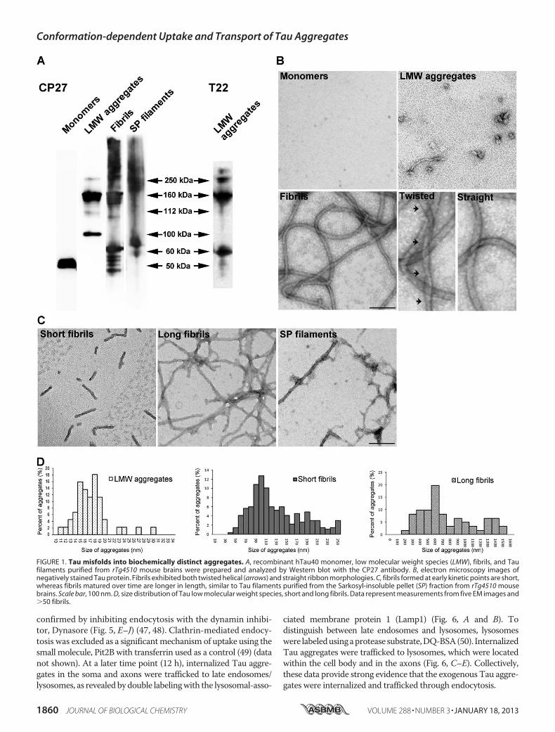
Properties—Tau is intrinsically disordered (37), and upon pro-longed incubation in high ionic-strength buffer such as PBS, itmisfolds into small, LMW aggregates. Alternatively, exposureto polyanions such as heparin induced Tau to form fibrils aspreviously described (38). Both LMW aggregates and fibrilsprepared this way are largely SDS-stable as assessed byWesternimmunoblot analysis and immunodetection with antibodyCP27 (humanTau specific) (Fig. 1A).Monomeric Taumigratesat�50 kDa, whereas LMWaggregates appearmainly as dimersand trimers. Both in vitro prepared Tau fibrils, and Sarkosylextracted, in vivo derived Tau filaments from severely affected(10 months old) rTg4510mice (35) appear as smears with highmolecularmass aggregates distributed from the top of the gel to150 kDa. Additionally, LMWTau aggregates are recognized bythe anti-Tau oligomer specific antibody, T22 (Fig. 1A) (29, 39).Furthermorphological examination ofTau aggregates by trans-mission electron microscopy revealed that LMW Tau aggre-gates are small spherical oligomers with diameters rangingfrom 10 to 30 nm (Fig. 1, B and D). Tau fibrils exhibit the char-acteristic straight and helical twist as previously described (40,41). Moreover, we observed that Tau fibrillization is largelytime dependent, with shorter fibrils (40–250 nm) appearing atan early time point (6–12 h) and longer, mature fibrils (200–1600 nm) forming at a later time point (24 h) (Fig. 1, C and D).Taken together, these data demonstrate that Tau protein canmisfold into a variety of aggregates with distinct biochemicaland morphological properties.Aggregation and Size-dependent Binding, Internalization, and
Anterograde Axonal Transport of Tau Species in Neurons—Next,the relationship between the folding state, size of extracellularTau, and cellular uptake was investigated. Previous researchhas shown that amyloidogenic proteins such as A� (42),expanded polyglutamine repeats (poly(Q)) (27), superoxide dis-mutase-1 (24), and the prion protein (19) can gain entry intocells upon aggregation. Moreover, Tau fibrils formed from theMTBR (amino acids 243–375) are readily taken up by cells (1,18, 43). However, it is unclear whether full-length Tau fibrilsbehave in a similar fashion, and furthermore, if additional vari-ables such as the size ormorphology of the aggregates influencethe uptake mechanism in a neuronal culture model. To exam-ine how wild type, full-length Tau conformers were internal-ized in different cellular compartments of wild type neurons, inthe absence of mediators of endocytosis (such as lipofectamineor wheat germ agglutinin), mouse primary hippocampal andcortical neurons were cultured in MF chambers. These cham-
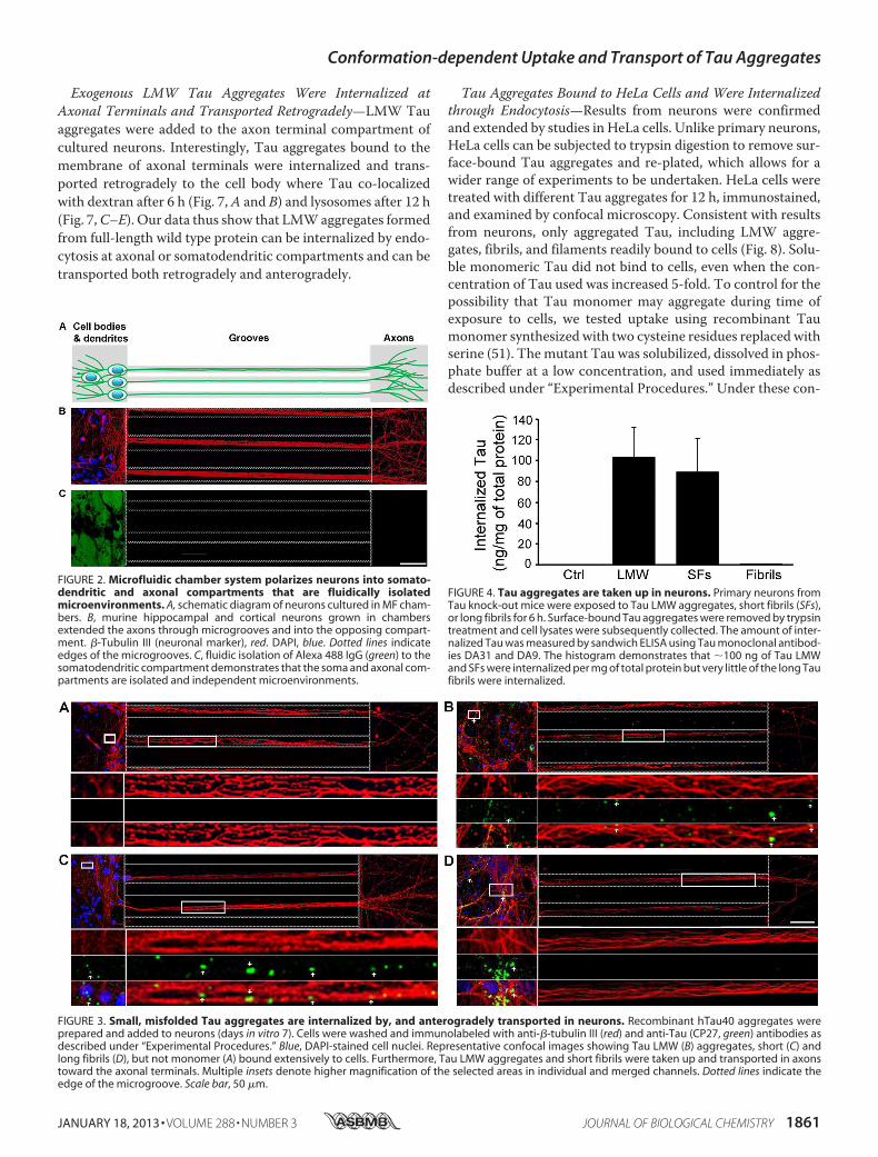
bers utilize microchannels to polarize and separate neuronalcell bodies and dendrites from axons (Fig. 2, A and B) intofluidically isolated microenvironments (Fig. 2C) (44). To con-firm the fluidic integrity of the chamber, Alexa 488 IgGs wereadded to the somatodendritic compartment and allowed toincubate with cells for over 12 h. No fluorescence signal wasobserved in microgrooves or in the opposing axonal compart-ment (Fig. 2C). Similar fluidic isolation was observed whenAlexa 488 IgG was added to the axonal compartment (data notshown). Addition of exogenous Tau monomer and differentaggregates to the somatodendritic compartment of neuronsrevealed that only aggregated Tau (including LMWaggregates,short and long fibrils) but not monomeric Tau was associatedwith neurons (Fig. 3, A–D), even when the dose of monomersadded was increased 5-fold (to 1 �m). Among the cell-boundTau aggregates, only LMW and short fibrils were observed inaxons projecting through microgrooves (Fig. 3, B and C). Ingeneral, more LMW Tau puncta were observed in axons ascompared with short fibrils as the fluidic integrity of the cham-ber does not allow free proteins to passively diffuse into themicrogrooves, the observation of Tau aggregates in axons pro-jecting through the grooves suggests that Tau internalized atthe somatodendritic compartment is anterogradely trans-ported down axons toward the axon terminals. To quantify theamount of misfolded Tau aggregates that were internalized,primary neurons from Tau knock-out mice were incubatedwith Tau aggregates, and extracellular bound Tau wasremoved by trypsin treatment. The amount of Tau internal-ized was quantified by Sandwich ELISA (45). Consistent withimmunofluorescence data, only LMW and SFs, but notfibrils were efficiently internalized by cells (100 � 30 ng/mgof total protein) (Fig. 4). Taken together, these data demon-strate that only Tau aggregates within a certain size rangebind efficiently to neurons, are internalized and transported.Despite the fact that long fibrils bind to cells, they do notappear to be internalized (or transported) as they are notreadily apparent in axons.Exogenous LMW Tau Aggregates Were Internalized in Neu-
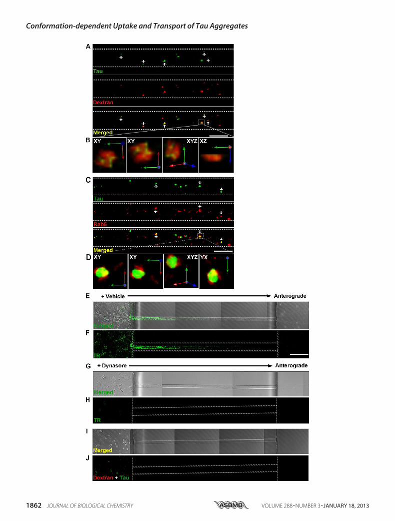
rons via Bulk Endocytosis—We focused on examining themechanism by which LMW Tau aggregates are taken up as ingeneral, more LMWTau puncta are observed in axons as com-pared with short fibrils. To examine how aggregates enter cells,7-day in vitro hippocampal and cortical neurons were exposedto LMW Tau aggregates for different lengths of time and mul-ticolor immunolabeling was performed to examine co-localiza-tion with proteins involved in the endosomal pathway (Fig. 5).When neurons were exposed to LMW Tau aggregates in thesomatodendritic compartment for 6 h, punctateTau aggregateswere observed in the axons. These aggregates co-localized withfluorescently labeled dextran, a glycan that is preferentiallytaken up through fluid-phase endocytosis (46) (Fig. 5A), andRab5, a GTPase that is enriched in early endosomes (Fig. 5C).Co-localization was further confirmed in three-dimensionalreconstructed image Z-stacks in which distinct co-localizationof Tau and dextran (yellow, Fig. 5B) or Tau with Rab5 (yellow,Fig. 5D) was evident from both linear and orthogonal perspec-tives. The majority of Tau aggregates in axons (83 � 1.6%) co-localized with dextran. Tau endocytosis-dependent uptake was
Conformation-dependent Uptake and Transport of Tau Aggregates
JANUARY 18, 2013 • VOLUME 288 • NUMBER 3 JOURNAL OF BIOLOGICAL CHEMISTRY 1859
confirmed by inhibiting endocytosis with the dynamin inhibi-tor, Dynasore (Fig. 5, E–J) (47, 48). Clathrin-mediated endocy-tosis was excluded as a significantmechanism of uptake using thesmall molecule, Pit2B with transferrin used as a control (49) (datanot shown). At a later time point (12 h), internalized Tau aggre-gates in the soma and axons were trafficked to late endosomes/lysosomes, as revealedbydouble labelingwith the lysosomal-asso-
ciated membrane protein 1 (Lamp1) (Fig. 6, A and B). Todistinguish between late endosomes and lysosomes, lysosomeswere labeledusingaprotease substrate,DQ-BSA(50). InternalizedTau aggregates were trafficked to lysosomes, which were locatedwithin the cell body and in the axons (Fig. 6, C–E). Collectively,these data provide strong evidence that the exogenous Tau aggre-gates were internalized and trafficked through endocytosis.
FIGURE 1. Tau misfolds into biochemically distinct aggregates. A, recombinant hTau40 monomer, low molecular weight species (LMW), fibrils, and Taufilaments purified from rTg4510 mouse brains were prepared and analyzed by Western blot with the CP27 antibody. B, electron microscopy images ofnegatively stained Tau protein. Fibrils exhibited both twisted helical (arrows) and straight ribbon morphologies. C, fibrils formed at early kinetic points are short,whereas fibrils matured over time are longer in length, similar to Tau filaments purified from the Sarkosyl-insoluble pellet (SP) fraction from rTg4510 mousebrains. Scale bar, 100 nm. D, size distribution of Tau low molecular weight species, short and long fibrils. Data represent measurements from five EM images and�50 fibrils.
Conformation-dependent Uptake and Transport of Tau Aggregates
1860 JOURNAL OF BIOLOGICAL CHEMISTRY VOLUME 288 • NUMBER 3 • JANUARY 18, 2013
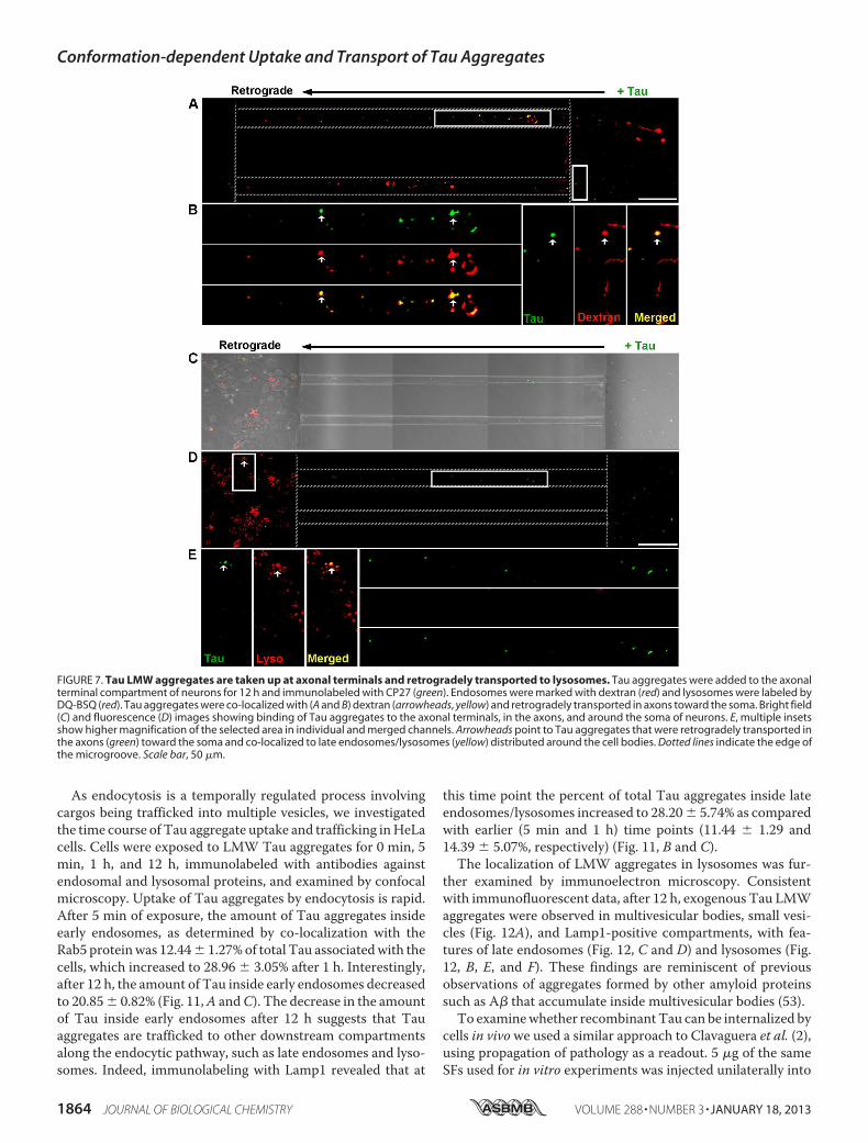
Exogenous LMW Tau Aggregates Were Internalized at
Axonal Terminals and Transported Retrogradely—LMW Tau
aggregates were added to the axon terminal compartment of
cultured neurons. Interestingly, Tau aggregates bound to the
membrane of axonal terminals were internalized and trans-
ported retrogradely to the cell body where Tau co-localized
with dextran after 6 h (Fig. 7,A and B) and lysosomes after 12 h
(Fig. 7,C–E). Our data thus show that LMWaggregates formed
from full-length wild type protein can be internalized by endo-
cytosis at axonal or somatodendritic compartments and can be
transported both retrogradely and anterogradely.
Tau Aggregates Bound to HeLa Cells and Were Internalized
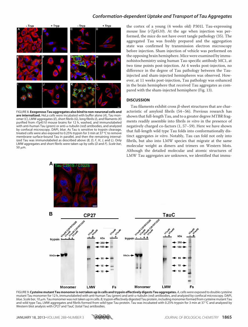
through Endocytosis—Results from neurons were confirmedand extended by studies in HeLa cells. Unlike primary neurons,HeLa cells can be subjected to trypsin digestion to remove sur-face-bound Tau aggregates and re-plated, which allows for awider range of experiments to be undertaken. HeLa cells weretreated with different Tau aggregates for 12 h, immunostained,and examined by confocal microscopy. Consistent with resultsfrom neurons, only aggregated Tau, including LMW aggre-gates, fibrils, and filaments readily bound to cells (Fig. 8). Solu-ble monomeric Tau did not bind to cells, even when the con-centration of Tau used was increased 5-fold. To control for thepossibility that Tau monomer may aggregate during time ofexposure to cells, we tested uptake using recombinant Taumonomer synthesized with two cysteine residues replaced withserine (51). Themutant Tau was solubilized, dissolved in phos-phate buffer at a low concentration, and used immediately asdescribed under “Experimental Procedures.” Under these con-
FIGURE 2. Microfluidic chamber system polarizes neurons into somato-dendritic and axonal compartments that are fluidically isolatedmicroenvironments. A, schematic diagram of neurons cultured in MF cham-bers. B, murine hippocampal and cortical neurons grown in chambersextended the axons through microgrooves and into the opposing compart-ment. �-Tubulin III (neuronal marker), red. DAPI, blue. Dotted lines indicateedges of the microgrooves. C, fluidic isolation of Alexa 488 IgG (green) to thesomatodendritic compartment demonstrates that the soma and axonal com-partments are isolated and independent microenvironments.
FIGURE 3. Small, misfolded Tau aggregates are internalized by, and anterogradely transported in neurons. Recombinant hTau40 aggregates wereprepared and added to neurons (days in vitro 7). Cells were washed and immunolabeled with anti-�-tubulin III (red) and anti-Tau (CP27, green) antibodies asdescribed under “Experimental Procedures.” Blue, DAPI-stained cell nuclei. Representative confocal images showing Tau LMW (B) aggregates, short (C) andlong fibrils (D), but not monomer (A) bound extensively to cells. Furthermore, Tau LMW aggregates and short fibrils were taken up and transported in axonstoward the axonal terminals. Multiple insets denote higher magnification of the selected areas in individual and merged channels. Dotted lines indicate theedge of the microgroove. Scale bar, 50 �m.
FIGURE 4. Tau aggregates are taken up in neurons. Primary neurons fromTau knock-out mice were exposed to Tau LMW aggregates, short fibrils (SFs),or long fibrils for 6 h. Surface-bound Tau aggregates were removed by trypsintreatment and cell lysates were subsequently collected. The amount of inter-nalized Tau was measured by sandwich ELISA using Tau monoclonal antibod-ies DA31 and DA9. The histogram demonstrates that �100 ng of Tau LMWand SFs were internalized per mg of total protein but very little of the long Taufibrils were internalized.
Conformation-dependent Uptake and Transport of Tau Aggregates
JANUARY 18, 2013 • VOLUME 288 • NUMBER 3 JOURNAL OF BIOLOGICAL CHEMISTRY 1861
Conformation-dependent Uptake and Transport of Tau Aggregates
1862 JOURNAL OF BIOLOGICAL CHEMISTRY VOLUME 288 • NUMBER 3 • JANUARY 18, 2013
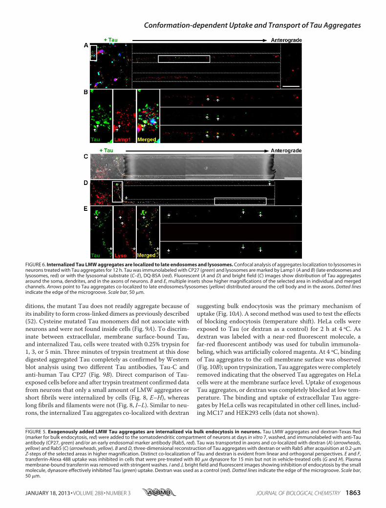
ditions, the mutant Tau does not readily aggregate because ofits inability to form cross-linked dimers as previously described(52). Cysteine mutated Tau monomers did not associate withneurons and were not found inside cells (Fig. 9A). To discrim-inate between extracellular, membrane surface-bound Tau,and internalized Tau, cells were treated with 0.25% trypsin for1, 3, or 5 min. Three minutes of trypsin treatment at this dosedigested aggregated Tau completely as confirmed by Westernblot analysis using two different Tau antibodies, Tau-C andanti-human Tau CP27 (Fig. 9B). Direct comparison of Tau-exposed cells before and after trypsin treatment confirmed datafrom neurons that only a small amount of LMW aggregates orshort fibrils were internalized by cells (Fig. 8, E–H), whereaslong fibrils and filaments were not (Fig. 8, I–L). Similar to neu-rons, the internalized Tau aggregates co-localized with dextran
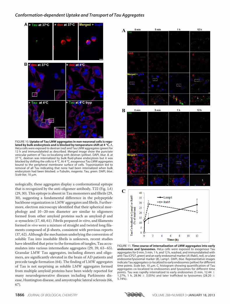
suggesting bulk endocytosis was the primary mechanism ofuptake (Fig. 10A). A secondmethod was used to test the effectsof blocking endocytosis (temperature shift). HeLa cells wereexposed to Tau (or dextran as a control) for 2 h at 4 ºC. Asdextran was labeled with a near-red fluorescent molecule, afar-red fluorescent antibody was used for tubulin immunola-beling, which was artificially colored magenta. At 4 ºC, bindingof Tau aggregates to the cell membrane surface was observed(Fig. 10B); upon trypsinization, Tau aggregateswere completelyremoved indicating that the observed Tau aggregates on HeLacells were at the membrane surface level. Uptake of exogenousTau aggregates, or dextran was completely blocked at low tem-perature. The binding and uptake of extracellular Tau aggre-gates by HeLa cells was recapitulated in other cell lines, includ-ing MC17 and HEK293 cells (data not shown).
FIGURE 5. Exogenously added LMW Tau aggregates are internalized via bulk endocytosis in neurons. Tau LMW aggregates and dextran-Texas Red(marker for bulk endocytosis, red) were added to the somatodendritic compartment of neurons at days in vitro 7, washed, and immunolabeled with anti-Tauantibody (CP27, green) and/or an early endosomal marker antibody (Rab5, red). Tau was transported in axons and co-localized with dextran (A) (arrowheads,yellow) and Rab5 (C) (arrowheads, yellow). B and D, three-dimensional reconstruction of Tau aggregates with dextran or with Rab5 after acquisition at 0.2-�mZ-steps of the selected areas in higher magnification. Distinct co-localization of Tau and dextran is evident from linear and orthogonal perspectives. E and F,transferrin-Alexa 488 uptake was inhibited in cells that were pre-treated with 80 �M dynasore for 15 min but not in vehicle-treated cells (G and H). Plasmamembrane-bound transferrin was removed with stringent washes. I and J, bright field and fluorescent images showing inhibition of endocytosis by the smallmolecule, dynasore effectively inhibited Tau (green) uptake. Dextran was used as a control (red). Dotted lines indicate the edge of the microgroove. Scale bar,50 �m.
FIGURE 6. Internalized Tau LMW aggregates are localized to late endosomes and lysosomes. Confocal analysis of aggregates localization to lysosomes inneurons treated with Tau aggregates for 12 h. Tau was immunolabeled with CP27 (green) and lysosomes are marked by Lamp1 (A and B) (late endosomes andlysosomes, red) or with the lysosomal substrate (C–E), DQ-BSA (red). Fluorescent (A and D) and bright field (C) images show distribution of Tau aggregatesaround the soma, dendrites, and in the axons of neurons. B and E, multiple insets show higher magnifications of the selected area in individual and mergedchannels. Arrows point to Tau aggregates co-localized to late endosomes/lysosomes (yellow) distributed around the cell body and in the axons. Dotted linesindicate the edge of the microgroove. Scale bar, 50 �m.
Conformation-dependent Uptake and Transport of Tau Aggregates
JANUARY 18, 2013 • VOLUME 288 • NUMBER 3 JOURNAL OF BIOLOGICAL CHEMISTRY 1863
As endocytosis is a temporally regulated process involvingcargos being trafficked into multiple vesicles, we investigatedthe time course of Tau aggregate uptake and trafficking inHeLacells. Cells were exposed to LMW Tau aggregates for 0 min, 5min, 1 h, and 12 h, immunolabeled with antibodies againstendosomal and lysosomal proteins, and examined by confocalmicroscopy. Uptake of Tau aggregates by endocytosis is rapid.After 5 min of exposure, the amount of Tau aggregates insideearly endosomes, as determined by co-localization with theRab5 proteinwas 12.44� 1.27%of total Tau associatedwith thecells, which increased to 28.96 � 3.05% after 1 h. Interestingly,after 12 h, the amount of Tau inside early endosomes decreasedto 20.85� 0.82% (Fig. 11,A andC). The decrease in the amountof Tau inside early endosomes after 12 h suggests that Tauaggregates are trafficked to other downstream compartmentsalong the endocytic pathway, such as late endosomes and lyso-somes. Indeed, immunolabeling with Lamp1 revealed that at
this time point the percent of total Tau aggregates inside lateendosomes/lysosomes increased to 28.20� 5.74% as comparedwith earlier (5 min and 1 h) time points (11.44 � 1.29 and14.39 � 5.07%, respectively) (Fig. 11, B and C).The localization of LMW aggregates in lysosomes was fur-
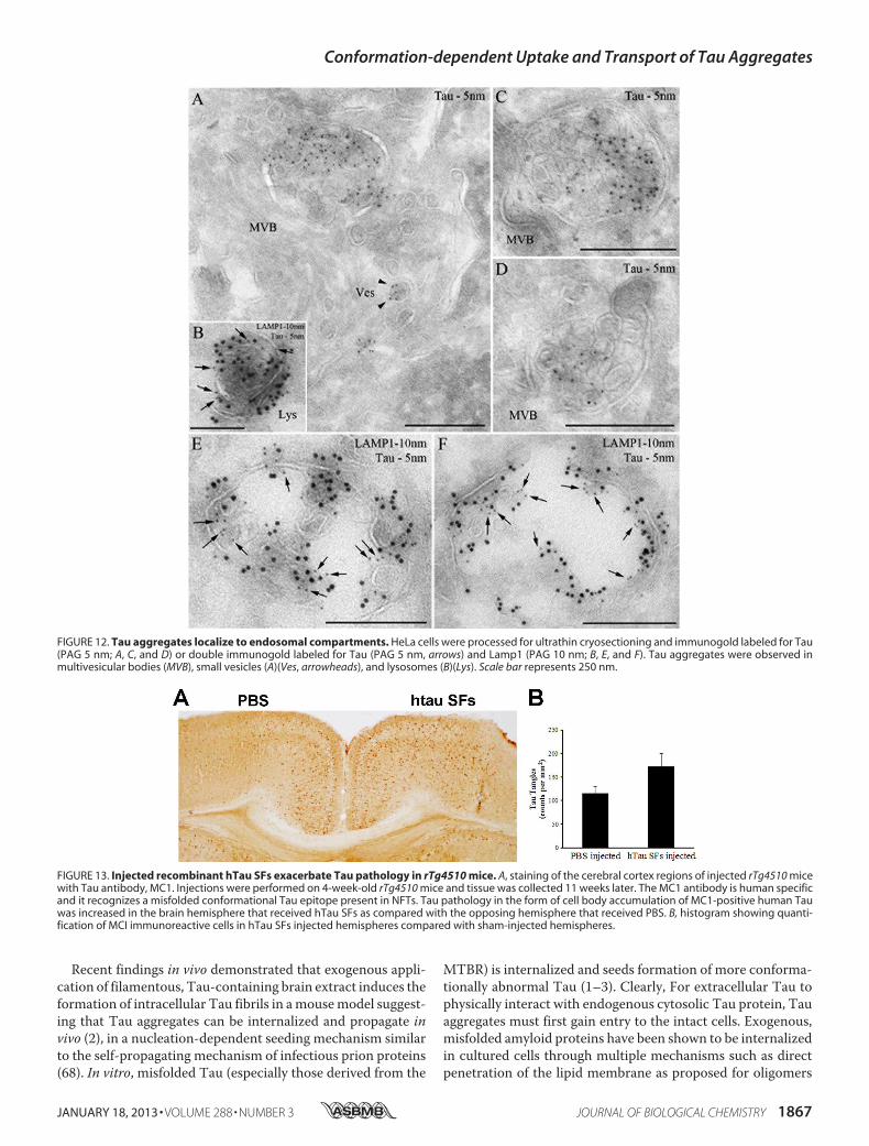
ther examined by immunoelectron microscopy. Consistentwith immunofluorescent data, after 12 h, exogenous Tau LMWaggregates were observed in multivesicular bodies, small vesi-cles (Fig. 12A), and Lamp1-positive compartments, with fea-tures of late endosomes (Fig. 12, C and D) and lysosomes (Fig.12, B, E, and F). These findings are reminiscent of previousobservations of aggregates formed by other amyloid proteinssuch as A� that accumulate inside multivesicular bodies (53).
To examinewhether recombinantTau can be internalized bycells in vivo we used a similar approach to Clavaguera et al. (2),using propagation of pathology as a readout. 5 �g of the sameSFs used for in vitro experiments was injected unilaterally into
FIGURE 7. Tau LMW aggregates are taken up at axonal terminals and retrogradely transported to lysosomes. Tau aggregates were added to the axonalterminal compartment of neurons for 12 h and immunolabeled with CP27 (green). Endosomes were marked with dextran (red) and lysosomes were labeled byDQ-BSQ (red). Tau aggregates were co-localized with (A and B) dextran (arrowheads, yellow) and retrogradely transported in axons toward the soma. Bright field(C) and fluorescence (D) images showing binding of Tau aggregates to the axonal terminals, in the axons, and around the soma of neurons. E, multiple insetsshow higher magnification of the selected area in individual and merged channels. Arrowheads point to Tau aggregates that were retrogradely transported inthe axons (green) toward the soma and co-localized to late endosomes/lysosomes (yellow) distributed around the cell bodies. Dotted lines indicate the edge ofthe microgroove. Scale bar, 50 �m.
Conformation-dependent Uptake and Transport of Tau Aggregates
1864 JOURNAL OF BIOLOGICAL CHEMISTRY VOLUME 288 • NUMBER 3 • JANUARY 18, 2013
the cortex of a young (4 weeks old) P301L Tau-expressingmouse line (rTg4510). At the age when injection was per-formed, the mice do not have overt tangle pathology (35). Theaggregated Tau was freshly prepared and the aggregationstate was confirmed by transmission electron microscopybefore injection. Sham injection of vehicle was performed onthe opposing brain hemisphere.Micewere examined by immu-nohistochemistry using human Tau-specific antibody MC1, attwo time points post-injection. At 4 weeks post-injection, nodifference in the degree of Tau pathology between the Tau-injected and sham-injected hemispheres was observed. How-ever, at 11 weeks post-injection, Tau pathology was enhancedin the brain hemisphere that received Tau aggregates as com-pared with the sham-injected hemisphere (Fig. 13).
DISCUSSION
Tau filaments exhibit cross �-sheet structures that are char-acteristic of amyloid fibrils (54–56). Previous research hasshown that full-length Tau, and to a greater degreeMTBR frag-ments readily assemble into fibrils in vitro in the presence ofnegatively charged co-factors (1, 57–59). Here we have shownthat full-length wild type Tau folds into conformationally dis-tinct aggregates in vitro. Notably, Tau can fold not only intofibrils, but also into LMW species that migrate at the samemolecular weight as dimers and trimers on Western blots.Although the detailed molecular and atomic structures ofLMW Tau aggregates are unknown, we identified that immu-
FIGURE 8. Exogenous Tau aggregates also bind to non-neuronal cells andare internalized. HeLa cells were incubated with buffer alone (A), Tau mon-omer (C), LMW aggregates (E), short fibrils (G), long fibrils (I), and filaments (K)purified from rTg4510 mouse brains for 12 h, washed, and immunolabeledwith anti-human Tau (green) or anti-�-tubulin (red) antibodies, and analyzedby confocal microscopy. DAPI, blue. As Tau is sensitive to trypsin cleavage,treated cells were also exposed to 0.25% trypsin for 3 min at 37 °C to removemembrane surface-bound Tau in parallel, and then the remaining internal-ized Tau was immunolabeled as described above (B, D, F, H, J, and L). OnlyLMW aggregates and short fibrils were taken up by cells (D and F). Scale bar,50 �m.
FIGURE 9. Cysteine mutant Tau monomer is not taken up in cells and trypsin effectively digests Tau aggregates. A, cells were exposed to double cysteinemutant Tau monomer for 12 h, immunolabeled with anti-human Tau (green) and anti-�-tubulin (red) antibodies, and analyzed by confocal microscopy. DAPI,blue. Scale bar, 10 �m. Tau monomer was not taken up in cells. B, trypsin effectively digested Tau protein, including monomer formed from cysteine mutant Tauand wild type Tau, LMW aggregates and fibrils formed from wild type Tau protein. Tau was incubated with 0.25% trypsin for 3 min at 37 °C and analyzed byWestern blot analysis with CP27 and TauC (total Tau) antibodies.
Conformation-dependent Uptake and Transport of Tau Aggregates
JANUARY 18, 2013 • VOLUME 288 • NUMBER 3 JOURNAL OF BIOLOGICAL CHEMISTRY 1865
nologically, these aggregates display a conformational epitopethat is recognized by the anti-oligomer antibody, T22 (Fig. 1A)(29, 30). This epitope is absent in Taumonomers and fibrils (29,30), suggesting a fundamental difference in the polypeptidebackbone organization in LMWaggregates and fibrils. Further-more, electron microscopy identified that their spherical mor-phology and 10–20-nm diameter are similar to oligomersformed from other amyloid proteins such as amyloid-� and�-synuclein (17, 60, 61). Fibrils prepared in vitro, and filamentsformed in vivo were a mixture of straight and twisted long fila-ments composed of �-sheets, consistent with previous reports(37, 62). Although themechanism underlying the conversion ofsoluble Tau into insoluble fibrils is unknown, recent studieshave identified that prior to the formation of tangles, Tau accu-mulates into various intermediate aggregates (29, 39, 63–65).Granular LMW Tau aggregates, including dimers and oligo-mers, are significantly elevated in the brain of AD patients andprecede tangle formation (64). The finding of LMW aggregatesof Tau is not surprising as soluble LMW aggregates formedfrom multiple amyloid proteins have been widely reported formany neurodegenerative diseases including Parkinsons dis-ease, Huntington disease, and amyotrophic lateral sclerosis (66,67).
FIGURE 10. Uptake of Tau LMW aggregates in non-neuronal cells is regu-lated by bulk endocytosis and is blocked by temperature shift at 4 °C. A,HeLa cells were exposed to dextran (red) and Tau LMW aggregates (green) for12 h and immunolabeled as described. Merged image show the punctatevesicular pattern of Tau co-localizing with dextran (yellow). DAPI, blue. B, at37 °C, dextran was internalized by bulk fluid-phase endocytosis but it wasblocked by shifting the cells to 4 °C. At 4 °C, exogenous Tau LMW aggregatesbound to the peripheral membrane surface of cells. Trypsinization led toremoval of all Tau indicating that none had been internalized when bulkendocytosis had been blocked. �-Tubulin, magenta. Tau, green. DAPI, blue.Scale bar, 10 �m.
FIGURE 11. Time course of internalization of LMW aggregates into earlyendosomes and lysosomes. HeLa cells were exposed to exogenous Tauaggregates for 0 min, 5 min, 1 h, and 12 h, washed, and immunolabeled withanti-Tau (CP27, green) and an early endosomal marker (A) (Rab5, red), or a lateendosome/lysosomal marker (B), Lamp1. DAPI, blue. Representative imagesindicate Tau aggregates co-localized to early endosomes (yellow) for differenttime points. Scale bar, 10 �m. C, histogram showing quantification of Tauaggregates co-localized to endosomes and lysosomes for different timepoints. Tau was rapidly internalized to early endosomes (5 min, 12.44 �
1.27%, 1 h, 28.96 � 3.05%) and later trafficked to lysosomes (28.20 �
5.74%).
Conformation-dependent Uptake and Transport of Tau Aggregates
1866 JOURNAL OF BIOLOGICAL CHEMISTRY VOLUME 288 • NUMBER 3 • JANUARY 18, 2013
Recent findings in vivo demonstrated that exogenous appli-cation of filamentous, Tau-containing brain extract induces theformation of intracellular Tau fibrils in amousemodel suggest-ing that Tau aggregates can be internalized and propagate in
vivo (2), in a nucleation-dependent seeding mechanism similarto the self-propagating mechanism of infectious prion proteins(68). In vitro, misfolded Tau (especially those derived from the
MTBR) is internalized and seeds formation of more conforma-tionally abnormal Tau (1–3). Clearly, For extracellular Tau tophysically interact with endogenous cytosolic Tau protein, Tauaggregates must first gain entry to the intact cells. Exogenous,misfolded amyloid proteins have been shown to be internalizedin cultured cells through multiple mechanisms such as directpenetration of the lipid membrane as proposed for oligomers
FIGURE 12. Tau aggregates localize to endosomal compartments. HeLa cells were processed for ultrathin cryosectioning and immunogold labeled for Tau(PAG 5 nm; A, C, and D) or double immunogold labeled for Tau (PAG 5 nm, arrows) and Lamp1 (PAG 10 nm; B, E, and F). Tau aggregates were observed inmultivesicular bodies (MVB), small vesicles (A)(Ves, arrowheads), and lysosomes (B)(Lys). Scale bar represents 250 nm.
FIGURE 13. Injected recombinant hTau SFs exacerbate Tau pathology in rTg4510 mice. A, staining of the cerebral cortex regions of injected rTg4510 micewith Tau antibody, MC1. Injections were performed on 4-week-old rTg4510 mice and tissue was collected 11 weeks later. The MC1 antibody is human specificand it recognizes a misfolded conformational Tau epitope present in NFTs. Tau pathology in the form of cell body accumulation of MC1-positive human Tauwas increased in the brain hemisphere that received hTau SFs as compared with the opposing hemisphere that received PBS. B, histogram showing quanti-fication of MCI immunoreactive cells in hTau SFs injected hemispheres compared with sham-injected hemispheres.
Conformation-dependent Uptake and Transport of Tau Aggregates
JANUARY 18, 2013 • VOLUME 288 • NUMBER 3 JOURNAL OF BIOLOGICAL CHEMISTRY 1867
formed from A� (42) and poly(Q) (27), or through the endo-somal pathway as shown for oligomers formed from �-sy-nuclein (25), the yeast prion protein (Sup35) (69), and fibrilsformed from the MTBR of Tau (1, 43). It is interesting that theinternalization of extracellular synuclein, whereas shown to beaggregation state dependent, has been reported to utilize recep-tor-mediated endocytosis (25). In contrast, conformation-de-pendent uptake of exogenous MTBR Tau fibrils has beenshown to be regulated by nonreceptormediated (bulk or adapt-ive) endocytosis (1, 18, 43). Here, we have used full-length Tauand shown that neurons internalize small aggregates such asLMW species and short fibrils with length in the range of 10 to100 �m. Longer fibrils bind peripherally to cell membranes butare not internalized. These results demonstrate that in additionto oligomeric status, the uptake of exogenous Tau is limited byother physical parameters such as the size of the aggregates.This is not surprising as the process of endocytosis involvesvesicles of different diameter and is highly selective toward pro-teins of different sizes (70).Endocytosis is a process by which cells absorb and engulf
largemolecules such as proteins that normally cannot cross thehydrophobic plasma membrane, and it has been proposed foruptake of multiple exogenous amyloid aggregates includingMTBRTau aggregates (1, 43),�-synuclein oligomers and fibrils(25). Alternatively, direct membrane penetration has also beendescribed for amyloids such as A� oligomers (42) and for otherproteins such as HIV Tat (71) and Drosophila ANT (72). Herewe show that exogenous Tau aggregates are taken up in cellsthrough an active process that is attenuated by dynamin inhi-bition, and low temperature shift, supporting endocytosis-me-diated internalization. Tau aggregates co-localized with dex-tran, the GTPase Rab5 and Lamp1 in neurons, and HeLa cellssuggesting that the internalized aggregates are transported inendosomal vesicles (Fig. 5) and trafficked through the endo-somal pathway to the lysosomes (Figs. 6 and 7). Whether Tauaggregates are degraded in lysosomes requires further investi-gation. As we observed no significant apoptosis in cells treatedwithTau aggregates (data not shown), it is likely that themajor-ity of Tau aggregates are degraded in lysosomes. Some, how-ever, could escape into the cytoplasm where they could act asseeds for templating. This is consistent with our observationthat injection of recombinant Tau aggregates into the brain ofmice enhances pathology most likely through uptake of aggre-gates and templating to endogenous Tau as described in a sim-ilar injection model (2). Additionally, our experiments suggestthat exogenously added Tau aggregates without brain lysate-derived co-factors are sufficient to enhance tauopathy anddrive propagation in vivo. That Tau aggregates can act as seedsis reminiscent of �-synuclein aggregate induced inclusion bodyformation in cells (18, 43, 73).Of particular interest was the observation that Tau aggre-
gates can be internalized at axonal terminals and retrogradelytransported toward the cell soma.Most of the aggregates co-lo-calized with lysosomal markers, especially as the aggregatesmoved from the distal tip back toward the cell soma. Both DQ-BSA and Lamp1 staining demonstrated a greater number oflysosomes toward the cell soma compartment, which agreeswith published data (74). Internalization of Tau at axonal ter-
minals is of particular interest as it may explain how tauopathycan move though the brain in both an antero- and retrogradedirection (75, 76).Taken together, these data provide a plausible molecular
mechanism for how physiologically relevant Tau can enter cellsto initiate propagation. Intracellular Tau aggregates, releasedvia secretion as a means of clearance, or upon degeneration ofaxons or somatodendritic compartments could be internalizedby anatomically connected cells, and then anterogradely andretrogradely transported to remote brain regions. Aggregatescould accumulate, clogging up cellular degradation machinerysuch as the ubiquitin-proteasome system, giving rise to ag-gresomes and autophagosomes (77). Subsequently, aggregatedproteins that failed to be sequestered or degraded may causelocal membrane rupture of degradative organelles, leading totheir release into the cytosol where they could physically inter-act with intracellular soluble protein and trigger endogenousTau protein misfolding. The cycle would be expected to berepeated leading to localized transneuronal spread, and trans-synaptic spread to distally connected regions. Thus the tempo-ral and spatially distinct distribution of Tau pathology thatdefines the early stages of AD could be explained by the uptake,templating, and release of aggregated Tau between neurons inneuroanatomically connected circuits.
Acknowledgments—We thank Li Shi for administrative support and
acknowledge the EM facilities at the New York Structural Biology
Center.
REFERENCES
1. Frost, B., Jacks, R. L., and Diamond, M. I. (2009) Propagation of Tau mis-
folding from the outside to the inside of a cell. J. Biol. Chem. 284,
12845–12852
2. Clavaguera, F., Bolmont, T., Crowther, R. A., Abramowski, D., Frank, S.,
Probst, A., Fraser, G., Stalder, A. K., Beibel,M., Staufenbiel,M., Jucker,M.,
Goedert, M., and Tolnay, M. (2009) Transmission and spreading of
tauopathy in transgenic mouse brain. Nat. Cell Biol. 11, 909–913
3. Lasagna-Reeves, C. A., Castillo-Carranza, D. L., Sengupta, U., Guerrero-
Munoz, M. J., Kiritoshi, T., Neugebauer, V., Jackson, G. R., and Kayed, R.
(2012) Alzheimer brain-derived tau oligomers propagate pathology from
endogenous tau. Sci. Rep. 2, 700
4. Ballatore, C., Lee, V. M., and Trojanowski, J. Q. (2007) Tau-mediated
neurodegeneration in Alzheimer’s disease and related disorders.Nat. Rev.
Neurosci. 8, 663–672
5. Hutton, M., Lendon, C. L., Rizzu, P., Baker, M., Froelich, S., Houlden, H.,
Pickering-Brown, S., Chakraverty, S., Isaacs, A., Grover, A., Hackett, J.,
Adamson, J., Lincoln, S., Dickson, D., Davies, P., Petersen, R. C., Stevens,
M., de Graaff, E., Wauters, E., van Baren, J., Hillebrand, M., Joosse, M.,
Kwon, J. M., Nowotny, P., Che, L. K., Norton, J., Morris, J. C., Reed, L. A.,
Trojanowski, J., Basun, H., Lannfelt, L., Neystat, M., Fahn, S., Dark, F.,
Tannenberg, T., Dodd, P. R., Hayward, N., Kwok, J. B., Schofield, P. R.,
Andreadis, A., Snowden, J., Craufurd, D., Neary, D., Owen, F., Oostra,
B. A., Hardy, J., Goate, A., van Swieten, J., Mann, D., Lynch, T., and Heu-
tink, P. (1998) Association of missense and 5�-splice-site mutations in tau
with the inherited dementia FTDP-17. Nature 393, 702–705
6. Spillantini, M. G., Crowther, R. A., Kamphorst, W., Heutink, P., and van
Swieten, J. C. (1998) Tau pathology in two Dutch families with mutations
in the microtubule-binding region of tau. Am. J. Pathol. 153, 1359–1363
7. Rizzu, P., Van Swieten, J. C., Joosse, M., Hasegawa, M., Stevens, M., Tib-
ben,A.,Niermeijer,M. F., Hillebrand,M., Ravid, R.,Oostra, B. A., Goedert,
M., vanDuijn, C.M., andHeutink, P. (1999) High prevalence ofmutations
in the microtubule-associated protein tau in a population study of fronto-
Conformation-dependent Uptake and Transport of Tau Aggregates
1868 JOURNAL OF BIOLOGICAL CHEMISTRY VOLUME 288 • NUMBER 3 • JANUARY 18, 2013
temporal dementia in the Netherlands. Am. J. Hum. Genet. 64, 414–421
8. Goedert, M., and Jakes, R. (2005) Mutations causing neurodegenerative
tauopathies. Biochim. Biophys. Acta 1739, 240–250
9. D’Souza, I., and Schellenberg, G. D. (2005) Regulation of tau isoform ex-
pression and dementia. Biochim. Biophys. Acta 1739, 104–115
10. Myers, A. J., Pittman, A.M., Zhao, A. S., Rohrer, K., Kaleem,M.,Marlowe,
L., Lees, A., Leung, D., McKeith, I. G., Perry, R. H., Morris, C. M., Tro-
janowski, J. Q., Clark, C., Karlawish, J., Arnold, S., Forman, M. S., Van
Deerlin, V., de Silva, R., and Hardy, J. (2007) The MAPT H1c risk haplo-
type is associated with increased expression of tau and especially of 4
repeat containing transcripts. Neurobiol. Dis. 25, 561–570
11. Braak, H., and Braak, E. (1991) Neuropathological stageing of Alzheimer-
related changes. Acta Neuropathol. 82, 239–259
12. Braak, H., Thal, D. R., and Del Tredici, K. (2011) Nerve cells immunore-
active for p62 in select hypothalamic and brainstem nuclei of controls and
Parkinson’s disease cases. J. Neural Transm. 118, 809–819
13. Liu, L., D. V., Wu, J. W., Witter, M. P., Small, S. A., Clelland, C., and Duff,
K. (2012) Trans-synaptic spread of Tau pathology in vivo. PLoS ONE 7,
doi: 10.1371/journal.pone.0031302
14. de Calignon, A., Polydoro, M., Suárez-Calvet, M., William, C., Adamow-
icz, D. H., Kopeikina, K. J., Pitstick, R., Sahara, N., Ashe, K. H., Carlson,
G. A., Spires-Jones, T. L., and Hyman, B. T. (2012) Propagation of Tau
pathology in a model of early Alzheimer’s disease. Neuron 73, 685–697
15. Danzer, K. M., Ruf, W. P., Putcha, P., Joyner, D., Hashimoto, T., Glabe, C.,
Hyman, B. T., and McLean, P. J. (2011) Heat-shock protein 70 modulates
toxic extracellular �-synuclein oligomers and rescues trans-synaptic tox-
icity. FASEB J. 25, 326–336
16. Danzer, K. M., Krebs, S. K., Wolff, M., Birk, G., and Hengerer, B. (2009)
Seeding induced by �-synuclein oligomers provides evidence for spread-
ing of �-synuclein pathology. J. Neurochem. 111, 192–203
17. Danzer, K.M., Haasen, D., Karow, A. R., Moussaud, S., Habeck, M., Giese,
A., Kretzschmar, H., Hengerer, B., and Kostka,M. (2007) Different species
of �-synuclein oligomers induce calcium influx and seeding. J. Neurosci.
27, 9220–9232
18. Kfoury, N., Holmes, B. B., Jiang, H., Holtzman, D. M., and Diamond, M. I.
(2012) Trans-cellular propagation of Tau aggregation by fibrillar species.
J. Biol. Chem. 287, 19440–19451
19. Prusiner, S. B. (1998) The prion diseases. Brain Pathol. 8, 499–513
20. Gousset, K., Schiff, E., Langevin, C., Marijanovic, Z., Caputo, A., Brow-
man, D. T., Chenouard, N., de Chaumont, F., Martino, A., Enninga, J.,
Olivo-Marin, J. C., Männel, D., and Zurzolo, C. (2009) Prions hijack tun-
nelling nanotubes for intercellular spread. Nat. Cell Biol. 11, 328–336
21. Gerdes, H. H. (2009) Prions tunnel between cells. Nat. Cell Biol. 11,
235–236
22. Fevrier, B., Vilette, D., Archer, F., Loew, D., Faigle, W., Vidal, M., Laude,
H., and Raposo, G. (2004) Cells release prions in association with exo-
somes. Proc. Natl. Acad. Sci. U.S.A. 101, 9683–9688
23. Saman, S., Kim, W., Raya, M., Visnick, Y., Miro, S., Saman, S., Jackson, B.,
McKee, A. C., Alvarez, V. E., Lee, N. C., and Hall, G. F. (2012) Exosome-
associated Tau is secreted in tauopathy models and is selectively phos-
phorylated in cerebrospinal fluid in early Alzheimer disease. J. Biol. Chem.
287, 3842–3849
24. Münch, C., O’Brien, J., and Bertolotti, A. (2011) Prion-like propagation of
mutant superoxide dismutase-1 misfolding in neuronal cells. Proc. Natl.
Acad. Sci. U.S.A. 108, 3548–3553
25. Lee, H. J., Suk, J. E., Bae, E. J., Lee, J. H., Paik, S. R., and Lee, S. J. (2008)
Assembly-dependent endocytosis and clearance of extracellular �-sy-
nuclein. Int. J. Biochem. Cell Biol. 40, 1835–1849
26. Lee, H. J., Suk, J. E., Bae, E. J., and Lee, S. J. (2008) Clearance and deposition
of extracellular �-synuclein aggregates in microglia. Biochem. Biophys.
Res. Commun. 372, 423–428
27. Ren, P. H., Lauckner, J. E., Kachirskaia, I., Heuser, J. E., Melki, R., and
Kopito, R. R. (2009) Cytoplasmic penetration and persistent infection of
mammalian cells by polyglutamine aggregates. Nat. Cell. Biol. 11,
219–225
28. Yamada, K., Cirrito, J. R., Stewart, F. R., Jiang,H., Finn,M. B.,Holmes, B. B.,
Binder, L. I.,Mandelkow, E.M., Diamond,M. I., Lee, V.M., andHoltzman,
D.M. (2011) In vivomicrodialysis reveals age-dependent decrease of brain
interstitial fluid Tau levels in P301S human Tau;Transgenic mice. J. Neu-
rosci. 31, 13110–13117
29. Lasagna-Reeves, C. A., Castillo-Carranza, D. L., Guerrero-Muoz, M. J.,
Jackson, G. R., and Kayed, R. (2010) Preparation and characterization of
neurotoxic Tau oligomers. Biochemistry 49, 10039–10041
30. Lasagna-Reeves, C. A., Castillo-Carranza, D. L., Sengupta, U., Clos, A. L.,
Jackson, G. R., and Kayed, R. (2011) Tau oligomers impair memory and
induce synaptic and mitochondrial dysfunction in wild-type mice. Mol.
Neurodegener 6, 39
31. Congdon, E. E., Wu, J. W., Myeku, N., Figueroa, Y. H., Herman, M., Mar-
inec, P. S., Gestwicki, J. E., Dickey, C. A., Yu, W. H., and Duff, K. (2012)
Methylthioninium chloride (methylene blue) induces autophagy and at-
tenuates tauopathy in vitro and in vivo. Autophagy 8, 609–622
32. Slot, J. W., and Geuze, H. J. (2007) Cryosectioning and immunolabeling.
Nat. Protoc. 2, 2480–2491
33. Raposo, G., Kleijmeer, M. J., Posthuma, G., Slot, J. W., and Geuze, H. J.
(1997) in Weirs Handbook of Experimental Immunology (Herzenberg,
L. A.,Weir, D.M., and Blackwell, C., eds) p. 208.1, Blackwell, Malden,MA
34. Brewer, G. J., Torricelli, J. R., Evege, E. K., and Price, P. J. (1993) Optimized
survival of hippocampal neurons in B27-supplementedNeurobasal, a new
serum-free medium combination. J. Neurosci. Res. 35, 567–576
35. Santacruz, K., Lewis, J., Spires, T., Paulson, J., Kotilinek, L., Ingelsson, M.,
Guimaraes, A., DeTure, M., Ramsden, M., McGowan, E., Forster, C., Yue,
M.,Orne, J., Janus, C.,Mariash, A., Kuskowski,M., Hyman, B., Hutton,M.,
and Ashe, K. H. (2005) Tau suppression in a neurodegenerative mouse
model improves memory function. Science 309, 476–481
36. Jicha, G. A., Bowser, R., Kazam, I. G., and Davies, P. (1997) Alz-50 and
MC-1, a new monoclonal antibody raised to paired helical filaments, rec-
ognize conformational epitopes on recombinant tau. J. Neurosci. Res. 48,
128–132
37. von Bergen, M., Barghorn, S., Biernat, J., Mandelkow, E. M., and Man-
delkow, E. (2005) Tau aggregation is driven by a transition from random
coil to � sheet structure. Biochim. Biophys. Acta. 1739, 158–166
38. Barghorn, S., Biernat, J., andMandelkow, E. (2005) Purification of recom-
binant Tau protein and preparation of Alzheimer-paired helical filaments
in vitro.Methods Mol. Biol. 299, 35–51
39. Lasagna-Reeves, C. A., Castillo-Carranza, D. L., Sengupta, U., Sarmiento,
J., Troncoso, J., Jackson, G. R., and Kayed, R. (2012) Identification of olig-
omers at early stages of Tau aggregation in Alzheimer’s disease. FASEB J.
26, 1946–1959
40. Barghorn, S., Davies, P., and Mandelkow, E. (2004) Tau paired helical
filaments fromAlzheimer’s disease brain and assembled in vitro are based
on �-structure in the core domain. Biochemistry 43, 1694–1703
41. Wischik, C. M., Novak, M., Edwards, P. C., Klug, A., Tichelaar, W., and
Crowther, R. A. (1988) Structural characterization of the core of the paired
helical filament of Alzheimer disease. Proc. Natl. Acad. Sci. U.S.A. 85,
4884–4888
42. Demuro, A., Mina, E., Kayed, R., Milton, S. C., Parker, I., and Glabe, C. G.
(2005) Calcium dysregulation and membrane disruption as a ubiquitous
neurotoxic mechanism of soluble amyloid oligomers. J. Biol. Chem. 280,
17294–17300
43. Guo, J. L., and Lee, V. M. (2011) Seeding of normal Tau by pathological
Tau conformers drives pathogenesis of Alzheimer-like tangles. J. Biol.
Chem. 286, 15317–15331
44. Taylor, A. M., Blurton-Jones, M., Rhee, S. W., Cribbs, D. H., Cotman,
C. W., and Jeon, N. L. (2005) A microfluidic culture platform for CNS
axonal injury, regeneration and transport. Nat. Methods 2, 599–605
45. Acker, C. M., Forest, S. K., Zinkowski, R., Davies, P., and d’Abramo, C.
(2013) Sensitive quantitative assays for Tau and phospho-Tau in trans-
genic mouse models. Neurobiol. Aging 34, 338–350
46. Oliver, J. M., Berlin, R. D., and Davis, B. H. (1984) Use of horseradish
peroxidase and fluorescent dextrans to study fluid pinocytosis in leuko-
cytes.Methods Enzymol. 108, 336–347
47. Macia, E., Ehrlich, M., Massol, R., Boucrot, E., Brunner, C., and Kirch-
hausen, T. (2006) Dynasore, a cell-permeable inhibitor of dynamin. Dev.
Cell 10, 839–850
48. Thompson, H. M., and McNiven, M. A. (2006) Discovery of a new “dyna-
sore.” Nat. Chem. Biol. 2, 355–356
Conformation-dependent Uptake and Transport of Tau Aggregates
JANUARY 18, 2013 • VOLUME 288 • NUMBER 3 JOURNAL OF BIOLOGICAL CHEMISTRY 1869
49. von Kleist, L., Stahlschmidt, W., Bulut, H., Gromova, K., Puchkov, D.,
Robertson,M. J.,MacGregor, K. A., Tomilin,N., Tomlin,N., Pechstein, A.,
Chau, N., Chircop, M., Sakoff, J., von Kries, J. P., Saenger, W., Kräusslich,
H. G., Shupliakov, O., Robinson, P. J., McCluskey, A., and Haucke, V.
(2011) Role of the clathrin terminal domain in regulating coated pit dy-
namics revealed by small molecule inhibition. Cell 146, 471–484
50. Voss, E. W., Jr., Workman, C. J., andMummert, M. E. (1996) Detection of
protease activity using a fluorescence-enhancement globular substrate.
BioTechniques 20, 286–291
51. Siddiqua, A., and Margittai, M. (2010) Three- and four-repeat Tau coas-
semble into heterogeneous filaments. An implication for Alzheimer dis-
ease. J. Biol. Chem. 285, 37920–37926
52. Schweers, O., Mandelkow, E. M., Biernat, J., and Mandelkow, E. (1995)
Oxidation of cysteine-322 in the repeat domain ofmicrotubule-associated
protein Tau controls the in vitro assembly of paired helical filaments. Proc.
Natl. Acad. Sci. U.S.A. 92, 8463–8467
53. Friedrich, R. P., Tepper, K., Rönicke, R., Soom,M.,Westermann, M., Rey-
mann, K., Kaether, C., and Fändrich, M. (2010) Mechanism of amyloid
plaque formation suggests an intracellular basis of A� pathogenicity. Proc.
Natl. Acad. Sci. U.S.A. 107, 1942–1947
54. Sawaya, M. R., Sambashivan, S., Nelson, R., Ivanova, M. I., Sievers, S. A.,
Apostol, M. I., Thompson, M. J., Balbirnie, M., Wiltzius, J. J., McFarlane,
H. T., Madsen, A. Ø., Riekel, C., and Eisenberg, D. (2007) Atomic struc-
tures of amyloid cross-� spines reveal varied steric zippers. Nature 447,
453–457
55. Kidd, M. (1963) Paired helical filaments in electronmicroscopy of Alzhei-
mer’s disease. Nature 197, 192–193
56. Pollock, N. J., Mirra, S. S., Binder, L. I., Hansen, L. A., and Wood, J. G.
(1986) Filamentous aggregates in Pick’s disease, progressive supranuclear
palsy, and Alzheimer’s disease share antigenic determinants with micro-
tubule-associated protein, Tau. Lancet 2, 1211
57. Goedert, M., Jakes, R., Spillantini, M. G., Hasegawa, M., Smith, M. J., and
Crowther, R. A. (1996) Assembly of microtubule-associated protein Tau
into Alzheimer-like filaments induced by sulphated glycosaminoglycans.
Nature 383, 550–553
58. Hasegawa, M., Smith, M. J., and Goedert, M. (1998) Tau proteins with
FTDP-17 mutations have a reduced ability to promote microtubule as-
sembly. FEBS Lett. 437, 207–210
59. Wilson, D. M., and Binder, L. I. (1997) Free fatty acids stimulate the po-
lymerization of Tau and amyloid � peptides. In vitro evidence for a com-
mon effector of pathogenesis in Alzheimer’s disease. Am. J. Pathol. 150,
2181–2195
60. Kayed, R., Head, E., Thompson, J. L., McIntire, T. M., Milton, S. C., Cot-
man, C.W., andGlabe, C.G. (2003) Common structure of soluble amyloid
oligomers implies common mechanism of pathogenesis. Science 300,
486–489
61. Wu, J. W., Breydo, L., Isas, J. M., Lee, J., Kuznetsov, Y. G., Langen, R., and
Glabe, C. (2010) Fibrillar oligomers nucleate the oligomerization of mo-
nomeric amyloid � but do not seed fibril formation. J. Biol. Chem. 285,
6071–6079
62. Crowther, R. A. (1991) Straight and paired helical filaments in Alzheimer
disease have a common structural unit. Proc. Natl. Acad. Sci. U.S.A. 88,
2288–2292
63. Patterson, K. R., Remmers, C., Fu, Y., Brooker, S., Kanaan, N.M., Vana, L.,
Ward, S., Reyes, J. F., Philibert, K., Glucksman, M. J., and Binder, L. I.
(2011) Characterization of prefibrillar Tau oligomers in vitro and in Al-
zheimer disease. J. Biol. Chem. 286, 23063–23076
64. Maeda, S., Sahara, N., Saito, Y., Murayama, S., Ikai, A., and Takashima, A.
(2006) Increased levels of granular tau oligomers. An early sign of brain
aging and Alzheimer’s disease. Neurosci. Res. 54, 197–201
65. Maeda, S., Sahara, N., Saito, Y., Murayama, M., Yoshiike, Y., Kim, H.,
Miyasaka, T., Murayama, S., Ikai, A., and Takashima, A. (2007) Granular
Tau oligomers as intermediates of Tau filaments. Biochemistry 46,
3856–3861
66. Banci, L., Bertini, I., Durazo, A., Girotto, S., Gralla, E. B., Martinelli, M.,
Valentine, J. S., Vieru, M., and Whitelegge, J. P. (2007) Metal-free super-
oxide dismutase forms soluble oligomers under physiological conditions.
A possible general mechanism for familial ALS. Proc. Natl. Acad. Sci.
U.S.A. 104, 11263–11267
67. Spillantini, M. G., Schmidt, M. L., Lee, V. M., Trojanowski, J. Q., Jakes, R.,
and Goedert, M. (1997) �-Synuclein in Lewy bodies. Nature 388,
839–840
68. Come, J. H., Fraser, P. E., and Lansbury, P. T., Jr. (1993) A kineticmodel for
amyloid formation in the prion diseases. Importance of seeding. Proc.
Natl. Acad. Sci. U.S.A. 90, 5959–5963
69. Narayanan, S., Bösl, B., Walter, S., and Reif, B. (2003) Importance of low-
oligomeric-weight species for prion propagation in the yeast prion system
Sup35/Hsp104. Proc. Natl. Acad. Sci. U.S.A. 100, 9286–9291
70. Schmid, E. M., and McMahon, H. T. (2007) Integrating molecular and
network biology to decode endocytosis. Nature 448, 883–888
71. Fawell, S., Seery, J., Daikh, Y., Moore, C., Chen, L. L., Pepinsky, B., and
Barsoum, J. (1994) Tat-mediated delivery of heterologous proteins into
cells. Proc. Natl. Acad. Sci. U.S.A. 91, 664–668
72. Derossi, D., Calvet, S., Trembleau, A., Brunissen, A., Chassaing, G., and
Prochiantz, A. (1996) Cell internalization of the third helix of the Anten-
napedia homeodomain is receptor-independent. J. Biol. Chem. 271,
18188–18193
73. Volpicelli-Daley, L. A., Luk, K. C., Patel, T. P., Tanik, S. A., Riddle, D. M.,
Stieber, A., Meaney, D. F., Trojanowski, J. Q., and Lee, V. M. (2011) Exog-
enous �-synuclein fibrils induce Lewy body pathology leading to synaptic
dysfunction and neuron death. Neuron 72, 57–71
74. Tai, H. C., and Schuman, E. M. (2008) Ubiquitin, the proteasome and
protein degradation in neuronal function and dysfunction.Nat. Rev. Neu-
rosci. 9, 826–838
75. Braak, H., and Del Tredici, K. (2011) Alzheimer’s pathogenesis. Is there
neuron-to-neuron propagation? Acta Neuropathol. 121, 589–595
76. Braak, H., and Del Tredici, K. (2011) The pathological process underlying
Alzheimer’s disease in individuals under thirty. Acta Neuropathol. 121,
171–181
77. Kopito, R. R. (2000) Aggresomes, inclusion bodies and protein aggrega-
tion. Trends Cell Biol. 10, 524–530
Conformation-dependent Uptake and Transport of Tau Aggregates
1870 JOURNAL OF BIOLOGICAL CHEMISTRY VOLUME 288 • NUMBER 3 • JANUARY 18, 2013


![Lesion of the subiculum reduces the spread of amyloid beta ... · amyloid-β (Aβ) [1,2] and tau [3-6] can seed aggregation of homologous proteins. Subsequently, the misfolded protein](https://img.pdfslide.us/doc/110x75/5fd7eedd533f052e695b66bb/lesion-of-the-subiculum-reduces-the-spread-of-amyloid-beta-amyloid-a.jpg)