Embed Size (px)
Citation preview
Murray et al. Neural Development 2010, 5:16http://www.neuraldevelopment.com/content/5/1/16
Open AccessR E S E A R C H A R T I C L E
Research articleSlit and Netrin-1 guide cranial motor axon pathfinding via Rho-kinase, myosin light chain kinase and myosin IIAilish Murray, Arifa Naeem, Sarah H Barnes, Uwe Drescher and Sarah Guthrie*
AbstractBackground: In the developing hindbrain, cranial motor axon guidance depends on diffusible repellent factors produced by the floor plate. Our previous studies have suggested that candidate molecules for mediating this effect are Slits, Netrin-1 and Semaphorin3A (Sema3A). It is unknown to what extent these factors contribute to floor plate-derived chemorepulsion of motor axons, and the downstream signalling pathways are largely unclear.
Results: In this study, we have used a combination of in vitro and in vivo approaches to identify the components of floor plate chemorepulsion and their downstream signalling pathways. Using in vitro motor axon deflection assays, we demonstrate that Slits and Netrin-1, but not Sema3A, contribute to floor plate repulsion. We also find that the axon pathways of dorsally projecting branchiomotor neurons are disrupted in Netrin-1 mutant mice and in chick embryos expressing dominant-negative Unc5a receptors, indicating an in vivo role for Netrin-1. We further demonstrate that Slit and Netrin-1 signalling are mediated by Rho-kinase (ROCK) and myosin light chain kinase (MLCK), which regulate myosin II activity, controlling actin retrograde flow in the growth cone. We show that MLCK, ROCK and myosin II are required for Slit and Netrin-1-mediated growth cone collapse of cranial motor axons. Inhibition of these molecules in explant cultures, or genetic manipulation of RhoA or myosin II function in vivo causes characteristic cranial motor axon pathfinding errors, including the inability to exit the midline, and loss of turning towards exit points.
Conclusions: Our findings suggest that both Slits and Netrin-1 contribute to floor plate-derived chemorepulsion of cranial motor axons. They further indicate that RhoA/ROCK, MLCK and myosin II are components of Slit and Netrin-1 signalling pathways, and suggest that these pathways are of key importance in cranial motor axon navigation.
BackgroundDeveloping axons are guided by a range of molecules,including diffusible chemorepellents [1-3]. In the devel-oping vertebrate hindbrain, the initial trajectory of cra-nial motor axons depends on repulsion from the ventralmidline floor plate [4]. Both dorsally projecting branchio-motor and visceral motor (BM/VM) neuron subtypes andventrally projecting somatic motor (SM) neuron subtypesrespond to floor plate repulsion.
In vitro experiments have shown that the diffusiblechemorepellents Netrin-1, Slit-1 and -2 andSemaphorin3A (Sema3A) repel BM/VM axons, whereasSM axons respond only to Sema3A [5-7]. Both Netrin-1
and Slits, but not Sema3A, are expressed by the hindbrainfloor plate at times of early motor axon extension [7-11].Slit-2 is also expressed by the rhombic lip, which bordersBM/VM axon pathways dorsally [7,12]. Thus, Netrin-1and the Slits are prime candidates to mediate BM axonrepulsion in vivo. Our hypothesis is that floor plate repul-sion drives BM axons away from the midline, whereas adorsal domain of repulsion might 'hem them in' to dorsalexit points. Consistent with this idea, BM neuronsexpress Unc5a (also known as UNC5H1) and Robo1/2receptors, which are required to mediate the repellenteffects of Netrin-1 and Slits, respectively [7,13-15].Whereas a role for Slit-Robo signalling in cranial motoraxon repulsion has been demonstrated in vivo [7], theextent of Netrin-1's contribution has been less clear. Inthis study, we therefore first examined the possible role ofNetrin-1, and then determined the relative contribution
* Correspondence: [email protected] MRC Centre for Developmental Neurobiology, 4th Floor New Hunt's House, King's College, Guy's Campus, London SE1 1UL, UKFull list of author information is available at the end of the article
© 2010 Murray et al; licensee BioMed Central Ltd. This is an Open Access article distributed under the terms of the Creative CommonsAttribution License (http://creativecommons.org/licenses/by/2.0), which permits unrestricted use, distribution, and reproduction inany medium, provided the original work is properly cited.
Murray et al. Neural Development 2010, 5:16http://www.neuraldevelopment.com/content/5/1/16
Page 2 of 15
of the putative molecular components of floor platerepulsion.
We have also investigated the signalling pathwaysinvolved in BM/VM (which we will now refer to as BM)axon guidance decisions, of which little is known. A keyprocess in repellent growth cone decisions is actin retro-grade flow, driven by myosin molecular motors, and inparticular, myosin II (reviewed by [16]). Myosin II func-tion is positively regulated by the phosphorylation of itsregulatory light chain (MRLC) by myosin light chainkinase (MLCK) and negatively regulated by myosin lightchain phosphatase. Both MLCK and RhoA kinase(ROCK) phosphorylate MRLC, while ROCK also indi-rectly activates myosin II by inhibiting myosin light chainphosphatase. Thus, RhoA acts via ROCK to control actinretrograde flow [17,18]. However, there is as yet no evi-dence to suggest that myosin II operates downstream ofNetrin-1 or Slits, and it has not been shown to play a rolein vertebrate axon pathfinding in vivo.
In this study we have used a floor plate-motor axondeflection assay, and present evidence that both Netrin-1and Slit chemorepellents contribute to BM axon deflec-tion. We also analysed Netrin-1 mutant mice [19], andchick embryos in which a dominant negative form of theNetrin receptor Unc5a was electroporated into cranialmotor neurons. In both cases cranial motor axon trajec-tories were altered in a manner consistent with a role forNetrin-1 in their pathfinding. We then used a combina-tion of in vitro assays and in vivo electroporations in chickembryos to investigate whether Netrin-1 and Slit signal-ling in cranial motor neurons depends on ROCK, MLCKand myosin II. Inhibitors of ROCK, MLCK or myosin IIabrogated or strongly attenuated cranial motor neurongrowth cone collapse, and produced pathfinding errors inexplant cultures. Electroporation of dominant-negativeforms of RhoA and MRLC, and of a constitutively activeform of MRLC also resulted in cranial motor axon path-finding defects in vivo. Taken together, these data suggestthat cranial motor axon repulsion by Slit and Netrin-1 isof crucial importance in governing axon pathfinding inthe hindbrain, and is mediated by ROCK, MLCK andmyosin II.
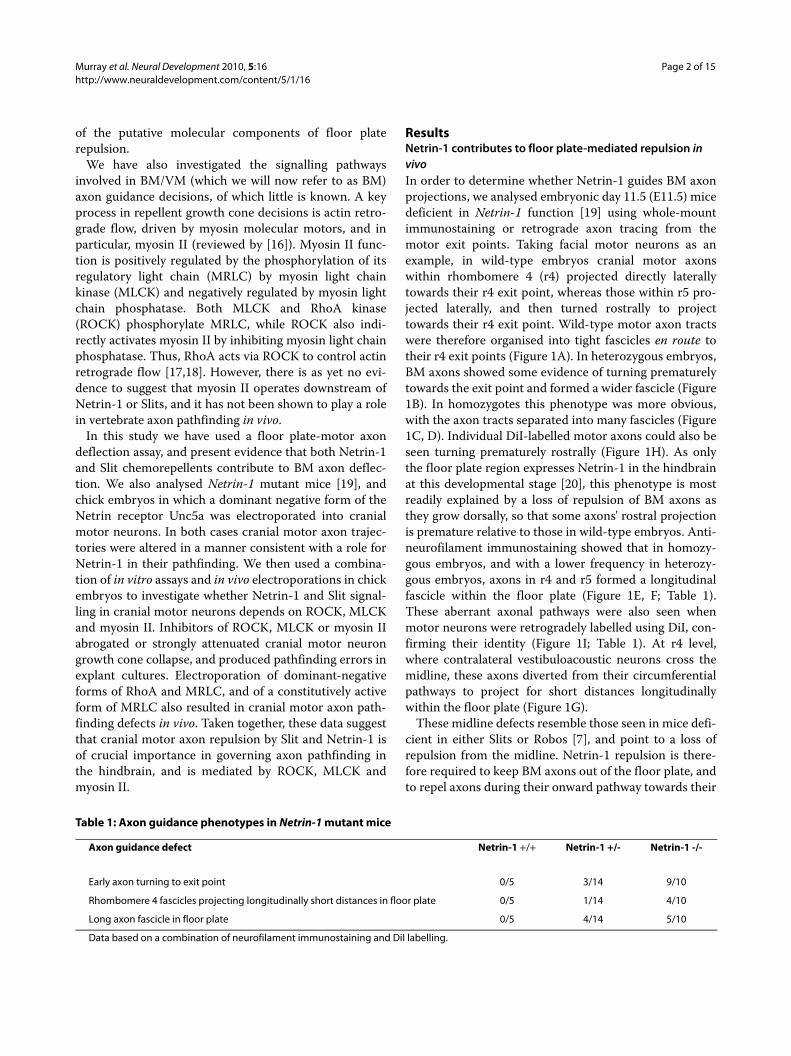
ResultsNetrin-1 contributes to floor plate-mediated repulsion in vivoIn order to determine whether Netrin-1 guides BM axonprojections, we analysed embryonic day 11.5 (E11.5) micedeficient in Netrin-1 function [19] using whole-mountimmunostaining or retrograde axon tracing from themotor exit points. Taking facial motor neurons as anexample, in wild-type embryos cranial motor axonswithin rhombomere 4 (r4) projected directly laterallytowards their r4 exit point, whereas those within r5 pro-jected laterally, and then turned rostrally to projecttowards their r4 exit point. Wild-type motor axon tractswere therefore organised into tight fascicles en route totheir r4 exit points (Figure 1A). In heterozygous embryos,BM axons showed some evidence of turning prematurelytowards the exit point and formed a wider fascicle (Figure1B). In homozygotes this phenotype was more obvious,with the axon tracts separated into many fascicles (Figure1C, D). Individual DiI-labelled motor axons could also beseen turning prematurely rostrally (Figure 1H). As onlythe floor plate region expresses Netrin-1 in the hindbrainat this developmental stage [20], this phenotype is mostreadily explained by a loss of repulsion of BM axons asthey grow dorsally, so that some axons' rostral projectionis premature relative to those in wild-type embryos. Anti-neurofilament immunostaining showed that in homozy-gous embryos, and with a lower frequency in heterozy-gous embryos, axons in r4 and r5 formed a longitudinalfascicle within the floor plate (Figure 1E, F; Table 1).These aberrant axonal pathways were also seen whenmotor neurons were retrogradely labelled using DiI, con-firming their identity (Figure 1I; Table 1). At r4 level,where contralateral vestibuloacoustic neurons cross themidline, these axons diverted from their circumferentialpathways to project for short distances longitudinallywithin the floor plate (Figure 1G).
These midline defects resemble those seen in mice defi-cient in either Slits or Robos [7], and point to a loss ofrepulsion from the midline. Netrin-1 repulsion is there-fore required to keep BM axons out of the floor plate, andto repel axons during their onward pathway towards their
Table 1: Axon guidance phenotypes in Netrin-1 mutant mice
Axon guidance defect Netrin-1 +/+ Netrin-1 +/- Netrin-1 -/-
Early axon turning to exit point 0/5 3/14 9/10
Rhombomere 4 fascicles projecting longitudinally short distances in floor plate 0/5 1/14 4/10
Long axon fascicle in floor plate 0/5 4/14 5/10
Data based on a combination of neurofilament immunostaining and DiI labelling.
Murray et al. Neural Development 2010, 5:16http://www.neuraldevelopment.com/content/5/1/16
Page 3 of 15
dorsal exit points. A previous study showed that trochlearmotor neuron cell bodies (originating in r1) were ectopi-cally located in the floor plate in Netrin-1 mutants [19].We focussed our attention on r2 to r8 levels but failed tosee any consistent misplacement of cranial motor neuroncell bodies.
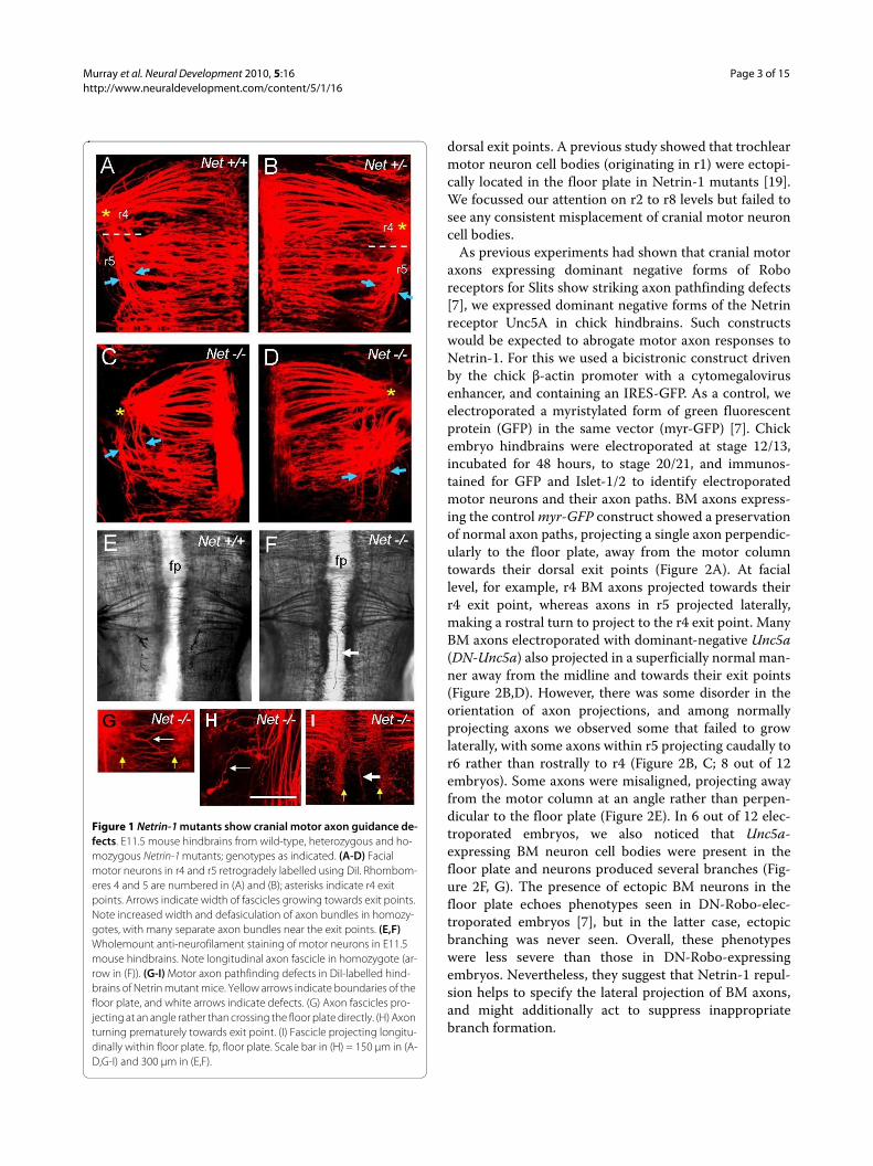
As previous experiments had shown that cranial motoraxons expressing dominant negative forms of Roboreceptors for Slits show striking axon pathfinding defects[7], we expressed dominant negative forms of the Netrinreceptor Unc5A in chick hindbrains. Such constructswould be expected to abrogate motor axon responses toNetrin-1. For this we used a bicistronic construct drivenby the chick β-actin promoter with a cytomegalovirusenhancer, and containing an IRES-GFP. As a control, weelectroporated a myristylated form of green fluorescentprotein (GFP) in the same vector (myr-GFP) [7]. Chickembryo hindbrains were electroporated at stage 12/13,incubated for 48 hours, to stage 20/21, and immunos-tained for GFP and Islet-1/2 to identify electroporatedmotor neurons and their axon paths. BM axons express-ing the control myr-GFP construct showed a preservationof normal axon paths, projecting a single axon perpendic-ularly to the floor plate, away from the motor columntowards their dorsal exit points (Figure 2A). At faciallevel, for example, r4 BM axons projected towards theirr4 exit point, whereas axons in r5 projected laterally,making a rostral turn to project to the r4 exit point. ManyBM axons electroporated with dominant-negative Unc5a(DN-Unc5a) also projected in a superficially normal man-ner away from the midline and towards their exit points(Figure 2B,D). However, there was some disorder in theorientation of axon projections, and among normallyprojecting axons we observed some that failed to growlaterally, with some axons within r5 projecting caudally tor6 rather than rostrally to r4 (Figure 2B, C; 8 out of 12embryos). Some axons were misaligned, projecting awayfrom the motor column at an angle rather than perpen-dicular to the floor plate (Figure 2E). In 6 out of 12 elec-troporated embryos, we also noticed that Unc5a-expressing BM neuron cell bodies were present in thefloor plate and neurons produced several branches (Fig-ure 2F, G). The presence of ectopic BM neurons in thefloor plate echoes phenotypes seen in DN-Robo-elec-troporated embryos [7], but in the latter case, ectopicbranching was never seen. Overall, these phenotypeswere less severe than those in DN-Robo-expressingembryos. Nevertheless, they suggest that Netrin-1 repul-sion helps to specify the lateral projection of BM axons,and might additionally act to suppress inappropriatebranch formation.
Figure 1 Netrin-1 mutants show cranial motor axon guidance de-fects. E11.5 mouse hindbrains from wild-type, heterozygous and ho-mozygous Netrin-1 mutants; genotypes as indicated. (A-D) Facial motor neurons in r4 and r5 retrogradely labelled using DiI. Rhombom-eres 4 and 5 are numbered in (A) and (B); asterisks indicate r4 exit points. Arrows indicate width of fascicles growing towards exit points. Note increased width and defasiculation of axon bundles in homozy-gotes, with many separate axon bundles near the exit points. (E,F) Wholemount anti-neurofilament staining of motor neurons in E11.5 mouse hindbrains. Note longitudinal axon fascicle in homozygote (ar-row in (F)). (G-I) Motor axon pathfinding defects in DiI-labelled hind-brains of Netrin mutant mice. Yellow arrows indicate boundaries of the floor plate, and white arrows indicate defects. (G) Axon fascicles pro-jecting at an angle rather than crossing the floor plate directly. (H) Axon turning prematurely towards exit point. (I) Fascicle projecting longitu-dinally within floor plate. fp, floor plate. Scale bar in (H) = 150 μm in (A-D,G-I) and 300 μm in (E,F).
Murray et al. Neural Development 2010, 5:16http://www.neuraldevelopment.com/content/5/1/16
Page 4 of 15
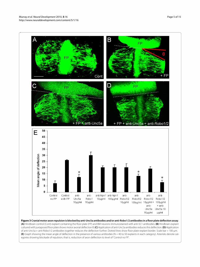
Both Slit and Netrin contribute to floor plate-mediated repulsion in vitroIn order to determine the molecular components of floorplate repulsion, we used a deflection assay in which bilat-eral ventral third hindbrain explants (which contain aninternal floor plate) were juxtaposed with a second floorplate explant at 90° in collagen gels and cultured for 24hours. In control explants cultured alone, BM axons,identified by SC1 immunostaining [21], followed a rea-sonably straight path laterally and away from the internalfloor plate (Figure 3A). However, in the presence of thejuxtaposed floor plate, motor axons deflected reproduc-ibly (Figure 3B). Confocal images of immunostainedexplants were used to measure the angle of BM axon bun-dles relative to the border with the additional floor plate,
and the mean angle for each explant category was deter-mined. The mean deflection angle was 7° for controlexplants and 26° in the presence of the additional floorplate.
We then applied to these co-cultures antibodies to theUnc5a, Robo1/2 and Neuropilin-1 receptors, whichmediate the repellent effects of Netrin-1, Slits andSema3A, respectively (reviewed in [1]). We found thatapplication of anti-Unc5a antibody at 10 μg/ml reducedfloor plate-dependent repulsion to 12° (Figure 3C, E), sig-nificantly different from controls with floor plate, but notsignificantly different from controls without floor plate.Therefore, this antibody completely blocked floor platerepulsion. Application of an anti-Robo1 antibody had noeffect, while anti-Robo1/2 antibodies elicited a dose-dependent reduction in repulsion at 10 μg/ml and 100 μg/ml, with the latter concentration producing completeblockade (Figure 3E). Both Robo1 and Robo2 participatein floor plate repulsion of BM axons in the rat [7], andthese data suggest that both receptors are also involved inthe chick. By contrast, anti-Neuropilin-1 antibodiesapplied at 10 μg/ml or 100 μg/ml did not significantlyreduce floor plate-mediated repulsion (Figure 1E), sug-gesting that Sema3A plays no role. Application of anti-Unc5a and anti-Robo1/2 antibodies together at 10 μg/mland 100 μg/ml, respectively, reduced the mean deflectionangle to 8° (Figure 3D, E). This is the largest reduction inrepulsion we observed, and is significantly different fromcontrols with floor plate but not significantly differentfrom controls without floor plate (see Additional file 1 forstatistics). Taken together, these data suggest that bothUnc5a and Robo1/2-dependent mechanisms mediatefloor plate repulsion.
To confirm that these antibodies block the repellenteffects of Netrin-1 and Slits in our culture system, we co-cultured hindbrain explants for 24 hours with their ros-tral/caudal borders juxtaposed to HEK293T cell clusterssecreting either Slit-1 or Netrin-1 or mock-transfected(control). In co-cultures with mock-transfected cells,most clusters were permissive, with motor axons growinginto them (Additional file 2A). By contrast, most Slit-1 orNetrin-1-secreting cell clusters were inhibitory, withmotor axons failing to grow into them (Additional file2B). Application of anti-Unc5a antibodies to Netrin-1 co-cultures, or of anti-Robo1/2 antibodies to Slit-1 co-cul-tures resulted in a significant increase in the number ofclusters that were invaded by axons (permissive; Addi-tional file 2). However, anti-Unc5a antibodies did notaffect Slit-mediated repulsion nor did anti-Robo1/2 anti-bodies block Netrin-1-mediated repulsion. This demon-strates that the anti-Unc5a and anti-Robo1/2 antibodiesblock BM axonal responses to Netrin-1 and Slit, respec-tively.
Figure 2 Expression of dominant-negative forms of Unc5a in the chick hindbrain causes BM axon pathfinding errors. (A-G) Flat-mount chick hindbrains that have been electroporated with plasmids encoding myristylated GFP (myr-GFP) or dominant-negative Unc5a (DN-Unc5a), as labelled. Immunostaining with anti-GFP (green) and anti-Islet1/2 (red) antibodies. In controls, axons in r5 grow rostrally and correctly towards exit point (orange arrow in (A)). In DN-Unc5a-ex-pressing embryos some axons also pathfind normally (orange arrow in (D)), whereas some axons in r5 turn caudally (arrowheads in (B,C)). Some DN-Unc5a-expressing BM axons are orientated at an angle to the floor plate rather than perpendicularly (arrowheads in (E)). DN-Unc5a-expressing BM neuron cell bodies are found ectopically within the floor plate (pink arrowheads in (F,G)) and produce multiple branches (white arrowheads in (F,G)). Inset in (F) shows cell body with higher Is-let-1/2 immunofluorescence. fp, floor plate. Scale bar: 50 μm (A,B); 25 μm (C-E); 20 μm (F,G).
Murray et al. Neural Development 2010, 5:16http://www.neuraldevelopment.com/content/5/1/16
Page 5 of 15
Figure 3 Cranial motor axon repulsion is blocked by anti-Unc5a antibodies and/or anti-Robo1/2 antibodies in a floor plate deflection assay. (A) Hindbrain control (Cont) explant containing the floor plate (FP) and BM neurons immunostained with anti-SC1 antibodies. (B) Hindbrain explant cultured with juxtaposed floor plate shows motor axonal deflection θ. (C) Application of anti-Unc5a antibodies reduces this deflection. (D) Application of anti-Unc5a + anti-Robo1/2 antibodies together reduces the deflection further. Dotted lines show floor plate-explant border. Scale bar = 100 μm. (E) Graph showing the mean angle of deflection in the presence of various antibodies (N = 40 to 50 explants in each category). Asterisks denote cat-egories showing blockade of repulsion, that is, reduction of axon deflection to level of 'Control no FP'.
Murray et al. Neural Development 2010, 5:16http://www.neuraldevelopment.com/content/5/1/16
Page 6 of 15
Taken together, these experiments indicate that bothNetrin-1 and Slits contribute to the floor plate repulsionof cranial BM axons. The finding that either antibodyalone reduces axon deflection to control levels indicates adegree of redundancy in the function of Netrin-1 andSlit-mediated repulsive systems.
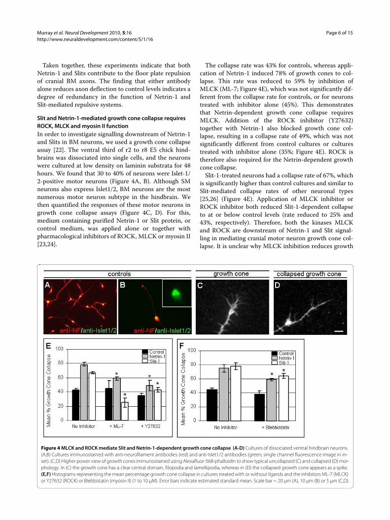
Slit and Netrin-1-mediated growth cone collapse requires ROCK, MLCK and myosin II functionIn order to investigate signalling downstream of Netrin-1and Slits in BM neurons, we used a growth cone collapseassay [22]. The ventral third of r2 to r8 E5 chick hind-brains was dissociated into single cells, and the neuronswere cultured at low density on laminin substrata for 48hours. We found that 30 to 40% of neurons were Islet-1/2-positive motor neurons (Figure 4A, B). Although SMneurons also express Islet1/2, BM neurons are the mostnumerous motor neuron subtype in the hindbrain. Wethen quantified the responses of these motor neurons ingrowth cone collapse assays (Figure 4C, D). For this,medium containing purified Netrin-1 or Slit protein, orcontrol medium, was applied alone or together withpharmacological inhibitors of ROCK, MLCK or myosin II[23,24].
The collapse rate was 43% for controls, whereas appli-cation of Netrin-1 induced 78% of growth cones to col-lapse. This rate was reduced to 59% by inhibition ofMLCK (ML-7; Figure 4E), which was not significantly dif-ferent from the collapse rate for controls, or for neuronstreated with inhibitor alone (45%). This demonstratesthat Netrin-dependent growth cone collapse requiresMLCK. Addition of the ROCK inhibitor (Y27632)together with Netrin-1 also blocked growth cone col-lapse, resulting in a collapse rate of 49%, which was notsignificantly different from control cultures or culturestreated with inhibitor alone (35%; Figure 4E). ROCK istherefore also required for the Netrin-dependent growthcone collapse.
Slit-1-treated neurons had a collapse rate of 67%, whichis significantly higher than control cultures and similar toSlit-mediated collapse rates of other neuronal types[25,26] (Figure 4E). Application of MLCK inhibitor orROCK inhibitor both reduced Slit-1-dependent collapseto at or below control levels (rate reduced to 25% and43%, respectively). Therefore, both the kinases MLCKand ROCK are downstream of Netrin-1 and Slit signal-ling in mediating cranial motor neuron growth cone col-lapse. It is unclear why MLCK inhibition reduces growth
Figure 4 MLCK and ROCK mediate Slit and Netrin-1-dependent growth cone collapse. (A-D) Cultures of dissociated ventral hindbrain neurons. (A,B) Cultures immunostained with anti-neurofilament antibodies (red) and anti-Islet1/2 antibodies (green; single channel fluorescence image in in-set). (C,D) Higher power view of growth cones immunostained using Alexafluor-568-phalloidin to show typical uncollapsed (C) and collapsed (D) mor-phology. In (C) the growth cone has a clear central domain, filopodia and lamellipodia, whereas in (D) the collapsed growth cone appears as a spike. (E,F) Histograms representing the mean percentage growth cone collapse in cultures treated with or without ligands and the inhibitors ML-7 (MLCK) or Y27632 (ROCK) or Blebbistatin (myosin II) (1 to 10 μM). Error bars indicate estimated standard mean. Scale bar = 20 μm (A), 10 μm (B) or 5 μm (C,D).
Murray et al. Neural Development 2010, 5:16http://www.neuraldevelopment.com/content/5/1/16
Page 7 of 15
cone collapse to below control levels. However, we specu-late that inhibiting the MLCK-dependent growth conecollapsing effects of Slit somehow unveils a second Slitactivity, which promotes growth cone protrusion.
In order to further test the involvement of myosin II incranial motor neuron growth cone collapse, we appliedthe myosin II inhibitor, blebbistatin. Application of bleb-bistatin at 1 μM did not enhance collapse beyond controllevels (38% compared with 45%), but in the presence ofSlit protein, blebbistatin attenuated collapse to 63% com-pared with 78% for Slit alone (Figure 4F). In addition,whereas Netrin-1 alone produced a collapse rate of 75%,blebbistatin reduced this rate to 58% (Figure 4F). In bothcases this represented an attenuation of collapse ratherthan complete blockade, but nevertheless demonstrates asignificant role of myosin II in Netrin-1 and Slit-mediatedcollapse. Higher concentrations of blebbistatin on disso-ciated neurons tended to increase growth cone collapseper se, and therefore make it problematic to test whethera higher dose might completely block collapse (data notshown).
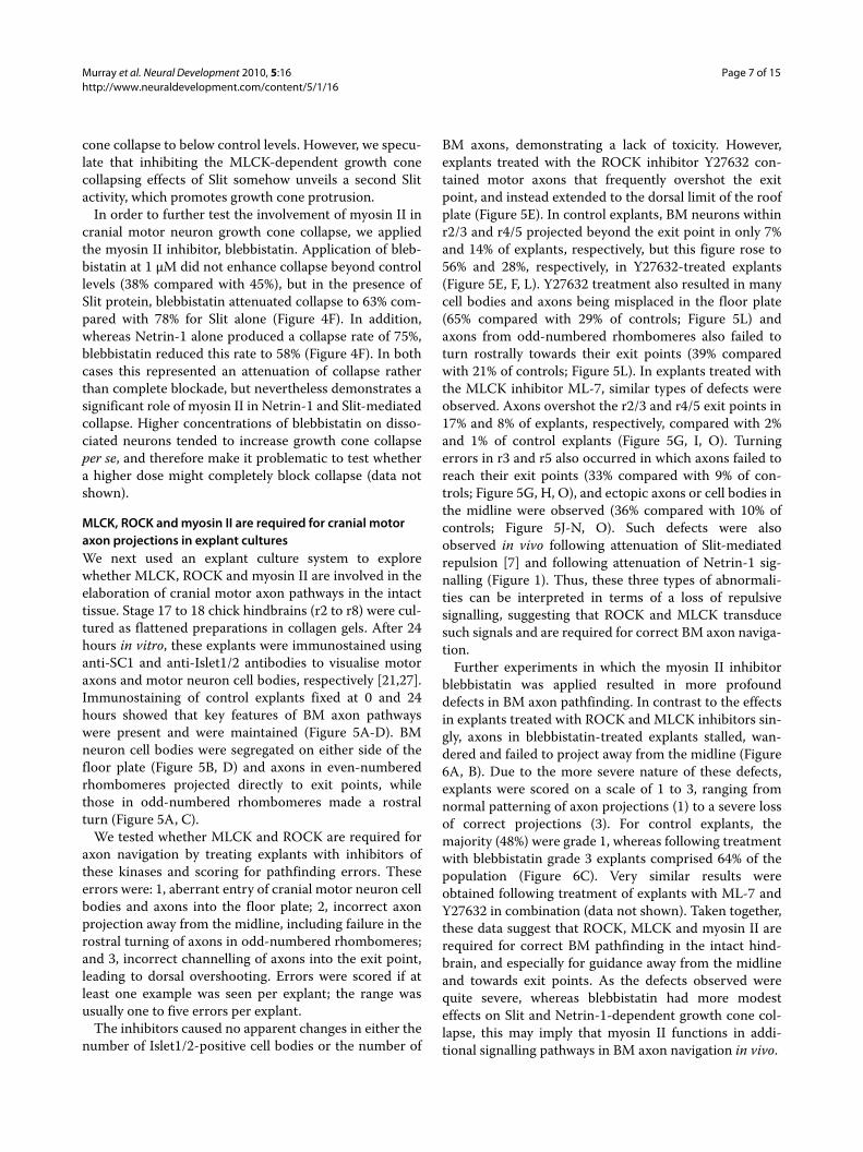
MLCK, ROCK and myosin II are required for cranial motor axon projections in explant culturesWe next used an explant culture system to explorewhether MLCK, ROCK and myosin II are involved in theelaboration of cranial motor axon pathways in the intacttissue. Stage 17 to 18 chick hindbrains (r2 to r8) were cul-tured as flattened preparations in collagen gels. After 24hours in vitro, these explants were immunostained usinganti-SC1 and anti-Islet1/2 antibodies to visualise motoraxons and motor neuron cell bodies, respectively [21,27].Immunostaining of control explants fixed at 0 and 24hours showed that key features of BM axon pathwayswere present and were maintained (Figure 5A-D). BMneuron cell bodies were segregated on either side of thefloor plate (Figure 5B, D) and axons in even-numberedrhombomeres projected directly to exit points, whilethose in odd-numbered rhombomeres made a rostralturn (Figure 5A, C).
We tested whether MLCK and ROCK are required foraxon navigation by treating explants with inhibitors ofthese kinases and scoring for pathfinding errors. Theseerrors were: 1, aberrant entry of cranial motor neuron cellbodies and axons into the floor plate; 2, incorrect axonprojection away from the midline, including failure in therostral turning of axons in odd-numbered rhombomeres;and 3, incorrect channelling of axons into the exit point,leading to dorsal overshooting. Errors were scored if atleast one example was seen per explant; the range wasusually one to five errors per explant.
The inhibitors caused no apparent changes in either thenumber of Islet1/2-positive cell bodies or the number of
BM axons, demonstrating a lack of toxicity. However,explants treated with the ROCK inhibitor Y27632 con-tained motor axons that frequently overshot the exitpoint, and instead extended to the dorsal limit of the roofplate (Figure 5E). In control explants, BM neurons withinr2/3 and r4/5 projected beyond the exit point in only 7%and 14% of explants, respectively, but this figure rose to56% and 28%, respectively, in Y27632-treated explants(Figure 5E, F, L). Y27632 treatment also resulted in manycell bodies and axons being misplaced in the floor plate(65% compared with 29% of controls; Figure 5L) andaxons from odd-numbered rhombomeres also failed toturn rostrally towards their exit points (39% comparedwith 21% of controls; Figure 5L). In explants treated withthe MLCK inhibitor ML-7, similar types of defects wereobserved. Axons overshot the r2/3 and r4/5 exit points in17% and 8% of explants, respectively, compared with 2%and 1% of control explants (Figure 5G, I, O). Turningerrors in r3 and r5 also occurred in which axons failed toreach their exit points (33% compared with 9% of con-trols; Figure 5G, H, O), and ectopic axons or cell bodies inthe midline were observed (36% compared with 10% ofcontrols; Figure 5J-N, O). Such defects were alsoobserved in vivo following attenuation of Slit-mediatedrepulsion [7] and following attenuation of Netrin-1 sig-nalling (Figure 1). Thus, these three types of abnormali-ties can be interpreted in terms of a loss of repulsivesignalling, suggesting that ROCK and MLCK transducesuch signals and are required for correct BM axon naviga-tion.
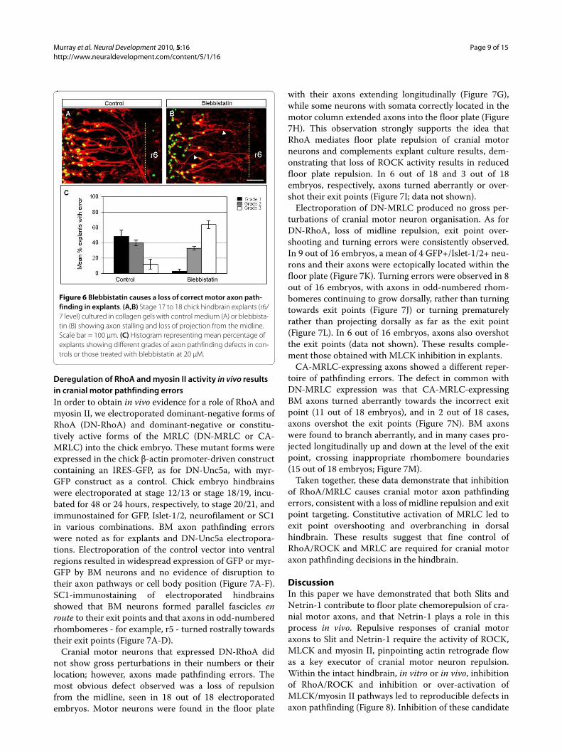
Further experiments in which the myosin II inhibitorblebbistatin was applied resulted in more profounddefects in BM axon pathfinding. In contrast to the effectsin explants treated with ROCK and MLCK inhibitors sin-gly, axons in blebbistatin-treated explants stalled, wan-dered and failed to project away from the midline (Figure6A, B). Due to the more severe nature of these defects,explants were scored on a scale of 1 to 3, ranging fromnormal patterning of axon projections (1) to a severe lossof correct projections (3). For control explants, themajority (48%) were grade 1, whereas following treatmentwith blebbistatin grade 3 explants comprised 64% of thepopulation (Figure 6C). Very similar results wereobtained following treatment of explants with ML-7 andY27632 in combination (data not shown). Taken together,these data suggest that ROCK, MLCK and myosin II arerequired for correct BM pathfinding in the intact hind-brain, and especially for guidance away from the midlineand towards exit points. As the defects observed werequite severe, whereas blebbistatin had more modesteffects on Slit and Netrin-1-dependent growth cone col-lapse, this may imply that myosin II functions in addi-tional signalling pathways in BM axon navigation in vivo.
Murray et al. Neural Development 2010, 5:16http://www.neuraldevelopment.com/content/5/1/16
Page 8 of 15
Figure 5 Inhibition of ROCK and MLCK signalling causes cranial motor axon guidance defects in chick hindbrain explants. (A-K,M,N) Stage 17 to 18 chick hindbrain explants cultured in collagen gels as controls or with the inhibitors ML-7 (MLCK) or Y27632 (ROCK) (10 to 20 μM), as labelled, and fixed at time 0 (A) or after 24 hours (all other panels), immunostained with anti-SC1 (red) and anti-Islet1/2 antibodies (green). In some cases (B,D,K,N) anti-Islet-1/2 immunostaining is shown as a single image of adjacent double-stained explant. (E,F,I) Rhombomere 4/5 level; (G,H) r6 level; (J,K,M,N) r6 to r8 level. Yellow arrows indicate boundaries of floor plate and arrowheads indicate axon pathfinding errors. Scale bar: (A-D) 100 μm; (E,F,J,K,M,N) 50 μm; (G-I) 25 μm. (L,O) Histograms representing the mean percentage of explants with three types of errors when treated with Y27632 (L) or ML-7 (O) compared with controls. Error bars represent standard error of the mean.
Murray et al. Neural Development 2010, 5:16http://www.neuraldevelopment.com/content/5/1/16
Page 9 of 15
Deregulation of RhoA and myosin II activity in vivo results in cranial motor pathfinding errorsIn order to obtain in vivo evidence for a role of RhoA andmyosin II, we electroporated dominant-negative forms ofRhoA (DN-RhoA) and dominant-negative or constitu-tively active forms of the MRLC (DN-MRLC or CA-MRLC) into the chick embryo. These mutant forms wereexpressed in the chick β-actin promoter-driven constructcontaining an IRES-GFP, as for DN-Unc5a, with myr-GFP construct as a control. Chick embryo hindbrainswere electroporated at stage 12/13 or stage 18/19, incu-bated for 48 or 24 hours, respectively, to stage 20/21, andimmunostained for GFP, Islet-1/2, neurofilament or SC1in various combinations. BM axon pathfinding errorswere noted as for explants and DN-Unc5a electropora-tions. Electroporation of the control vector into ventralregions resulted in widespread expression of GFP or myr-GFP by BM neurons and no evidence of disruption totheir axon pathways or cell body position (Figure 7A-F).SC1-immunostaining of electroporated hindbrainsshowed that BM neurons formed parallel fascicles enroute to their exit points and that axons in odd-numberedrhombomeres - for example, r5 - turned rostrally towardstheir exit points (Figure 7A-D).
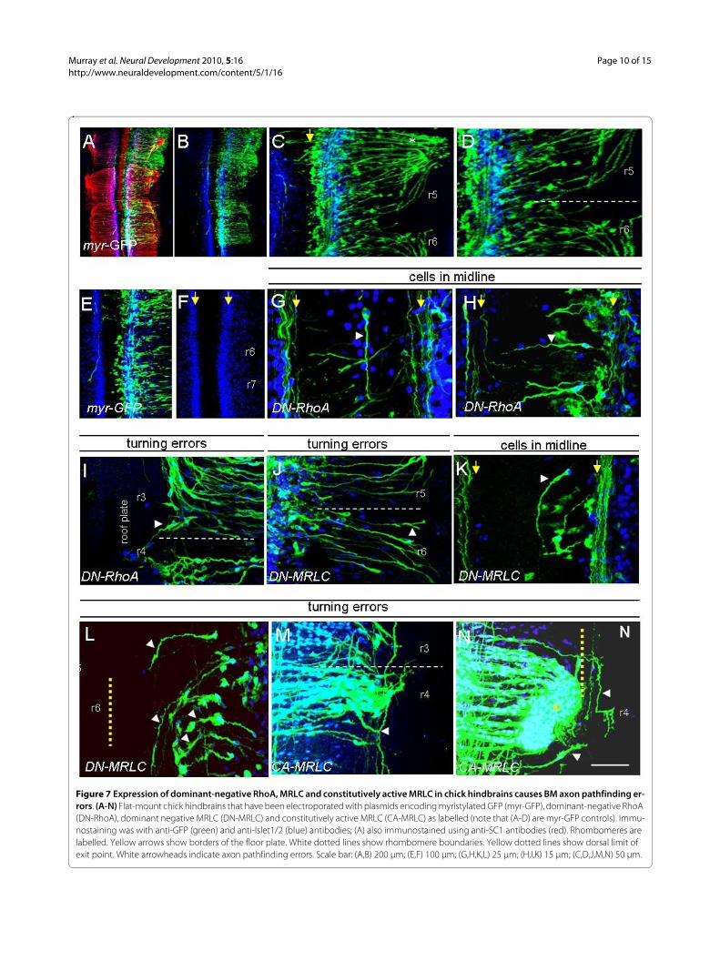
Cranial motor neurons that expressed DN-RhoA didnot show gross perturbations in their numbers or theirlocation; however, axons made pathfinding errors. Themost obvious defect observed was a loss of repulsionfrom the midline, seen in 18 out of 18 electroporatedembryos. Motor neurons were found in the floor plate
with their axons extending longitudinally (Figure 7G),while some neurons with somata correctly located in themotor column extended axons into the floor plate (Figure7H). This observation strongly supports the idea thatRhoA mediates floor plate repulsion of cranial motorneurons and complements explant culture results, dem-onstrating that loss of ROCK activity results in reducedfloor plate repulsion. In 6 out of 18 and 3 out of 18embryos, respectively, axons turned aberrantly or over-shot their exit points (Figure 7I; data not shown).
Electroporation of DN-MRLC produced no gross per-turbations of cranial motor neuron organisation. As forDN-RhoA, loss of midline repulsion, exit point over-shooting and turning errors were consistently observed.In 9 out of 16 embryos, a mean of 4 GFP+/Islet-1/2+ neu-rons and their axons were ectopically located within thefloor plate (Figure 7K). Turning errors were observed in 8out of 16 embryos, with axons in odd-numbered rhom-bomeres continuing to grow dorsally, rather than turningtowards exit points (Figure 7J) or turning prematurelyrather than projecting dorsally as far as the exit point(Figure 7L). In 6 out of 16 embryos, axons also overshotthe exit points (data not shown). These results comple-ment those obtained with MLCK inhibition in explants.
CA-MRLC-expressing axons showed a different reper-toire of pathfinding errors. The defect in common withDN-MRLC expression was that CA-MRLC-expressingBM axons turned aberrantly towards the incorrect exitpoint (11 out of 18 embryos), and in 2 out of 18 cases,axons overshot the exit points (Figure 7N). BM axonswere found to branch aberrantly, and in many cases pro-jected longitudinally up and down at the level of the exitpoint, crossing inappropriate rhombomere boundaries(15 out of 18 embryos; Figure 7M).
Taken together, these data demonstrate that inhibitionof RhoA/MRLC causes cranial motor axon pathfindingerrors, consistent with a loss of midline repulsion and exitpoint targeting. Constitutive activation of MRLC led toexit point overshooting and overbranching in dorsalhindbrain. These results suggest that fine control ofRhoA/ROCK and MRLC are required for cranial motoraxon pathfinding decisions in the hindbrain.
DiscussionIn this paper we have demonstrated that both Slits andNetrin-1 contribute to floor plate chemorepulsion of cra-nial motor axons, and that Netrin-1 plays a role in thisprocess in vivo. Repulsive responses of cranial motoraxons to Slit and Netrin-1 require the activity of ROCK,MLCK and myosin II, pinpointing actin retrograde flowas a key executor of cranial motor neuron repulsion.Within the intact hindbrain, in vitro or in vivo, inhibitionof RhoA/ROCK and inhibition or over-activation ofMLCK/myosin II pathways led to reproducible defects inaxon pathfinding (Figure 8). Inhibition of these candidate
Figure 6 Blebbistatin causes a loss of correct motor axon path-finding in explants. (A,B) Stage 17 to 18 chick hindbrain explants (r6/7 level) cultured in collagen gels with control medium (A) or blebbista-tin (B) showing axon stalling and loss of projection from the midline. Scale bar = 100 μm. (C) Histogram representing mean percentage of explants showing different grades of axon pathfinding defects in con-trols or those treated with blebbistatin at 20 μM.
Murray et al. Neural Development 2010, 5:16http://www.neuraldevelopment.com/content/5/1/16
Page 10 of 15
Figure 7 Expression of dominant-negative RhoA, MRLC and constitutively active MRLC in chick hindbrains causes BM axon pathfinding er-rors. (A-N) Flat-mount chick hindbrains that have been electroporated with plasmids encoding myristylated GFP (myr-GFP), dominant-negative RhoA (DN-RhoA), dominant negative MRLC (DN-MRLC) and constitutively active MRLC (CA-MRLC) as labelled (note that (A-D) are myr-GFP controls). Immu-nostaining was with anti-GFP (green) and anti-Islet1/2 (blue) antibodies; (A) also immunostained using anti-SC1 antibodies (red). Rhombomeres are labelled. Yellow arrows show borders of the floor plate. White dotted lines show rhombomere boundaries. Yellow dotted lines show dorsal limit of exit point. White arrowheads indicate axon pathfinding errors. Scale bar: (A,B) 200 μm; (E,F) 100 μm; (G,H,K,L) 25 μm; (H,I,K) 15 μm; (C,D,J,M,N) 50 μm.
Murray et al. Neural Development 2010, 5:16http://www.neuraldevelopment.com/content/5/1/16
Page 11 of 15
molecules led to the ectopic positioning of cell bodies andaxons in the floor plate, and the failure of axons to projector to target exit points correctly. All of these defects areconsistent with a loss of repulsion by the floor plate and/or the dorsal neuroepithelium, and closely resembledefects resulting from attenuation of Slit or Netrin-1 sig-nalling. Therefore, Slits and Netrin-1 play key roles in BMaxon repulsion, acting via ROCK, MLCK and myosin IIto regulate the growth cone cytoskeleton.
Slits and Netrin-1 collaborate in floor plate repulsionThe results of BM axon deflection assays suggest thatboth Slits and Netrin-1 but not Sema3A play a role infloor plate repulsion. Abrogation of either Slit or Netrin-1-dependent mechanisms was sufficient to block repul-sion. Supportive evidence that both mechanisms partici-pate comes from attenuating repulsive function in Slit1/2double mutants, Robo1 or 2 single mutants or Netrin-1mutants ([7] and this study). In all cases, phenotypesappear to reflect a partial elimination of floor plate repul-sion. There is a discrepancy, therefore, between the pres-ence of defects in single mutants, apparently reflecting anadditive effect of Slit and Netrin-1 repulsion, and theredundancy of both mechanisms in our floor plate deflec-tion assay. We think that this might reflect technical limi-tations of the deflection assay, and indeed in this systemblocking both Netrin-1 and Slit signalling produces thestrongest effect.
It would be challenging to abrogate both Slit and Netrinrepulsive mechanisms genetically. But in view of the factthat we previously found that Slit-3 was not repulsive [7],analysis of Slit-1/Slit-2/Netrin-1 triple mutants wouldtherefore be very interesting. Although we found thatSema3A did not play a role in floor plate repulsion, itremains possible that Sema3A derived from the noto-chord might contribute to cranial motor axon repulsionfrom outside the central nervous system [10]. Floor plate-derived SM axon repellents remain to be identified, asneither Slits nor Netrin-1 repel these axons [6,7].
Our finding that antibodies to Unc5a and Robo1/2block BM axon repulsion, and that Unc5a loss-of-func-tion produces pathfinding defects, implicates thesereceptors in responses to Netrin-1 and Slits, respectively.While Robo1 and 2 have previously been established asresponding to Slits in BM neurons, Netrin-1 signalling viaUnc5a is less well understood. Whereas DCC (Deleted incolorectal cancer) alone can mediate Netrin-dependentattraction, Unc5a can function alone or with DCC tomediate repulsion [28-30]. We have previously shownthat in the rat, Unc5a is expressed early during cranialmotor axon projection away from the midline, while DCCis expressed later [13]. This suggests that Unc5a alonemediates the initial phase of cranial motor axon out-growth; however, we have not formally tested the role ofDCC.
Slit and Netrin-1-mediated growth cone collapse requires MLCK, ROCK and myosin IIOur experiments show that ROCK (and by implicationRhoA) is required for both Netrin-1 and Slit-dependentmotor neuron growth cone collapse. RhoA has been pro-posed to be involved in repulsive signalling in several sys-tems (reviewed in [17,18]) - for example, in Robo-dependent repulsive signalling at the midline in Droso-
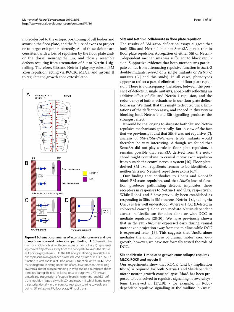
Figure 8 Schematic summaries of axon guidance errors and role of repulsion in cranial motor axon pathfinding. (A) Schematic dia-gram of chick hindbrain with grey axons on control (right) represent-ing correct trajectories, away from the floor plate towards the dorsal exit points (grey ellipses). On the left side (pathfinding errors) blue ax-ons represent axon guidance errors induced by loss of ROCK or MLCK function in vitro and loss of RhoA or MRLC function in vivo. (B-D) Sche-matic diagrams showing operation of repulsive mechanisms during BM cranial motor axon pathfinding in even and odd-numbered rhom-bomeres during (B) initial polarisation and outgrowth, (C) onward growth and suppression of ectopic branching/turning, and (D) roof plate repulsion (especially via MLCK and myosin II), which hems in axon trajectories dorsally and ensures correct axon turning towards exit points. EP, exit point; FP, floor plate; RP, roof plate.
Murray et al. Neural Development 2010, 5:16http://www.neuraldevelopment.com/content/5/1/16
Page 12 of 15
phila [31] and in growth cone collapse [32]. Sema3A-dependent growth cone collapse in dorsal root ganglionneurons was found to be partially blocked by inhibition ofROCK [24]. However, a role for ROCK downstream ofNetrin-1 and Slit in growth cone collapse has not previ-ously been demonstrated. ROCK phosphorylates andactivates MRLC [33,34], and our results implicating bothROCK and myosin II in growth cone collapse suggest thatMRLC is an important target of ROCK phosphorylationin our system. However, ROCK can also phosphorylateLIM kinase and thence affect the activity of cofilin, whichis involved in actin severing/depolymerisation (reviewedin [18]). Indeed, cofilin has been implicated as a target ofSlit signalling in Xenopus retinal axons [26], and itremains an interesting question as to whether cofilin isinvolved in the repulsive guidance events downstream ofSlit in BM neurons. Our evidence for the roles of MLCKand myosin II in Slit and Netrin-1 signalling is consistentwith studies in Drosophila suggesting that MLCK actsdownstream of both Netrin and Slit-dependent axonpathfinding decisions at the midline [35].
RhoA, ROCK, MLCK and myosin II regulate cranial motor neuron pathfinding in vivoTaken together, data from explant cultures and in vivoexperiments suggest a crucial role of RhoA acting viaROCK and MLCK/myosin II in BM pathfinding deci-sions. In explants or in vivo, attenuation of either ROCKor MLCK function led to reproducible pathfinding errorsthat bore a close resemblance to axon pathfinding defectsproduced by attenuation of Slit-Robo signalling [7] (Fig-ure 8A) and in Netrin-1 mutant/DN-Unc5a-expressingembryos. Inhibition of myosin II led to a loss of projec-tions away from the midline, suggesting that myosin II isa crucial target of ROCK and MLCK to regulate cranialmotor axon outgrowth and guidance (Figure 8).
RhoA/ROCK and MLCK/myosin II pathways thusensure segregation of cranial motor neurons and theiraxon projections ipsilateral to the floor plate during nor-mal development. Turning errors of r3/r5 axons wereobserved following attenuation of MLCK or MRLC func-tion in vitro or in vivo, and axons overshot exit points. Wespeculate, therefore, that ROCK and MLCK act via myo-sin II to ensure axon deflection at the dorsal side of theneuroepithelium. Indeed, it has previously been shownthat actin retrograde flow mediated by myosin II isrequired to suppress inappropriate protrusions on theside facing away from an attractant cue during growthcone turning [36]. Experiments demonstrating a role forMLCK and myosin II in dorsal root ganglion axons turn-ing at borders between a laminin substratum and a non-permissive substratum support this idea [23]. Our inter-pretation is that BM axon turning towards exit pointsdepends on localised repulsion at the dorsal edge of the
neuroepithelium, possibly dependent on a narrowdomain of Slit expression, which hems axons in to theirexit points [7,12,34]. Indeed, when isolated from its adja-cent mesenchyme, the dorsal tissue of the hindbrain actsas a source of chemorepulsion [37]. We cannot formallyexclude, however, that attenuation of ROCK/MLCK/myosin II function blocks responses to exit point-derivedchemoattraction [21]. However, the nature of such puta-tive chemoattraction is unknown, and the most likelysource, the boundary cap cells [38], are absent from ourexplant cultures. We also found that expression of CA-MRLC produced defects in BM axon guidance, includingaxons that overshot their exit points and branched ectop-ically. Fine regulation of MRLC activity therefore appearsto be required for accurate BM axon guidance.
ConclusionsModel of repellent signalling pathways in cranial motor axon guidanceOur model, based on the current and previously pub-lished data, can be summarised as three phases of axonpathfinding during the dorsal projection of BM hindbrainmotor neurons (Figure 8B-D). In the initial phase, Slit andNetrin-1 signalling from the floor plate, acting via RhoA/ROCK and MLCK/myosin II, would act to polarise theinitial projection of motor axons away from the midline,and to exclude their axons and cell bodies from thisregion (Figure 8B). Secondly, the gradient of repulsionwould steer axons away from the midline and suppressinappropriate branches (Figure 8C). Thirdly, a repellentborder of Slit dorsally, acting via ROCK and MLCK,would mediate turning of axons in odd-numbered rhom-bomeres towards their exit point, and ensure the correcttermination of both odd- and even-numbered axons atthe correct dorsoventral level of the exit point (Figure8D). An exit point-derived chemoattractant and/oranteroposterior polarisation of the neuroepithelium isrequired to explain the rostral (rather than caudal) pro-jection. Nevertheless, key features of cranial motor axonnavigation appear to depend on repulsive signallingmechanisms.
Materials and methodsEmbryosFertilised hens' eggs (Winter farm or Henry Stewart farm,UK) were incubated to relevant stages [39] at 37°C.
Netrin-1 mutant miceNetrin-1 mutant mice were a kind gift of Dr Marc Tessier-Lavigne and were genotyped as previously described [19].Embryos were obtained at E11.5; DiI retrograde labellingand whole-mount immunohistochemistry were per-formed using anti-neurofilament antibody 2H3 (Develop-mental Studies Hybridoma Bank, Iowa City, Iowa, USA;
Murray et al. Neural Development 2010, 5:16http://www.neuraldevelopment.com/content/5/1/16
Page 13 of 15
1:100) as previously described [40,41]. Hindbrains weredissected out, flat-mounted and axon pathways wereimaged using a laser-scanning confocal microscope.
BM axon deflection assaysFor BM axon deflection assays, ventral explants were dis-sected from stage 18 to 21 embryo chick hindbrains aspreviously described [7,42]. Floor plate explants were dis-sected separately and juxtaposed to the rostral or caudalhindbrain explant borders in collagen gels, in mediumbased on OptiMEM (Invitrogen, Paisley, UK) [7]. Theantibodies used were anti-Unc5a, anti-Robo1 and anti-Neuropilin-1 (R & D Systems, Abingdon, Oxon., UK) andan anti-peptide antibody recognising Robo1 and Robo2('S3'; kind gift of Dr V Sundaresan) [40]. Binding to chickproteins was confirmed using western blots on hindbrainprotein extracts (data not shown). Antibodies were usedat concentrations of 10 to 100 μg/ml.
After 24 hours in vitro, gels were immunostained usingantibodies to the SC1/BEN glycoprotein present onmotor neurons/axons (anti-SC1 at 1:10 or anti-BEN at1:70; Developmental Studies Hybridoma Bank) [21,43].Secondary antibodies were Alexa Fluor-conjugated 488,568 or 633 at 1:800 (Invitrogen, Paisley, UK) or Cy3-con-jugated antibody (Stratech Scientific, Suffolk, UK). Theangle of deflection of axon bundles was measured onconfocal images (Scion Image programme, NIH; Figure2B) to derive a mean axon deflection. Two angles werequantified per explant and a mean was derived for thewhole population of explants in a particular condition, asit would not have been valid to choose one angle perexplant.
To verify that antibodies blocked BM axon responses toSlit and Netrin, HEK293T cells were mock-transfected(control) or transfected with full-length human myc-tagged Slit expression constructs (hSlit-1; kind gift of DrS Sakano, Asahi Kasei Corporation, Japan) in pcDNA3.1(Invitrogen) [44] or a Netrin-1-secreting cell line wasused (kind gift of Dr C Holt). Cell clusters were made inhanging drops and were co-cultured adjacent to the ros-tral/caudal explant borders in collagen gels for 48 hoursas previously described [6,37]. Immunohistochemistrywas as above and cultures were scored as to whether SC-1-positive BM axons entered the cell cluster (permissive),or avoided it (inhibitory) (Additional file 2).
Dissociated motor neuron cultures for growth cone collapseGlass coverslips were coated with poly-D-ornithine andlaminin (15 μg/ml and 1 mg/ml, respectively; Sigma, Dor-set, UK). E5 (stage 25 to 26) hindbrains were isolated asdescribed above and the ventral third of the neuroepithe-lium was dissected out, removing the floor plate, in L15medium (Gibco). Calcium and magnesium-free Hank'sBalanced Salt Solution (HBSS; Invitrogen, Paisley, UK)
was added for 1 to 2 minutes, replaced with 1 ml trypsin(Gibco) and incubated for 15 minutes at 37°C. Trypsinwas then replaced with trypsin inhibitor solution and thetissue dissociated by triturating 15 times. The superna-tant was added to 1 ml of medium containing Neurobasalmedium with 2% B27 supplement, 2% horse serum, 0.1%β-2-mercaptoethanol, 0.35% Glutamax, 1% chick embryoextract, 1% penicillin/streptomycin and 50 ng/ml ciliaryneurotrophic factor (CNTF) (all reagents from Sigma orGibco). Cells were counted and resuspended for platingat 75 × 103 per coverslip in 200 μl of medium.
Axon guidance molecules and/or inhibitors werediluted in pre-warmed media and applied for 30 minutesbefore fixation. Both Slit and Netrin proteins (R & D Sys-tems) were applied to neuronal cultures at concentrationsof 750 ng/ml to 2 μg/ml, while inhibitors were used at 1 to10 μM. In some experiments Drosophila Slit (dSlit) wastransfected into HEK293 cells and the Slit protein waspurified from the conditioned medium [45]. Controlswere treated with normal medium, or controls for dSlitexperiments were treated with conditioned medium frommock-transfected HEK293T cells. Coverslips were thenfixed for 10 minutes in warmed 4% paraformaldehyde,rinsed in phosphate-buffered saline and blocked using 1%bovine serum albumin/0.5% TritonX100 in phosphate-buffered saline for 1 hour. Mouse anti-Islet1/2 (4D5;Developmental Studies Hybridoma Bank) and rabbitanti-neurofilament antibodies (AB1991; Chemicon, Mil-lipore, Watford, UK) were then added in blocking solu-tion for 24 to 48 hours at 4°C. Cultures were washed for 1hour three times in blocking solution, before Cy5-conju-gated goat anti-rabbit, AlexaFluor-488 anti-mouse andAlexa-Fluor-568-phalloidin in blocking solution wereadded overnight at 4°C. After further washes, coverslipswere mounted in Fluorsave (Chemicon). At least 30growth cones per condition were scored in three separateexperiments for collapsed or uncollapsed morphology.Growth cones were only scored if they belonged to neu-rons containing an Islet-1/2-positive cell body, and theneuron was not in contact with any other neuron. Thegrowth cones on axons that were at least three times thelength of the cell body were scored, and a growth conewas scored as collapsed if there were fewer than four tofive filopodia or if it had no clearly defined centraldomain. The Chi-squared test was used to compare datastatistically.
Whole hindbrain culturesStage 17 to 18 hindbrains (r2 to r8) were isolated, flat-tened and cultured in collagen gels in OptiMEM-basedmedium (Gibco) as previously described [37] for 24hours. Immunohistochemistry was as above using anti-SC1/BEN antibodies in combination with the 321 poly-clonal antibody to Islet-1/2 or A8 polyclonal antibody toIslet-1 (1:2,000 and 1:1,000, respectively; kind gifts of Dr
Murray et al. Neural Development 2010, 5:16http://www.neuraldevelopment.com/content/5/1/16
Page 14 of 15
T Jessell). Inhibitors used were Y27632 (ROCK), ML-7(MLCK) and blebbistatin (myosin II) (Calbiochem, Not-tingham, UK; 10 to 20 μM) [23,24]. Controls wereuntreated, except those for blebbistatin, which weretreated with vehicle (1 μl/ml dimethyl sulfoxide; DMSO).
Explant cultures were scored for BM neuron/axonguidance defects, namely: 1, ectopic BM neurons/axonsin the floor plate; 2, turning errors of r3/r5 axons en routeto their exit points; and 3, overshooting of exit points byBM axons. For assessment of cell bodies in the floor plate,rhombomere levels 4/5 were excluded due to the contral-ateral vestibuloacoustic neurons, a subset of which crossthe midline [46]. After blebbistatin treatment (20 μM),explants were scored on a scale of 1 to 3: 1 = normaldevelopment; 2 = intermediate development; and 3 = dis-rupted development, with few axons projecting awayfrom the floor plate.
Electroporation of chick embryos in ovoHens' eggs were incubated to stage 10 to 11 and pro-cessed after [47]. The fourth ventricle was microinjectedwith the appropriate DNA construct. Control constructswere GFP or myristylated GFP (myr-GFP), each regulatedby a ß-actin promoter with a cytomegalovirus enhancer,and incorporating an IRES. Experimental constructsemployed the same vector containing GFP, and includedDN-Unc5a (dominant negative form lacking the cytoplas-mic domain; kind gift of Dr M Tessier-Lavigne), and DN-RhoA (dominant negative form that competes with theendogenous molecule for binding to cellular guaninenucleotide exchange factors (GEFs) and cannot activate adownstream response; kind gift of Dr C Nobes) [48,49].DN-MRLC and CA-MRLC (dominant-negative and con-stitutively active forms of MRLC) were also used, inwhich an alanine substitution for Ser19 and Thr18 resultsin an unphosphorylatable form, while an asparagine sub-stitution at the same position produces a pseudophos-phorylated, consitutively active form (kind gift of Dr YRao) [50].
Embryos were incubated for 24 or 48 hours to stage 20/21 (depending on the age at electroporation) and immu-nohistochemistry was performed as described previously[7], using anti-GFP (Molecular Probes, Invitrogen; rabbitor chick; 1:800), anti-SC1 mouse monoclonal (Develop-mental Studies Hybridoma Bank; 1:10), anti-Islet1/2mouse monoclonal (4D5; 1:100), and anti-neurofilamentrabbit polyclonal Ab1991 (1:1,000; Chemicon). Second-ary antibodies and microscopy were as above.
Additional material
AbbreviationsBM: branchiomotor; CA: constitutively active; DCC: DCC, Deleted in colorectalcancer; DN: dominant-negative; E: embryonic day; GFP: green fluorescent pro-tein; MLCK: myosin light chain kinase; MRLC: myosin regulatory light chain;myr-GFP: myristylated GFP; r: rhombomere; ROCK: RhoA kinase; Sema3A:Semaphorin3A; SM: somatic motor; VM: visceral motor.
Competing interestsThe authors declare that they have no competing interests.
Authors' contributionsAM performed the majority of experiments in this study; other experimentswere performed by AN and SB. AM had substantial input into experimentaldesign and performed all statistical analysis, as well as contributed to writing ofthe paper. SG conceived the study and experimental design, took a major rolein writing the paper, with collaborative input and extensive discussions withUD, who is joint grant holder.
AcknowledgementsThanks to Britta Eickholt for her help and advice during this project. This work was funded by a Wellcome Trust Project grant to SG and UD and a BBSRC PhD studentship to AM.
Author DetailsMRC Centre for Developmental Neurobiology, 4th Floor New Hunt's House, King's College, Guy's Campus, London SE1 1UL, UK
References1. Huber AB, Kolodkin AL, Ginty DD, Cloutier JF: Signaling at the growth
cone: ligand-receptor complexes and the control of axon growth and guidance. Annu Rev Neurosci 2003, 26:509-563.
2. Chilton JK: Molecular mechanisms of axon guidance. Dev Biol 2006, 292:13-24.
3. Guthrie S: Patterning and axon guidance of cranial motor neurons. Nat Rev Neurosci 2007, 8:859-871.
4. Guthrie S, Pini A: Chemorepulsion of developing motor axons by the floor plate. Neuron 1995, 14:1117-1130.
5. Colamarino SA, Tessier-Lavigne M: The axonal chemoattractant netrin-1 is also a chemorepellent for trochlear motor axons. Cell 1995, 81:621-629.
6. Varela-Echavarria A, Tucker A, Puschel AW, Guthrie S: Motor axon subpopulations respond differentially to the chemorepellents netrin-1 and semaphorin D. Neuron 1997, 18:193-207.
7. Hammond R, Vivancos V, Naeem A, Chilton J, Mambetisaeva E, Andrews W, Sundaresan V, Guthrie S: Slit-mediated repulsion is a key regulator of motor axon pathfinding in the hindbrain. Development 2005, 132:4483-4495.
8. Kennedy TE, Serafini T, de la Torre JR, Tessier-Lavigne M: Netrins are diffusible chemotropic factors for commissural axons in the embryonic spinal cord. Cell 1994, 78:425-435.
9. Melendez-Herrera E, Varela-Echavarria A: Expression of secreted semaphorins and their receptors in specific neuromeres, boundaries, and neuronal groups in the developing mouse and chick brain. Brain Res 2006, 1067:126-137.
10. Anderson CN, Ohta K, Quick MM, Fleming A, Keynes R, Tannahill D: Molecular analysis of axon repulsion by the notochord. Development 2003, 130:1123-1133.
Additional file 1 Statistical comparisons of effects of antibodies in floor plate deflection assay.
Additional file 2 Confirmation that antibodies to Unc5a and Robo1/Robo2 block the effects of Netrin-1 and Slit, respectively. (A,B) Exam-ples of chick hindbrain explants in collagen gels (immunostained with anti-SC1 antibodies after 24 hours in vitro) with their rostral/caudal borders fac-ing clusters of HEK293 cells that were either mock-transfected (A) or trans-fected with Slit-1 (B). Axons enter the cluster (permissive) (A) or avoid the cluster (inhibitory) (B). (C) Table showing effects of control and Netrin-1 or Slit-1-secreting HEK293T cell clusters in permitting or inhibiting cranial motor axon outgrowth and effects of anti-Unc5a or anti-Robo1/2 antibod-ies. Scale bar = 100 μm.
Received: 23 March 2010 Accepted: 22 June 2010 Published: 22 June 2010This article is available from: http://www.neuraldevelopment.com/content/5/1/16© 2010 Murray et al; licensee BioMed Central Ltd. This is an Open Access article distributed under the terms of the Creative Commons Attribution License (http://creativecommons.org/licenses/by/2.0), which permits unrestricted use, distribution, and reproduction in any medium, provided the original work is properly cited.Neural Development 2010, 5:16
Murray et al. Neural Development 2010, 5:16http://www.neuraldevelopment.com/content/5/1/16
Page 15 of 15
11. Chilton JK, Guthrie S: Cranial expression of class 3 secreted semaphorins and their neuropilin receptors. Dev Dyn 2003, 228:726-733.
12. Gilthorpe JD, Papantoniou EK, Chedotal A, Lumsden A, Wingate RJ: The migration of cerebellar rhombic lip derivatives. Development 2002, 129:4719-4728.
13. Barrett C, Guthrie S: Expression patterns of the netrin receptor UNC5H1 among developing motor neurons in the embryonic rat hindbrain. Mech Dev 2001, 106:163-166.
14. Leonardo ED, Hinck L, Masu M, Keino-Masu K, Ackerman SL, Tessier-Lavigne M: Vertebrate homologues of C. elegans UNC-5 are candidate netrin receptors. Nature 1997, 386:833-838.
15. Brose K, Bland KS, Wang KH, Arnott D, Henzel W, Goodman CS, Tessier-Lavigne M, Kidd T: Slit proteins bind Robo receptors and have an evolutionarily conserved role in repulsive axon guidance. Cell 1999, 96:795-806.
16. Brown J, Bridgman PC: Role of myosin II in axon outgrowth. J Histochem Cytochem 2003, 51:421-428.
17. Dent EW, Gertler FB: Cytoskeletal dynamics and transport in growth cone motility and axon guidance. Neuron 2003, 40:209-227.
18. Dickson BJ: Rho GTPases in growth cone guidance. Curr Opin Neurobiol 2001, 11:103-110.
19. Serafini T, Colamarino SA, Leonardo ED, Wang H, Beddington R, Skarnes WC, Tessier-Lavigne M: Netrin-1 is required for commissural axon guidance in the developing vertebrate nervous system. Cell 1996, 87:1001-1014.
20. Serafini T, Kennedy TE, Galko MJ, Mirzayan C, Jessell TM, Tessier-Lavigne M: The netrins define a family of axon outgrowth-promoting proteins homologous to C. elegans UNC-6. Cell 1994, 78:409-424.
21. Guthrie S, Lumsden A: Motor neuron pathfinding following rhombomere reversals in the chick embryo hindbrain. Development 1992, 114:663-673.
22. Raper JA, Kapfhammer JP: The enrichment of a neuronal growth cone collapsing activity from embryonic chick brain. Neuron 1990, 4:21-29.
23. Turney SG, Bridgman PC: Laminin stimulates and guides axonal outgrowth via growth cone myosin II activity. Nat Neurosci 2005, 8:717-719.
24. Gallo G: RhoA-kinase coordinates F-actin organization and myosin II activity during semaphorin-3A-induced axon retraction. J Cell Sci 2006, 119:3413-3423.
25. Nguyen Ba-Charvet KT, Brose K, Marillat V, Kidd T, Goodman CS, Tessier-Lavigne M, Sotelo C, Chedotal A: Slit2-Mediated chemorepulsion and collapse of developing forebrain axons. Neuron 1999, 22:463-473.
26. Piper M, Anderson R, Dwivedy A, Weinl C, van Horck F, Leung KM, Cogill E, Holt C: Signaling mechanisms underlying Slit2-induced collapse of Xenopus retinal growth cones. Neuron 2006, 49:215-228.
27. Varela-Echavarria A, Pfaff SL, Guthrie S: Differential expression of LIM homeobox genes among motor neuron subpopulations in the developing chick brain stem. Mol Cell Neurosci 1996, 8:242-257.
28. Keino-Masu K, Masu M, Hinck L, Leonardo ED, Chan SS, Culotti JG, Tessier-Lavigne M: Deleted in Colorectal Cancer (DCC) encodes a netrin receptor. Cell 1996, 87:175-185.
29. Hong K, Hinck L, Nishiyama M, Poo MM, Tessier-Lavigne M, Stein E: A ligand-gated association between cytoplasmic domains of UNC5 and DCC family receptors converts netrin-induced growth cone attraction to repulsion. Cell 1999, 97:927-941.
30. Bartoe JL, McKenna WL, Quan TK, Stafford BK, Moore JA, Xia J, Takamiya K, Huganir RL, Hinck L: Protein interacting with C-kinase 1/protein kinase Calpha-mediated endocytosis converts netrin-1-mediated repulsion to attraction. J Neurosci 2006, 26:3192-3205.
31. Fritz JL, VanBerkum MF: Regulation of rho family GTPases is required to prevent axons from crossing the midline. Dev Biol 2002, 252:46-58.
32. Wu KY, Hengst U, Cox LJ, Macosko EZ, Jeromin A, Urquhart ER, Jaffrey SR: Local translation of RhoA regulates growth cone collapse. Nature 2005, 436:1020-1024.
33. Kimura K, Ito M, Amano M, Chihara K, Fukata Y, Nakafuku M, Yamamori B, Feng J, Nakano T, Okawa K, Iwamatsu A, Kaibuchi K: Regulation of myosin phosphatase by Rho and Rho-associated kinase (Rho-kinase). Science 1996, 273:245-248.
34. Totsukawa G, Yamakita Y, Yamashiro S, Hartshorne DJ, Sasaki Y, Matsumura F: Distinct roles of ROCK (Rho-kinase) and MLCK in spatial regulation of MLC phosphorylation for assembly of stress fibers and focal adhesions in 3T3 fibroblasts. J Cell Biol 2000, 150:797-806.
35. Kim YS, Fritz JL, Seneviratne AK, VanBerkum MF: Constitutively active myosin light chain kinase alters axon guidance decisions in Drosophila embryos. Dev Biol 2002, 249:367-381.
36. Loudon RP, Silver LD, Yee HF Jr, Gallo G: RhoA-kinase and myosin II are required for the maintenance of growth cone polarity and guidance by nerve growth factor. J Neurobiol 2006, 66:847-867.
37. Caton A, Hacker A, Naeem A, Livet J, Maina F, Bladt F, Klein R, Birchmeier C, Guthrie S: The branchial arches and HGF are growth-promoting and chemoattractant for cranial motor axons. Development 2000, 127:1751-1766.
38. Niederlander C, Lumsden A: Late emigrating neural crest cells migrate specifically to the exit points of cranial branchiomotor nerves. Development 1996, 122:2367-2374.
39. Hamburger V, Hamilton HL: A series of normal stages in the development of the chick embryo. 1951. Dev Dyn 1992, 195:231-272.
40. Sundaresan V, Mambetisaeva E, Andrews W, Annan A, Knoll B, Tear G, Bannister L: Dynamic expression patterns of Robo (Robo1 and Robo2) in the developing murine central nervous system. J Comp Neurol 2004, 468:467-481.
41. Jacob J, Tiveron MC, Brunet JF, Guthrie S: Role of the target in the pathfinding of facial visceral motor axons. Mol Cell Neurosci 2000, 16:14-26.
42. Naeem A, Abbas L, Guthrie S: Comparison of the effects of HGF, BDNF, CT-1, CNTF, and the branchial arches on the growth of embryonic cranial motor neurons. J Neurobiol 2002, 51:101-114.
43. Chedotal A, Pourquie O, Sotelo C: Initial tract formation in the brain of the chick embryo: selective expression of the BEN/SC1/DM-GRASP cell adhesion molecule. Eur J Neurosci 1995, 7:198-212.
44. Itoh A, Miyabayashi T, Ohno M, Sakano S: Cloning and expressions of three mammalian homologues of Drosophila slit suggest possible roles for Slit in the formation and maintenance of the nervous system. Brain Res Mol Brain Res 1998, 62:175-186.
45. Stein E, Tessier-Lavigne M: Hierarchical organization of guidance receptors: silencing of netrin attraction by slit through a Robo/DCC receptor complex. Science 2001, 291:1928-1938.
46. Simon H, Lumsden A: Rhombomere-specific origin of the contralateral vestibulo-acoustic efferent neurons and their migration across the embryonic midline. Neuron 1993, 11:209-220.
47. Momose T, Tonegawa A, Takeuchi J, Ogawa H, Umesono K, Yasuda K: Efficient targeting of gene expression in chick embryos by microelectroporation. Dev Growth Differ 1999, 41:335-344.
48. Ridley AJ, Hall A: The small GTP-binding protein rho regulates the assembly of focal adhesions and actin stress fibers in response to growth factors. Cell 1992, 70:389-399.
49. Ridley AJ, Paterson HF, Johnston CL, Diekmann D, Hall A: The small GTP-binding protein rac regulates growth factor-induced membrane ruffling. Cell 1992, 70:401-410.
50. Fumoto K, Uchimura T, Iwasaki T, Ueda K, Hosoya H: Phosphorylation of myosin II regulatory light chain is necessary for migration of HeLa cells but not for localization of myosin II at the leading edge. Biochem J 2003, 370:551-556.
doi: 10.1186/1749-8104-5-16Cite this article as: Murray et al., Slit and Netrin-1 guide cranial motor axon pathfinding via Rho-kinase, myosin light chain kinase and myosin II Neural Development 2010, 5:16


![A two-step actin polymerization mechanism drives dendrite … · 2021. 7. 19. · Netrin receptor, DCC, [7–9] to promote polarized lopo - dia formation during axon guidance. Ena/VASP](https://img.pdfslide.us/doc/110x75/6138eaeda4cdb41a985b5e18/a-two-step-actin-polymerization-mechanism-drives-dendrite-2021-7-19-netrin.jpg)