Embed Size (px)
Citation preview
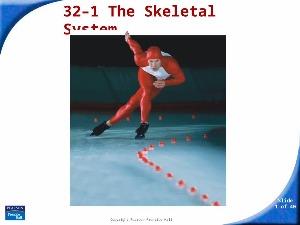
Slide 1 of 40
Copyright Pearson Prentice Hall
32–1 The Skeletal System
36–1 The Skeletal System
Slide 2 of 40
Copyright Pearson Prentice Hall
The Skeleton
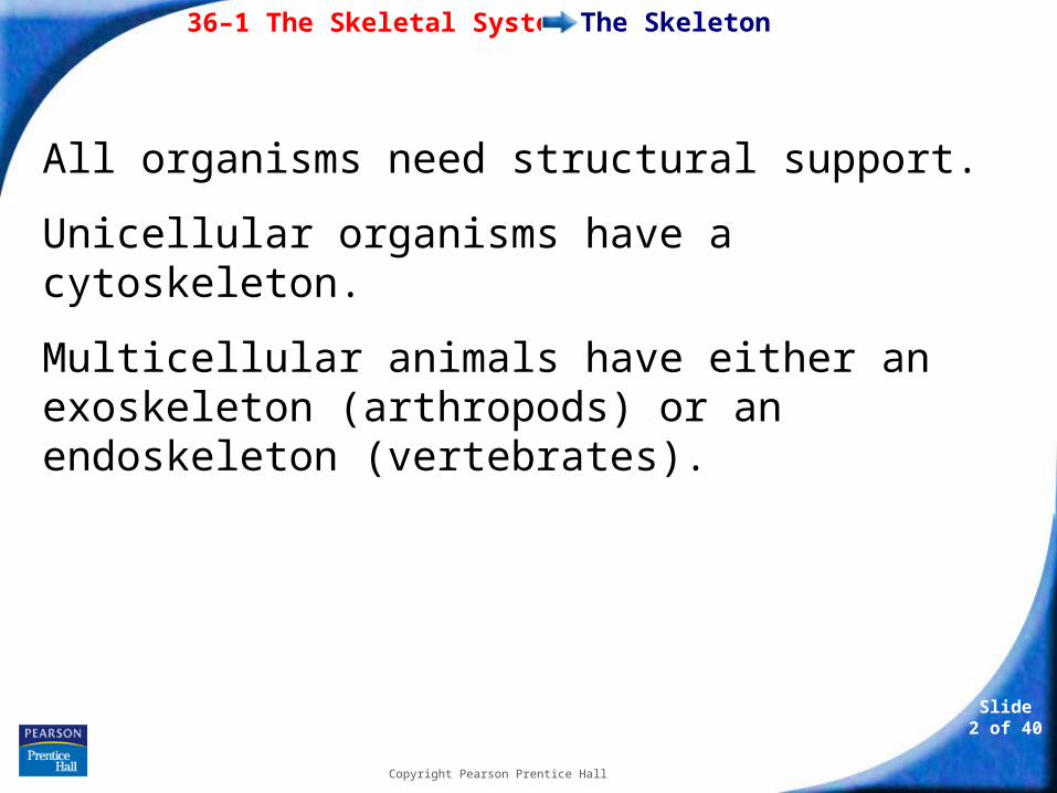
All organisms need structural support.
Unicellular organisms have a cytoskeleton.
Multicellular animals have either an exoskeleton (arthropods) or an endoskeleton (vertebrates).
36–1 The Skeletal System
Slide 3 of 40
Copyright Pearson Prentice Hall
The Skeleton
The human skeleton is composed of bone.
Bones and other connective tissues, such as cartilage and ligaments, form the skeletal system.
36–1 The Skeletal System
Slide 4 of 40
Copyright Pearson Prentice Hall
The Skeleton
The skeleton:
• supports the body.
• protects internal organs.
• provides for movement.
• stores mineral reserves.
• provides a site for blood cell formation.
36–1 The Skeletal System
Slide 5 of 40
Copyright Pearson Prentice Hall
The Skeleton
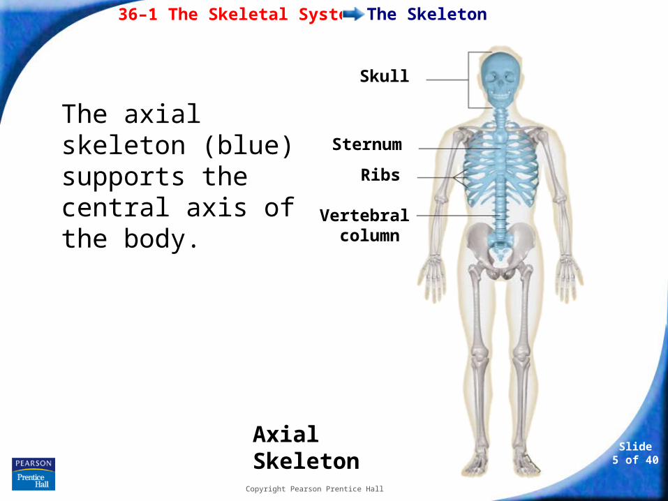
Axial Skeleton
Skull
Sternum
Ribs
Vertebral column
The axial skeleton (blue) supports the central axis of the body.
36–1 The Skeletal System
Slide 6 of 40
Copyright Pearson Prentice Hall
The Skeleton
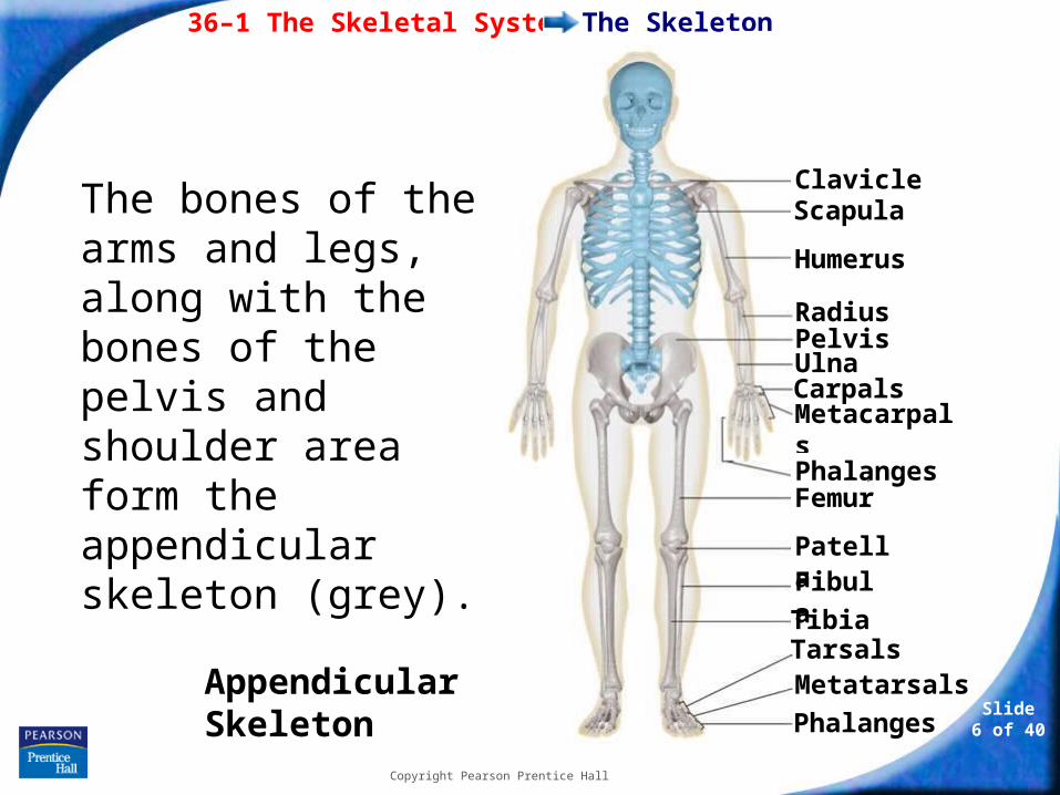
Clavicle Scapula
Humerus
Radius Pelvis Ulna Carpals Metacarpals
Phalanges Femur
Patella Fibula
Tibia Tarsals Metatarsals
Phalanges
Appendicular Skeleton
The bones of the arms and legs, along with the bones of the pelvis and shoulder area form the appendicular skeleton (grey).
36–1 The Skeletal System
Slide 7 of 40
Copyright Pearson Prentice Hall
Structure of Bones
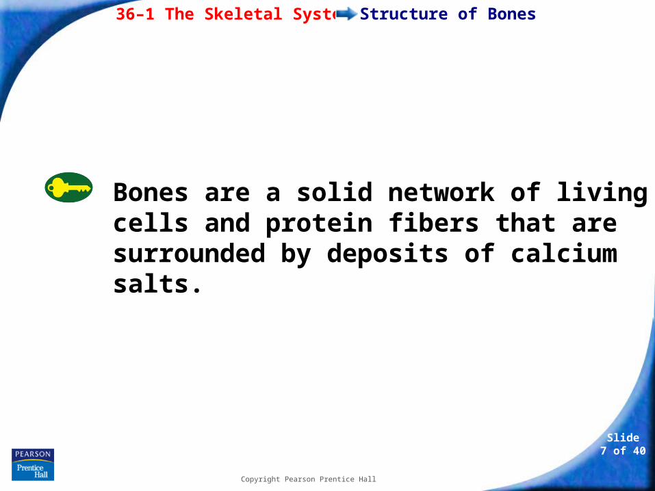
Bones are a solid network of living cells and protein fibers that are surrounded by deposits of calcium salts.
36–1 The Skeletal System
Slide 8 of 40
Copyright Pearson Prentice Hall
Structure of Bones
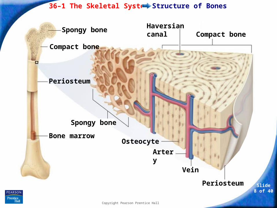
Bone marrow
Periosteum
Spongy bone
Compact bone
Haversian canalCompact bone
Spongy bone
Periosteum
Osteocyte
Artery
Vein
36–1 The Skeletal System
Slide 9 of 40
Copyright Pearson Prentice Hall
Structure of Bones
The bone is surrounded by a tough layer of connective tissue called the periosteum.
Blood vessels in the periosteum carry oxygen and nutrients to the bone.
36–1 The Skeletal System
Slide 10 of 40
Copyright Pearson Prentice Hall
Structure of Bones
Beneath the periosteum is a thick layer of compact bone.
Running through compact bone is a network of tubes called Haversian canals that contain blood vessels and nerves.
36–1 The Skeletal System
Slide 11 of 40
Copyright Pearson Prentice Hall
Structure of Bones
Spongy bone is found inside the outer layer of compact bone.
Spongy bone is also found in the ends of long bones and in the middle of short, flat bones.
Spongy bone adds strength without adding mass.

36–1 The Skeletal System
Slide 12 of 40
Copyright Pearson Prentice Hall
Structure of Bones
Osteocytes, or mature bone cells, are embedded in the bone matrix.
Other bone cells—osteoclasts and osteoblasts—line the Haversian canals and the surfaces of compact and spongy bone.
• Osteoclasts break down bone.
• Osteoblasts produce bone.
36–1 The Skeletal System
Slide 13 of 40
Copyright Pearson Prentice Hall
Structure of Bones
Bone marrow is a soft tissue inside the cavities within bones.
There are two types of bone marrow:
• Yellow marrow is made up of fat cells.
• Red marrow produces red blood cells, some kinds of white blood cells, and platelets.
36–1 The Skeletal System
Slide 14 of 40
Copyright Pearson Prentice Hall
Development of Bones
Development of Bones
The skeleton of an embryo is composed of cartilage.
Cartilage is a strong connective tissue that supports the body and is softer and more flexible than bone.
36–1 The Skeletal System
Slide 15 of 40
Copyright Pearson Prentice Hall
Development of Bones
Cartilage is replaced by bone during the process of bone formation called ossification.
Bone tissue forms as osteoblasts secrete mineral deposits.
When the osteoblasts become surrounded by bone tissue, they mature into osteocytes.
36–1 The Skeletal System
Slide 16 of 40
Copyright Pearson Prentice Hall
Development of Bones
Many long bones have growth plates at either end.
Growth of cartilage at these plates causes bones to lengthen. Gradually, this cartilage is replaced by bone tissue.
By early adulthood, cartilage in the growth plates is replaced by bone, the bones become ossified, and growth stops.
36–1 The Skeletal System
Slide 17 of 40
Copyright Pearson Prentice Hall
Types of Joints
Types of Joints
A place where one bone attaches to another bone is called a joint.
Joints permit bones to move without damaging each other.
36–1 The Skeletal System
Slide 18 of 40
Copyright Pearson Prentice Hall
Types of Joints
Depending on its type of movement, a joint is classified as immovable, slightly movable, or freely movable.
36–1 The Skeletal System
Slide 19 of 40
Copyright Pearson Prentice Hall
Types of Joints
Immovable Joints
Immovable joints, called fixed joints, allow no movement.
The bones are interlocked and held together by connective tissue, or they are fused together.
Places where bones in the skull meet are examples of immovable joints.
36–1 The Skeletal System
Slide 20 of 40
Copyright Pearson Prentice Hall
Types of Joints
Slightly Movable Joints
Slightly movable joints permit a small amount of restricted movement.
Slightly movable joints are found in the joints between adjacent vertebrae.
36–1 The Skeletal System
Slide 21 of 40
Copyright Pearson Prentice Hall
Types of Joints
Freely Movable Joints
Freely movable joints permit movement in one or more directions.
Four common freely movable joints are:
•ball-and-socket joints
•hinge joints
•pivot joints
•saddle joints
36–1 The Skeletal System
Slide 22 of 40
Copyright Pearson Prentice Hall
Types of Joints
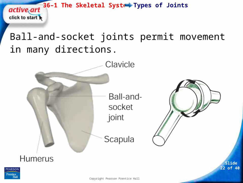
Ball-and-socket joints permit movement in many directions.

36–1 The Skeletal System
Slide 23 of 40
Copyright Pearson Prentice Hall
Types of Joints
Hinge joints permit back-and-forth motion.
36–1 The Skeletal System
Slide 24 of 40
Copyright Pearson Prentice Hall
Types of Joints
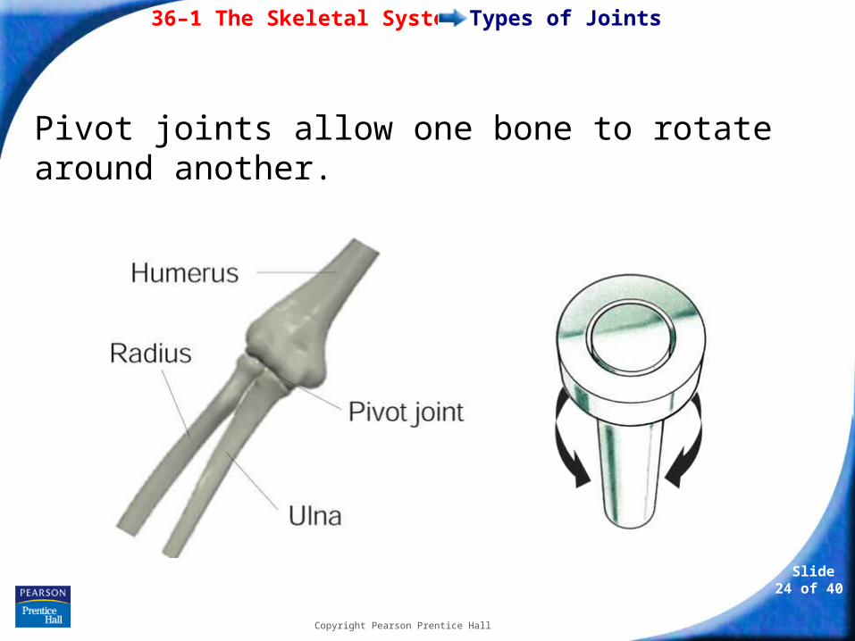
Pivot joints allow one bone to rotate around another.
36–1 The Skeletal System
Slide 25 of 40
Copyright Pearson Prentice Hall
Types of Joints
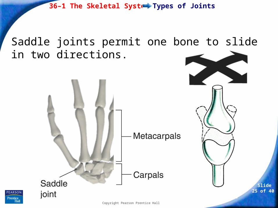
Saddle joints permit one bone to slide in two directions.
36–1 The Skeletal System
Slide 26 of 40
Copyright Pearson Prentice Hall
Structure of Joints
Structure of Joints
In freely movable joints, cartilage covers the surfaces where two bones come together.
Joints are also surrounded by a fibrous capsule that holds the bones together while still allowing them to move.
36–1 The Skeletal System
Slide 27 of 40
Copyright Pearson Prentice Hall
Structure of Joints
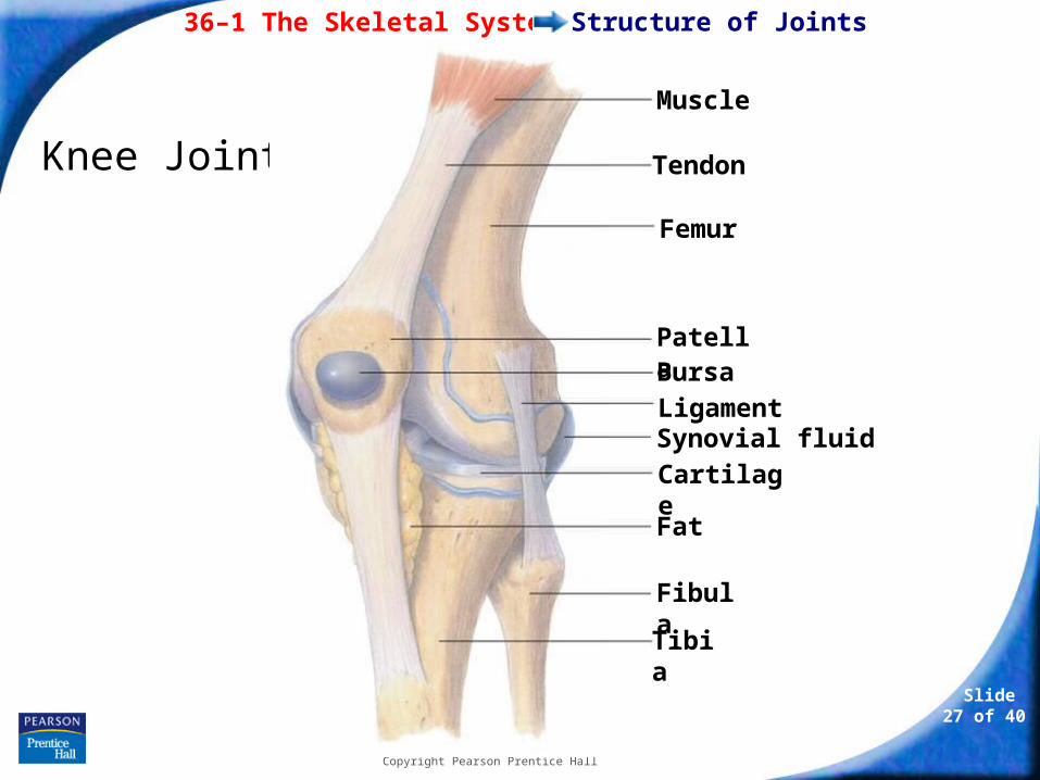
Knee Joint
Muscle
Tendon
Femur
Patella Bursa Ligament Synovial fluidCartilage
Fat
Fibula
Tibia
36–1 The Skeletal System
Slide 28 of 40
Copyright Pearson Prentice Hall
Structure of Joints
Connective tissue called ligaments hold bones together in joints and are attached to membranes that surround bones.
Synovial fluid forms a thin lubricating film over the surface of the joint.
Synovial fluid enables the bones to slide past each other more smoothly.
36–1 The Skeletal System
Slide 29 of 40
Copyright Pearson Prentice Hall
Structure of Joints
In some freely movable joints small sacs of synovial fluid called bursae form.
A bursa reduces the friction between bones of a joint and also acts as a shock absorber.