Embed Size (px)
Citation preview
Skin & Wound CarePrevention & Treatment
By Candy Houk, RNSkin & Wound Program Manager
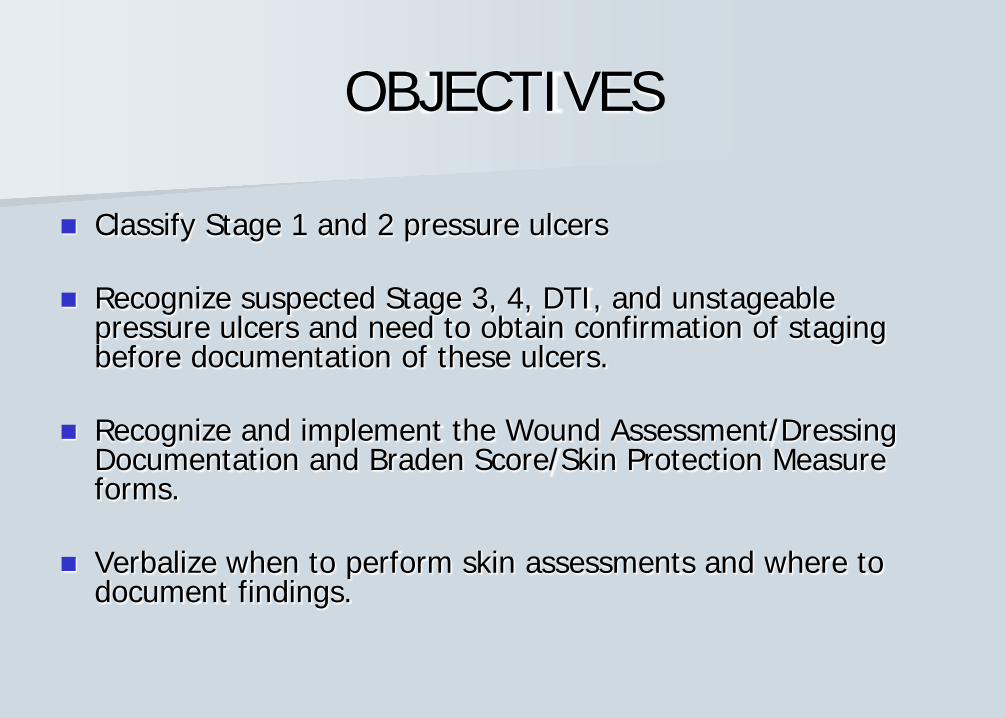
OBJECTIVES
Classify Stage 1 and 2 pressure ulcers
Recognize suspected Stage 3, 4, DTI, and unstageable pressure ulcers and need to obtain confirmation of staging before documentation of these ulcers.
Recognize and implement the Wound Assessment/Dressing Documentation and Braden Score/Skin Protection Measure forms.
Verbalize when to perform skin assessments and where to document findings.
Skin Facts -Skin is the largest organ in the body. -From birth to maturity skin undergoes about a 7-fold
expansion. -Average adult has about 2 square meters of skin. -Skin weighs about 6 pounds. -The skin receives about 1/3 of the body’s circulating
blood volume. -Skin is capable of self generation. -Can withstand limited mechanical & chemical
assaults.
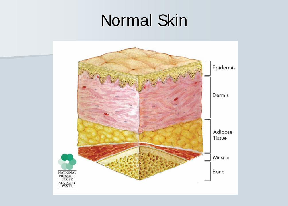
Normal Skin
Factors altering skin characteristics
-Age -Sun-Hydration-Soaps-Nutrition-Medications
Types of Wounds
Vascular – arterial ischemic, venous ischemic, lymphatic wounds
Neuropathic – diabetic woundsPressure/Friction – pressure ulcersSurgery/Trauma –clean or contaminatedOther – anorectal fistulas, stoma-related,
neoplasmic, vasculitic, inflammatory
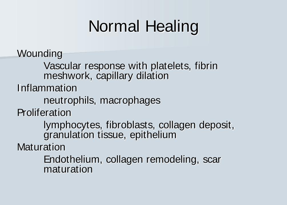
Normal HealingWounding
Vascular response with platelets, fibrin meshwork, capillary dilation
Inflammationneutrophils, macrophages
Proliferationlymphocytes, fibroblasts, collagen deposit, granulation tissue, epithelium
MaturationEndothelium, collagen remodeling, scar maturation
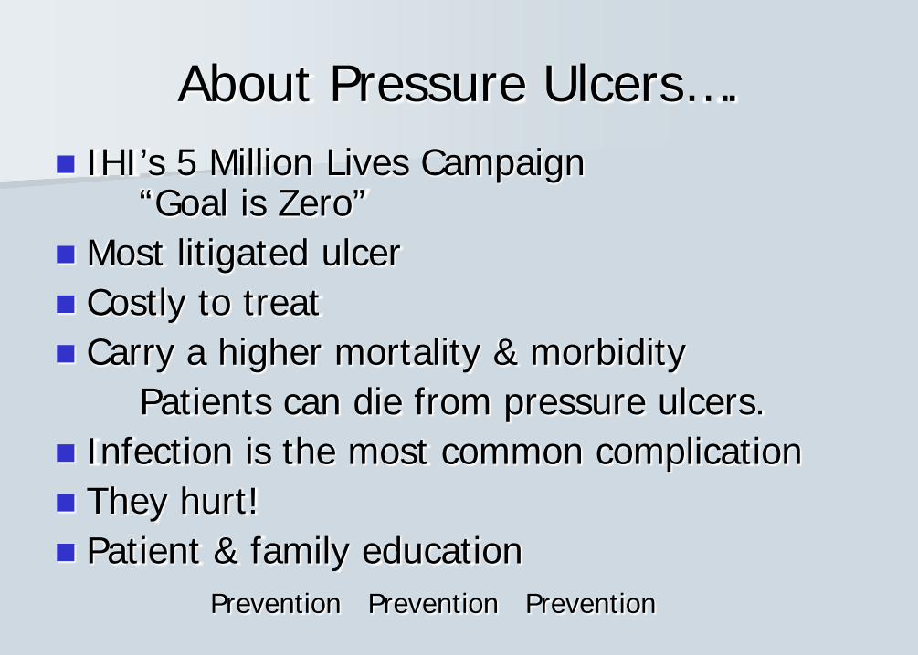
About Pressure Ulcers…. IHI’s 5 Million Lives Campaign
“Goal is Zero” Most litigated ulcer Costly to treat Carry a higher mortality & morbidity
Patients can die from pressure ulcers. Infection is the most common complication They hurt! Patient & family education
Prevention Prevention Prevention
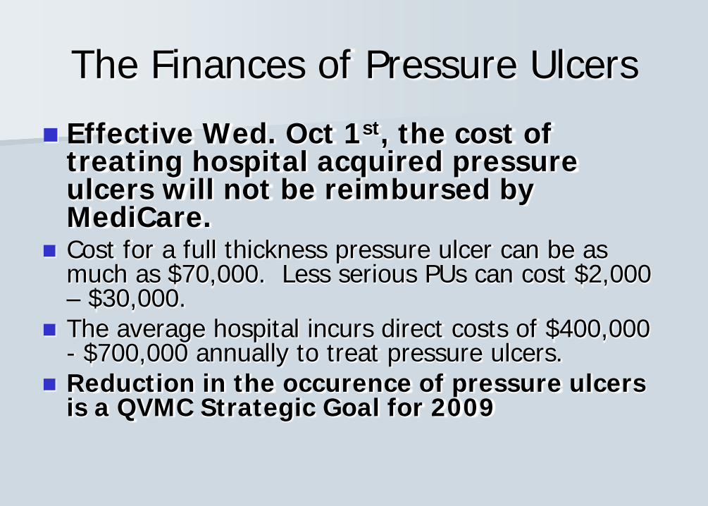
The Finances of Pressure Ulcers
Effective Wed. Oct 1st, the cost of treating hospital acquired pressure ulcers will not be reimbursed by MediCare.
Cost for a full thickness pressure ulcer can be as much as $70,000. Less serious PUs can cost $2,000 – $30,000.
The average hospital incurs direct costs of $400,000 - $700,000 annually to treat pressure ulcers.
Reduction in the occurence of pressure ulcers is a QVMC Strategic Goal for 2009
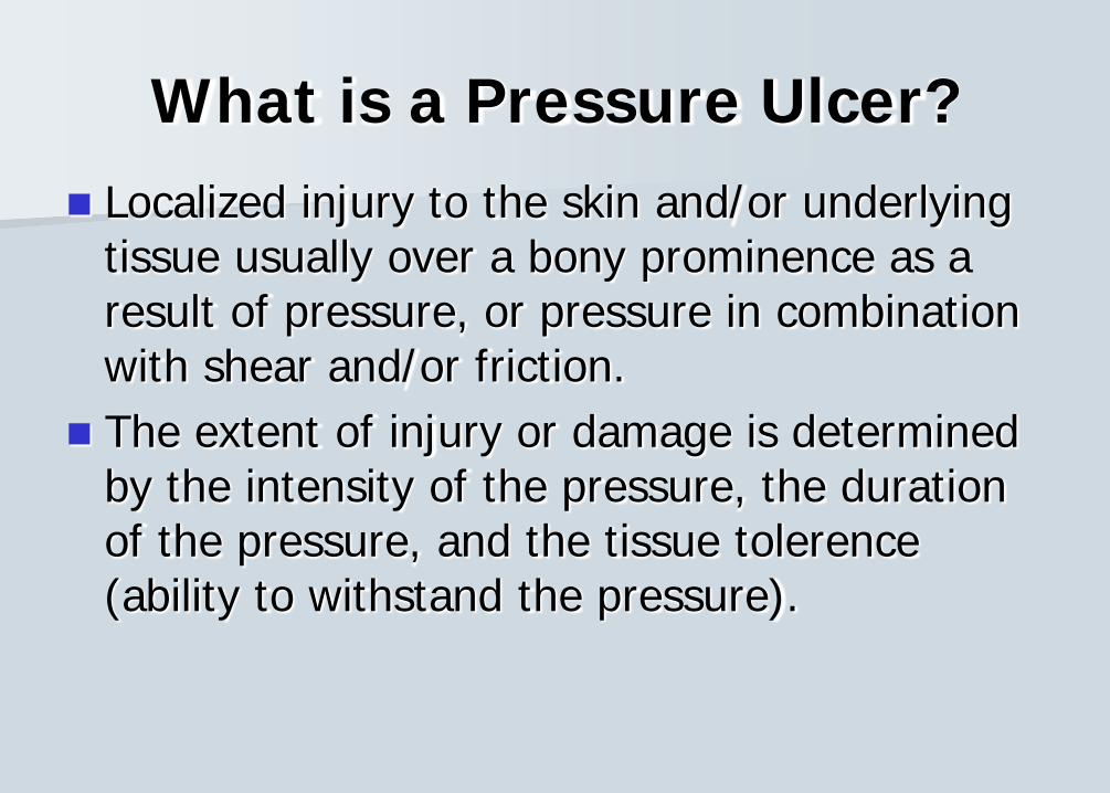
What is a Pressure Ulcer? Localized injury to the skin and/or underlying
tissue usually over a bony prominence as a result of pressure, or pressure in combination with shear and/or friction.
The extent of injury or damage is determined by the intensity of the pressure, the duration of the pressure, and the tissue tolerence (ability to withstand the pressure).
Definitions
Pressure - Pressure compresses underlying tissue and small blood vessels against the surface beneath. Pressure is exerted vertically. Tissues become ischemic and die.
Friction/Shear - Friction is the resistance created when one surface moves horizontally across another surface to create the tissue injury. – (e.g. dragging a patient across bed linen). – Shear occurs when one layer of tissue slides horizontally over
another, deforming and disrupting blood flow (e.g. when the head of the bed is raised > 30 degrees & patient slides down in bed).
Both require pressure exerted by the body against an external surface.
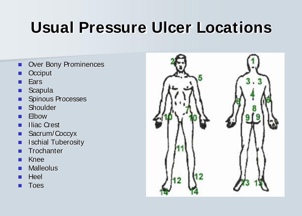
Usual Pressure Ulcer Locations
Over Bony Prominences Occiput Ears Scapula Spinous Processes Shoulder Elbow Iliac Crest Sacrum/Coccyx Ischial Tuberosity Trochanter Knee Malleolus Heel Toes
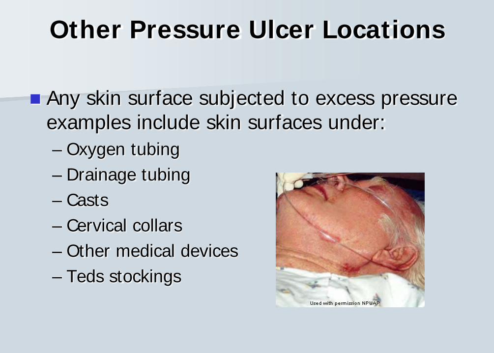
Other Pressure Ulcer Locations
Any skin surface subjected to excess pressure examples include skin surfaces under: – Oxygen tubing – Drainage tubing – Casts – Cervical collars – Other medical devices – Teds stockings
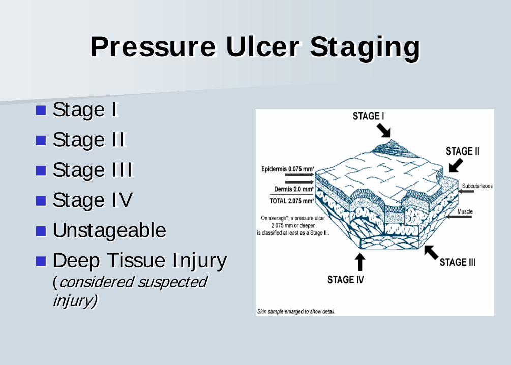
Pressure Ulcer Staging
Stage I Stage II Stage III Stage IV Unstageable Deep Tissue Injury
(considered suspected injury)
Staging of Pressure Ulcers Only pressure ulcers are staged. Other ulcer types
are not staged: diabetic, venous, etc. RNs may stage pressure ulcers that are Stage
1 or 2. Any pressure ulcer suspected to be more serious than Stage 2 requires a Wound Nurse consult.
Accurate staging requires visualization & identification of the tissues in the wound bed.
As the ulcer heals, it cannot be “reverse” staged because the original tissue does not regenerate, ie., a Stage 4 ulcer does not become a Stage 2. One would document “healing Stage 4 ulcer”.
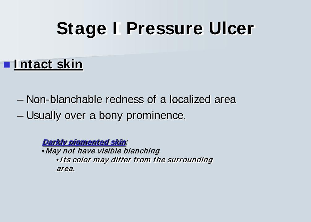
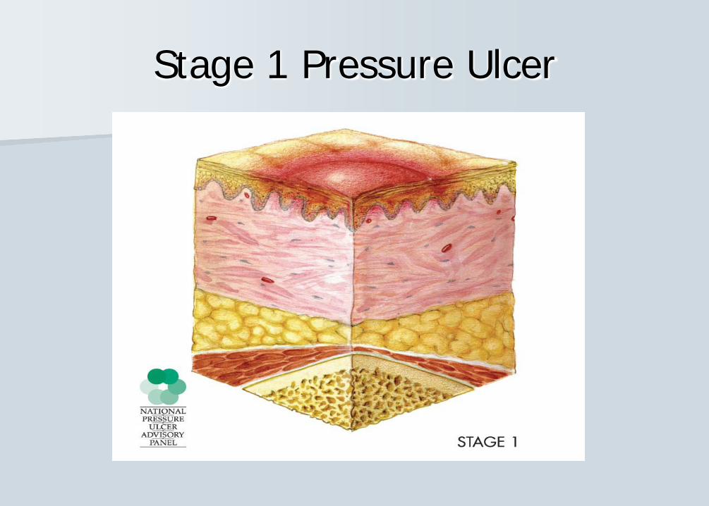
Stage I Pressure Ulcer
Intact skin
– Non-blanchable redness of a localized area – Usually over a bony prominence.
Darkly pigmented skin:•May not have visible blanching
•Its color may differ from the surrounding area.
Stage 1 Pressure Ulcer
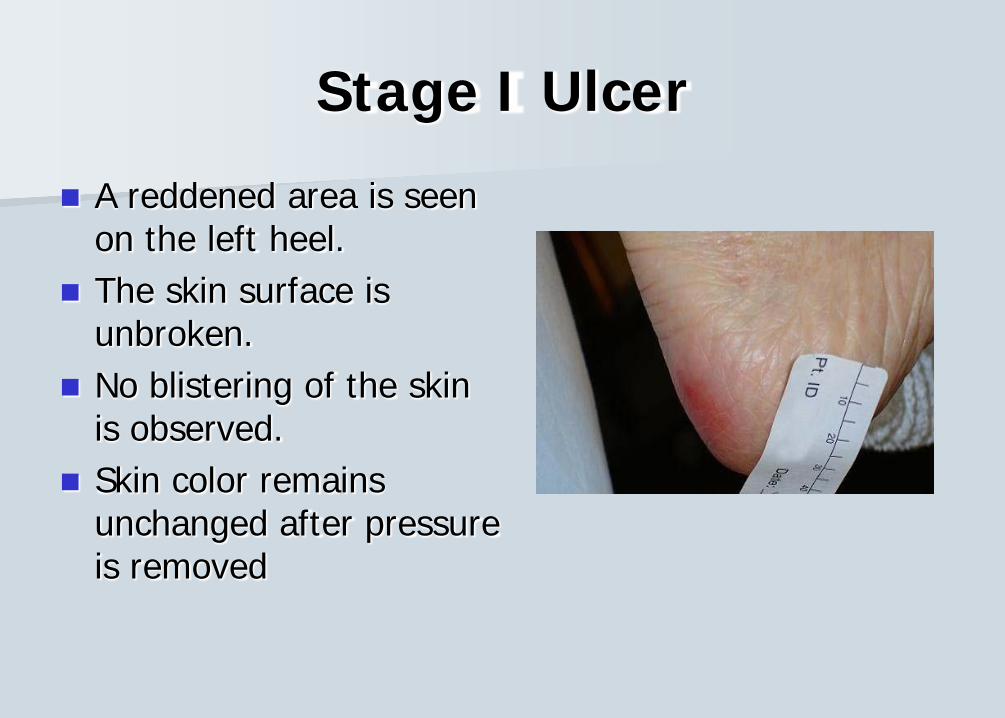
Stage I Ulcer
A reddened area is seen on the left heel.
The skin surface is unbroken.
No blistering of the skin is observed.
Skin color remains unchanged after pressure is removed
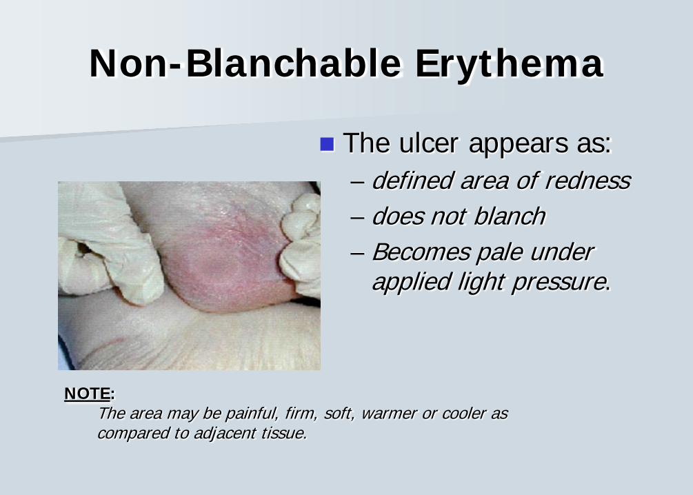
Non-Blanchable Erythema
The ulcer appears as:– defined area of redness – does not blanch– Becomes pale under
applied light pressure.
NOTE:The area may be painful, firm, soft, warmer or cooler as compared to adjacent tissue.
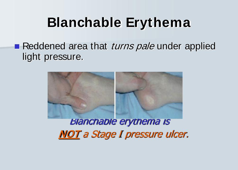
Blanchable Erythema
Reddened area that turns pale under applied light pressure.
Blanchable erythema is NOT a Stage I pressure ulcer.
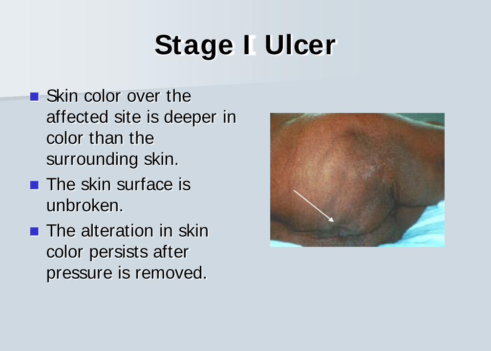
Stage I Ulcer
Skin color over the affected site is deeper in color than the surrounding skin.
The skin surface is unbroken.
The alteration in skin color persists after pressure is removed.
Stage II Pressure Ulcer
Partial thickness loss of dermis Presents as:
Shallow open ulcerRed-pink wound bedWithout sloughIntact or ruptured/open serum-filled blister.
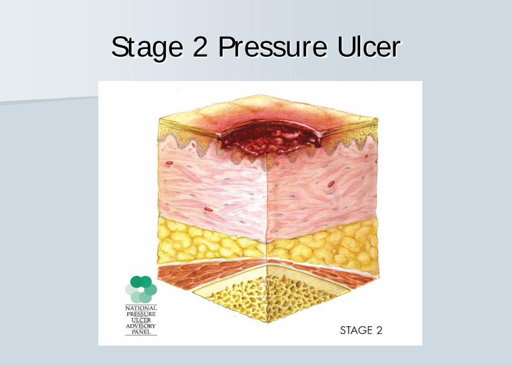
Stage 2 Pressure Ulcer
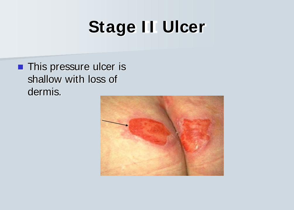
Stage II Ulcer
This pressure ulcer is shallow with loss of dermis.
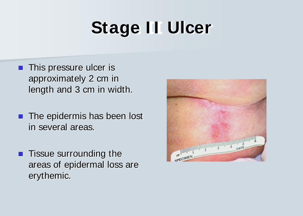
Stage II Ulcer
This pressure ulcer is approximately 2 cm in length and 3 cm in width.
The epidermis has been lost in several areas.
Tissue surrounding the areas of epidermal loss are erythemic.
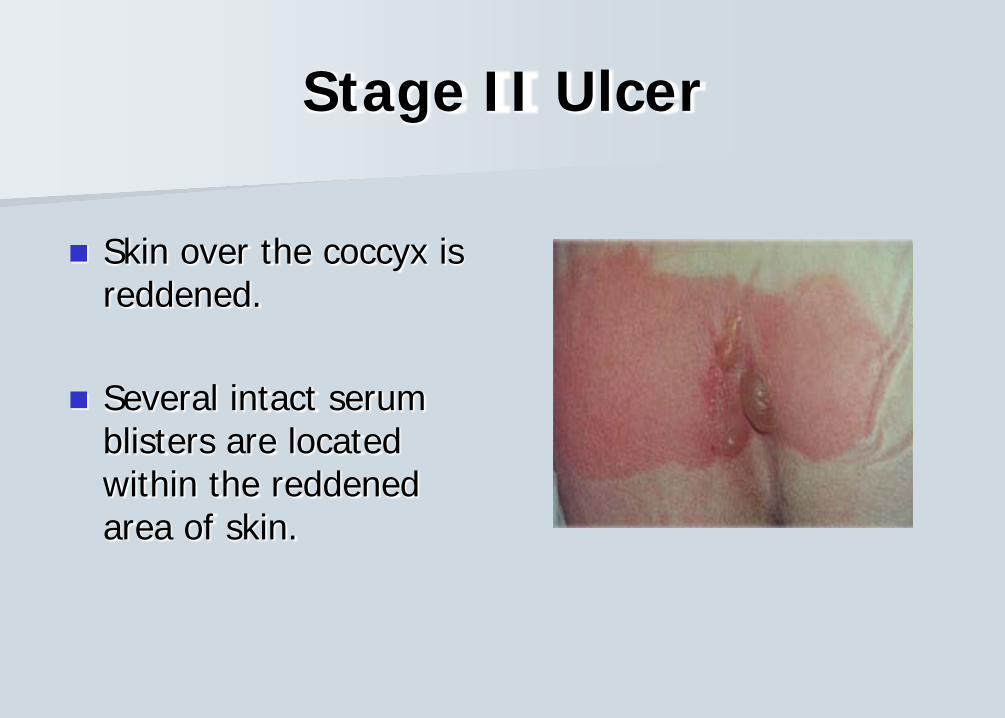
Stage II Ulcer
Skin over the coccyx is reddened.
Several intact serum blisters are located within the reddened area of skin.
Stage III Pressure Ulcer
Full thickness tissue loss.
Subcutaneous fat may be visible, but bone, tendon, or muscle are not exposed
Slough may be present but does not obscure the depth of tissue loss.
May include undermining and tunneling.
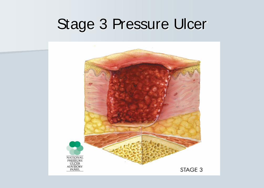
Stage 3 Pressure Ulcer
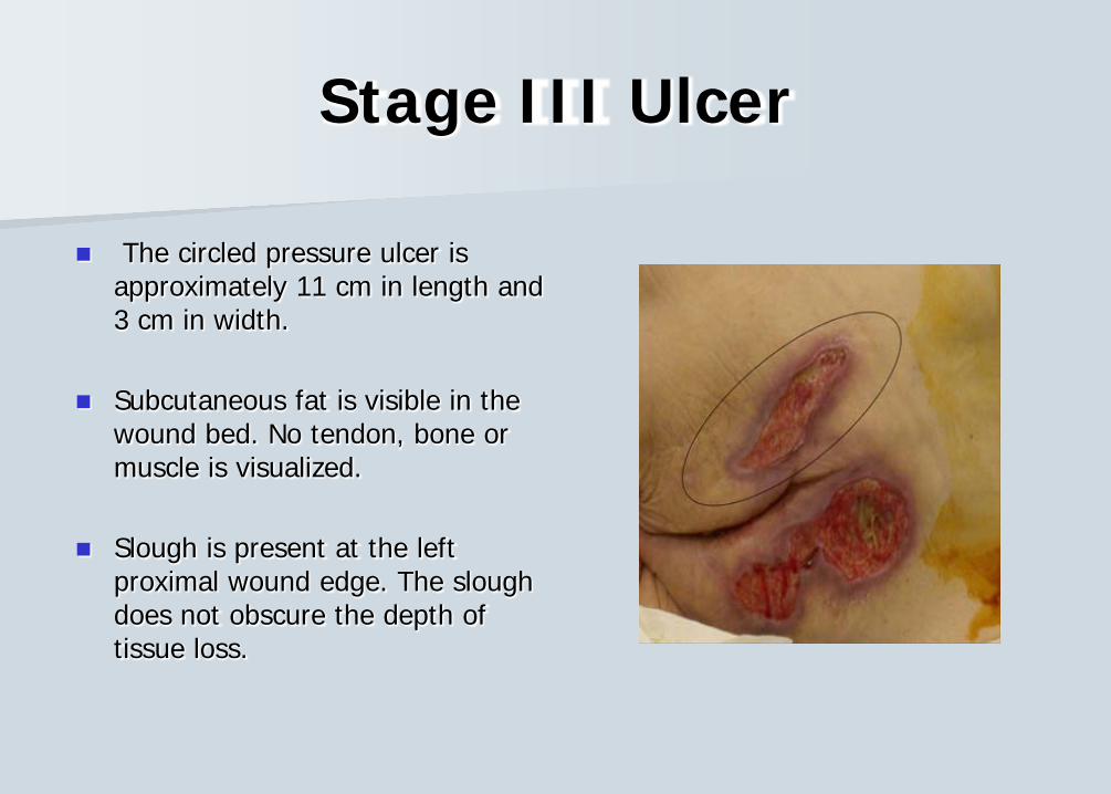
Stage III Ulcer
The circled pressure ulcer is approximately 11 cm in length and 3 cm in width.
Subcutaneous fat is visible in the wound bed. No tendon, bone or muscle is visualized.
Slough is present at the left proximal wound edge. The slough does not obscure the depth of tissue loss.
Stage IV Pressure Ulcer
Full thickness tissue loss with exposed bone, tendon or muscle.
Slough or eschar may be present on some parts of the wound bed.
Often includes undermining and tunneling.
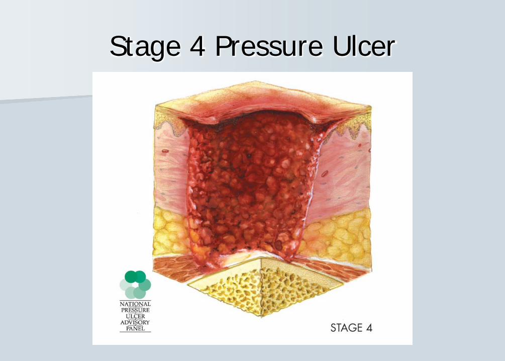
Stage 4 Pressure Ulcer
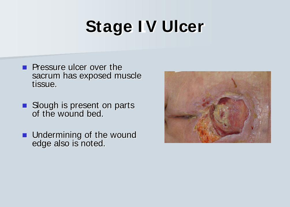
Stage IV Ulcer
Pressure ulcer over the sacrum has exposed muscle tissue.
Slough is present on parts of the wound bed.
Undermining of the wound edge also is noted.
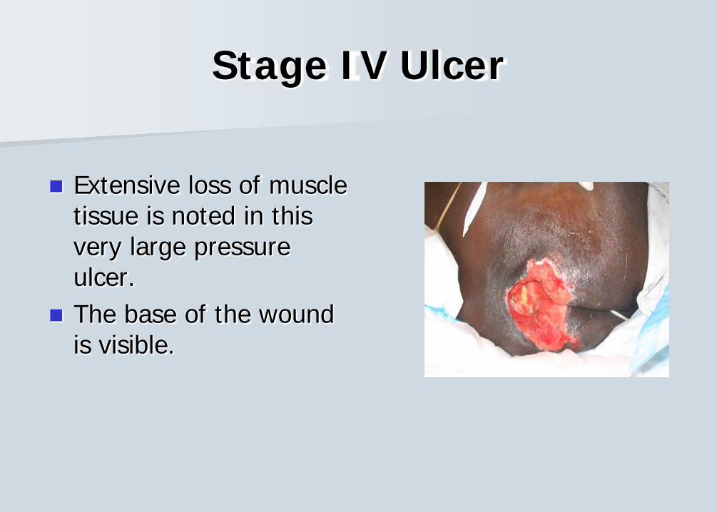
Stage IV Ulcer
Extensive loss of muscle tissue is noted in this very large pressure ulcer.
The base of the wound is visible.
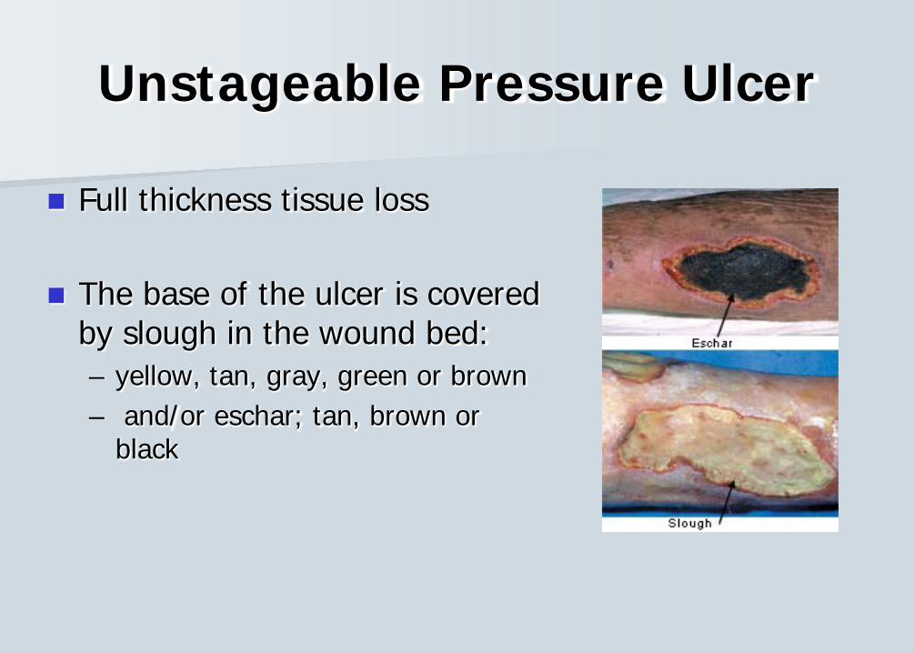
Unstageable Pressure Ulcer
Full thickness tissue loss
The base of the ulcer is covered by slough in the wound bed: – yellow, tan, gray, green or brown– and/or eschar; tan, brown or
black
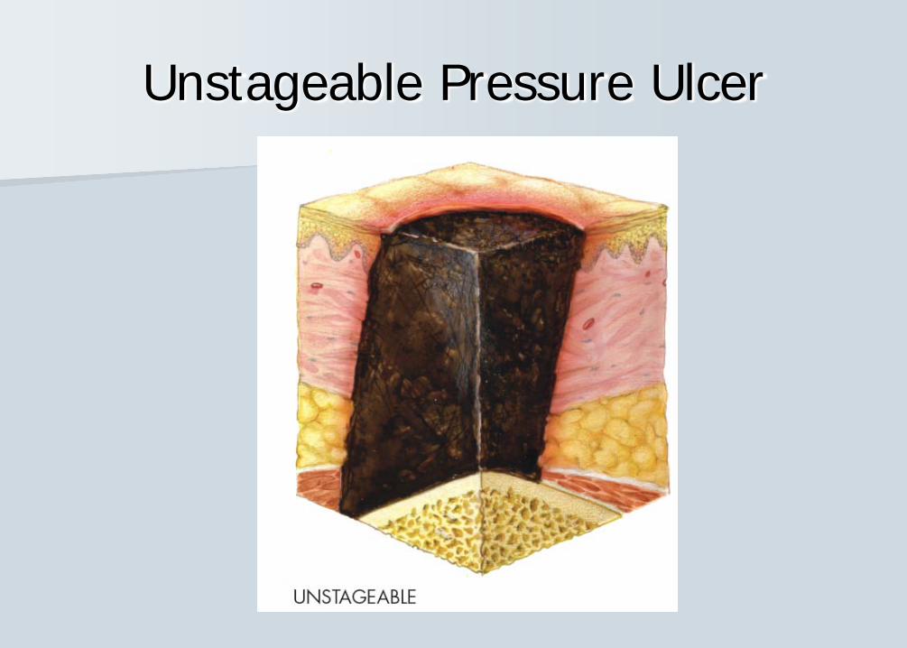
Unstageable Pressure Ulcer
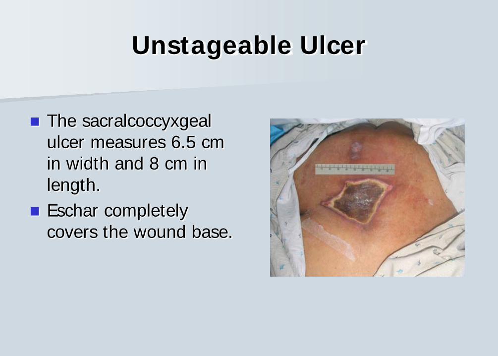
Unstageable Ulcer
The sacralcoccyxgeal ulcer measures 6.5 cm in width and 8 cm in length.
Eschar completely covers the wound base.
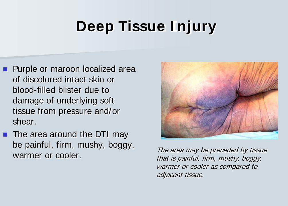
Deep Tissue Injury
Purple or maroon localized area of discolored intact skin or blood-filled blister due to damage of underlying soft tissue from pressure and/or shear.
The area around the DTI may be painful, firm, mushy, boggy, warmer or cooler.
The area may be preceded by tissue that is painful, firm, mushy, boggy, warmer or cooler as compared to adjacent tissue.
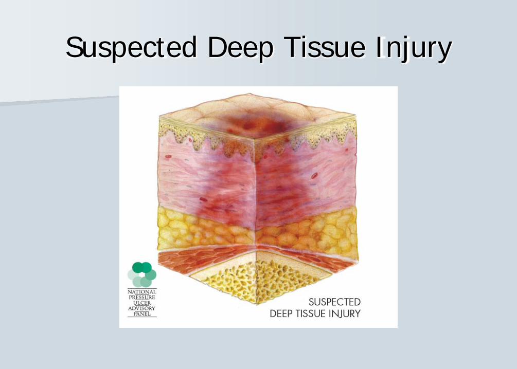
Suspected Deep Tissue Injury
Proposed Etiology of DTI
Pressure to the skin and soft tissue and ischemia
Muscle injury associated with a decrease in nutrient supply
Vasopresser use
Injury or damage to the fascia from shearing injury or torsion of the perforating vessels
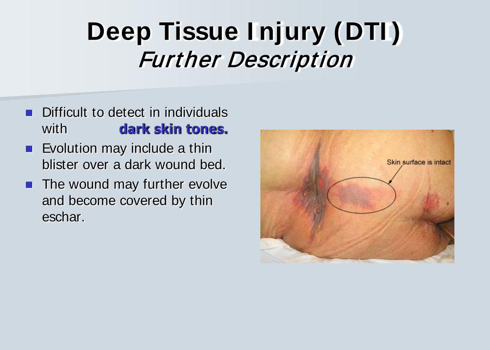
Deep Tissue Injury (DTI)Further Description
Difficult to detect in individuals with dark skin tones.
Evolution may include a thin blister over a dark wound bed.
The wound may further evolve and become covered by thin eschar.
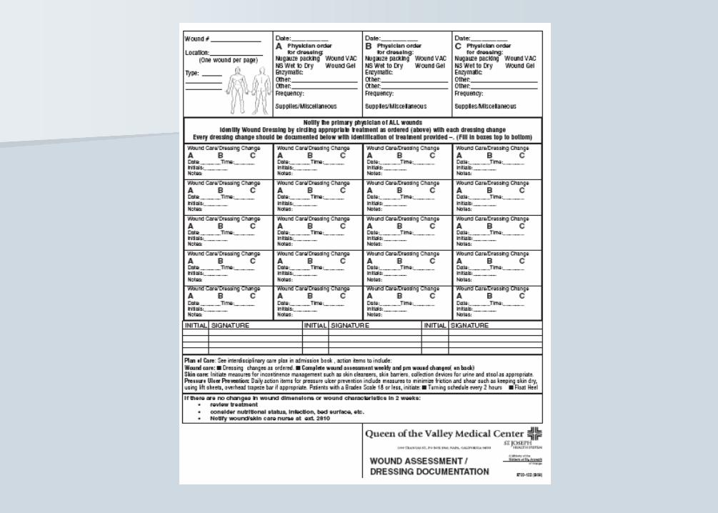
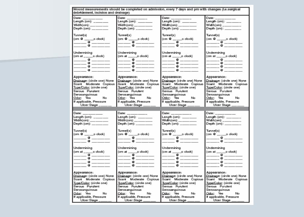
Documentation All patients have their skin assessed daily. All wounds, including pressure ulcers, are measured on
admission and at least weekly on “Wound Wednesday” and documented on the “Wound Assessment/Dressing Documentation” form, including any dressing changes, one wound per form.
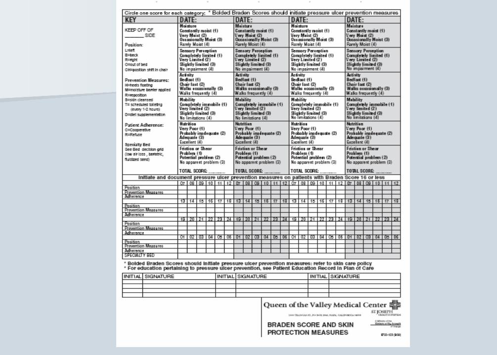
Preventive measures: turning every 2 hours, cleansing skin, incontinence care, floating heels, moisture barrier applications are documented on the “Braden Score & Skin Protection Measures” form.
Any pressure ulcers present on admission must be documented on the Interdisciplinary Assessment form.
The POC should be updated to reflect any wound during the course of hospitalization.
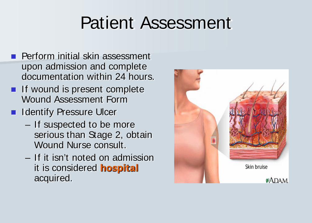
Patient Assessment
Perform initial skin assessment upon admission and complete documentation within 24 hours.
If wound is present complete Wound Assessment Form
Identify Pressure Ulcer– If suspected to be more
serious than Stage 2, obtain Wound Nurse consult.
– If it isn’t noted on admission it is considered hospitalacquired.
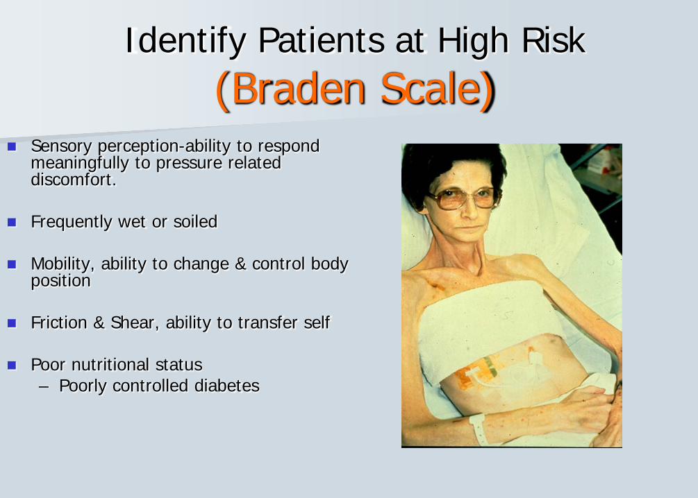
Identify Patients at High Risk(Braden Scale)
Sensory perception-ability to respond meaningfully to pressure related discomfort.
Frequently wet or soiled
Mobility, ability to change & control body position
Friction & Shear, ability to transfer self
Poor nutritional status– Poorly controlled diabetes
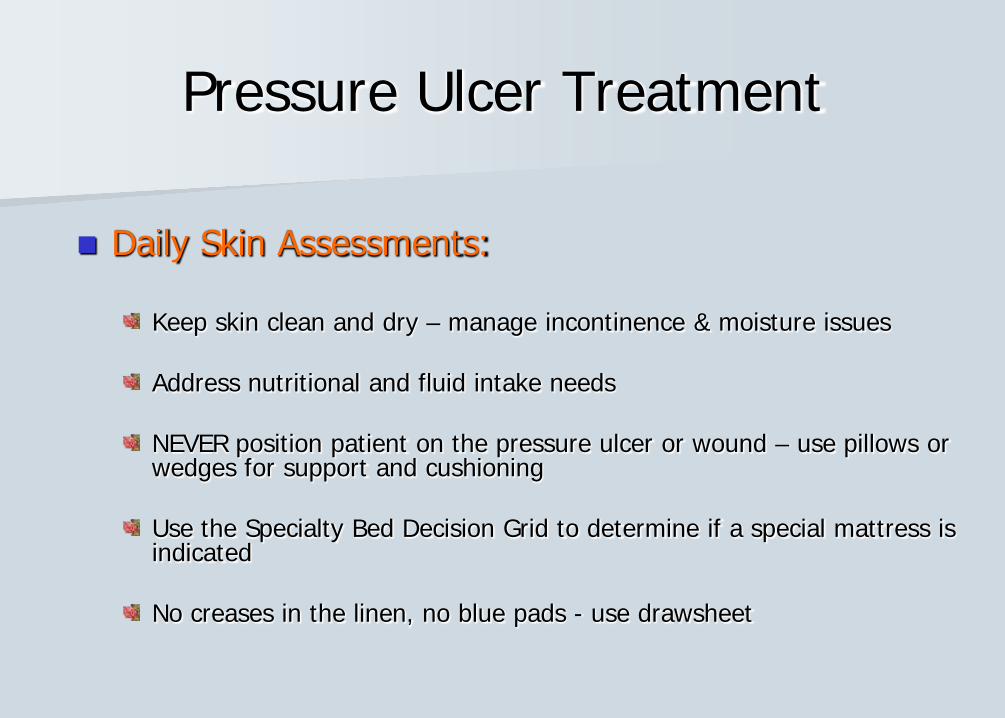
Pressure Ulcer Treatment
Daily Skin Assessments:
Keep skin clean and dry – manage incontinence & moisture issues
Address nutritional and fluid intake needs
NEVER position patient on the pressure ulcer or wound – use pillows or wedges for support and cushioning
Use the Specialty Bed Decision Grid to determine if a special mattress is indicated
No creases in the linen, no blue pads - use drawsheet
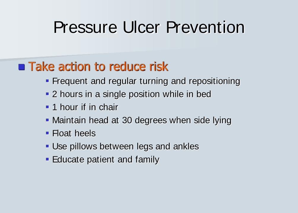
Pressure Ulcer Prevention
Take action to reduce risk Frequent and regular turning and repositioning 2 hours in a single position while in bed 1 hour if in chair Maintain head at 30 degrees when side lying Float heels Use pillows between legs and ankles Educate patient and family
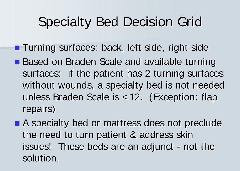
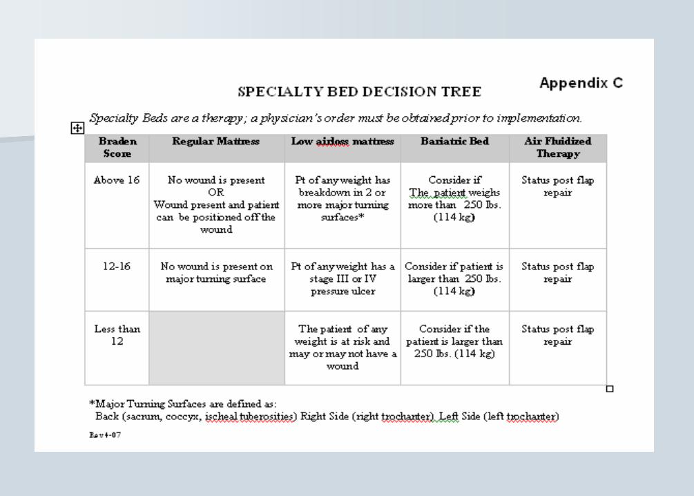
Specialty Bed Decision Grid
Turning surfaces: back, left side, right side Based on Braden Scale and available turning
surfaces: if the patient has 2 turning surfaces without wounds, a specialty bed is not needed unless Braden Scale is <12. (Exception: flap repairs)
A specialty bed or mattress does not preclude the need to turn patient & address skin issues! These beds are an adjunct - not the solution.
Prevention! Prevention! Prevention!
The most impactful step we can take toward achieving a higher quality of care and reducing skin management costs.
Proactive skin care:-improves staff time efficiency-supply reductions-improved infection control
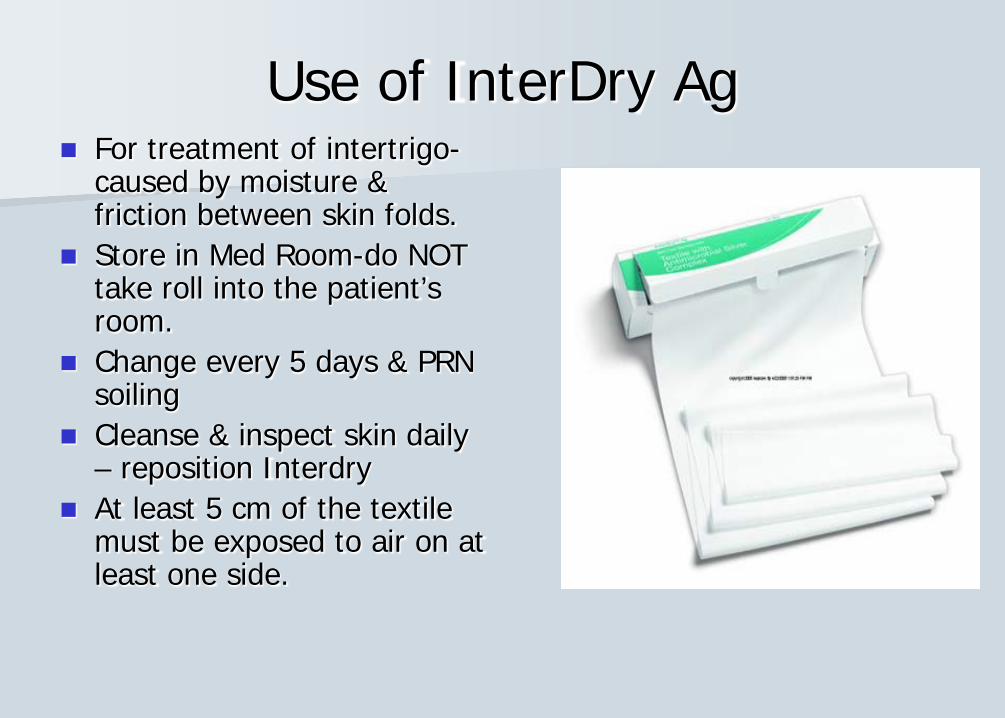
Use of InterDry Ag For treatment of intertrigo-
caused by moisture & friction between skin folds.
Store in Med Room-do NOT take roll into the patient’s room.
Change every 5 days & PRN soiling
Cleanse & inspect skin daily – reposition Interdry
At least 5 cm of the textile must be exposed to air on at least one side.