Embed Size (px)
Citation preview
In TIME wounds will heal
Kate Gray
Clinical Nurse Specialist Wound Care
Hutt Valley DHB
Overview • Introduction and overview
• Acute versus chronic and classification of wounds
• Risk Assessment
• History
• Examination
- wound bed preparation (TIME)
• Investigation
• Diagnosis
• Intervention
• Summary

Acute Wounds
• Trauma
• Surgery
• Abrasions
• Surgical incisions
• Tears
• Penetrating injuries
• Burns
Short healing times
Pass through stages of healing in timely manner
Healing often by primary intention
Carville, K. (2012). Wound care manual. Western Australia: Silver Chain Foundation Casey, G. (2011). Wound healing – repair at the expense of function. Kia Tiaki Nursing New Zealand, 17(6)22-27
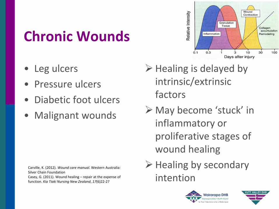
Chronic Wounds
• Leg ulcers
• Pressure ulcers
• Diabetic foot ulcers
• Malignant wounds
Healing is delayed by intrinsic/extrinsic factors
May become ‘stuck’ in inflammatory or proliferative stages of wound healing
Healing by secondary intention
Carville, K. (2012). Wound care manual. Western Australia: Silver Chain Foundation Casey, G. (2011). Wound healing – repair at the expense of function. Kia Tiaki Nursing New Zealand, 17(6)22-27

All wounds have the potential to become chronic if the
treatment regime is incorrect or inappropriate (p.14)
Eagle, M. (2009). Wound assessment: The patient and the wound. Wound Essentials, 4, 14-24
Key Message
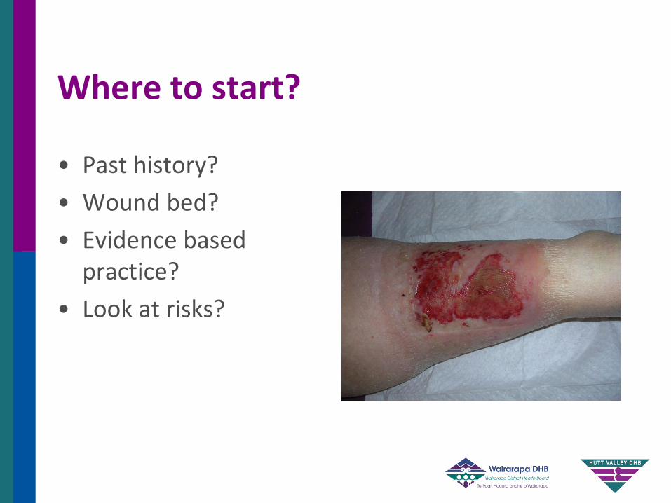
Where to start?
• Past history?
• Wound bed?
• Evidence based practice?
• Look at risks?
“Examine the whole patient. Treat the cause
and patient-centred concerns before the hole
in the patient” p. 25
Sibbald, R. G. et al (2012). Special considerations in wound bed preparation 2011: an update. WCET Journal, 32(2), 10-30 Sibbald, R,G,, Woo, K. & Ayello, E. (2007). Increased bacterial burden and infection: NERDS and STONES. Wounds UK, 3(2), 25-46
Wound Assessment Mnemonic
• H History
• E Examination
• I Investigations
• D Diagnosis
• I Intervention
Wound Prevention is through Management of Risk
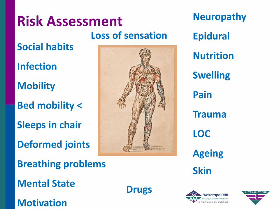
Risk Assessment
Social habits
Infection
Mobility
Bed mobility <
Sleeps in chair
Deformed joints
Breathing problems
Mental State
Motivation
Neuropathy
Epidural
Nutrition
Swelling
Pain
Trauma
LOC
Ageing
Skin
Loss of sensation
Drugs
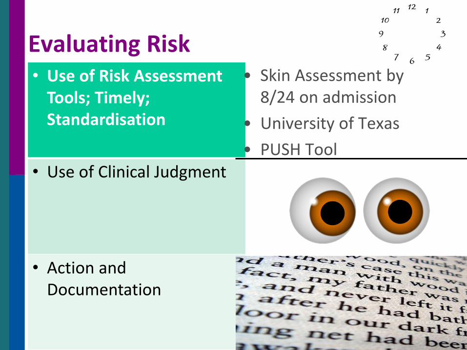
Evaluating Risk
• Use of Risk Assessment Tools; Timely; Standardisation
• Use of Clinical Judgment
• Action and Documentation
• Skin Assessment by 8/24 on admission
• University of Texas
• PUSH Tool
Wound Assessment – History (Risk assessment and factors affecting wound healing)
• Medical
• Surgical
• Pharmacological
• Social
• History of Wound

Wound Assessment – Medical History • Advanced age
• Immobility
• Systemic malignancy, radiotherapy, chemotherapy, terminal illness
• Malnutrition
• Systemic inflammatory diseases e.g. RA, diabetes, autoimmune
• Neurological
Casey, G. (2011). Wound healing-repair at the expense of function. Kia Tiaki Nursing New Zealand, 17(6) 22-27

Wound Assessment – Surgical History • Previous varicose vein
surgery
• Sclerotherapy/
endovascular
• Previous revascularisation
• Previous skin lesion and skin graft
• Previous skin condition
Wound Assessment – Pharmacological • Steroids
• Specifics – e.g. hydroxyurea
• Anticoagulants
• Immunosuppressive
• Smoking
• Allergic Reactions

Wound Assessment – Psychosocial
• Low self esteem
• Altered body image
• Depression
• Social isolation
• Loss of independence
• Financial
• Loss of family role
• Interpersonal relationships

Holistic Wound Assessment DOMAINS OF WELLBEING
• Physical wellbeing
• Mental wellbeing
• Social wellbeing
• Spiritual/cultural wellbeing
International consensus. Optimising wellbeing in people living with a wound. An expert working group review. London: Wounds International, 2012.

Delayed Wound Healing
Systemic Factors
Regional Factors
Local Factors
White, R. (2008). Delayed wound healing: in whom, what, when and why?. Primary Health Care, 18(2), 40-46
Checklist Wound
• Wound bed/stage of healing
• Wound site/location
• Wound size
• Amount/type of exudate
• Odour
• Pain
• Wound edge/margin
• Surrounding skin
Eagle, M. (2009). Wound assessment: The patient and the wound. Wound Essentials, 4, 14-24
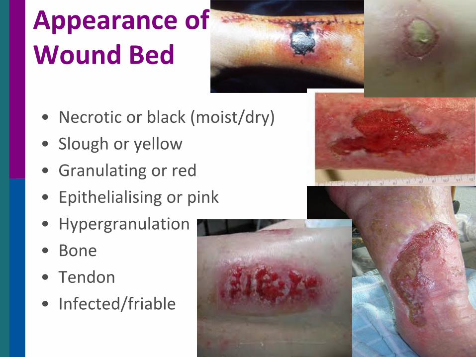
Appearance of Wound Bed
• Necrotic or black (moist/dry)
• Slough or yellow
• Granulating or red
• Epithelialising or pink
• Hypergranulation
• Bone
• Tendon
• Infected/friable
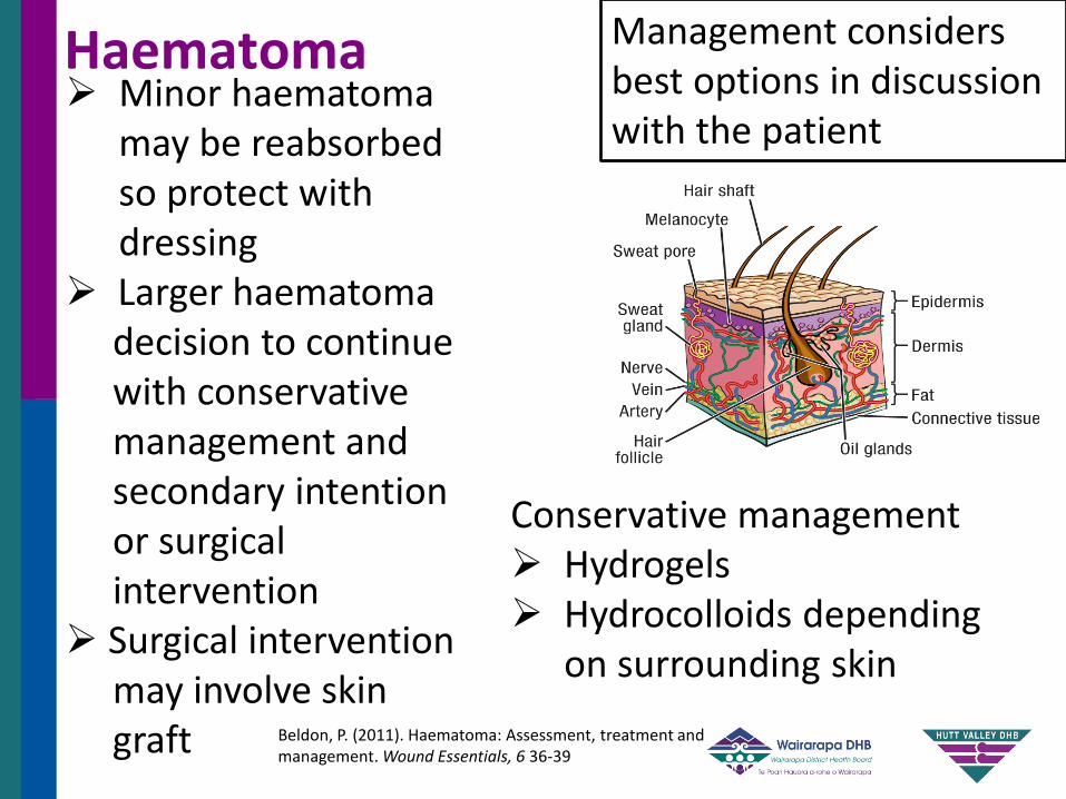
Haematoma Minor haematoma
may be reabsorbed so protect with dressing
Larger haematoma decision to continue with conservative management and secondary intention or surgical intervention Surgical intervention may involve skin graft
Management considers best options in discussion with the patient
Conservative management Hydrogels Hydrocolloids depending
on surrounding skin
Beldon, P. (2011). Haematoma: Assessment, treatment and management. Wound Essentials, 6 36-39

Wound Site/Location Point of Reference
Specifics to Aetiology
Choice of Dressing
Healing Time
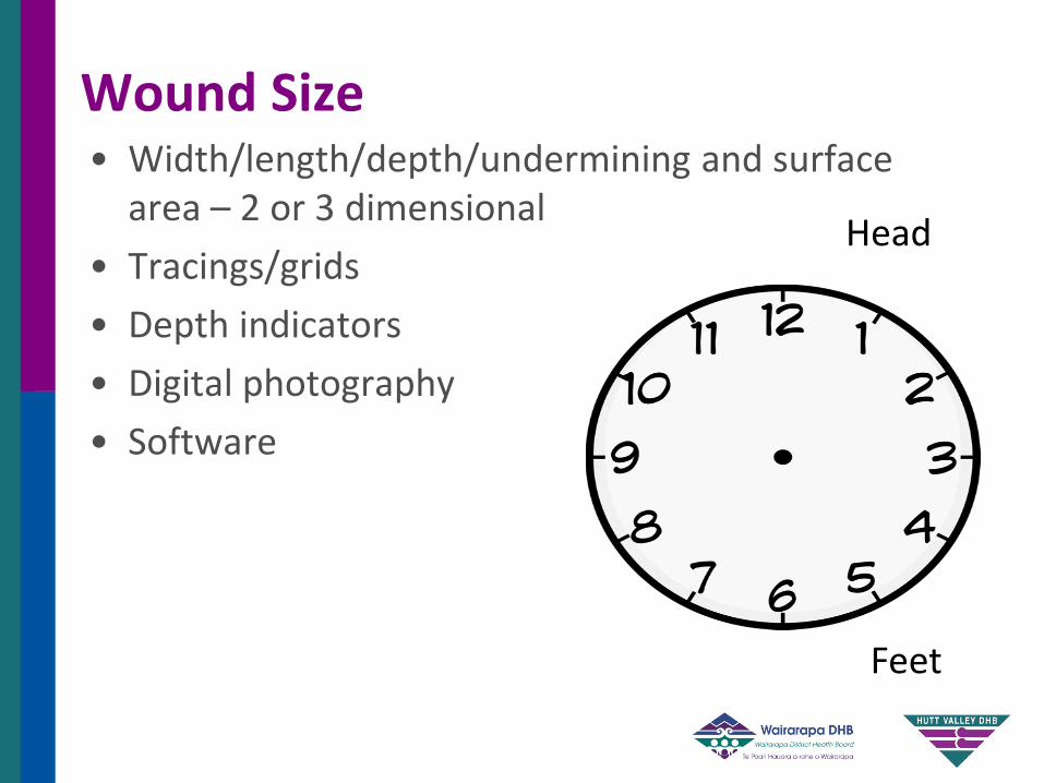
Wound Size • Width/length/depth/undermining and surface
area – 2 or 3 dimensional
• Tracings/grids
• Depth indicators
• Digital photography
• Software
Head
Feet
Odour
• Establish the cause
• Review QOL impact for the patient use of assessment tools for odour
• Treat and manage
Treat infection – topical/systemic
Debridement of devitalised tissue
Manage exudate
Odour control dressings

Surrounding Skin • Healthy
• Dry/scaly
• Erythema
• Blisters
• Discolouration
• Fragile/thin
• Pale
• Shiny/hairless
• Cellulitis
• Oedema
• Eczema (dry/wet)
• Maceration • Excoriation • Vascularity –
colour, warmth, capillary return
“Pain is an under-recognised and under-treated component of chronic wound care …….”p.18
Sibbald, R. et al., (2012). Special considerations in wound bed preparation 2011: an update. WCET Journal, 32(2), April/June, 10-30

Pain
• When?
• Where?
• Description?
• How relieve?
• Rating scale?
• Clinical observation
• ?Disease
• ?Surgery
• ?Trauma
• ?Infection
• ?Retained foreign body
• ?Wound care practices/products
“Pain is whatever the patient says it is, but
sometimes the patient doesn’t say” p.4
Principles of best practice: Minimising pain at wound dressing-related procedures. A consensus document. London: MEP Ltd, 2004
Wound Bed Preparation
Aim
“To create an optimal wound healing environment by producing a well vascularised, stable wound bed with little or no exudate”
Vowden, K. & Vowden, P. (2002). Wound bed preparation. http://www.worldwidewounds.com.

Wound Bed Preparation
T Tissue non-viable or deficient
I Infection or inflammation
M Moisture balance
E Edge of wound – non-advancing
or undermined
Sibbald, R.G., Orsted, H.L., Coutts, P.M. & Keast, D.H. (2007). Best practice recommendations for preparing the wound bed: Update 2006. Advances in Skin & Wound Care 20, 390-405

“T” Tissue
Why debride?
• Provide a physical barrier to physiology of healing
• Interfere with topical delivery of e.g. antimicrobials, pain relief
• Contribute to infection
• Prolong inflammatory process
• Unable to assess the wound accurately
• Increase production of exudate and odour
Effective debridement in a changing NHS: a UK consensus. London: Wounds UK, 2013.
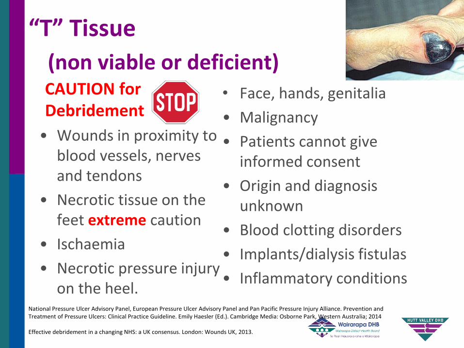
“T” Tissue
(non viable or deficient) • Face, hands, genitalia
• Malignancy
• Patients cannot give informed consent
• Origin and diagnosis unknown
• Blood clotting disorders
• Implants/dialysis fistulas
• Inflammatory conditions
CAUTION for Debridement
• Wounds in proximity to blood vessels, nerves and tendons
• Necrotic tissue on the feet extreme caution
• Ischaemia
• Necrotic pressure injury on the heel.
National Pressure Ulcer Advisory Panel, European Pressure Ulcer Advisory Panel and Pan Pacific Pressure Injury Alliance. Prevention and Treatment of Pressure Ulcers: Clinical Practice Guideline. Emily Haesler (Ed.). Cambridge Media: Osborne Park, Western Australia; 2014 Effective debridement in a changing NHS: a UK consensus. London: Wounds UK, 2013.
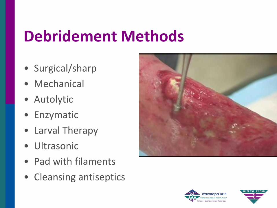
• Surgical/sharp
• Mechanical
• Autolytic
• Enzymatic
• Larval Therapy
• Ultrasonic
• Pad with filaments
• Cleansing antiseptics
Debridement Methods
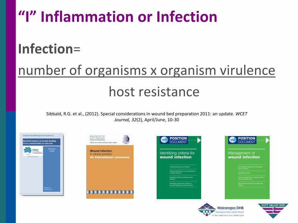
“I” Inflammation or Infection
Infection=
number of organisms x organism virulence
host resistance
Sibbald, R.G. et al., (2012). Special considerations in wound bed preparation 2011: an update. WCET Journal, 32(2), April/June, 10-30
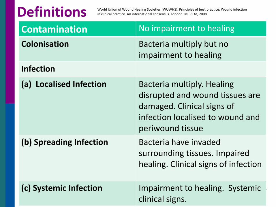
Definitions Contamination No impairment to healing
Colonisation Bacteria multiply but no impairment to healing
Infection
(a) Localised Infection Bacteria multiply. Healing disrupted and wound tissues are damaged. Clinical signs of infection localised to wound and periwound tissue
(b) Spreading Infection Bacteria have invaded surrounding tissues. Impaired healing. Clinical signs of infection
(c) Systemic Infection Impairment to healing. Systemic clinical signs.
World Union of Wound Healing Societies (WUWHS). Principles of best practice: Wound infection in clinical practice. An international consensus. London: MEP Ltd, 2008.
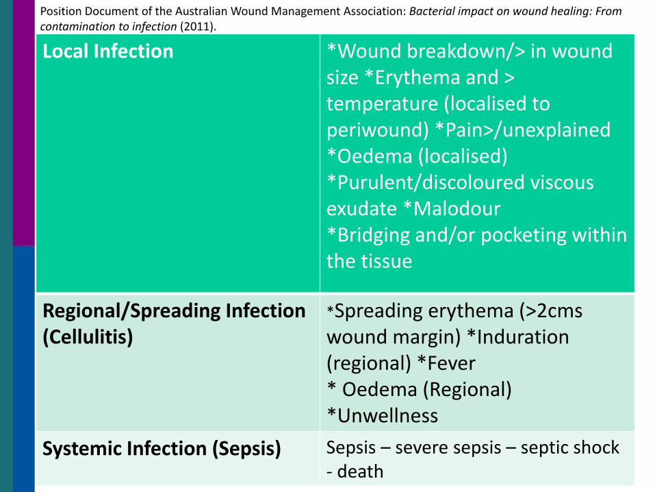
Local Infection *Wound breakdown/> in wound size *Erythema and > temperature (localised to periwound) *Pain>/unexplained *Oedema (localised) *Purulent/discoloured viscous exudate *Malodour *Bridging and/or pocketing within the tissue
Regional/Spreading Infection (Cellulitis)
*Spreading erythema (>2cms wound margin) *Induration (regional) *Fever * Oedema (Regional) *Unwellness
Systemic Infection (Sepsis) Sepsis – severe sepsis – septic shock - death
Position Document of the Australian Wound Management Association: Bacterial impact on wound healing: From contamination to infection (2011).

• Clean the wound with normal saline
• Swab only in contact with
wound surface
• Levine technique
Taking a Wound Swab
Gardner, S. (2010). Expert commentary. Wounds International 1(3), 20
Biofilm
• What is biofilm?
• Which wounds does biofilm form in?
• Can you see a biofilm in a wound?
• How do you remove a biofilm?
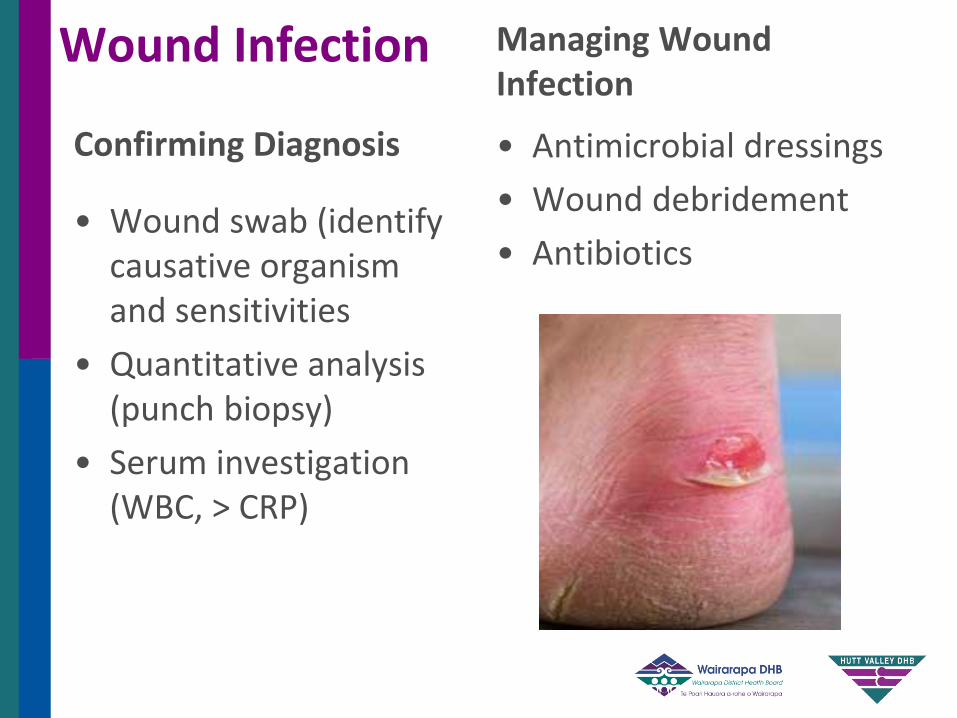
Wound Infection
Confirming Diagnosis
• Wound swab (identify causative organism and sensitivities
• Quantitative analysis (punch biopsy)
• Serum investigation (WBC, > CRP)
Managing Wound Infection
• Antimicrobial dressings
• Wound debridement
• Antibiotics

Recognise
• Stages of wound healing
• Excessive inflammation caused by underlying comorbidities
• Increasing bacterial burden
• Infection
Use risk assessment and
management
Antibiotic Resistance ‘How can nurses contribute to antimicrobial stewardship?’
Sussman, G., Swanson, T., Black, J., Cooper, R., Schultz, G., Fletcher, J. & Smith, D. (2014). Ten top tips: reducing antibiotic resistance. Wounds International, 5(4), 4-8
“ Exudate – understand, assess and manage..” p. 12
World Union of Wound Healing societies (WUWHS). Principles of best practice: Wound exudate and the role of dressings. A consensus document.
London: MEP Ltd, 2007

“M” Moisture Balance Colour e.g.Serous (clear); Purulent (cloudy,
creamy); Haemoserous (pink or red); Infection (green, yellow or brown); other
Consistency e.g. High viscosity (high protein from infection, inflammation) e.g. Low viscosity (low protein from CCF, malnutrition)
Odour e.g. infection or bacterial growth; necrotic tissue; dressing
Amount (subjective)
Depend on surface area of wound; indicate systemic problem; type of wound World Union of Wound Healing societies (WUWHS). Principles of best practice:
Wound exudate and the role of dressings. A consensus document. London: MEP Ltd, 2007
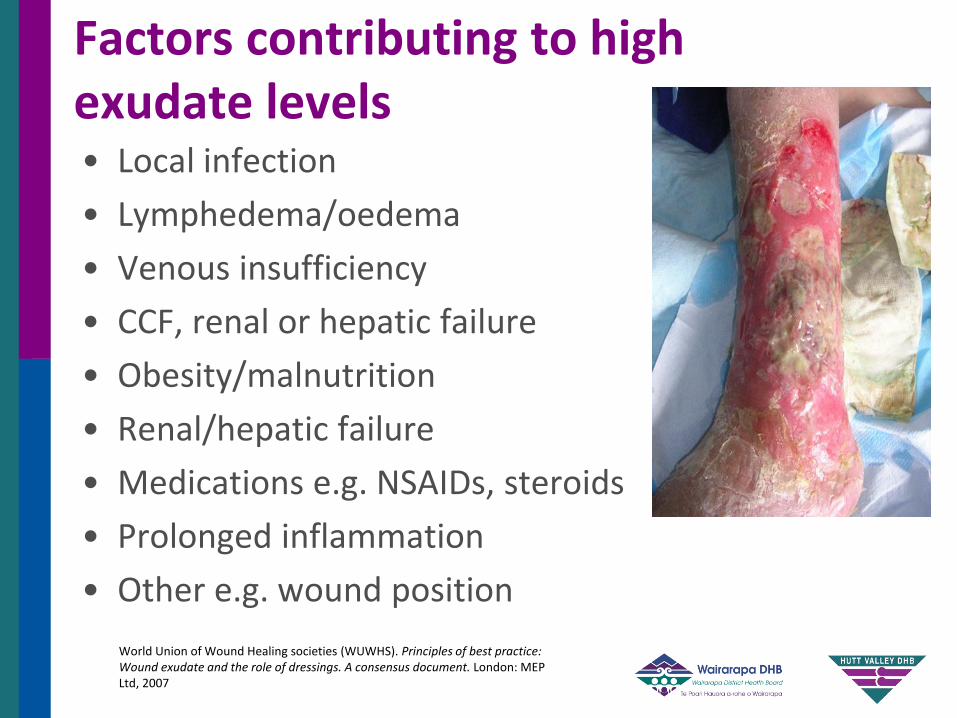
Factors contributing to high exudate levels • Local infection
• Lymphedema/oedema
• Venous insufficiency
• CCF, renal or hepatic failure
• Obesity/malnutrition
• Renal/hepatic failure
• Medications e.g. NSAIDs, steroids
• Prolonged inflammation
• Other e.g. wound position
World Union of Wound Healing societies (WUWHS). Principles of best practice: Wound exudate and the role of dressings. A consensus document. London: MEP Ltd, 2007

Exudate Assessment
Assess patient
Assess regional factors
Assess dressing
Assess wound bed/edge; size; stage of healing; fistula or sinus
Assess periwound
Assess exudate
World Union of Wound Healing societies (WUWHS). Principles of best practice: Wound exudate and the role of dressings. A consensus document. London: MEP Ltd, 2007
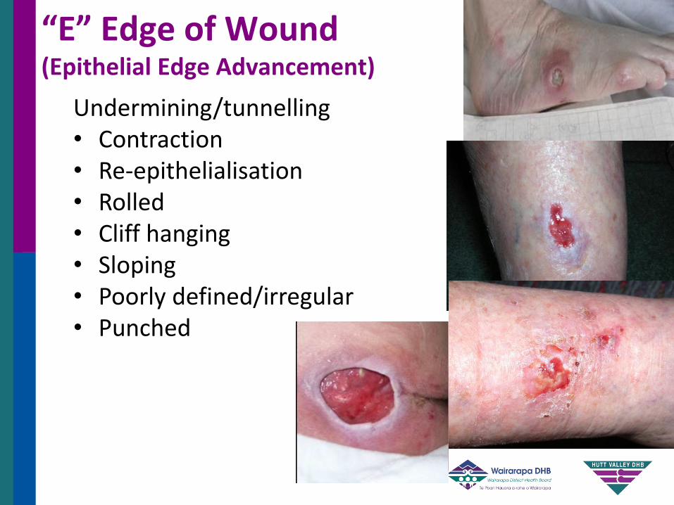
“E” Edge of Wound (Epithelial Edge Advancement)
Undermining/tunnelling • Contraction • Re-epithelialisation • Rolled • Cliff hanging • Sloping • Poorly defined/irregular • Punched
“Evidence-based medicine is the integration of best research evidence with clinical expertise and
patient values”
Sackett, D.L. Centre Evidence Based Medicine 2010 http://www.cebm.net/index

Investigation and Diagnosis
• Physical tests and observations
• Biological tests
• Biochemical tests
• Others

Do we have a Diagnosis
• History – Medical – Surgical - Pharmacological
Social
• Examination – Regional
Local – TIME +
• Investigations
• Diagnosis
• Intervention/Planning
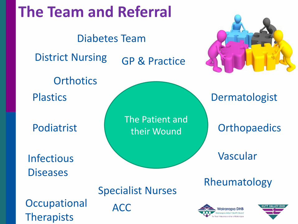
The Patient and their Wound
The Team and Referral
District Nursing
Plastics
GP & Practice
Dermatologist
Podiatrist Orthopaedics
Vascular
Rheumatology
Infectious Diseases
Orthotics
Occupational Therapists
Diabetes Team
Specialist Nurses
ACC
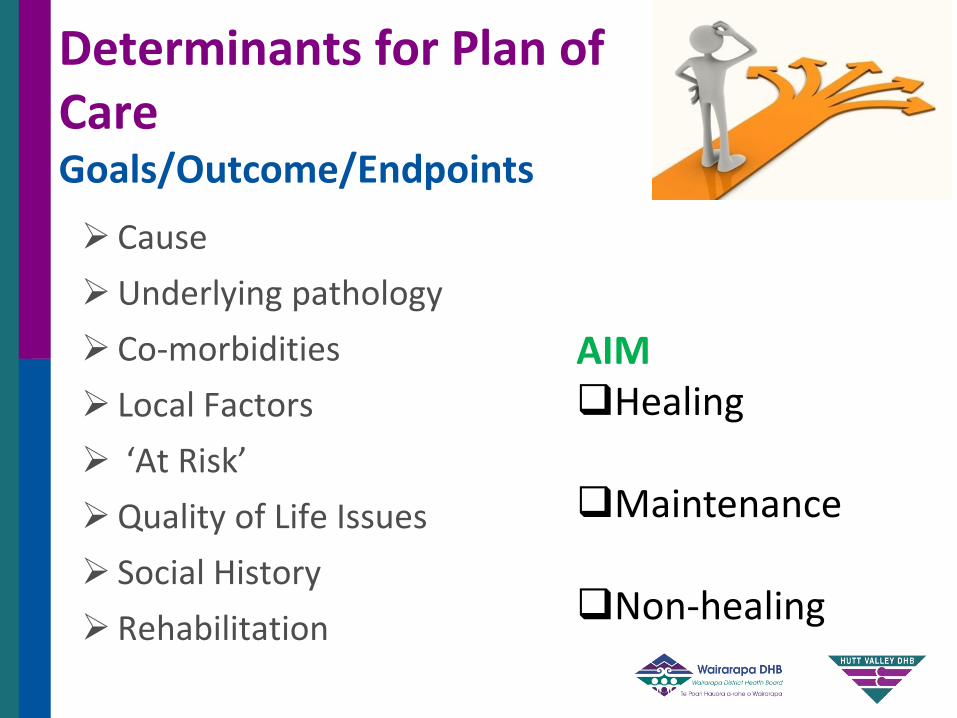
Cause
Underlying pathology
Co-morbidities
Local Factors
‘At Risk’
Quality of Life Issues
Social History
Rehabilitation
Determinants for Plan of Care Goals/Outcome/Endpoints
AIM Healing
Maintenance
Non-healing

Goals of Care (Local)
Short Term
Long Term
• Haemostasis
• Debride
• Remove foreign bodies
• Protect surrounding skin
• Reduce bacterial load
• To improve physical function

Documentation • Provides
communication
• Provides evidence in litigation
• Used for research and statistical evidence
• Aids education
• Used in clinical audit and quality assurance
• Contributes to care planning
• Provides evidence of continuity of care
• Supports service delivery
• Supports effective clinical judgment
• Supports decision making
Benbow, M. (2011). Documentation: Keeping accurate patient records. Wound Essentials, 6, 90-92

“Good records= Good defence Poor records = Poor defence No records= No defence” p. 23
Eagle, M. (2009). Wound assessment: The patient and the wound. Wound Essentials, 4, 14-24
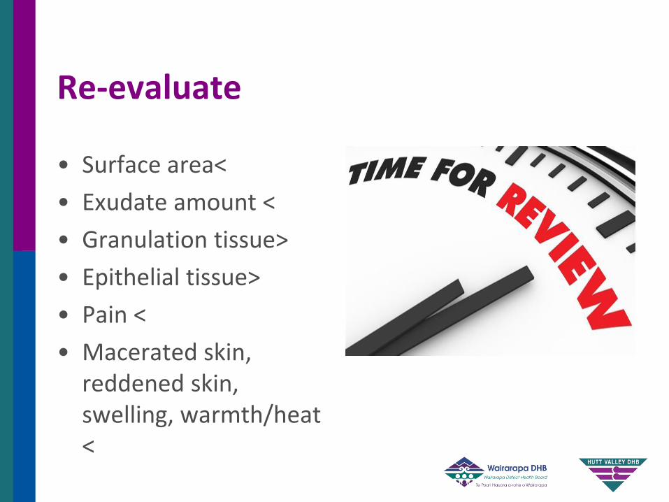
Re-evaluate
• Surface area<
• Exudate amount <
• Granulation tissue>
• Epithelial tissue>
• Pain <
• Macerated skin, reddened skin, swelling, warmth/heat <
• Wound should be 30% smaller (surface area) at week 4 to heal in 12 weeks (Falanga & Sabolinski, 1999)
• 20% to 40% reduction in two and four weeks is likely to be a reliable predictor of healing (Falanga, 2005;
Margolis et al, 2004)
• 50% reduction at week 4 a good predictor for persons with DFU (Sheehan et al, 2003)
• Any wound greater than six weeks old is considered chronic (Bowler & Davies, 1999)
Referral – Parameters in Wound Healing

Where to start?
• Past history?
• Wound Bed?
• Evidence based practice?
• Look at risks?
• Understand factors which influence wound healing
• Understand general and health issues that may influence ability of wound to heal
• Holistic wound assessment assesses physical; social; psychological and spiritual/cultural domains of wellbeing
• Identify the specific aetiology/causal factor of the wound and concurrent disease processes p.24
Summary
Eagle, M. (2009). Wound assessment: The patient and the wound. Wound Essentials, 4, 14-24

• Identify type of wound, stage of healing; consider wound bed and peri-wound skin (p. 24)
• Assess baseline information using logical systematic assessment tools and document findings
• Identify factors that may delay healing
• Re-evaluate current wound management; change according to local wound assessment
• Recognise limitation of knowledge and make appropriate referrals p.24
Summary (contd)
Eagle, M. (2009). Wound assessment: The patient and the wound. Wound Essentials, 4, 14-24
Other References
• Bowler, P.G. & Davies, B.J. (1999). The microbiology of acute and chronic wounds, Wounds, 11, 72-99
• Falanga, V. & Sabolinski, M. (1999). A bilayered living skin construct (APILGRAF) accelerates complete closure of hard-to-heal venous ulcers. Wound Repair Regeneration, 7, 201-7
• Margolis, D.J., Allen-Taylor, L. & Hoffstad, O. (2004). The accuracy of venous leg ulcer prognostic models in a wound care system. Wound Repair Regeneration, 12, 163-168