Embed Size (px)
Citation preview

Single-molecule Förster resonance energy transfer (FRET)analysis discloses the dynamics of the DNA–topoisomerase II(Top2) interaction in the presence of TOP2-targeting agentsReceived for publication, April 24, 2017, and in revised form, June 7, 2017 Published, Papers in Press, June 19, 2017, DOI 10.1074/jbc.M117.792861
Wan-Chen Huang‡1, Chun-Ying Lee‡§, and Tao-shih Hsieh‡§¶†
From the ‡Institute of Cellular and Organismic Biology, Academia Sinica, Taipei 115, Taiwan, the §Department of Chemistry,National Taiwan University, Taipei 106, Taiwan, and the ¶Department of Biochemistry, Duke University Medical Center,Durham, North Carolina 27710
Edited by Patrick Sung
Topoisomerases play crucial roles in DNA replication, tran-scription, and recombination. For instance, topoisomerase II(Top2) is critically important for resolving DNA tangles duringcell division, and as such, it is a broad anticancer drug target.Top2 regulates DNA topology by transiently breaking one dou-ble-stranded DNA molecule (cleavage), allowing a second dou-ble strand to pass through the opened DNA gate (opening), andthen closing the gate by rejoining the broken ends. Drugs thatmodulate Top2 catalysis may therefore affect enzymatic activityat several different steps. Previous studies have focused onexamining DNA cleavage and ligation; however, the dynamicopening and closing of the DNA gate has been less explored.Here, we used the single-molecule Förster resonance energytransfer (smFRET) method to observe the open and closed stateof the DNA gate and to measure dwell times in each state. Ourresults show that Top2 binds and bends DNA to increase theenergy transfer efficiency (EFRET), and ATP treatment furtherinduces the fluctuation of EFRET, representing the gate openingand closing. Additionally, our results demonstrate that bothtypes of Top2-targeting anticancer drugs, the catalytic inhibitordexrazoxane (ICRF187) and mechanistic poison teniposide(VM26), can interfere with DNA gate dynamics and shorten thedwell time in the closed state. Moreover, Top2 bound to thenonhydrolyzable ATP analog 5�-adenylyl-�,�-imidodiphos-phate exhibits altered DNA gate dynamics, but the DNA gateappears to open and close even after N-gate closure. In sum-mary, we have utilized single-molecule detection to unravelTop2 DNA gate dynamics and reveal previously unknowneffects of Top2 drugs on these dynamics.
Topoisomerases play crucial roles in DNA replication, tran-scription, and recombination (1– 4). Frequent unwinding andrewinding of the DNA duplex during these processes can createtopological obstructions to DNA transactions, and failure to
properly resolve these obstructions can induce cell death(4 – 6). Topoisomerases resolve topological strain by breaking aDNA strand and allowing a second strand to pass throughbefore rejoining the broken ends (7). Depending on the sub-strate specificity, topoisomerases can be categorized into twotypes, types I and II, which process single-stranded and double-stranded DNA, respectively. Whereas type I topoisomerasesare specifically involved in transcription, recombination, andDNA repair, type II topoisomerases are required for DNA rep-lication and segregation of catenated chromosomes (5, 7).Because cancer cell growth is heavily dependent on type IItopoisomerase functions, they have become critical targets foranticancer drugs (4, 8).
The catalytic cycle of topoisomerase II (Top2) obeys a two-gate mechanism and requires both ATP and Mg2� to triggerthe strand passage reaction (5, 9). The cycle is initiated whenthe core domains of a Top2 homodimer bind to a DNA frag-ment (G-segment or G-DNA), which is then bent to a 150°angle (10). The N-gate will close and trap the T-segment uponATP binding to the ATPase domain. Concurrently, the tyrosineresidues inside the core domain participate in a transesterifica-tion reaction and form covalent adducts with the DNA back-bone, breaking the G-segment (cleavage) and pulling the bro-ken ends apart (opening) to produce a double-stranded DNAbreak with 4-bp overhangs. This conformation is referred to asthe open DNA gate. The hydrolysis of one ATP is accompaniedby the passage of the T-segment through the opened gate (11).Next, the DNA gate is closed, bringing the ends of the G-seg-ment into close proximity for rejoining (ligation) via the secondtransesterification reaction. Finally, a second ATP hydrolysisdissociates the closed N-gate, which allows the release of theG-segment from Top2 (12). Thus, the opening and closure ofthe DNA gate are closely related to, but functionally distinctfrom cleavage and ligation reactions.
The Top2 complex with cleaved DNA can be stabilized bythe anticancer drugs, etoposide (VP16) and teniposide (VM26),which interfere with DNA ligation. This action results in accu-mulation of DNA breaks that are poisonous to cells, and thedrugs are therefore referred to as DNA poisons (11, 13). More-over, two bisdioxopiperazine-type antitumor agents, ICRF193and ICRF187 (dexrazoxane), which are termed catalytic inhib-itors, block ATPase activity and promote the closed clamp for-mation, resulting in impaired enzyme turnover and T-segment
This work was supported by Ministry of Science and Technology TaiwanMOST Grant 105-2321-B-001-006 and Academia Sinica intramural fundingGrant AS-105-TP-B04 (to T. S. H.). The authors declare that they have noconflicts of interest with the contents of this article.
This article contains supplemental Figs. S1 and S2.† Deceased August 4, 2016. Original corresponding author.1 To whom correspondence should be addressed: 128 Academia Rd., Section
2, Nankang, Taipei 11529, Taiwan. Tel.: 886-2-2787-1536; Fax: 886-2-2787-1533; E-mail: [email protected].
crosARTICLE
J. Biol. Chem. (2017) 292(30) 12589 –12598 12589© 2017 by The American Society for Biochemistry and Molecular Biology, Inc. Published in the U.S.A.
by guest on Decem
ber 29, 2019http://w
ww
.jbc.org/D
ownloaded from

passage (14). Although biochemical studies on Top2-targetingdrugs have elucidated many features of Top2 inhibition, a com-plete delineation of the mechanism, in terms of the interferencewith gate dynamics, remains to be uncovered. For example,whether Top2 drugs alter DNA gate dynamics and how thisalteration may correlate with N-gate activity to perturb Top2function is previously unknown.
Currently, several high-resolution fluorescence techniquesare available for the precise physicochemical characterizationof elemental processes at the level of a single protein or nucleicacid. Similar FRET methods have been used to monitor theDNA gate recently (15–18). However, previous studies did notaddress the ability of Top2 drugs to interfere with DNA gatedynamics. Here, we used single-molecule Förster resonanceenergy transfer (smFRET),2 a technique based on total internalreflection fluorescence (TIRF) microscopy, to observe the DNAgate dynamics of Top2. Dynamic measurements were made inthe presence of cofactors, ATP and AMPPNP, as well as anti-cancer drugs ICRF187 and VM26. Based on our experiments,we report that (i) DNA gate dynamics are altered by treatmentwith either Top2 poisons or catalytic inhibitors, which bothshorten the gate closed time, and (ii) DNA gate opening andclosing may persist after N-gate closure.
Results
smFRET detection of DNA opening and closing
To observe the dynamics of the DNA gate as it goes throughdifferent conformational states during the catalytic cycle, weperformed TIRF microscopy-based smFRET measurements.The DNA substrate was designed such that the fluorophoreadducts were located 2 bp outside the DNA footprint of Top2(Fig. 1B) (15). The DNA sequence used for smFRET experi-ments had an additional biotinylated 23-nt poly(T) sequence atthe 5�-end of the same strand with AF488, which allowed theDNA molecule to be immobilized on the coverglass surface.The FRET measurement was performed after reaction bufferwas infused into the reaction chamber. The distribution ofFRET efficiency and the dynamics of single molecules were col-lected in the absence or presence of DmTop2, ATP, and VM26.Five separate recordings of single molecules were made in ran-domly selected areas, and the average FRET efficiency betweenthe first 10 and 20 frames was taken as the average FRET value.The FRET-distribution histograms were then fitted with a dou-ble-Gaussian peak-fit function (Fig. 2A). When only DNA waspresent (top panel), the FRET peak was centered at E � 0.36,indicating the FRET value for the native B-DNA conformation.After 300 nM DmTop2 was infused (second panel), the FRETvalue shifted to E1 � 0.49. The increased FRET value is consis-tent with bending of the DNA duplex, which should reduce thedistance between the two fluorophores and increase the ener-gy-transfer efficiency. Intriguingly, a small population of mole-cules exhibited a lower FRET peak (at E2 � 0.29), and after ATPaddition, this population increased from 10 to 17% (thirdpanel). The opening of the DNA gate would create a longer
distance between the fluorophores and thus a lower FRET effi-ciency. Moreover, the fraction of molecules in the lower peakincreased to 23% when 200 �M teniposide (VM26, which pre-vents DNA ligation) was added to the observation chamber(bottom panel). This observation may suggest that by keepingthe DNA backbone unlocked, there is a greater chance for theopened DNA gate to be observed.
Mg2� is thought to be essential for the DNA cleavage andligation steps (19). Therefore, to determine whether the lowerFRET peak (Fig. 2A) was caused by DNA cleavage-opening afterATP addition, we obtained a parallel set of measurements usingreaction buffer without Mg2� (Fig. 2B). When 300 nM DmTop2was added to the reaction chamber, the FRET value increasedfrom 0.34 to 0.47, which suggests that DNA binding and bend-ing by DmTop2 occurred in the absence of Mg2�. However, thelower FRET peak (EFRET � 0.29) was not detected, further sup-porting the notion that the FRET value at 0.29 represents thecleavage-opening of the DNA gate. When the unlocking of theDNA backbone is inhibited (by removing Mg2�), the openedstate cannot be achieved.
We further analyzed each single-molecule time traceobtained from the treatment with DmTop2, ATP, and/orVM26. We aimed to determine whether we could observethe transient/intermediate states with the smFRET method.Therefore, we used the hidden Markov model (HMM) to fit allof the FRET trajectories with the maximum of five states usingvbFRET in a Matlab environment. No assumptions were madeabout the values or distributions. Hundreds of trajectories werefitted to generate each distribution, and from the fitted FRETresults, we found the trajectories with transitions are almostentirely between two states. According to the EFRET and �EFRETvalues for each FRET trajectory, we were able to classify thepossible types of transitions. The representative FRET trajec-tory in Fig. 2C show multiple transitions between high and lowFRET and variable dwell times in each state. The distribution ofdwell times calculated from the fitted FRET values is plotted inFig. 2D. High FRET panels show the distribution of dwell timesat the higher FRET value (E1), 0.5, which represents bending ofDNA. Low FRET panels show the distribution of dwell times atthe lower FRET value (E2), 0.29, which is taken to represent theopen state of the DNA gate. Rate constants were determined byfitting a single exponential function to the dwell time histo-grams, and the average dwell time was calculated. Intriguingly,in the Top2-treatment group without ATP, FRET transitionswere detected in some of the single molecules (Fig. 2C, upperpanel) and exhibited a relatively long dwell time, �2.3 s, in theclosed-gate state and a short opening time of �0.2 s (Fig. 2D),however, the FRET value of most molecules (more than 90%)remained constant throughout the observation period (supple-mental Fig. S1). With ATP treatment, the dwell time at highFRET was shortened to �1.2 s and the low-FRET dwell timewas 0.4 s. In the VM26-treatment group, we observed singlemolecules displayed either one of two possible transition pat-terns. In the slow-transition group, the dwell time was similarto that of the ATP-treatment group (supplemental Fig. S2).However, in the fast-transition group, the high-FRET dwelltime was shortened to 0.5 s, and the low-FRET dwell time wasslightly reduced to 0.3 s (Fig. 2D, bottom panel). This result
2 The abbreviations used are: smFRET, single-molecule Förster resonanceenergy transfer; TIRF, total internal reflection fluorescence; AMPPNP,5�-adenylyl-�,�-imidodiphosphate; HMM, hidden Markov model.
Single-molecule study of topoisomerase 2 gate dynamics
12590 J. Biol. Chem. (2017) 292(30) 12589 –12598
by guest on Decem
ber 29, 2019http://w
ww
.jbc.org/D
ownloaded from

suggested that Top2 tends to open the DNA gate more readilyin the presence of VM26 than in its absence.
Interference effect of AMPPNP and Top2 drugs on DNA gateopen and closed states
AMPPNP, a nonhydrolyzable analog of ATP, has beenreported to bind to the ATP-binding pocket of Top2 and allowa single turnover of T-DNA transport through the Top2 DNAgate (20, 21). Thus, we sought to examine DNA gate dynamicsafter AMPPNP binding. Fig. 3A shows the FRET-distributionhistograms of the DNA substrate alone, with DmTop2, andwith DmTop2 plus AMPPNP. We observed that the low-FRETpopulation was slightly increased in the AMPPNP-treatmentgroup. Upon further analyzing the single-molecule FRET tra-jectories (Fig. 3B), we observed frequent gate opening and clos-ing events indicated by the transitions between two FRET effi-
ciency levels. Fig. 3C shows that after AMPPNP treatment, thedwell times in the high-FRET and low-FRET states were simi-lar, 0.57 and 0.43 s, respectively. Observing FRET transitions insingle molecules with AMPPNP treatment was unexpected,and we initially considered these findings may be contrary toprevious reports, which have generally supported the view thatAMPPNP induces N-gate closure and the trapping of the DNAgate (22, 23). It was previously reported that DNA cleavage canoccur in the presence of AMPPNP (24). Therefore, we per-formed a gel-based DNA-cleavage assay to test whether DNAcleavage occurs in our experimental system. The gel results(Fig. 3D) showed that whereas ATP induced the relaxation ofsupercoiled DNA into open-circular form, AMPPNP did not.Furthermore, AMPPNP induced a faint band corresponding tothe linear-form DNA. VM26, a Top2 poison, impedes Top2ligation and thereby generates DNA break. When we applied
Figure 1. Schematic of the smFRET experimental platform for studying Top2 dynamics. A, catalytic cycle of Top2. The FRET pair-labeled DNA substratewas used. Upon G-DNA binding to Top2, the DNA is bent, bringing the fluorophores closer together, and increasing energy-transfer efficiency. When ATP bindsto the ATPase domain, N-gate closure is accompanied by cleavage of G-DNA and opening of the DNA gate. Upon opening of the DNA gate, the fluorophoresare separated and the energy-transfer efficiency decreases. The FRET efficiency can therefore be used to measure the distance between the two fluorophoresthat represents the DNA native state, DNA bending, and opening of the DNA gate. B, fluorescent DNA substrate designed for the experiment. A 28-bpoligonucleotide-based DNA substrate was synthesized, containing a FRET pair, Alexa Fluor 488 and Alexa Fluor 555 with an additional biotinylated 23-nt poly(T)sequence at the 5�-end of the AF488-labeled strand; the fluorophores were situated on complementary strands and separated by 15 bp (�1.5 turns).
Single-molecule study of topoisomerase 2 gate dynamics
J. Biol. Chem. (2017) 292(30) 12589 –12598 12591
by guest on Decem
ber 29, 2019http://w
ww
.jbc.org/D
ownloaded from

Single-molecule study of topoisomerase 2 gate dynamics
12592 J. Biol. Chem. (2017) 292(30) 12589 –12598
by guest on Decem
ber 29, 2019http://w
ww
.jbc.org/D
ownloaded from

Figure 2. DmTop2 induces DNA bending, as well as DNA gate opening and closing. A, FRET efficiency-distribution histograms of the interaction betweenDmTop2 and DNA. The distribution of FRET efficiency values (EFRET) was determined from hundreds of single molecules in the absence or presence of DmTop2,ATP, and VM26. B, Mg2� was removed from the reaction buffer and similar experiments were performed in parallel with A. C, representative intensities andcorresponding FRET trajectories from single-molecule measurements. I (A.U.) panels, red line, acceptor intensity; green line, donor intensity. EFRET panels, blueline, raw FRET efficiency; red line, HMM fitted FRET efficiency. D, the histogram of dwell time at each state (gate closed dwell time, �close, high EFRET; gate opendwell �open time, low EFRET) was fitted with a single exponential function.
Figure 3. AMPPNP and Top2 poisons interfere with DNA gate dynamics. A, FRET-distribution histograms of DNA alone and DmTop2 with or withoutAMPPNP. B, representative time and FRET trajectories after AMPPNP treatment. I (A.U.) panel, red line: acceptor intensity; green line: donor intensity. EFRET panel,blue line: raw FRET efficiency; red line: HMM fitted FRET efficiency. C, dwell time distribution of the high- and low-FRET states after AMPPNP treatment. Rateconstants were determined by fitting an exponential decay function to the histogram derived from a normalized population of dwell times. D, results of aDNA-cleavage assay showing increased relaxed circular DNA (RC) by DmTop2 with ATP but not with AMPPNP. In the presence of VM26 both ATP and AMPPNPtreatments showed increased linear DNA. Lanes are labeled as follows: D, DNA; L, linearized DNA; M, AMPPNP; T, ATP, with and without VM26. NC, nicked circularDNA; L, linear DNA; NSC, negative supercoil DNA; RC, relaxed circular DNA. E, FRET-distribution histogram showing that the low-FRET population was increasedwhen gate ligation was impaired by VM26.
Single-molecule study of topoisomerase 2 gate dynamics
J. Biol. Chem. (2017) 292(30) 12589 –12598 12593
by guest on Decem
ber 29, 2019http://w
ww
.jbc.org/D
ownloaded from

VM26 to both the ATP- and AMPPNP-treated groups, a con-siderable amount of linearized DNA was observed in the pres-ence of ATP, indicating DNA cleavage. However, an increase inlinear DNA also appeared in the AMPPNP plus VM26 co-treat-ment group. This result indicated that DNA gate opening andclosing could still occur after AMPPNP treatment, and thatVM26 interfered with subsequent ligation steps resulting in theincreased generation of linear DNA. The FRET-distributionhistogram revealed that the low-FRET population was furtherincreased after AMPPNP treatment when gate ligation was hin-dered by VM26 (Fig. 3E).
Impairment of DNA gate opening and closing by Top2catalytic inhibitors
Previous biochemical data have provided evidence thattwo Top2 catalytic inhibitors, ICRF193 and ICRF187, canreduce Top2-mediated DNA-relaxation activity and pro-mote the closed-clamp formation of the protein (14). However,whether these inhibitors interfere with DNA gate opening andclosing is unclear. Thus, we performed smFRET measurementswith various concentrations of Top2 catalytic inhibitors (Fig. 4).Whereas the dwell time at the low-FRET state increased whenthe ICRF187 concentration was raised from 200 to 400 �M, thedwell time at the high-FRET state remained unchanged. Simi-larly, in the ICRF193-treatment group, the dwell time was lon-ger in the low-FRET state than at the high-FRET state (Fig. 4B).We fitted the histogram of low FRET dwell time distribution inFig. 4B with a double exponential decay function. This couldbetter fit the dwell times, suggesting the possibility that twocomponents exist: one component is transient, at around 0.25–0.3 s, and one is longer, at around 1.3–1.6 s, gate opening time(data not shown). However, fitting with multiexponential decayfunctions can always produce better fitted results, and mightgenerate false states. Therefore, we conclude that fitting withsingle exponential decay function is preferred. These resultssuggest that Top2 catalytic inhibitors might slightly prolong theopen state of the DNA gate.
Discussion
We used the smFRET system to study the dynamics of Top2interaction with DNA at different steps of the catalytic cycle,including DNA bending, DNA gate opening, and DNA gateclosing. With this methodology, we determined the effect ofions, cofactors, and Top2 anticancer drugs on protein dynam-ics. Our data demonstrate the utility of this novel tool for study-ing the effects of Top2 drugs on the conformational dynamicsof the enzyme. The use of smFRET complements previouslyestablished methods in probing the mechanisms by whichdrugs might exert their effects on Top2.
When DNA alone was present, the FRET efficiency peak wascentered at 0.36. After a saturating amount of DmTop2 wasintroduced into the reaction chamber, our data suggest thebinding and bending of the DNA duplex brought the two fluo-rophores closer together, resulting in an increase in the energy-transfer efficiency, whereas a small population of moleculesexhibited a lower FRET peak (at 0.29) meaning that addingTop2 alone can initiate opening of the DNA gate. Moreover,with ATP, the FRET efficiency decreased in a subpopulation of
molecules, representing the state when the DNA gate is open.Mg2� can unlock the DNA gate and form a triad with conservedacidic amino acid residues that are situated in the core domain(13, 25). In the group treated with only Top2, a small proportionof the single molecules showed FRET transitions with a shortopen gate dwell time of �0.2 s, but the DNA gate of most of thesingle molecules was closed throughout the entire observationperiod. When Mg2� was removed from the reaction buffer (Fig.2B), DNA bending was not affected, but DNA gate openingcould no longer occur. Suggesting the essential role of Mg2� forthe Top2-induced DNA gate opening.
According to our control experiments with DNA only andTop2 plus DNA, we found that these two states have close butdistinct EFRET values of 0.36 and 0.5, respectively. Therefore, inour experimental design, we are able to tell the differencebetween Top2 dissociation from DNA and Top2 binding-in-duced DNA bending, according to the �EFRET values and FRETtrajectories. Within our smFRET observation period, which isaround 20 s on average, we readily observed FRET fluctuationsbetween 0.5 and 0.29 (with �E ranging from 0.18 to 0.25). Onlya few trajectories showed FRET transitions between 0.36 and0.5 (�E � 0.1�0.15), but we do not observe the combination ofFRET transitions among these three states. Therefore, we areconfident that we were able to extract properly the dwell timeinformation. However, we are not able to exclude the possibilitythat random association of Top2 and DNA occurs. In this case,FRET values would not change with bending, and the DNAcleavage-related opening would also not be observed.
ATP is a cofactor that can promotes T-segment capture andstimulates G-segment cleavage (9, 22, 26 –28). Pre-steady-stateanalysis of ATP hydrolysis using Saccharomyces cerevisiae (Sc)Top2 and DNA has previously indicated that the enzyme bindsand hydrolyzes two ATP molecules per reaction cycle, with thehydrolysis of the ATP molecules occurring sequentially (27,29). In the smFRET data obtained here, we did not observe anyconsistent pause between the two cycles of DNA gate openingand closing, but instead observed a continuous opening andclosing in the presence of ATP. This result suggests that ATP iscontinuously hydrolyzed to open and close the DNA gate, withan average dwell time in the open and closed states that can bemodified by the presence of Top2-targeting drugs. The sum ofdwell times in the high- and low-FRET states can describe thecompletion of a single cycle of DNA opening and closing. With-out T-segment, the average time for a full cycle can be esti-mated from the DmTop2 plus ATP condition, and is �1.54 s.
Top2-targeting drugs are divided into two classes, based ontheir mode of action. One class is Top2 poisons, such as VM26and VP16, which result in DNA double-strand breaks. Theother class, which includes inhibitors such as ICRF193 andICRF187, suppresses ATPase activity. In the presence of ATP,the reaction equilibrium strongly favors ligation over cleavage,and thus at any given time, only a small fraction of the enzymeis covalently attached to cleaved DNA (30). However, whenVP16 impedes the ligation at the DNA gate, a comparativelylarger fraction of the enzyme remains covalently attached tocleaved DNA. Our smFRET results demonstrated that theVM26-treatment group showed considerably faster openingkinetics compared with the group treated with ATP alone.
Single-molecule study of topoisomerase 2 gate dynamics
12594 J. Biol. Chem. (2017) 292(30) 12589 –12598
by guest on Decem
ber 29, 2019http://w
ww
.jbc.org/D
ownloaded from

Figure 4. Top2 catalytic inhibitors modulate DNA gate dynamics. Dwell time distribution of high- and low-FRET states after treatment with ICRF187 (A) andICRF193 (B) at two concentrations. The histogram of the dwell time at each state (gate closed dwell time: �close, high EFRET; gate open dwell time: �open, low EFRET)was fitted with a single exponential function.
Single-molecule study of topoisomerase 2 gate dynamics
J. Biol. Chem. (2017) 292(30) 12589 –12598 12595
by guest on Decem
ber 29, 2019http://w
ww
.jbc.org/D
ownloaded from
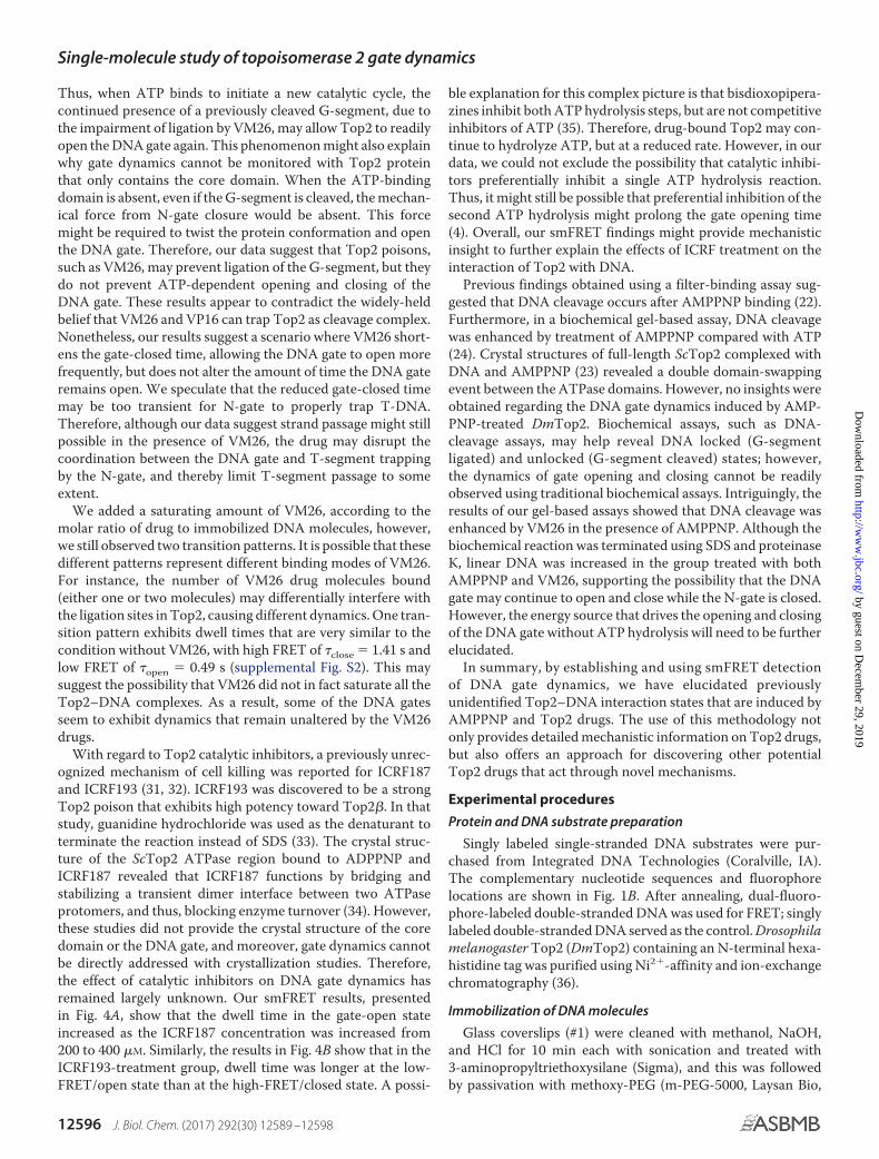
Thus, when ATP binds to initiate a new catalytic cycle, thecontinued presence of a previously cleaved G-segment, due tothe impairment of ligation by VM26, may allow Top2 to readilyopen the DNA gate again. This phenomenon might also explainwhy gate dynamics cannot be monitored with Top2 proteinthat only contains the core domain. When the ATP-bindingdomain is absent, even if the G-segment is cleaved, the mechan-ical force from N-gate closure would be absent. This forcemight be required to twist the protein conformation and openthe DNA gate. Therefore, our data suggest that Top2 poisons,such as VM26, may prevent ligation of the G-segment, but theydo not prevent ATP-dependent opening and closing of theDNA gate. These results appear to contradict the widely-heldbelief that VM26 and VP16 can trap Top2 as cleavage complex.Nonetheless, our results suggest a scenario where VM26 short-ens the gate-closed time, allowing the DNA gate to open morefrequently, but does not alter the amount of time the DNA gateremains open. We speculate that the reduced gate-closed timemay be too transient for N-gate to properly trap T-DNA.Therefore, although our data suggest strand passage might stillpossible in the presence of VM26, the drug may disrupt thecoordination between the DNA gate and T-segment trappingby the N-gate, and thereby limit T-segment passage to someextent.
We added a saturating amount of VM26, according to themolar ratio of drug to immobilized DNA molecules, however,we still observed two transition patterns. It is possible that thesedifferent patterns represent different binding modes of VM26.For instance, the number of VM26 drug molecules bound(either one or two molecules) may differentially interfere withthe ligation sites in Top2, causing different dynamics. One tran-sition pattern exhibits dwell times that are very similar to thecondition without VM26, with high FRET of �close � 1.41 s andlow FRET of �open � 0.49 s (supplemental Fig. S2). This maysuggest the possibility that VM26 did not in fact saturate all theTop2–DNA complexes. As a result, some of the DNA gatesseem to exhibit dynamics that remain unaltered by the VM26drugs.
With regard to Top2 catalytic inhibitors, a previously unrec-ognized mechanism of cell killing was reported for ICRF187and ICRF193 (31, 32). ICRF193 was discovered to be a strongTop2 poison that exhibits high potency toward Top2�. In thatstudy, guanidine hydrochloride was used as the denaturant toterminate the reaction instead of SDS (33). The crystal struc-ture of the ScTop2 ATPase region bound to ADPPNP andICRF187 revealed that ICRF187 functions by bridging andstabilizing a transient dimer interface between two ATPaseprotomers, and thus, blocking enzyme turnover (34). However,these studies did not provide the crystal structure of the coredomain or the DNA gate, and moreover, gate dynamics cannotbe directly addressed with crystallization studies. Therefore,the effect of catalytic inhibitors on DNA gate dynamics hasremained largely unknown. Our smFRET results, presentedin Fig. 4A, show that the dwell time in the gate-open stateincreased as the ICRF187 concentration was increased from200 to 400 �M. Similarly, the results in Fig. 4B show that in theICRF193-treatment group, dwell time was longer at the low-FRET/open state than at the high-FRET/closed state. A possi-
ble explanation for this complex picture is that bisdioxopipera-zines inhibit both ATP hydrolysis steps, but are not competitiveinhibitors of ATP (35). Therefore, drug-bound Top2 may con-tinue to hydrolyze ATP, but at a reduced rate. However, in ourdata, we could not exclude the possibility that catalytic inhibi-tors preferentially inhibit a single ATP hydrolysis reaction.Thus, it might still be possible that preferential inhibition of thesecond ATP hydrolysis might prolong the gate opening time(4). Overall, our smFRET findings might provide mechanisticinsight to further explain the effects of ICRF treatment on theinteraction of Top2 with DNA.
Previous findings obtained using a filter-binding assay sug-gested that DNA cleavage occurs after AMPPNP binding (22).Furthermore, in a biochemical gel-based assay, DNA cleavagewas enhanced by treatment of AMPPNP compared with ATP(24). Crystal structures of full-length ScTop2 complexed withDNA and AMPPNP (23) revealed a double domain-swappingevent between the ATPase domains. However, no insights wereobtained regarding the DNA gate dynamics induced by AMP-PNP-treated DmTop2. Biochemical assays, such as DNA-cleavage assays, may help reveal DNA locked (G-segmentligated) and unlocked (G-segment cleaved) states; however,the dynamics of gate opening and closing cannot be readilyobserved using traditional biochemical assays. Intriguingly, theresults of our gel-based assays showed that DNA cleavage wasenhanced by VM26 in the presence of AMPPNP. Although thebiochemical reaction was terminated using SDS and proteinaseK, linear DNA was increased in the group treated with bothAMPPNP and VM26, supporting the possibility that the DNAgate may continue to open and close while the N-gate is closed.However, the energy source that drives the opening and closingof the DNA gate without ATP hydrolysis will need to be furtherelucidated.
In summary, by establishing and using smFRET detectionof DNA gate dynamics, we have elucidated previouslyunidentified Top2–DNA interaction states that are induced byAMPPNP and Top2 drugs. The use of this methodology notonly provides detailed mechanistic information on Top2 drugs,but also offers an approach for discovering other potentialTop2 drugs that act through novel mechanisms.
Experimental procedures
Protein and DNA substrate preparation
Singly labeled single-stranded DNA substrates were pur-chased from Integrated DNA Technologies (Coralville, IA).The complementary nucleotide sequences and fluorophorelocations are shown in Fig. 1B. After annealing, dual-fluoro-phore-labeled double-stranded DNA was used for FRET; singlylabeled double-stranded DNA served as the control. Drosophilamelanogaster Top2 (DmTop2) containing an N-terminal hexa-histidine tag was purified using Ni2�-affinity and ion-exchangechromatography (36).
Immobilization of DNA molecules
Glass coverslips (#1) were cleaned with methanol, NaOH,and HCl for 10 min each with sonication and treated with3-aminopropyltriethoxysilane (Sigma), and this was followedby passivation with methoxy-PEG (m-PEG-5000, Laysan Bio,
Single-molecule study of topoisomerase 2 gate dynamics
12596 J. Biol. Chem. (2017) 292(30) 12589 –12598
by guest on Decem
ber 29, 2019http://w
ww
.jbc.org/D
ownloaded from

Inc.) and 1% biotin-PEG (biotin-PEG-5000, Laysan Bio, Inc.) tominimize nonspecific binding. After assembling the coverslipsby using a 6-channel Ibidi sticky slide (Sticky-slide VI 0.4, Ibidi,GmbH, Germany), the chamber was filled with streptavidinsolution, and the coverslips were incubated for 5 min. Afterwashing with TEN buffer (10 mM Tris-HCl, pH 7.9, 0.1 mM
EDTA, 50 mM NaCl), 15 pM biotinylated DNA was surface-immobilized, and then excess DNA was removed by washingthe chamber with TEN buffer.
Single-molecule FRET measurement
All single-molecule measurements were carried out at roomtemperature in the presence of 2 mM Trolox in addition to theDmTop2 reaction buffer, which contained 20 mM Tris-HCl, pH7.9, 50 mM KCl, 0.1 mM EDTA, and 10 mM MgCl2. Data acqui-sition was initiated after the reaction buffer containingDmTop2 was injected. ATP and/or Top2 drugs were subse-quently added to the chamber and data were acquired. Single-molecule fluorescence studies were performed using automaticobjective-type TIRF microscopy (Celltirf, Olympus) with aninverted Olympus IX81 microscope equipped with an NA1.49UAPON �100 oil-immersion TIRF objective. Molecules wereexcited with a 488-nm laser (diode laser, 60 milliwatt), and thefluorescence emission was passed through a set of opticsthat included a long-pass filter to reject scattered laser light(DM505lp) and dichroic mirrors to split donor and acceptorchannels (ET535/30m and ET575/40m, Chroma). Movies wererecorded with an iXon3 897 electron-multiplying charge-cou-pled device camera (Andor) at a 50-ms time resolution for 2min, and then processed using IDL (Interactive Data Language)and a custom data-acquisition and analysis software package(Center for the Physics of Living Cells, University of Illinoisat Urban-Champaign) to produce and analyze fluorescence-intensity time traces. After correction of the donor (ID) andacceptor (IA) intensities for cross-talk between the two chan-nels, FRET efficiencies (EFRET) were calculated as EFRET �IA/(ID � IA). For unbiased identification of multiple states,each time trace was then processed using vbFRET software(37), with the maximum of 5 states based on the HMM andmaximum evidence. FRET-efficiency values were binned foreach selected time trace to generate histograms. The FRET-distribution histograms were fitted with the peak-fit functionusing Origin 8.5 software.
Dwell time analysis
Dwell times from high FRET states and low FRET stateswere obtained after analysis of FRET time traces with theHMM, and were integrated and normalized to produce dis-tribution functions that were then fitted to single exponen-tial decay function, exp(kt), or double exponential decayfunction, A1 exp(k1t) � A2 exp(k2t).
DNA-cleavage assay
The reaction buffer contained 8.5 nM negatively supercoiledpUC19 DNA and 50 nM DmTop2, added with either 1 mM ATPor AMPPNP in the presence or absence of 32 �M teniposide(VM26). Reaction mixtures were incubated at 30 °C for 30 min,and the reactions were terminated by adding SDS to a final
concentration of 0.25%, EDTA to 10 mM, and proteinase K to0.4 mg/ml. Incubation was continued at 50 °C for 1 h and thenthe samples were analyzed by electrophoresis in 1.2% agarosegels containing 0.5 �g/ml of ethidium bromide. As a positivecontrol for DNA-cleavage, linearized pUC19 was generated byScaI digestion and loaded onto the gels alongside the reactionproducts.
Author contributions—W. C. H. and T. S. H. designed the study;W. C. H. carried out the research; W. C. H. and C. Y. L. analyzed thedata; and W. C. H. and T. S. H. wrote the paper.
Acknowledgments—We are grateful to Dr. Nei-Li Chan (Departmentof Biochemistry and Molecular Biology, National Taiwan University,Taipei, Taiwan) for helpful discussions. We thank Dr. Marcus J.Calkins at the editorial office of the Institute of Cellular and Organ-ismic Biology, Academia Sinica, for manuscript editing.
References1. Wang, J. C. (2002) Cellular roles of DNA topoisomerases: a molecular
perspective. Nat. Rev. Mol. Cell Biol. 3, 430 – 4402. Champoux, J. J. (2001) DNA topoisomerases: structure, function, and
mechanism. Annu. Rev. Biochem. 70, 369 – 4133. Nitiss, J. L. (2009) DNA topoisomerase II and its growing repertoire of
biological functions. Nat. Rev. Cancer 9, 327–3374. Pommier, Y., Sun, Y., Huang, S. N., and Nitiss, J. L. (2016) Roles of eukary-
otic topoisomerases in transcription, replication and genomic stability.Nat. Rev. Mol. Cell Biol. 17, 703–721
5. Chen, S. H., Chan, N. L., and Hsieh, T. S. (2013) New mechanistic andfunctional insights into DNA topoisomerases. Annu. Rev. Biochem. 82,139 –170
6. Ashour, M. E., Atteya, R., and El-Khamisy, S. F. (2015) Topoisomerase-mediated chromosomal break repair: an emerging player in many games.Nat. Rev. Cancer 15, 137–151
7. Schoeffler, A. J., and Berger, J. M. (2008) DNA topoisomerases: harnessingand constraining energy to govern chromosome topology. Q Rev. Biophys.41, 41–101
8. Nitiss, J. L. (2009) Targeting DNA topoisomerase II in cancer chemother-apy. Nat. Rev. Cancer 9, 338 –350
9. Roca, J., Berger, J. M., Harrison, S. C., and Wang, J. C. (1996) DNA trans-port by a type II topoisomerase: direct evidence for a two-gate mechanism.Proc. Natl. Acad. Sci. U.S.A. 93, 4057– 4062
10. Dong, K. C., and Berger, J. M. (2007) Structural basis for gate-DNA rec-ognition and bending by type IIA topoisomerases. Nature 450, 1201–1205
11. Morris, S. K., and Lindsley, J. E. (1999) Yeast topoisomerase II is inhibitedby etoposide after hydrolyzing the first ATP and before releasing the sec-ond ADP. J. Biol. Chem. 274, 30690 –30696
12. Olland, S., and Wang, J. C. (1999) Catalysis of ATP hydrolysis by twoNH2-terminal fragments of yeast DNA topoisomerase II. J. Biol. Chem.274, 21688 –21694
13. Wu, C. C., Li, T. K., Farh, L., Lin, L. Y., Lin, T. S., Yu, Y. J., Yen, T. J., Chiang,C. W., and Chan, N. L. (2011) Structural basis of type II topoisomeraseinhibition by the anticancer drug etoposide. Science 333, 459 – 462
14. Roca, J., Ishida, R., Berger, J. M., Andoh, T., and Wang, J. C. (1994) Anti-tumor bisdioxopiperazines inhibit yeast DNA topoisomerase II by trap-ping the enzyme in the form of a closed protein clamp. Proc. Natl. Acad.Sci. U.S.A. 91, 1781–1785
15. Smiley, R. D., Collins, T. R., Hammes, G. G., and Hsieh, T. S. (2007) Single-molecule measurements of the opening and closing of the DNA gate byeukaryotic topoisomerase II. Proc. Natl. Acad. Sci. U.S.A. 104, 4840 – 4845
16. Collins, T. R., Hammes, G. G., and Hsieh, T. S. (2009) Analysis of theeukaryotic topoisomerase II DNA gate: a single-molecule FRET and struc-tural perspective. Nucleic Acids Res. 37, 712–720
17. Lee, S., Jung, S. R., Heo, K., Byl, J. A., Deweese, J. E., Osheroff, N., andHohng, S. (2012) DNA cleavage and opening reactions of human topoi-
Single-molecule study of topoisomerase 2 gate dynamics
J. Biol. Chem. (2017) 292(30) 12589 –12598 12597
by guest on Decem
ber 29, 2019http://w
ww
.jbc.org/D
ownloaded from

somerase II� are regulated via Mg2�-mediated dynamic bending of gate-DNA. Proc. Natl. Acad. Sci. U.S.A. 109, 2925–2930
18. Hardin, A. H., Sarkar, S. K., Seol, Y., Liou, G. F., Osheroff, N., and Neuman,K. C. (2011) Direct measurement of DNA bending by type IIA topoi-somerases: implications for non-equilibrium topology simplification. Nu-cleic Acids Res. 39, 5729 –5743
19. Deweese, J. E., and Osheroff, N. (2010) The use of divalent metal ions bytype II topoisomerases. Metallomics 2, 450 – 459
20. Sugino, A., Higgins, N. P., Brown, P. O., Peebles, C. L., and Cozzarelli, N. R.(1978) Energy coupling in DNA gyrase and the mechanism of action ofnovobiocin. Proc. Natl. Acad. Sci. U.S.A. 75, 4838 – 4842
21. Osheroff, N., Shelton, E. R., and Brutlag, D. L. (1983) DNA topoisomeraseII from Drosophila melanogaster: relaxation of supercoiled DNA. J. Biol.Chem. 258, 9536 –9543
22. Roca, J., and Wang, J. C. (1992) The capture of a DNA double helix by anATP-dependent protein clamp: a key step in DNA transport by type IIDNA topoisomerases. Cell 71, 833– 840
23. Schmidt, B. H., Osheroff, N., and Berger, J. M. (2012) Structure of a topoi-somerase II-DNA-nucleotide complex reveals a new control mechanismfor ATPase activity. Nat. Struct. Mol. Biol. 19, 1147–1154
24. Robinson, M. J., and Osheroff, N. (1991) Effects of antineoplastic drugs onthe post-strand-passage DNA cleavage/religation equilibrium of topoi-somerase II. Biochemistry 30, 1807–1813
25. Schmidt, B. H., Burgin, A. B., Deweese, J. E., Osheroff, N., and Berger, J. M.(2010) A novel and unified two-metal mechanism for DNA cleavage bytype II and IA topoisomerases. Nature 465, 641– 644
26. Baird, C. L., Harkins, T. T., Morris, S. K., and Lindsley, J. E. (1999) Topoi-somerase II drives DNA transport by hydrolyzing one ATP. Proc. Natl.Acad. Sci. U.S.A. 96, 13685–13690
27. Harkins, T. T., Lewis, T. J., and Lindsley, J. E. (1998) Pre-steady-stateanalysis of ATP hydrolysis by Saccharomyces cerevisiae DNA topoisomer-ase II: 2. kinetic mechanism for the sequential hydrolysis of two ATP.Biochemistry 37, 7299 –7312
28. Wendorff, T. J., Schmidt, B. H., Heslop, P., Austin, C. A., and Berger, J. M.(2012) The structure of DNA-bound human topoisomerase II�: confor-mational mechanisms for coordinating inter-subunit interactions withDNA cleavage. J. Mol. Biol. 424, 109 –124
29. Harkins, T. T., and Lindsley, J. E. (1998) Pre-steady-state analysis of ATPhydrolysis by Saccharomyces cerevisiae DNA topoisomerase II: 1. a DNA-dependent burst in ATP hydrolysis. Biochemistry 37, 7292–7298
30. Osheroff, N., Zechiedrich, E. L., and Gale, K. C. (1991) Catalytic functionof DNA topoisomerase II. Bioessays 13, 269 –273
31. Jensen, L. H., Nitiss, K. C., Rose, A., Dong, J., Zhou, J., Hu, T., Osheroff, N.,Jensen, P. B., Sehested, M., and Nitiss, J. L. (2000) A novel mechanism ofcell killing by anti-topoisomerase II bisdioxopiperazines. J. Biol. Chem.275, 2137–2146
32. Wang, L., and Eastmond, D. A. (2002) Catalytic inhibitors of topoisomer-ase II are DNA-damaging agents: induction of chromosomal damage bymerbarone and ICRF-187. Environ. Mol. Mutagen. 39, 348 –356
33. Huang, K. C., Gao, H., Yamasaki, E. F., Grabowski, D. R., Liu, S., Shen, L. L.,Chan, K. K., Ganapathi, R., and Snapka, R. M. (2001) Topoisomerase IIpoisoning by ICRF-193. J. Biol. Chem. 276, 44488 – 44494
34. Classen, S., Olland, S., and Berger, J. M. (2003) Structure of the topoi-somerase II ATPase region and its mechanism of inhibition by thechemotherapeutic agent ICRF-187. Proc. Natl. Acad. Sci. U.S.A. 100,10629 –10634
35. Morris, S. K., Baird, C. L., and Lindsley, J. E. (2000) Steady-state and rapidkinetic analysis of topoisomerase II trapped as the closed-clamp interme-diate by ICRF-193. J. Biol. Chem. 275, 2613–2618
36. Hu, T., Sage, H., and Hsieh, T. S. (2002) ATPase domain of eukaryoticDNA topoisomerase II: inhibition of ATPase activity by the anti-cancerdrug bisdioxopiperazine and ATP/ADP-induced dimerization. J. Biol.Chem. 277, 5944 –5951
37. Bronson, J. E., Fei, J., Hofman, J. M., Gonzalez, R. L., Jr, and Wiggins, C. H.(2009) Learning rates and states from biophysical time series: a Bayesianapproach to model selection and single-molecule FRET data. Biophys. J.97, 3196 –3205
Single-molecule study of topoisomerase 2 gate dynamics
12598 J. Biol. Chem. (2017) 292(30) 12589 –12598
by guest on Decem
ber 29, 2019http://w
ww
.jbc.org/D
ownloaded from

Wan-Chen Huang, Chun-Ying Lee and Tao-shih HsiehTOP2-targeting agents
topoisomerase II (Top2) interaction in the presence of−dynamics of the DNA Single-molecule Förster resonance energy transfer (FRET) analysis discloses the
doi: 10.1074/jbc.M117.792861 originally published online June 19, 20172017, 292:12589-12598.J. Biol. Chem.
10.1074/jbc.M117.792861Access the most updated version of this article at doi:
Alerts:
When a correction for this article is posted•
When this article is cited•
to choose from all of JBC's e-mail alertsClick here
Supplemental material:
http://www.jbc.org/content/suppl/2017/06/19/M117.792861.DC1
http://www.jbc.org/content/292/30/12589.full.html#ref-list-1
This article cites 37 references, 15 of which can be accessed free at
by guest on Decem
ber 29, 2019http://w
ww
.jbc.org/D
ownloaded from