Embed Size (px)
Citation preview

Single-Molecule Fluorescence and in Vivo Optical Traps: HowMultiple Dyneins and Kinesins InteractBenjamin H. Blehm and Paul R. Selvin*
Physics Department and Center for Physics of the Living Cell, University of Illinois at Urbana-Champaign, 1110 West Green Street,Urbana, Illinois 61802, United States
CONTENTS
1. Introduction 33351.1. Kinesin and Dynein Interaction: Tug-of-War
versus Coordinated Model 33362. Controlling the Transport Complex from the
Bottom Up 33392.1. Mixing Multiple Motors in Vitro: Tug-of-War
Motion, Cofactors, and Teams 33392.1.1. Cofactors 33392.1.2. Multiple Motors 3340
2.2. Beyond Beads: Synthetic Cargos 33402.2.1. Motor Diffusion on Cargo Surfaces 33402.2.2. Building a Scaffold to Control Motor
Number 33412.2.3. Kinesin and NCD on a DNA Scaffold 33412.2.4. Kinesin and Yeast Dynein on a DNA
Origami Scaffold 33422.2.5. Issues with DNA Scaffolds and Origami 33422.2.6. Dynein’s Stall Force 3342
2.3. Cellular Organelles in Vitro: Examining Partsof the Transport Complex 3343
2.3.1. Effect of Viscoelasticity in a Cell 33433. Transport Complex in Vivo: Examining Organ-
elles in Living Cells 33443.1. Tracking Organelles in Living Cells 3344
3.1.1. Motor Concentration Regulates Tugs ofWar 3344
3.1.2. Opposite Polarity Motors Are BothRequired for in Vivo Transport 3344
3.1.3. Kinesin and Dynein Interact to BypassObstacles 3344
3.1.4. Myosin Can Modulate Microtubule-Based Transport 3344
3.1.5. Regulatory Factors Modulate Transport 3344
3.1.6. Controlling in Vivo Motility with Exter-nal Signals 3344
3.2. New Techniques for Tracking Organelles inLiving Cells 3345
3.3. Optical Trapping in Vivo 33463.3.1. Optical Trap Calibration 33463.3.2. In Vivo Optical Trapping Results 3347
4. Conclusions 3349Author Information 3350
Corresponding Author 3350Notes 3350Biographies 3350
Acknowledgments 3350References 3350
1. INTRODUCTIONKinesin and dynein walking on microtubules are the two maindrivers of long-distance intracellular transport in a huge varietyof systems, from neurons to melanophores. These motors,however, are oppositely directed, with (most) kinesin drivingcargos toward the plus ends of microtubules whereas dyneindrives cargos toward the minus ends.1 There are only two typesof dynein, cytoplasmic and axonemal, with only cytoplasmicdynein being used for organelle transport.2 In this review, whenwe use the term dynein, we are referring to cytoplasmic dynein.Dynein is generally associated with a large multisubunitcomplex, dynactin, in vivo, which appears to be necessary formany types of transport.3 Kinesins make up a large family ofmotors involved in organelle transport, ranging from conven-tional kinesin (kinesin-1), which is a typical processive, plus-end-directed kinesin, to NCD, a nonprocessive, minus-end-directed kinesin.4 In addition to dynein and kinesin, there is athird motor, myosin, which walks on actin. Often, myosin isalso present on the cargo, and the cargo is made to switchbetween microtubules and actin; the latter is often for finalplacement of the cargo.5
In this review, we describe experimental systems at multiplelevels of complexity, including single-motor-type in vitro assays,multimotor in vitro assays, purified-organelle in vitro assays,and finally in vivo cellular assays (Figure 1). This spread ofexperiments allows an unprecedented view of the transportcomplex, as kinesin and dynein can be observed with differingcomponents of the transport complex (i.e., different levels ofaccessory proteins) and in different environments. Through the
Special Issue: 2014 Single Molecule Mechanisms and Imaging
Received: October 7, 2013Published: January 23, 2014
Review
pubs.acs.org/CR
© 2014 American Chemical Society 3335 dx.doi.org/10.1021/cr4005555 | Chem. Rev. 2014, 114, 3335−3352

combination of measurements at all of these levels ofcomplexity, the ability to parse out the function of parts ofthe transport complex, and reconstitute it in vitro, becomes areal possibility.In addition, new techniques, from in vivo optical trapping, to
high-resolution imaging, are discussed. Such techniques allowfor the detailed examination of all of these systems in multipledomains: force, orientation, position, and velocity, amongothers. They will also allow for the development and testing oftheoretical models that describe intracellular transport andmultimotor interactions. This review is organized such that,after one has read the first section for an overview, each sectioncan be read more or less independently.
1.1. Kinesin and Dynein Interaction: Tug-of-War versusCoordinated Model
An initial question is why multimotor models are needed. Afterall, a single motor type is all that is needed for transport in onedirection. Most motors appear to be recruited to cargos byspecific binding factors, so the cell can control the presence ofmotors on a specific cargo.6 However, it is known that, in manysystems, both kinesins and dyneins are simultaneously presenton cargo.7 Often, seemingly erratic up-and-back behavior isobserved.8 How multiple motors, and different motor types,interact and are regulated is fundamental to understandingintracellular transport. (For excellent reviews covering intra-cellular transport, see refs 1, 5, and 9.)There is currently a wide array of models describing the
interaction between kinesin and dynein. In this review, the terminteraction means any interplay between kinesin and dyneindynamics, such as through a cargo, not necessarily a direct,physical interaction. These models typically fall into two maincategories: coordinated motion, which involves a secondaryprotein or complex that controls the states of kinesin anddynein, regulating their activity and determining the cargo’sdirectionality on the microtubule, and tug-of-war motion, whichpostulates that kinesin and dynein interact directly by forcetransductions through the cargo that determine directionality(Figure 2). Historically, the definitions of coordinated versus
tug-of-war motion have varied somewhat.1a Today, however,there is general agreement. Coordinated motion typicallyinvolves only one particular type of motor being active at anytime (kinesin or dynein). Tug-of-war models have severalpossible states, for example, both motors are pulling and theone that is pulling with more force wins out. Another tug-of-war scenario can have the “losing” motor come off themicrotubule or stay bound but walk or diffuse backward. It is
Figure 1. Molecular motor interactions at different levels ofcomplexity. (A) The simplest level of complexity is a single motorwith a cargo or label attached and a microtubule track in an in vitroenvironment. This has been the predominant type of experiment inthe study of molecular motors. It has revealed their stepping behavior,stall force, and other characteristics. However, it has little to say aboutmotor−motor interactions. (B) Complexity can be increased byadding extra motors, either multiple kinesins, multiple dyneins. orkinesin and dynein. This is the most basic way to study motor−motorinteractions and has been used to study the cooperativity of groupsmotors. Knowing the absolute number of each motor can be difficult.(C) Adding in accessory proteins and parts of the transport complex,such as dynactin, is the next level of complexity. How accessoryproteins and signaling molecules (such as cAMP or a kinase) modulatekinesin−dynein interactions can be studied in this system. (D) Theliving cell is the most complex system in which to study motor−motorinteractions. Cellular gradients, accessory proteins, microtubule-associated proteins (MAPs), organelles, and filament meshes are justa few of the things present that could affect transport. This complexitymakes it very difficult to isolate specific causes of transport behaviorbut also allows for the study of motor−motor interactions in theirnative settings.
Figure 2. Models of kinesin and dynein interaction. (A) Coordinationcomplex model. In this model, there exists a complex that regulateskinesin and dynein’s activity such that they never interfere with eachother. The complex turns kinesin on while keeping dynein off and viceversa. The complex is visualized here as a secondary protein with bothmotors attached, but it could be a signaling molecule or other factorthat activates and deactivates the motors. Under this model, kinesinand dynein should never interfere with each other or besimultaneously active. (B) Tug-of-war model. This model postulatesthat there is no external control of the motors. They regulatethemselves and each other by force transduction through the cargo.Both motors can be active simultaneously and interfere with eachother. Directionality is determined by the different motors interactingby force transduction through the cargo.
Chemical Reviews Review
dx.doi.org/10.1021/cr4005555 | Chem. Rev. 2014, 114, 3335−33523336

possible that which set of motors “wins” depends on theparticular number of the motors pulling and that number mightbe regulated. In this review, determination of cargodirectionality by strain sensitivity is the definition used fortug-of-war motion. A tug of war can lead to stalling (e.g., yeastdynein and mammalian kinesin, as discussed later), inefficientmotility, or highly efficient motility (mammalian kinesin andmammalian dynein) depending on motor properties.10
Coordinated motion would be any other type of regulatorymechanism of cargo directionality that prevents motors frombeing simultaneously active (the existence of some external“coordinator” outside the motors and cargo). Higher-ordermechanisms could exist that modulate both of these models.For many years, a coordinated model was popular because a
tug-of-war model seemed unable to explain organelle motility.This is because, if both dynein and kinesin were activesimultaneously, one would expect the organelle to stall (notmove) quite often. Whenever both motors became active, theorganelle would stop, and it would only restart when onlyeither kinesin or dynein remained active. Such a scenarioassumed that simultaneously active kinesin and dynein wouldact as anchors against one another. This sort of behavior can beseen in experimental systems with yeast dynein and mammaliankinesin.11 However, this particular case is an artificial system:yeast and mammalian motors are not designed to worktogether.Recent theoretical arguments10b,c and several experi-
ments7d,12 have shown that a tug of war can lead to motilitywithout constant stalling and with efficient directional switch-ing. For example, in 1992, Vale et al. showed that attachingkinesin and dynein to a surface and laying down microtubuleson them caused bidirectional gliding of the microtubules withreversals in direction occurring routinely (Figure 3A,B).12c In asomewhat different arrangement, Blehm et al.12a and DeBerg etal.13 attached kinesin and dynein to a polystyrene bead andwatched it walk on a microtubule. They showed similarreversals and bidirectional motion, with saltations and direc-tional switching (Figure 3C). The fact that directional switchingcan be seen in both of these systems without any externalcoordinating complex is strong support for a local tug of war,although it is difficult to say how similar other transportproperties are between in vitro and in vivo systems.These experimental results are supported by several
theoretical articles showing that, by varying motor properties,such as stall force, on/off rate, and velocity, among others,different directionalities and types of motility can be engineer-ed.10b,c,14 Interestingly, the theoretical results showed that, byhaving a detachment force (the force needed to pull a motor offthe microtubule) that is small compared to the stall force (theforce needed to prevent the cargo from proceeding), tug-of-warinteractions could occur that result in minimal stalling. Tug-of-war events would happen quickly, with one set of motorsquickly detaching while the other took control and transportedthe cargo. However, a large detachment force relative to thestall force would lead to a situation with both motors attachedto the cargo and microtubule and no motility occurring.10b
Bidirectional switching of purified organelles without anycytoplasmic signaling factors (instead of isolated motors, asdiscussed previously) has also been observed, further adding tothe tug-of-war hypothesis.7d,15 Organelles were purified fromliving cells, with a small complement of motors still attached.When placed on microtubules, they exhibited bidirectional,saltatory motion similar to that predicted by tug-of-war models
Figure 3. In vitro bidirectional motion. (A) Trace of a microtubule’sposition while gliding on a surface coated with mammalian kinesin andmammalian dynein. Adapted with permission from ref 12c. Copyright1992 The Rockefeller University Press. (B) Polarity of microtubulegliding is dependent on kinesin surface density in microtubule glidingassays. The black circles indicate kinesin direction, black squaresindicate dynein direction, and white squares indicate bidirectionalmotion. Microtubules observed either more than 1 min or greater than30 μm were scored. Only within a specific range of kinesin densitiescan bidirectional motion be seen. Adapted with permission from ref12c. Copyright 1992 The Rockefeller University Press. (C) 500-nmfluorescent beads coated with kinesin and dynein displayed bidirec-tional, saltatory motion in vitro, if the correct ratio of dynein andkinesin was used.13 The plus end of the microtubule was determinedby observing a green-fluorescent-protein- (GFP-) labeled kinesin walktoward the positive end. Also, mammalian dynein must be used, asyeast dynein and kinesin do not typically display bidirectionalmotion.11 The ability of a simplified system, containing only a beadwith kinesin and dynein attached, to display bidirectional, saltatorymotion indicates that kinesin and dynein can interact solely throughforce transduction through the cargo, a basic tug-of-war interaction.Data from Figure 6B of DeBerg et al.13
Chemical Reviews Review
dx.doi.org/10.1021/cr4005555 | Chem. Rev. 2014, 114, 3335−33523337
and similar to that seen in the cell. This bidirectional motility ofcargos in vitro shows that cytoplasmic factors are not necessaryfor directional switching,7d,15b although a large array ofaccessory proteins could still be attached to the transportcomplex, making a firm conclusion impossible to reach.Evidence for a tug of war also comes from work on vesicle
fission, in Dictyostelium and rat liver cells, both in vitro and invivo.7e,12b In the case of fission, it is clear that a tug of war ishappening because pulling by both motors is causing theendosome to stretch.7e,12b In addition, in vivo trapping in bothDictyostelium and human epithelium cells has suggested a modelin which dynein remains attached to the microtubule duringplus- and minus-end-directed transport whereas kinesin isactive during plus-end-directed motion.12a This “dynein-dragstug-of-war model”7d,12a (Figure 8B) posits that, during kinesin-driven motion, dynein is dragged backward along themicrotubule, effectively reducing kinesin’s stall force. However,during dynein-driven motion, kinesin is detached from themicrotubule.Coordinated motion also has a large amount of support. A
huge array of accessory proteins affect intracellular transport,16
there are known regulatory factors that bias directionality,17 andthere is a lack of competition between opposite-directedmotors.7c,18 The first two methods of coordination, accessoryproteins and regulatory factors, do not necessarily exclude a tugof warthey could potentially modify the way the local tug ofwar worksbut a lack of competition between motors seemsto directly contradict any tug-of-war model. If the motors werenot pulling against one another, how could a tug of war beoccurring? For example, in many situations, eliminating a motorreduces motility in all directions,7c implying that there is acoordination factor that requires both motors to be present toinitiate motility. If a tug of war were occurring, eliminating onemotor would naively be expected to increase the motility of theopposite motor (eliminating kinesin, for example, wouldincrease dynein-driven motion). When dynein or dynactinfunction was disrupted in Drosophila embryos, plus-end-directed motility was adversely affected: Decreasing dynein-driven motion negatively affected kinesin-driven motion,opposite what would be expected in a tug of war.7c However,it is possible that impairing a motor in one direction couldimpair motion in all directions because of the presence ofobstacles in vivo.1b
Other experiments in Ustilago maydis (yeastlike fungus) andXenopus melanophores have shown that down-regulatingdynein or kinesin-driven motion had no effect on the oppositemotor’s motility.18b This result clearly indicates that the motorsare not interfering with one another.18a In vivo optical trappingalso provides support for coordinated-motion models. InDrosophila embryos, organelles that are detached frommicrotubules using an optical trap tend to move in the samedirection as they had been moving when they reactivated.19
This could indicate that only one set of motors is active at atime during transport, which clashes with the idea of bothmotors being active simultaneously, that is, a tug of war.It is also clear that cells must have a way to regulate cargo
directionality in the cell and that higher-order mechanismsother than motor-copy number and tug of war between motorsmight regulate transport.7f,20 A comparison of the predictionsof unregulated tug-of-war models (transport models in whichonly tug of war regulates directionality) to in vivo transportbehavior revealed discrepancies, indicating that additional levelsof regulation are required on top of tug of war.20 For example,
changes in motor-copy number in Drosophila embryos hadminimal effect on transport behavior, indicating that amechanism other than tug of war regulates transport.7f Invivo trapping work with Chlamydomonas also indicated thatcoordinated motion occurs during intraflagellar transport.21 Inthis case, large groups of motors of one type appeared to worktogetherstall forces of 50 pN were generated!with no tugof war occurring. The generation of these large forces, with raredirectional changes and saltations (commonly seen in othersystems), suggests coordinated motion, with minimal competi-tion between motor types. In the same study, the knockout ofone motor was found to have no effect on transport by theother motor, again opposing tug of war.21
Fu and Holzbaur examined another mammalian system,namely, mouse neurons, and found evidence for coordinatedmotion.22 They showed that phosphorylation of an adaptorprotein, the JNK interacting protein 1 (JIP1), acted as amolecular switch to control the direction of axonal amyloidprecursor protein (APP) cargo transport, involved inAlzheimer’s disease. When JIP1 was unphosphorylated, dyneinwas bound to the microtubule and kinesin was not; after JIP1phosphorylation, the opposite was true, providing a clearexample of coordinated motion, as kinesin and dynein werenever simultaneously active.Characteristics of the tug-of-war and coordinated-motion
models are now being merged into more sophisticated modelsin which transport is regulated at the level of motor propertiessuch as stall force, release force, microtubule binding andunbinding rates, and the relationship between load and motorvelocity.20 These models assume a local tug of war in whichmotors engage stochastically with the microtubule, withrandom binding and force events determining directionalitybut motors rarely engaging in a prolonged tug of war. Instead,their properties are such that, when one set of motors has anupper hand, the other motors stop interfering with transport,either by unbinding or by simply getting pulled along behindthe “winners”.7d,10b,12a These more complex models postulatean interplay between local tug-of-war interactions on the cargosand larger regulatory events, such as changes in motor number,motor properties (through phosphorylation or accessoryprotein binding), or even microtubule modification.20,12a
Conclusion: Evidence for local tugs of war occurring in mosttransport systems is very strong.7d,12,15b,20,23 Similarly, regu-lation of motility and directionality at higher levels than a localtug of war has been demonstrated in several systems.7f,20,21
Some of the current debate between which model is correct isbased on different groups focusing on different behaviors.1a,9b,20
In vitro systems with just kinesin and dynein clearly display tug-of-war behavior,12a,c,23b but this simple behavior cannot explainall transport behavior in vivo.20 Most systems might have a tug-of-war method for regulating directionality, with a higher levelof regulation and control set on top that controls motornumber, type of motor, phosphorylation of motor(s), and soon. In addition, the diversity of transport systems also seems toindicate that both coordinated-motion systems and tug-of-warsystems exist, with some model systems (Chlamydomonas,Ustilago maydis, APP transport) showing mostly coordina-tion18b,21a,22 whereas others (A549 cells, Dictyostelium phag-osomes, and others) show clear evidence of tug-of-warbehavior.7d,12a,b
Chemical Reviews Review
dx.doi.org/10.1021/cr4005555 | Chem. Rev. 2014, 114, 3335−33523338

2. CONTROLLING THE TRANSPORT COMPLEX FROMTHE BOTTOM UP
This section describes techniques that have been used to studykinesin−dynein interactions in vitro, how these techniqueswork, and what results have been obtained using them. Wehave organized the discussion from the simplest, two- or three-component systems, to the most complex systems, consisting oflarge constructs or entire purified organelles.Various motors and accessory proteins associated with the
transport complex have been purified24 and are being addedpiecemeal to in vitro systems to study their effects in isolation.By altering the motor(s), accessory proteins, and cargo types,specific interactions between components of the transportcomplex can be observed. Tug-of-war interactions of groups ofkinesin,24b,25 dynein,26 and kinesin and dynein7d,12a have beenobserved using this type of assay.These assays have strongly demonstrated that pairs of motors
(kinesin and mammalian dynein12a,c,13 or kinesin and yeastdynein)11 undergo a tug of war in vitro without external signalsor cofactors. In addition, when cofactors are examined in vitrowith a motor (e.g., dynactin−dynein), these cofactors modulatemotor properties, which then could influence the tug of warbetween motors.27 Finally, different teams of a single motortype (teams of kinesin-only, dynein-only, or NCD-only) havebeen shown to have different cooperative behaviors.26,28
Kinesin-only teams appear to be poor cooperators, particularlyon fixed surfaces (that exist on polystyrene beads, for example).In contrast, dynein and NCD appear to be particularly good atsharing the load equally between motors to generate forcesgreater than that of a single motor.26,28 This impacts dual-motor transport in that teams of dynein would apparently beable to overwhelm kinesin in a tug of war, another method ofmodulating transport behavior.
2.1. Mixing Multiple Motors in Vitro: Tug-of-War Motion,Cofactors, and Teams
Single-motor behavior for kinesin and dynein has beenindividually well characterized using various fluorescence andforce spectroscopy techniques. Stepping behavior,29 force−velocity curves,30 and interactions of various structural elementsin the motors have been observed.31 More remains to be done,but observations of the interplay between multiple motors insimplified in vitro environments have also started to revealinteresting information about the motors.The most basic dual-motor experiments began with gliding
assays, in which kinesin and dynein were attached to a surfaceand the gliding of microtubules over the surface wasobserved.12c In 1992, Vale et al. showed that coupling kinesinand dynein through a microtubule led not to stalled motion butinstead to bidirectional motility of the microtubule, withstochastic directional switching (Figure 3A). The motor−motorforce interactions through the microtubule affected motorbinding and unbinding events, leading to the hypothesis thatthe motors’ mechanical properties, as opposed to outsideactivating factors, played a key role in determining direction-ality. In addition, motor density on the surface tightly regulatedthe directionality of the microtubules. That is, more kinesin ledto more plus-end-directed motion (Figure 3B), and the velocityof microtubule gliding was decreased when opposite-polaritymotors were present. This is clear evidence that a tug of warwas occurring.Gliding assays have also shown how two different kinesin
motors (OSM-II and kinesin-2 from C. elegens) interact to drive
plus-end-directed intraflagellar transport (IFT).32 These resultssupport previous in vivo data indicating that OSM-II andkinesin-2 are simultaneously active during plus-end-directedIFT.33 When the ratios of the two motors attached to thesurface were varied, the motors were able to continuously varythe velocity of the gliding microtubules between the velocitiesof the individual motors. This finding strongly indicated that,during IFT, the motors were undergoing a mechanicalcompetition to drive plus-end-directed motion. Therefore, acoordination mechanism beyond mechanical coupling throughthe cargo is unnecessary for two different kinesin motors todrive plus-end-directed motion, that is, a tug of war can alsodrive interactions between motors going in the samedirection.32
A somewhat more physiological situation, with a bead labeledwith kinesin and dynein walking on microtubules attached to acoverslip surface, was also tested.12a,13,23b In experiments byMuresan et al. using latex beads coated with kinesin and dynein,the beads always walked in the kinesin direction, that is,bidirectional motility was not observed.23b However, whenkinesin was inhibited (by an antikinesin antibody), dyneinwould take over, indicating that directionality was regulated bythe presence of kinesin. Similarly, DeBerg et al. observed themotility of polystyrene beads coated with kinesin and dynein(without dynactin).13 They, however, found that the cargosunderwent bidirectional, saltatory motion (motion withdirectional reversals and repeated pauses; see Figure 3C).The direction of motion and stall-force behavior could bebiased by altering the ratio of dynein and kinesin but onlywithin a fairly tight range.12a,c,13 This indicates that one way toregulate directionality in a tug of war is by altering the motorratio and that the ratio of kinesin to dynein must be tightlycontrolled if bidirectional motion is to occur.In vitro stall-force measurements were also made on beads
with kinesin and dynein attached by Blehm et al.,12a who usedan optical trap to measure stall forces in both the plus andminus directions. A single kinesin and also a single dynein on abead showed the “normal” stall forces of individual kinesin anddynein (6 and 1 pN, respectively; Figure 8A,C). A bead pulledby kinesin and dynein, however, showed a reduced stall force inthe plus direction (that is, with kinesin “winning’) and a stallforce equal to or greater than that of a single dynein in theminus, dynein-driven direction (presumably because multipledyneins were working together; see Figure 8D).12a Theseresults indicate that, when moving toward the microtubule plusend, kinesin has to drag dynein behind it, with dyneinpresumably still bound to the microtubule even though dyneinis forced to move backwards. When the cargo is moving towardthe microtubule minus end, dynein operates freely as a team,presumably with kinesin detached from the microtubulethatis, the kinesin(s) adds no drag. These results provide supportfor a dynein-drags tug-of-war model (see Figure 8E).
2.1.1. Cofactors. Several elaborations on in vitro motor−cargo systems have also been explored. For example, cofactors,such as the dynactin complex and Lis1, a regulator ofdynein,24c,27,34 have been added to in vitro systems. Thesecofactors have been shown to have significant effects on thebehavior of dynein alone and have also been suggested to linkkinesin and dynein during intracellular transport.Lis1 appears to act as a “clutch”, causing dynein to remain
attached to the microtubule for extended periods of time,particularly increasing its binding time (time attached to themicrotubule) under load.34,35 This behavior would theoretically
Chemical Reviews Review
dx.doi.org/10.1021/cr4005555 | Chem. Rev. 2014, 114, 3335−33523339
help teams of dynein motors apply large forces for themovement of large objects such as nuclei.Dynactin was shown to increase dynein’s processivity and
enhance microtubule binding in vitro,27 for dynein from yeastand from chick embryo brains. In addition, Ross et al.24c
showed that dynein with dynactin in vitro can undergobidirectional motion. This surprising result was suggested to bea method of modifying dynein’s properties so as to allowobstacles on microtubules to be bypassed.24c However,experiments by Ross et al. also showed that groups ofdynein−dynactin complexes displayed only unidirectionalmotion.24c This finding indicates that multiple dyneins workingtogether (thought to be typical)7d,12a,b,26 would not displaybidirectional behavior without kinesin present. In addition, it ispossible that the plus-end-directed motion of the dynein−dynactin complex was diffusive, although its velocity was ATP-dependent.24c If the plus-end-directed motion were diffusive,dynactin−dynein complexes could not drive net plus-end-directed motion, indicating that dynactin acts merely as a tetherto keep dynein attached to the microtubule. Dynactin’s effectson dynein and bidirectional motility in general are still unclear,although they are definitely significant.3
Another cofactor is JIP1, which interferes with kinesinautoinhibition in cultured mouse neurons, thereby activatingkinesin when bound to it in vitro. In addition, when p150Glued
(a dynactin subunit) binds to JIP1, it counteracts JIP1’s effectson kinesin, inhibiting it again. Thus, it appears that JIP1 andp150Glued can act as a switch to regulate transport.22 This is aclear case of a cofactor directing coordinated motion. Thiscofactor can turn kinesin on and off, thereby preventing it frombeing simultaneously active with dynein.These experiments show that cofactor modulation of
intracellular transport through the alteration of motor proper-ties appears to be a very significant method of transportregulation. It is particularly important for dynein, as there isonly one type of cytoplasmic dynein whereas there are manytypes of kinesins.36 Different kinesins could therefore be used indifferent situations in the cell, but a single type of cytoplasmicdynein must play several roles, and cofactors could help modifyits behavior. Cofactor modulation is an example of a higher-order method of regulating motion, with a tug of war oftenoccurring between the local motors on the organelle whilecofactors modulate the individual motors’ behavior so as to biasthe outcome of the tug of war or even, as in the case of JIP1,prevent a tug of war from occurring altogether.2.1.2. Multiple Motors. The interaction between several
motors of the same type is also of interest, as it appears thatmany cargos carry several kinesins and dyneins. Whetherdyneins cooperate with other dyneins, and kinesins withkinesins, will have a large effect on transport. These same-motor interactions could potentially enhance or impair cargotransport by a single motor type. This, in turn, could be animportant method of influencing the outcome of a tug of war.For example, it is well-known that increasing the number ofdyneins or kinesins can increase processivity under conditionsof no load.37 However, when the motors have to share loadsand actively exert force, how well they cooperate is not clear.Several articles have shown that multiple kinesins do notcooperate well24b,26,28,38 whereas multiple dyneins workextremely well as a team.26 In addition, a minus-end-directedkinesin, NCD, also behaves cooperatively as a group.28 Thecooperativity within groups of a single type of motor nowappears to be a major component of the tug-of-war model, with
the significant differences between kinesin and mammaliandynein playing a large part in the intracellular tug of war.26,28
Why dynein is more cooperative as a group than kinesin isdriven by the fact that a single dynein can function as a gear inresponse to load, taking smaller steps as the load increases.39
To show this functionality, Mallik et al. applied force to dyneinattached to a bead (using an optical trap in vitro). As the forceincreased, the step size of dynein decreased (from 32 to 8 nm).This allowed motors with reduced loads to catch up andincrease their loads , so as to share the load more equitably. Incontrast, kinesin reduces its velocity under load (while its stepsize remains constant) but not in the same fashion as dynein.Consequently, kinesins do not cooperate well with otherkinesins to generate large forces.26,28 However, some studieshave shown that kinesin can cooperate more effectively at lowervelocities, achieved in this case by lowering the ATPconcentration.40 The fact that dynein is more cooperative asa team than kinesin is another high-order method for regulatingthe tug-of-war interaction between the two motor types. Asingle kinesin is likely to win out over a few dynein motors, butas the number of dynein motors increases, their ability tocooperate will overwhelm the kinesin, driving motion towardthe minus end of microtubules.Conclusion: Simple in vitro systems consisting of just
kinesin, dynein, and cargo, with no external coordinatingfactors, have shown that bidirectional motility is possiblewithout a coordinating complex and that a tug-of-warmechanism is how motors interact in vitro.12a,c,23b Additionalstudies on groups of either dyneins or kinesins have revealedhow they cooperate and the potential impact this will have on adirectional tug of war. Kinesin typically wins tugs of war with adynein, but because dyneins are better at working as a team,enough dynein can overwhelm a kinesin.24b,26,28 Finally, theeffects of cofactors and how they modulate motor behaviorhave also been studied, such as the effects of dynactin, Lis1, andJIP1 on dynein, indicating that accessory proteins cansignificantly modulate motor behavior in vitro.27,34 These invitro experiments have clearly shown that, although a tug of waroccurs with just kinesin and dynein present, cofactors andmotor-copy number are means of modifying the outcome of atug of war, sometimes completely eliminating it (JIP1), and aretherefore higher-order methods of regulating cargo direction-ality.
2.2. Beyond Beads: Synthetic Cargos
It is possible that some of the in vitro systems used to studymotors create artifacts because of their artificial natures. Inparticular, beads restrain the bound motors to remain in onespot along the bead surface, whereas on lipid vesicles andpresumably organelles in the cell, the free flow of motors allowsmultiple motors to be recruited to one spot on a vesicle. Inaddition, the number of motors bound to beads is very difficultto ascertain, as even at a specific concentration of motors,different numbers will bind to different beads and only some ofthe motors will be bound to microtubules at one time. Toovercome these issues, synthetic cargos have been developed tomore closely mimic in vivo cargos, and others have beencreated to control the number and type of motors present.
2.2.1. Motor Diffusion on Cargo Surfaces. The idea thatthe properties of the cargo influence kinesin cooperativity wasdemonstrated recently on giant unilamellar vesicles coated withkinesin.41 Studies in which kinesins were firmly tethered inplace (attached to a solid surface, such as a polystyrene bead)
Chemical Reviews Review
dx.doi.org/10.1021/cr4005555 | Chem. Rev. 2014, 114, 3335−33523340
showed poor kinesin-1 cooperativity in vitro.24b,28,42 Hereagain, we mean the ability of motors of a single type to worktogether (as opposed to kinesin and dynein cooperating witheach other). However, on giant unilamellar vesicles, kinesinswere recruited to the microtubule-binding site, as they freelydiffused around the vesicle until they attached to themicrotubule.41 This increased the number of available motorsat the microtubule and enhanced their cooperativity. Otherevidence that motor cooperativity can be altered by the cargo’sproperties is shown in cooperative pulling of nanotubes,25,43
microtubule gliding,44 and intraflagellar transport.21a Thesestudies all showed that groups of kinesins would generate forcesgreater than a single kinesin could against different types ofloads (membrane elasticity, magnetic traps, and optical traps,respectively).2.2.2. Building a Scaffold to Control Motor Number. A
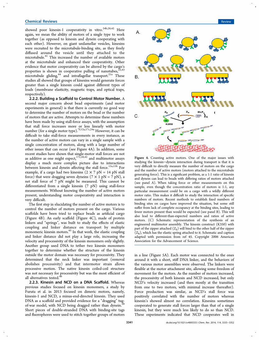
second major concern about bead experiments (and motorexperiments in general) is that there is currently no good wayto determine the number of motors on the bead or the numberof motors that are active. Attempts to determine these numbershave been made by using stall-force assays, with the assumptionthat stall force increases more or less linearly with motornumber (for a single motor type).7f,12a,17c,24a However, it can bedifficult to take stall-force measurements in every instance, asthe number of active motors can vary in a single sample with asingle concentration of motors, along with a large number ofother issues that can occur (see Figure 4A). In addition, somerecent studies have shown that single-motor stall forces are notas additive as one might expect,17c,28,42 and multimotor assaysdisplay a much more complex picture due to interactionsbetween kinesin and dynein affecting the stall force.12a,15b Forexample, if a cargo had two kinesins (2 × 7 pN = 14 pN stallforce) that were dragging seven dyneins (7 × 1 pN = 7 pN), anet stall force of 7 pN might be measured. This cannot bedifferentiated from a single kinesin (7 pN) using stall-forcemeasurements. Without knowing the number of active motorspresent, understanding motor cooperativity and interaction isvery difficult.The first step in elucidating the number of active motors is to
control the number of motors present on the cargo. Variousscaffolds have been tried to replace beads as artificial cargo(Figure 4B). An early scaffold (Figure 4C), made of proteinlinkers and “springs”, was built to study the effects of elasticcoupling and linker distance on transport by multiplemonomeric kinesin motors.45 In that work, the elastic couplingand linker distance did not play a large role, increasing thevelocity and processivity of the kinesin monomers only slightly.Another group used DNA to tether two kinesin monomerstogether to determine whether the structure of the kinesinoutside the motor domain was necessary for processivity. Theydetermined that the neck linker was important (removalabolishes processivity) and that intermotor strain allowsprocessive motion. The native kinesin coiled-coil structurewas not necessary for processivity but was the most efficient ofall alternatives tested.46
2.2.3. Kinesin and NCD on a DNA Scaffold. Whereasprevious studies focused on kinesin monomers, a study byFuruta et al. in 2013 focused on dimeric motors, namely,kinesin-1 and NCD, a minus-end-directed kinesin. They usedDNA as a scaffold and provided evidence for a “dragging” tug-of-war model, with NCD being dragged rather than dynein.28
Short pieces of double-stranded DNA with binding-site tagsand fluorophores were used to stitch together groups of motors
in a line (Figure 5A). Each motor was connected to the onesaround it with a short, stiff DNA linker, and the behaviors ofthe various motor assemblies were observed. The linkers wereflexible at the motor attachment site, allowing some freedom ofmovement for the motors. As the number of motors increased,the processivity of both kinesin and NCD increased, but onlyNCD’s velocity increased (and then mostly at the transitionfrom one to two motors, with minimal increase thereafter).Force production was similar, as NCD’s stall force waspositively correlated with the number of motors whereaskinesin’s showed almost no correlation. Kinesins sometimescooperated to generate stall forces larger than that of a singlekinesin, but they were much less likely to do so than NCD.These experiments indicated that NCD cooperates well in
Figure 4. Counting active motors. One of the major issues withstudying the kinesin−dynein interaction during transport is that it isvery difficult to directly measure the number of motors on the cargoand the number of active motors (motors attached to the microtubulegenerating force). This is a significant problem, as a 1:1 ratio of kinesinand dynein can lead to beads with differing ratios of motors attached(see panel A). When taking force or other measurements on thissample, even though the concentration ratio of motors is 1:1, anyparticular measurement could be on a cargo with a wildly differentmotor ratio. This makes it difficult to study the interaction of specificnumbers of motors. Recent methods to establish fixed numbers ofbinding sites on cargos have improved the situation, but some stillsuffer from lack of complete occupancy at the binding sites, leading tofewer motors present than would be expected (see panel B). This willalso lead to different-than-expected numbers and ratios of activemotors. (C) Schematic representation of the synthesis of anengineered multimotor assembly. The kinesin construct (K350) withpart of the zipper attached (ZE) will bind to the other half of the zipper(ZR), which has the elastic spring attached to it. Schematic and captionadapted with permission from ref 45. Copyright 2006 AmericanAssociation for the Advancement of Science.
Chemical Reviews Review
dx.doi.org/10.1021/cr4005555 | Chem. Rev. 2014, 114, 3335−33523341

groups whereas kinesin gains little advantage from increasingmotor number.A final experiment by Furuta et al. tethered kinesin and NCD
together with the DNA scaffold previously used to coupletogether only kinesin or only NCD (Figure 5A). The ratios ofthe motors (kinesin/NCD) were changed, and the behaviors ofcoupled plus- and minus-end-directed motors were observed.28
These experiments showed that the velocity of plus-end-directed motion decreased with increasing numbers of NCDmotors, indicating that some sort of tug of war was going on,reducing kinesin’s velocity. It is interesting that NCD, as aweak, cooperative, minus-end-directed motor, behaved sosimilarly to mammalian dynein when coupled to kinesin.12a,26
This is evidence that motors with certain properties functiontogether better in tug-of-war situations, as NCD andmammalian dynein behave similarly and both display bidirec-tional motility when coupled with kinesin.2.2.4. Kinesin and Yeast Dynein on a DNA Origami
Scaffold. A final, more complex, but also more versatile,synthetic cargo also uses DNA, in the form of DNA“origami”.47 This allowed the construction of more or lessarbitrary structures that can be used to determine specificnumbers, types, and placements of binding sites on a singlecargo, up to 90 unique sites on the DNA constructdemonstrated in this article.11 In this technique, a large,cylindrical scaffold consisting of 12 helical pieces of DNA wasthe synthetic cargo, and 21-base-pair DNA handles were left atspecific sites of the scaffold (Figure 5B). Then, appropriateantihandles could be attached to the motors of interest (in thiscase, kinesin-1 and yeast dynein). The effects of increasingnumbers of kinesin and yeast dynein were observed, and runlength increased with motor number, whereas velocity did notchange (kinesin) or decreased (yeast dynein) with increasingnumber. In these experiments, mixed-motor ensembles ofkinesin and yeast dynein were routinely immobile; whenmotile, they moved more slowly than single-motor ensembles,
and yeast dynein in general “won” over (mammalian) kinesin interms of the direction of motility. This is very different from(mammalian) kinesin’s interaction with mammalian dynei-n.12a,b,13 Finally, the authors incorporated a photocleavablehandle that allowed severance of specific motors. They usedthis handle to detach one motor type or the other and showedthat the immobile cargos became mobile again after removal ofone of the motor types, directly demonstrating that a tug of warwas going on between the yeast dynein and the mammaliankinesin.11
2.2.5. Issues with DNA Scaffolds and Origami. There isa significant issue with the DNA origami technique, in that,currently, the binding sites available for each motor type are notcompletely filled. The authors saw about 80% occupancy ofeach site, leaving uncertainty in the actual number of motorspresent on the complex.11 This occupancy issue could be due tothe fact the authors attached DNA linkers to the motors (with aSNAP-tag), which were then annealed to the chassis. Thereforethe final step was the binding of an oligonucleotide alreadyattached to the motor. When using kinesin and NCD heldtogether by a DNA scaffold, however, Furuta et al.28 annealedthe whole scaffold together and then attached the motors withSNAP-tags, which showed essentially 100% occupancy. Furutaet al. did have difficulty obtaining full occupancy withHaloTags, however. In addition, although the DNA for bothof these techniques is commercially available, it still requiresdesign and assembly, which appears to be quite complicated, inparticular for the larger origami structures.
2.2.6. Dynein’s Stall Force. Another important note aboutthe experiments by Derr et al.11 and other experiments is that,in vitro, single yeast dynein and single mammalian dynein havebeen shown to have very different behaviors. A single yeastdynein has been shown to be a slow, strong (7-pN stall force)motor in vitro,48 while a single mammalian dynein in vitro hasbeen measured to be weak (1−2 pN) and of a similar speed tomammalian kinesin.12a,b,15b,26,39 Mammalian dynein’s stall force
Figure 5. Synthetic DNA cargos. (A) Schematic representation of DNA−motor construction (not drawn to scale). The typical spacing betweenmotors is 22.7 nm, and the lengths of kinesin, SNAP-tag, and HaloTag are ∼17, 4.3, and 4.8 nm, respectively. Note that the DNA was fully ligatedand annealed before the motors were attached enzymatically with a HaloTag or SNAP-tag. By altering the number of each tag, the motor ratio couldbe controlled. Caption and figure adapted with permission from ref 28. Copyright 2006 American Association for the Advancement of Science. (B)Schematic of the 12-helix-bundle chassis structure with 6 inner and 6 outer helices. Each outer helix contains up to 15 optional handles, yielding 90uniquely addressable sites. Each handle consists of an unpaired 21-bp (∼7-nm) oligonucleotide sequence for hybridization to complementaryantihandle sequences covalently attached to motors or fluorophores. The inset shows an orthogonal cross section. The chassis is substantially largerand more complex than in the work of Furuta et al.,28 although it is more customizable. (C) Schematic of a chassis labeled with five fluorophores(red) at handle position 14 on each of five outer helices and dynein at handles at positions 1, 5, 9, and 13 on a single outer helix. Oligonucleotide-labeled dynein is also shown. Note that the motors are attached to a piece of single-stranded DNA through a SNAP-tag and then the DNA isattached to the chassis. Figure and caption adapted with permission from ref 11. Copyright 2012 American Association for the Advancement ofScience.
Chemical Reviews Review
dx.doi.org/10.1021/cr4005555 | Chem. Rev. 2014, 114, 3335−33523342
and behavior are disputed: Some contend it has a stall force of5−7 pN.49 A substantial comparison of mammalian and yeastdynein will not be undertaken here as it is outside the purviewof this review. However, we note that, in the experiments withyeast dynein and mammalian kinesin, it was routine for cargosto be stationary as the motors were engaged in a balanced tugof war, preventing motility.11 This might indicate that thesemotors’ properties are not well balanced for each other. This isin contrast to mammalian kinesin and mammalian dynein,which rarely stall as a result of a tug of war in vitro and appearto have complementary properties: Dynein is dragged bykinesin, but it is weaker and can cooperate in groups asopposed yeast dynein.12a,b,26,39
Conclusion: Synthetic cargos have shown that groups of onlykinesin motors can cooperate if they are attached to fluidmembranes that allow free motor diffusion.41 In addition,synthetic DNA cargos can be constructed that control thenumber of motors attached (along with allowing detachmentwith specific signals) and have clearly shown that a tug of waroccurs between kinesin and dynein.11,28 Interestingly, dyneinswith different properties (yeast vs mammalian) displayextremely different behaviors, indicating that motor properties(stall force, velocity, detachment force) play a significant role inregulating intracellular transport.
2.3. Cellular Organelles in Vitro: Examining Parts of theTransport Complex
Purifying intact organelles with the entire transport complexpresent and active and examining their behavior is another wayto observe transport behavior in a controlled environment.However, it is difficult to ensure that all factors involved intransport are present and functional. Nonetheless, evenorganelles with part of the transport complex can reveal usefulinformation about intracellular transport. Purified organelleshave shown tug-of-war behavior, and the viscoelasticity of thecellular cytoplasm has been found to have minimal effect onorganelle transport.7d,12a,b,50
The power of examining intact organelles can be seen in thatkinesin and cytoplasmic dynein were both discovered as themotors that powered directional transport using thistechnique.51 Organelles have long been purified and studiedto see which proteins will copurify with them, in an attempt todetermine what proteins are part of the transport complex. Oneof the first nonmotor parts of the transport complex identifiedwas dynactin, a separate protein from dynein but one thatevidently plays an important role in the transport complex. Itwas examined by in vitro motility assays.52 A more recentexample is in vitro studies of organelle fission properties. Forexample, studies on the transition from early- to late-endocyticvesicles have compared the motile properties of the twopopulations to characterize the difference in proteins present inthe transport complex.15a Similarly, Huntingtin protein hasbeen shown to be necessary for dynein-mediated transport invitro.53 Herpes simplex virus transport in vitro was shown to bepredominantly mediated by kinesin and associated with thetrans-Golgi network marker TGN46.54
Purified organelles can also display motile behavior verysimilar to that seen in the cell, with bidirectional, saltatorymotion.7d Some of the first evidence for a tug of war was shownusing purified organelles.23b That study showed that thepresence of kinesin determined directionality. Only plus-end-directed purified vesicles had kinesin present, whereas bothplus- and minus-end-directed vesicles had dynein. Furthermore,
when kinesin was inhibited, plus-end-directed vesicles uni-formly became minus-end-directed. The study saw no bidirec-tional movement, but clearly some sort of local tug of war orinteraction between kinesin and dynein was occurring, assimilar behavior occurred when the motors were bound tobeads.23b
Optical trapping of purified organelles has recently been usedto measure the behavior of the transport complex without theinterference of the highly viscoelastic and complex cellularenvironment. Organelles purified from a wide array of cells,including Dictyostelium,55 A549,12a and mouse macrophages,J774A.1,15b have been trapped. Different methods ofpurification led to different parts of the motor complex beingpresent; specifically, some of the harsher purification techniquesstripped away dynein,12a,b whereas others did not.12b Trappingof these organelles has shown that kinesin’s stall force (withdynein stripped away) is not affected by the remainingtransport complex, but kinesin and dynein together on acargo can interact in surprising ways, leading at times tostretching of the organelle and effectively reducing kinesin’sstall force.12a,b The stretching reveals that kinesin and dyneindo engage in a tug of war, as only kinesin and dynein pullingsimultaneously would be able to cause the stretching of theorganelle.
2.3.1. Effect of Viscoelasticity in a Cell. In addition, intwo independent studies, it was found that that there is littledifference in the forces exerted by purified organelles thatdisplay bidirectional motion in vitro and their behavior invivo.12a,b,15b Given that viscoelastic behavior is extremelydifferent in vitro and in vivo, this is a surprising result.(Viscoelastic moduli of a cell range from 102 to 105 dyn/cm,12a,15b,56 leading to viscoelasticities several orders ofmagnitude higher than that of water, which has a viscosity of1 cP and essentially no elasticity50,57). Apparently the highviscoelasticity of the cell has minimal effect on trans-port.12a,15b,50
The interplay of the components of the transport complexappears to be the major factor determining transport behavior(see also section 3.3). Organelles purified with only one motorbehave similarly to single-motor experiments. In contrast,having both motors present on purified cargos can lead tostretching of the cargo and a reduction in kinesin-driven stallforces. Because the simple addition of dynein causes stretchingof the cargo and reduces kinesin’s stall force, the motors mustbe engaging one another in a tug of war. When directionalmotion occurs with purified organelles, the lack of a differencebetween their in vivo and in vitro force behaviors also indicatesthat external factors to regulate motility are not necessary forshort-range bidirectional motility. The standardization ofpurification techniques58 and their increasing use is creating amiddle ground between in vivo and in vitro studies. Purifiedorganelles analyzed by in vitro techniques might be more suitedto exploring the exact components of the transport complexand, thus, lead to clearer conclusions than can be extractedfrom in vivo studies.Overall, the preponderance of in vitro data indicates that a
tug of war occurs between motors present on in vitrocargos.11,12,23b,28 Motors’ properties, particularly those ofdynein, are regulated by the presence of regulatory cofactors,and this regulation could potentially influence the outcome of atug of war.22,24c,27b,34,36 In addition, the outcome of the tug ofwar can be determined by the number of each motor typepresent and how well these motors can cooperate among
Chemical Reviews Review
dx.doi.org/10.1021/cr4005555 | Chem. Rev. 2014, 114, 3335−33523343
themselves.11−13,23b,26,28 Assays with motor-coated beads,12a,13
DNA scaffolds,11,28 and purified organelles7d,12a,b all supportthese conclusions.
3. TRANSPORT COMPLEX IN VIVO: EXAMININGORGANELLES IN LIVING CELLS
Observation of organelle motility in the living cell is a huge areawith decades of research behind it. A great variety of techniquesexist for observing organelle behavior, from basic lightmicroscopy, phase contrast and differential interferencecontrast (DIC), to modern fluorescence (total-internal-reflection fluorescence microscopy, TIRFM,59 and fluorescencerecovery after photobleaching, FRAP)60 and super-resolutiontechniques (stimulated emission depletion, STED,61 andstructured illumination microscopy, SIM).62 In vivo opticaltrapping studies have shown that a tug of war is likelyoccurring7d,12a,b but have also revealed that higher-ordermechanisms must exist to regulate and coordinate motorbehavior in the cell.7f,20
3.1. Tracking Organelles in Living Cells
Recent discoveries in the area of kinesin−dynein interactions inliving cells have revealed a wide variety of behaviors acrossorganelle and cell types. How cells regulate directionality is oneof the major questions about bidirectional intracellulartransport, and the experiments discussed in this section coverthe wide variety of mechanisms observed in the cell: motorconcentration, opposite polarity motors, myosin, regulatoryfactors, and external signaling. Many of these mechanismsmight be unique to a specific cellular system, whereas othersmight be more broadly applied.3.1.1. Motor Concentration Regulates Tugs of War.
During endosome transport in the fungus Ustilago maydis,kinesin-1, kinesin-3, and dynein, have been shown to exhibit acomplex interaction that alternates between cooperation andcompetition.18b Dynein dominates transport near the cell tip,but kinesin-3 takes over outside the cell tip for long-rangetransport, necessitating a cooperative “hand-off” between themotors for transport across the cell.18b However, near the plusends of microtubules, motion driven by kinesin-3 toward theplus end appears to be stopped by a high dynein concentration.The high dynein concentration eventually overwhelms kinesin-3 and prevents it from running endosomes off the plus end. Asonly minus-end-directed endosomes had dynein present,whereas both plus- and minus-end-directed endosomes hadkinesin-3, the presence of dynein apparently regulatesdirectionality in this system.63 This high concentration ofdynein is maintained by dynein transport to the plus end of themicrotubule by kinesin-1.64 This supports a model in which thecell regulates the dynein concentration, thereby regulatingoverall motion. Locally, at the cargo, the decision of whichdirection to travel is driven by a tug of war in which net force ormotor number is intermittently tested to determine direction-ality.3.1.2. Opposite Polarity Motors Are Both Required for
in Vivo Transport. In Drosophila S2 cells, it has been shownthat opposite-polarity motors must both be present on certainorganelles for motility to occur.65 However, it apparently doesnot matter what the specific motor is.65b During the tracking offluorescently labeled peroxisomes (GFP was targeted to theorganelles), it was observed that knocking out kinesin-1 orcytoplasmic dynein caused all motility to stop. Knocking outkinesin-1 and replacing it with kinesin-3 (specifically unc104
from Caenorhabditis elegans, in this case) restored motility.65b
Similarly, replacing dynein with NCD, a minus-end-directedkinesin, would restore motility. (In this case, motility washeavily plus-end-biased, as NCD has different properties thandynein.) If kinesin or dynein was replaced by a nonmotilemutant, motility would not resume. This indicates that activeplus- and minus-end-directed motors are simultaneouslyrequired during intracellular transport.65b What this means isunclear. It could indicate that there is a coordinating complexthat requires the presence of both motors to function.
3.1.3. Kinesin and Dynein Interact to BypassObstacles. Another potential reason for the simultaneouspresence of kinesin and dynein on many cargos is that theymight aid in bypassing obstacles on microtubules, such as tauprotein patches and microtubule intersections.1b,9a,66 Researchon nuclear migration in C. elegans has indicated that, eventhough kinesin is the driver for nuclear movement, the deletionof dynein seriously slows and impairs transport, likely becauseof the inability to back the nucleus up to bypass obstacles.67
Also, kinesin and dynein are differentially regulated by amicrotubule-associated protein, tau. Kinesin generally detachesin the presence of tau-decorated microtubules, whereas dyneinreverses at patches of tau. This indicates that dynein andkinesin interact to allow more efficient intracellular transportand could explain why knocking out one motor stops transportin all directions.
3.1.4. Myosin Can Modulate Microtubule-BasedTransport. Myosin is often also present on organelles withkinesin and dynein, and its interaction with microtubule-basedtransport is an open question. Mitochondrial motility onmicrotubules in Drosophila neurons appears to be inhibited bymyosin V in both directions and by myosin VI duringretrograde transport, whereas myosin II has no effect onmicrotubule-based transport. This appears to indicate thatmyosin inhibits long-range microtubule-based motility andmight aid organelle docking and pausing.68 Myosin’s effect ontransport is still unclear, but it could have an override effect onkinesin−dynein tugs of war, with myosin perhaps supersedingkinesin and dynein to allow switching to actin-based transport.
3.1.5. Regulatory Factors Modulate Transport. A levelof complexity typically present only in vivo is externalregulatory factors, such as phosphatases and kinases. Forexample, in the Xenopus laevis melanophore system, melano-phores (pigment granules) can be dispersed or aggregated byexternal signals.69 Recently, it was shown that this control ofdirectionality is mediated by phosphorylation of the dyneinintermediate chain, where phosphorylation of dynein appa-rently stimulates its activity.70 This type of regulation has alsobeen seen in Huntington’s disease, as one of the downstreameffects of pathogenic huntingtin protein is kinesin phosphor-ylation, which appears to inhibit kinesin.71 In addition,phosphorylation of the scaffolding protein JIP-1 modulatesthe directionality of amyloid precursor protein (APP) motilityin mouse axons. JIP-1 phosphorylation may fully coordinateAPP motility, as experiments indicated that kinesin and dyneinwere not simultaneously active in this system.22,72
3.1.6. Controlling in Vivo Motility with ExternalSignals. An exciting new method for studying intracellulartransport is the creation of an in vivo transport system that canbe induced to move upon addition of an external ligand. Thisallows much greater control of in vivo transport and controlover the motors attached to the tracked organelle. This createsan artificial system where the scientist has control over an
Chemical Reviews Review
dx.doi.org/10.1021/cr4005555 | Chem. Rev. 2014, 114, 3335−33523344
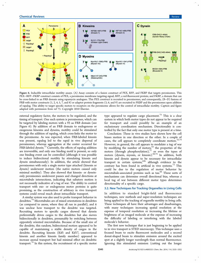
external regulatory factor, the motors to be regulated, and thetiming of transport. One such system is peroxisomes, which canbe targeted by labeling motors with a 93 aa FRB domain (seeFigure 6). By addition of an FRB domain to endogenous orexogenous kinesins and dyneins, motility could be stimulatedthrough the addition of rapalog, which cross-links the motor tothe peroxisome. As was expected, when FRB-labeled kinesinwas present, rapalog led to the rapid in vivo dispersal ofperoxisomes, whereas aggregation at the center occurred forFRB-labeled dynein.73 Currently, the effects of rapalog additionare irreversible, and only one binding motif is present, so onlyone binding event can be controlled (although it was possibleto induce bidirectional motility by stimulating kinesin anddynein simultaneously). In addition, the article showed thatperoxisomes with only a single motor type attached (kinesin ordynein) underwent motion (the native motors caused onlyminimal motility). They also showed that kinesin- or dynein-only peroxisomes underwent pauses and changed directions atmicrotubule intersections, indicating that saltatory motion isnot necessarily indicative of a tug of war. The ability to controltransport with exo- or endogenous motor proteins is quitepromising, as the construction of arbitrary in vivo transportsystems could reveal much about intracellular transport.A similar system was also used to probe transport behavior in
dendrites.74 Microtubules are of mixed orientations in dendrites(as compared to axons, where they all run in parallel), and itwas unclear how transport to the dendrite was targeted.Recruiting motors to specific cargos showed that dyneinpreferentially drives cargos to the dendrites but also movesbidirectionally in dendrites, presumably by switching betweenoppositely oriented microtubules. Because of the small size ofthe dendrite, this random, bidirectional motion appeared to becapable of maintaining a stable density of cargos in thedendrite. Recruiting kinesin (Kif5 and Kif17, conventionalkinesin and another kinesin family member) appeared toincrease axonal transport but had minimal effect on dendritictransport.74 In this system, the recruitment of a specific motor
type appeared to regulate cargo placement.75 This is a clearsystem in which both motor types do not appear to be requiredfor transport and could possibly be an example of anexclusionary coordination mechanism. Directionality is con-trolled by the fact that only one motor type is present at a time.Conclusion: These in vivo studies have shown how the cell
biases motion in one direction or the other. In a couple ofcases, the cell appears to completely coordinate motion.22,74
However, in general, the cell appears to modulate a tug of warby modifying the number of motors,63 the properties of themotors (through phosphorylation),71 or even the types ofmotors (dynein, myosin, or kinesin).68,74 In addition, bothkinesin and dynein appear to be necessary for intracellulartransport in certain systems,65b although evidence to thecontrary has been found in artificial in vivo systems.73 Thiscould be due to the regulation of motor behavior bymicrotubule-associated proteins such as tau.66 These sorts ofmechanisms can determine overall directional bias, whereas alocal tug of war between different motor types determinesdirectionality of a specific cargo.
3.2. New Techniques for Tracking Organelles in Living Cells
In addition to standard bright-field and fluorescencetechniques, new methods and super-resolution techniques arebeing applied to the tracking of organelle motility in living cells.These techniques all have their advantages and disadvantages,with many techniques increasing spatial resolution at theexpense of temporal resolution or increasing the lifetime orbrightness of an imaged molecule at the expense of increasingthe difficulty of labeling or interfering with the labeledmolecule’s behavior.The first new technique that is just beginning to be applied
to in vivo transport is STED microscopy. This technique uses afocused beam to excite fluorescent molecules and a seconddonut-shaped beam to stimulate emission around the excitedspot at a slightly longer wavelength than normal fluorescence.Ignoring this stimulated emission (cutting out the longer
Figure 6. Inducible intracellular motility assays. (A) Assay consists of a fusion construct of PEX, RFP, and FKBP that targets peroxisomes. ThisPEX−RFP−FKBP construct consists of PEX, a peroxisome membrane targeting signal; RFP, a red-fluorescent protein; and FKBP, a domain that canbe cross-linked to an FRB domain using rapamycin analogues. This PEX construct is recruited to peroxisomes, and consequently, (B−D) fusions ofFRB with motor constructs (1, 2, 4, 5, 7, and 8) or adaptor protein fragments (3, 6, and 9) are recruited to FKBP and the peroxisomes upon additionof rapalog. This ability to target specific motors to receptors on the peroxisome allows for the control of intracellular motility. Caption and figureadapted with permission from ref 73. Copyright 2010 Elsevier.
Chemical Reviews Review
dx.doi.org/10.1021/cr4005555 | Chem. Rev. 2014, 114, 3335−33523345
wavelength) allows the size of the fluorescent spot to bereduced, yielding subdiffraction spatial information.76 Thistechnique has been used to image several organelle types incellular areas smaller than the diffraction limit. In particular, ithas imaged synaptic vesicle motility in synaptic boutons, areasso small that typical wide-field techniques have troubleaccurately tracking organelles.77 Synaptic vesicles were mostlydiffusive, but there was a strong flux of vesicles through theaxon before reaching the synaptic bouton. The small area beingimaged allowed this scanning technique to image vesicledynamics with a reasonable time resolution (18 Hz). Theresolution, however, is still somewhat limited because of thephotostability of dyes, leading to a 50−100-nm resolution.77
Another subdiffraction imaging technique is structuredillumination microscopy (SIM), with a resolution of about100 nm.78 A nonlinear version of the technique, known assaturated SIM, has theoretically unlimited resolution, althoughonly 50-nm resolution has been achieved.79 SIM uses patternedillumination light to transfer information from high spatialfrequencies past the diffraction limit to lower spatial frequencieswithin the limit. This technique requires computationalprocessing after image acquisition, as well as multiple imagesof a wide-field area under different illumination patterns.Although it only doubles the lateral resolution, it is moreamenable to vesicle tracking than STED, as it requires less timeto take a complete image. This technique has successfullytracked vesicles and kinesin-driven transport in retinal pigmentepithelium cells and Drosophila S2 cells.80 Saturated SIM,although providing higher resolution, requires more imagesunder alternating illumination patterns, thereby requiring moretime per complete image.79,81 It also requires saturation of thefluorophores being imaged, which quickly photobleaches mostfluorophores.Other new imaging techniques use novel labels, although
these labels tend to be large, generally 20−40 nm in diameter.Quantum dots or gold/silver nanoparticles are two ways toincrease brightness and reduce photobleaching. Gold nano-particles have been used in A549 cells to track endosomes atextremely high spatial and temporal resolution (1.5 nm and 25μs).82 This was achieved using dark-field imaging and aquadrant photodiode to record the nanoparticles’ images. Thistechnique was able to resolve both dynein and kinesin steps anddid not suffer from photobleaching.82a Similar work was doneearlier on quantum dots with a fast CCD camera instead of aquadrant photodiode.82b Upconverting nanoparticles (UCNPs)also have been shown to display excellent properties for cellulartracking, as they do not photobleach, they require minimalexcitation power, and they are minimally cytotoxic. In addition,because UCNPs upconvert two long-wavelength photons, theexcitation causes minimal cellular autofluorescence.83 Anothertechnique, bFIONA (bright-field imaging with 1-nm accuracy),uses bright-field imaging of highly absorbing particles. It wasdemonstrated on melanophores, which contain dark melanin-containing vesicles.17d By saturating the CCD everywhereexcept the melanophore, it leaves an organelle-sized spot thatcan be fit with a 2D Gaussian, creating the opposite situation totypical FIONA (fluorescence imaging with 1-nm accuracy).29
This allows highly accurate fits to organelles and will notphotobleach, although it is limited to highly light-absorbentorganelles.Conclusion: These new techniques have allowed imaging of
in vivo intracellular transport at higher resolution and for longerperiods of time.17d,82 In addition, they have imaged in vivo
motor stepping, revealing that motors step similarly in vivo asthey do in vitro.82 All of these techniques, however, involveserious tradeoffs. Both STED and SIM trade temporalresolution for spatial resolution. STED, being a scanningtechnique, allows high speed (full frame rate of 28 Hz)scanning of small areas at high spatial resolutions (50 nm), butit is fairly complex to setup and loses time resolution as onescans larger areas, similar to confocal microscopy.76,77 SIM, onthe other hand, can take large wide-field images at the same rateas smaller image areas, but it is limited to a doubling ofresolution and requires significant postprocessing.78 New labelscan increase photostability and brightness but come with theirown costs. Most are significantly larger than fluorophores(quantum dots, gold nanoparticles, UCNPs) and presentsignificant difficulties when labeling.82 bFIONA is inherentlylimited to absorbent particles (or organelles).17d
3.3. Optical Trapping in Vivo
Although one of the first uses of an optical trap was in a livingcell, when Ashkin et al. attempted to measure the forces exertedby mitochondria,84 over the next 20 years, trapping was rarelyused in the cell. This was because making quantitative forcemeasurements in the cell was extremely difficult becaue of thecomplex nature of the cellular environment; the initial articlemade many assumptions that might or might not have beenappropriate when taking in vivo measurements. In the cell, theviscous and elastic properties of the cytoplasm are unknownand can vary both temporally and spatially, makingexperimental results difficult to interpret. In contrast, in vitro,the spring constant of the trap (the stiffness) can be easilymeasured, largely because water’s viscous properties areconstant and well-known, yielding nanometer and subnan-ometer results with a millisecond or greater time resolution.85
3.3.1. Optical Trap Calibration. In the past few years, invivo optical trapping has been rejuvenated thanks to newcalibration techniques.7f,12a,b,15b,26,86 As a result, new questionsabout motor transport in the cell can now be answered.87 Thespring constant or stiffness (k) of an optical trap in a purelyviscous medium (i.e., an in vitro optical trap) can be calibratedusing a wide variety of methods. All of these methods measureessentially the same thing: the strength of the trap whencompared to a known force acting on it. This can be done bymeasuring the trap strength against viscous drag (drag method)or by measuring the trap strength against Brownian motion (byeither the equipartition method or the power spectrummethod).88 For a more thorough review of these techniques,see Neuman et al.85a
The most common technique used to calibrate traps in livingcells is to estimate the trap stiffness by measuring the index ofrefraction of the cellular cytoplasm, the index of refraction, andthe size of the trapped organelle and then to create a viscous, invitro environment that replicates these indices of refraction, andmeasure the trap stiffness in it.7f,19,26,86 In this index-of-refraction matched environment, standard in vitro calibrationtechniques are used. Then one creates an organelle size versusstiffness calibration in this in vitro environment. After that, thestiffness of the trap is assumed to be directly related to the sizeof the organelle being trapped, that is, trap stiffness is linearlyrelated to r, the radius of the trapped object (assumed to bespherical). This technique is more straightforward than theactual in vivo calibrations described later, but it suffers from thefact that it relies on many more assumptions. This type oftechnique and the in vivo calibration technique described below
Chemical Reviews Review
dx.doi.org/10.1021/cr4005555 | Chem. Rev. 2014, 114, 3335−33523346
appear to measure very similar trap stiffnesses, indicating thatthis technique might be sufficient for most measurements.Actual results (force measurements) are quite variable betweendiffering calibrations and systems, however, so it is difficult todetermine how well various techniques work relative to eachother.For fully in vivo optical trap calibration, the assumptions
going into a calibration must be minimized. There are twodifferent in vivo calibration techniques that were developed andtested in vitro and in vivo15b,89 and are based on the fluctuationdissipation theorem (FDT). Compared to in vitro techniques,they make minimal assumptions about the environment. Theminimal assumptions are as follows: The trapped particle’senvironment must be locally homogeneous, and the responseof the system to a small-applied force must be the same as itsresponse to a similar spontaneous thermal force. Thesetechniques require more complex setups and analysis, but inreturn, they allow trap calibration in situ, calibrating the trap oneach organelle being observed. In addition, these techniquesactively measure the local viscoelasticity.The fundamental issue in vivo is that the trap is in an
environment with three variables: the environment’s elasticity,the environment’s viscosity, and the trap’s stiffness (effectively,a second form of elasticity). Both techniques take threemeasurements, one from pasive observation of the trappedobject and two from actively applying force to the trappedobject: the trapped object’s oscillation in-phase and out-of-phase relative to the trap’s oscillation. The passive measures thecombination of the trap’s damping and environment’s dampingof the organelle’s thermal vibration. The active measures thetrap’s ability to apply force against the local environment. Theratio of the in-phase and out-of-phase measurementscorresponds to the relative strengths of the environment’selasticity and viscosity versus the trap’s stiffness. Because thereare three measurements and variables, all three variables can bedetermined. Therefore, the trap stiffness can be extracted.The method developed by Hendricks et al.15b is very similar
to the FDT method,89a with the main differences being in someslightly different mathematical terminology and transformationsand the fact that Hendricks et al. performed global fits to theactive (or forced) and passive (or thermal) spectra. Fisher et al.applied their equations individually at each frequency. Theactual calibration was very similar, with a measurement of theresponse to an applied force by moving the trapping laser orstage and a measurement of the passive response of the systemto thermal fluctuations. These in vivo calibration techniquesdiffer from in vitro methods mainly in adding one extra step forthe in vivo case, namely, the active calibration, where theresponse of the trapped object to the trap’s applied force istested. This extra measurement allows the measurement of thelocal viscoelasticity of the trapped object. This then allows theremoval of the effect of the local environment on the trap andthe calibration of the trap’s stiffness.3.3.2. In Vivo Optical Trapping Results. The wide
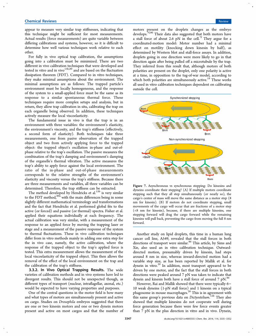
varieties of calibration methods and in vivo systems have led todivergent results. This should not be unexpected though, asdifferent types of transport (nuclear, intraflagellar, axonal, etc.)would be expected to have varying properties and purposes.One of the central questions in the motor field is how many
and what types of motors are simultaneously present and activeon cargo. Studies on Drosophila embryos suggested that thereare one or two kinesin motors and one or two dynein motorspresent and active on most cargos and that the number of
motors present on the droplets changes as the embryodevelops.7f,90 Their data also suggested that both motors havea stall force of about 2.6 pN in the cell.7f They argue for acoordinated-motion model: Motor number had a minimaleffect on motility (knocking down kinesin by half), asdetermined by Western blot and stall-force assays. In addition,droplets going in one direction were more likely to go in thatdirection again after being pulled off a microtubule by the trap.They inferred from this result that, although motors of bothpolarities are present on the droplet, only one polarity is activeat a time, in opposition to the tug-of-war model, according towhich both polarities are simultaneously active.19 These worksall used in vitro calibration techniques dependent on calibratingoutside the cell.
Another study on lipid droplets, this time in a human lungcancer cell line, A549, revealed that the stall forces in bothdirections of transport were similar.86 This article, by Sims andXie, also used an in vitro calibration technique. Outward-directed motion, presumably driven by kinesin, had stepsaround 8 nm in size, whereas inward-directed motion had avariable step size, as has been reported by Mallik et al. fordynein in vitro.39 In addition, most transport appeared to bedriven by one motor, and the fact that the stall forces in bothdirections were peaked around 7 pN was taken to indicate thatdynein and kinesin both have a stall force of around 7 pN.86
However, Rai and Mallik showed that there were typically 6−10 weak dyneins (1-pN stall force) and 1 kinesin on a typicalphagosome in mouse macrophages.26 This is in agreement withthis same group’s previous data on Dictyostelium.12b They alsoshowed that multiple kinesins do not cooperate well duringforce generation because there were few force events greaterthan 7 pN in the plus direction in vitro and in vivo. Dynein,
Figure 7. Asynchronous vs synchronous stepping: Do kinesins anddyneins coordinate their stepping? (A) If multiple motors coordinatestepping such that they all step simultaneously (or nearly so), thecargo’s center of mass will move the same distance as a motor step (8nm for kinesin). (B) If motors do not coordinate stepping, smallmovements of the cargo will occur that are fractions of a motor step(<8 nm for kinesin), because, if there are multiple kinesins, onestepping forward will drag the cargo forward while the remainingkinesins will pull back, preventing the cargo from moving the full 8 nmforward.
Chemical Reviews Review
dx.doi.org/10.1021/cr4005555 | Chem. Rev. 2014, 114, 3335−33523347

however, appears to be excellent at collectively generating forceand appears to form a catch bond (stronger bond as the loadincreases) to the microtubule under high opposing forces,making it unlikely to detach.35 Catch-bond behavior was alsoseen in Drosophila embryos.19
Dynein’s catch-bond behavior was shown by pulling back ongroups of dyneins attached to organelles in the cell. The largerthe stall force of the cargo (i.e., the more dyneins attached), thelonger the dyneins would hold onto the microtubule beforerelease under a superstall force (a force pulling back greaterthan that required to stall dynein-driven motion). They alsoobserved dynein’s variable step size in the cell and kinesin’s 8-nm step size.26 This group calibrated outside the cell. Althoughcatch-bond behavior might look like it would prevent dyneinfrom being dragged backward by kinesin, (dynein-drags model;Figure 8E), it is important to note that the behaviordemonstrated here is not what would be expected for anorganelle undergoing a tug of war. In the experiments in whichcatch-bond behavior was observed, a vesicle was moving in thedynein-driven direction when a sudden, large force was appliedto it; the vesicle was suddenly jerked backward against thedynein-driven motion. However, in a tug-of-war situation,kinesin would reattach during dynein-driven motion, and theamount of force applied against the dynein would slowly growas kinesin stepped away. This should not initiate catch-bondbehavior. Catch-bond behavior would prevent dynein detach-ment in the case of sudden, large forces and would aid inpreventing organelle detachment, but it would not be expectedto be significant during typical tug-of-war behavior.
Two groups have also demonstrated optical trap calibrationin living cells.12a,15b In these studies, all calibration was done inthe cell, accounting for any effects of a viscoelastic environment.Hendricks et al. examined phagocytosed latex beads in a mousemacrophage line, J774A.1.15b They discovered that inward- andoutward-directed forces were nearly balanced and found that, athigh loads (>10 pN), 8-nm stepping occurred, indicating thatteams of motors (dyneins, kinesins, or dyneins and kinesins)had correlated stepping (Figure 7).The other work, by Blehm et al., compared inward and
outward forces exerted by lipid droplets in a human lung cancercell line (A549), phagocytosed polystyrene beads in Dictyoste-lium, and dynein- and kinesin-coated beads in vitro.12a Thisarticle indicated that there were typically several dyneins andgenerally one kinesin on most cargos. This was concludedbecause the stall force in the minus direction was typicallygreater than 1 pN: Because a single dynein was shown to have astall force ∼1 pN in vitro, several dyneins were necessary togenerate this large force (Figure 8A,B). In the positivedirection, the stall force was rarely greater than 6 pN, andhence, only one kinesin was pulling at a time (Figure 8C).Blehm et al. also found that the plus-end-directed stalls in the
cell (i.e., outward or presumably kinesin-driven) looked verysimilar to the in vitro plus-end-directed stalls for kinesin- anddynein-coated beads (Figure 8D). In particular, a range of stallforces from 1 to 7 pN was found, equal to or more often lessthan the 7-pN in vitro stall force of a kinesin alone (Figure 8C).This finding is important, as it indicates that kinesin’s stall forcewas reduced by the presence of dynein, not by some other
Figure 8. Dynein drags. (A) In vitro (red) beads with a single dynein have a lower stall force than minus-end-directed lipid droplets in vivo (blue).(B) Beads coated with multiple dynein in vitro have stall forces similar to those of minus-end-directed lipid droplets in vivo. (C) A single kinesin ona bead in vitro has a narrower and higher stall-force distribution than plus-end-directed lipid droplets in vivo. (D) In vitro, beads coated with kinesinand dynein stall at similar forces while walking toward the microtubule plus end as lipid droplets and phagosomes in vivo (less than the stall force ofa single kinesin).12a (E) Dynein being dragged by kinesin during plus-end-directed motion appears to be the simplest explanation for why the beadswith kinesin and dynein in vitro and organelles in vivo have reduced plus-end-directed stall forces. Adapted with permssion from ref 12a. Copyright2013 National Academy of Sciences.
Chemical Reviews Review
dx.doi.org/10.1021/cr4005555 | Chem. Rev. 2014, 114, 3335−33523348

effect of the cell. This suggests that dynein reduces kinesin’sstall force, perhaps by remaining attached to the microtubuleduring plus-end-directed motion and acting as a drag (Figure8E). This behavior could potentially help bypass obstacles,allowing for quicker directional switching or stronger attach-ment to the microtubule if kinesin detaches. It could alsosimply be the most efficient way for these two motors tointeract to implement a tug of war. Data from both the Blehmet al. and Hendricks et al. articles support a weak 1-pN dyneinand a strong 7-pN kinesin in vivo.12a,15b They also stronglysupport a tug-of-war model, with cooperative dynein working ina group against a single kinesin (or perhaps two kinesins)during intracellular transport.In contrast, Laib et al. measured extremely high in vivo stall
forces in an intraflagellar transport (IFT) system, indicatinglarge, coordinated groups of kinesins or dyneins. They bound amicrosphere to the outside of Chlamydomonas’ flagella byattaching the sphere to a transmembrane protein undergoingIFT (Figure 9).21a They could then measure the force themolecular motors inside the cell were exerting, even though thebead was outside the cell. This allowed typical in vitro stiffnesscalibration techniques to work.21a This system appears to be
quite different than most others studied, as the forces measuredwere extremely high, around 60 pN in both directions. Thisindicates upward of 10 motors cooperating during transportassuming a stall force similar to kinesin’s, 5−7 pN. Using thesame technique and system, Shih et al. also saw similarbehavior, with a somewhat lower peak stall force of 20−30pN.21b The reason for the difference in peak forces betweenthese articles was unclear but could potentially be due to themicrosphere being nonspecifically bound in the Laib et al. work,whereas Shih et al. specifically bound the bead with anantibody. In addition, temperature-sensitive kinesin-2 anddynein-1b were used to measure the effects of each motor ontransport. When kinesin-2 was knocked out, dynein-drivenmotion continued unaffected, with the typical pauses thataccompanied two-motor transport still occurring.21 Similarbehavior was observed with dynein-1b.21b This was taken asevidence for reciprocal coordination as opposed to a tug-of-warmodel; in a tug-of-war model, knocking out kinesin should haveimproved dynein-driven transport and removed most saltations,and vice versa.21a
The Shih et al.21b and the Laib et al.21a articles seem to showthat there are perhaps many ways in which kinesin and dyneincan interact. That kinesin (and dynein) could yield up to 60 pNof force indicates excellent cooperativity in their systems, inaddition to coordination between the two motors to preventthem from interfering with each other. Other systems seem toindicate poor kinesin cooperativity and rarely if ever saw motorcooperation on this scale.Conclusion: From the wide variety of in vivo optical trapping
results, the different kinesin−dynein transport systems presentquite a bit of complexity. All of the different techniques used toobserve and measure force in these in vivo systems might ormight not be entirely compatible. In addition to differingcalibration techniques, some groups looked at maximumforce,15b,86 some looked at stalls,7f,12a,39 and some looked atescape forces (the minimum force required to escape thetrap).12b Each of these methods has its merits, as does eachcellular system, but it will be difficult to compare and contrastthese systems until more data are collected, so it can beunderstood how and why these systems act so differently. Itmight be expected that transport systems with differentfunctions and purposes, for example, intraflagellar transport21a
as opposed to neuronal transport, would have differentcharacteristics, which could explain much of the variabilityencountered in different systems.
4. CONCLUSIONSRecent interest in how kinesin and dynein interact duringbidirectional motion has led to many new systems andtechniques that can be used to explore this interaction. Newdata and theoretical works have also led to much more nuancedmodels of the kinesin−dynein interaction, with evidence forlocal directional regulation (individual organelles) being mainlythrough tug of war between motors, whereas intermediate- andlarge-scale regulation (regional and whole cell) are dominatedby other factors (motor number, phosphorylation state,microtubule modifications, etc.).Motors also seem to be somewhat specialized in working
with each other, in that mammalian kinesin and dynein appearto have characteristics allowing them to work together whensimultaneously present on a cargo, or at least not interfere witheach other to the point of preventing transport.7d,12b,15b Dyneinapparently is dragged behind kinesin during some plus-end-
Figure 9. Schematic of the assay developed by Laib et al.21a for thestudy of the transport of FMG-1-bound beads on the surface of aflagellum. The flagellum of a single Chlamydomonas cell is immobilizedon a coverslip, and a polystyrene bead held in an optical trap islowered onto the flagellum. A cluster of FMG-1 proteins on theflagellar surface is believed to bind to the bead (red box; enlargedregion). Back-and-forth surface motion of the bead thus representsFMG-1 motion driven by opposing microtubule motors (cytoplasmicdynein 1b and kinesin 2; arrows indicate directions of motion) withinthe flagellum. The optical trap exerts a controlled force (“load”) on thebead (and connected motors), directed toward the trap center andproportional to the bead’s displacement. Thus, the displacement isused to determine the force produced by the motors. Caption andfigure reprinted with permission from ref 23a. Copyright 2009Elsevier.
Chemical Reviews Review
dx.doi.org/10.1021/cr4005555 | Chem. Rev. 2014, 114, 3335−33523349

directed transport, whereas kinesin routinely releases to allowunhindered minus-end-directed transport.7d,12a This behaviorhas also been shown with mammalian NCD and kinesin.28 Thisis in contrast to yeast dynein and human kinesin,11 which seemto actively work against each other, leading to stalled cargos.Compatible motors seem to be able to coordinate with eachother naturally through a local tug of war,7d with weak,cooperative dyneins acting as a foil to strong, weaklycooperative kinesin. Noncompatible motors (yeast dyneinand mammalian kinesin) act similarly to early tug-of-warpredictions in that both motors appear to be active and neithercan win out, leading to stalling and failed transport.11
Whereas evidence in a majority of systems investigated so farseems to point to a local tug of war to determine directionalityand motility behavior for individual cargos7d,10b,c,11,12,15b andgeneral transport bias being regulated by some higher signalingmechanism,7c,f,17a,b,18,23a,37a only a few systems have beenexamined. In particular, there are systems that seem to showstrong coordination (no tug of war occurring at all).Intraflagellar transport21 and amyloid precursor protein trans-port22 both displayed heavily coordinated motion: kinesin ordynein was active, but rarely, if ever, were they both activesimultaneously.More complex systems, such as systems with more motor
types present,68 or other specialized types of transport promiseto add more complex regulatory mechanisms. Intraflagellartransport systems have already shown this to be the case,displaying very different behavior than that seen so far in otherin vivo systems.18b,63,64
New techniques are being developed to count motor numberin vitro and observe organelle behavior in the living cell and invarious purified in vitro systems. As these techniques improveand more systems are explored, a more complete understandingof how the interplay between motor−motor interactions (thelocal tug of war) and cellular regulation (motor number,phosphorylation, signaling) controls bidirectional transport willbe uncovered.
AUTHOR INFORMATIONCorresponding Author
*E-mail: [email protected]
The authors declare no competing financial interest.
Biographies
Benjamin H. Blehm is currently a postdoctoral fellow at the NationalInstitutes of Health and previously received his Ph.D. at the Universityof Illinois, Urbana-Champaign. Current research interests include the
role of force and tension in mediating cell−microenvironmentinteractions and developing new optical trapping and imagingtechniques.
Paul R. Selvin is a Professor of Physics, Biophysics and Cell andDevelopmental Biology at the University of Illinois, Urbana-Champaign. He has developed many single-molecule fluorescencetechniques, as well as optical trap techniques, with the goal ofunderstanding molecular motors and, more recently, neuronalsynapses.
ACKNOWLEDGMENTSFunding for this work was provided by Grants NIH GM068625and NSF 0822613
REFERENCES(1) (a) Gross, S. P. Phys. Biol. 2004, 1, R1. (b) Welte, M. A. Curr.Biol. 2004, 14, R525.(2) Mallik, R.; Gross, S. P. Curr. Biol. 2004, 14, R971.(3) Schroer, T. A. Annu. Rev. Cell Dev. Biol. 2004, 20, 759.(4) (a) Hirokawa, N.; Noda, Y.; Tanaka, Y.; Niwa, S. Nat. Rev. Mol.Cell Biol. 2009, 10, 682. (b) Walker, R. A.; Salmon, E. D.; Endow, S. A.Nature 1990, 347, 780.(5) Barlan, K.; Rossow, M. J.; Gelfand, V. I. Curr. Opin. Cell Biol.2013, 25, 483.(6) Kamal, A.; Goldstein, L. S. Curr. Opin. Cell Biol. 2002, 14, 63.(7) (a) Jolly, A. L.; Gelfand, V. I. Biochem. Soc. Trans. 2011, 39, 1126.(b) Deacon, S. W.; Nascimento, A.; Serpinskaya, A. S.; Gelfand, V. I.Curr. Biol. 2005, 15, 459. (c) Gross, S. P.; Welte, M. A.; Block, S. M.;Wieschaus, E. F. J. Cell Biol. 2002, 156, 715. (d) Hendricks, A. G.;Perlson, E.; Ross, J. L.; Schroeder, H. W.; Tokito, M.; Holzbaur, E. L.F. Curr. Biol. 2010, 20, 697. (e) Murray, J. W.; Sarkar, S.; Wolkoff, A.W. Traffic 2008, 9, 833. (f) Shubeita, G. T.; Tran, S. L.; Xu, J.;Vershinin, M.; Cermelli, S.; Cotton, S. L.; Welte, M. A.; Gross, S. P.Cell 2008, 135, 1098.(8) Rebhun, L. I. J. Gen. Physiol. 1967, 50, 223.(9) (a) Ross, J. L.; Ali, M. Y.; Warshaw, D. M. Curr. Opin. Cell Biol.2008, 20, 41. (b) Lipowsky, R.; Klumpp, S. Physica A 2005, 352, 53.(10) (a) Berger, F.; Keller, C.; Klumpp, S.; Lipowsky, R. Phys. Rev.Lett. 2012, 108, 208101. (b) Muller, M. J. I.; Klumpp, S.; Lipowsky, R.Proc. Natl. Acad. Sci. U.S.A. 2008, 105, 4609. (c) Muller, M. J. I.;Klumpp, S.; Lipowsky, R. J. Stat. Phys. 2008, 133, 1059.(11) Derr, N.; Goodman, B.; Jungmann, R.; Leschziner, A.; Shih, W.;Reck-Peterson, S. Science 2012, 338, 662.(12) (a) Blehm, B. H.; Schroer, T. A.; Trybus, K. M.; Chemla, Y. R.;Selvin, P. R. Proc. Natl. Acad. Sci. U.S.A. 2013, 110, 3381. (b) Soppina,V.; Rai, A. K.; Ramaiya, A. J.; Barak, P.; Mallik, R. Proc. Natl. Acad. Sci.U.S.A. 2009, 106, 19381. (c) Vale, R. D.; Malik, F.; Brown, D. J. CellBiol. 1992, 119, 1589.(13) DeBerg, H. A.; Blehm, B. H.; Sheung, J.; Thompson, A. R.;Bookwalter, C. S.; Torabi, S. F.; Schroer, T. A.; Berger, C. L.; Lu, Y.;Trybus, K. M.; Selvin, P. R. J. Biol. Chem. 2013, 288, 32612.
Chemical Reviews Review
dx.doi.org/10.1021/cr4005555 | Chem. Rev. 2014, 114, 3335−33523350
(14) Zhang, Y. Phys. Rev. E: Stat. Phys., Plasmas, Fluids 2009, 79,061918.(15) (a) Bananis, E.; Nath, S.; Gordon, K.; Satir, P.; Stockert, R. J.;Murray, J. W.; Wolkoff, A. W. Mol. Biol. Cell 2004, 15, 3688.(b) Hendricks, A. G.; Holzbaur, E. L. F.; Goldman, Y. E. Proc. Natl.Acad. Sci. U.S.A. 2012, 109, 18447.(16) (a) Deacon, S. W.; Serpinskaya, A. S.; Vaughan, P. S.; Fanarraga,M. L.; Vernos, I.; Vaughan, K. T.; Gelfand, V. I. J. Cell Biol. 2003, 160,297. (b) Haghnia, M.; Cavalli, V.; Shah, S. B.; Schimmelpfeng, K.;Brusch, R.; Yang, G.; Herrera, C.; Pilling, A.; Goldstein, L. S. B. Mol.Biol. Cell 2007, 18, 2081.(17) (a) Rozdzial, M. M.; Haimo, L. T. J. Cell Biol. 1986, 103, 2755.(b) Sammak, P.; Adams, S.; Harootunian, A.; Schliwa, M.; Tsien, R. J.Cell Biol. 1992, 117, 57. (c) Vershinin, M.; Carter, B. C.; Razafsky, D.S.; King, S. J.; Gross, S. P. Proc. Natl. Acad. Sci. U.S.A. 2007, 104, 87.(d) Kural, C.; Serpinskaya, A. S.; Chou, Y. H.; Goldman, R. D.;Gelfand, V. I.; Selvin, P. R. Proc. Natl. Acad. Sci. U.S.A. 2007, 104,5378. (e) Nascimento, A. A.; Roland, J. T.; Gelfand, V. I. Annu. Rev.Cell Dev. Biol. 2003, 19, 469.(18) (a) Levi, V.; Serpinskaya, A. S.; Gratton, E.; Gelfand, V. Biophys.J. 2006, 90, 318. (b) Schuster, M.; Kilaru, S.; Fink, G.; Collemare, J.;Roger, Y.; Steinberg, G. Mol. Biol. Cell 2011, 22, 3645.(19) Leidel, C.; Longoria, R. A.; Gutierrez, F. M.; Shubeita, G. T.Biophys. J. 2012, 103, 492.(20) Kunwar, A.; Tripathy, S. K.; Xu, J.; Mattson, M. K.; Anand, P.;Sigua, R.; Vershinin, M.; McKenney, R. J.; Yu, C. C.; Mogilner, A.;Gross, S. P. Proc. Natl. Acad. Sci. U.S.A. 2011, 108, 18960.(21) (a) Laib, J. A.; Marin, J. A.; Bloodgood, R. A.; Guilford, W. H.Proc. Natl. Acad. Sci. U.S.A. 2009, 106, 3190. (b) Shih, S. M.; Engel, B.D.; Kocabas, F.; Bilyard, T.; Gennerich, A.; Marshall, W. F.; Yildiz, A.Elife 2013, 2, e00744.(22) Fu, M. M.; Holzbaur, E. L. F. J. Cell Biol. 2013, 202, 495.(23) (a) Mallik, R.; Gross, S. P. Curr. Biol. 2009, 19, R416.(b) Muresan, V.; Godek, C. P.; Reese, T. S.; Schnapp, B. J. J. Cell Biol.1996, 135, 383.(24) (a) Mallik, R.; Petrov, D.; Lex, S. A.; King, S. J.; Gross, S. P.Curr. Biol. 2005, 15, 2075. (b) Jamison, D. K.; Driver, J. W.; Diehl, M.R. J. Biol. Chem. 2012, 287, 3357. (c) Ross, J. L.; Wallace, K.; Shuman,H.; Goldman, Y. E.; Holzbaur, E. L. F. Nat. Cell Biol. 2006, 8, 562.(25) Campas, O.; Leduc, C.; Bassereau, P.; Casademunt, J.; Joanny,J.-F.; Prost, J. Biophys. J. 2008, 94, 5009.(26) Rai, A.; Rai, A.; Ramaiya, A.; Jha, R.; Mallik, R. Cell 2012, 152,172.(27) (a) Kardon, J. R.; Reck-Peterson, S. L.; Vale, R. D. Proc. Natl.Acad. Sci. U.S.A. 2009, 106, 5669. (b) King, S. J.; Schroer, T. A. Nat.Cell Biol. 2000, 2, 20.(28) Furuta, K.; Furuta, A.; Toyoshima, Y. Y.; Amino, M.; Oiwa, K.;Kojima, H. Proc. Natl. Acad. Sci. U.S.A. 2013, 110, 501.(29) Yildiz, A.; Tomishige, M.; Vale, R. D.; Selvin, P. R. Science 2004,303, 676.(30) Svoboda, K.; Block, S. M. Cell 1994, 77, 773.(31) Vale, R. D.; Milligan, R. A. Science 2000, 288, 88.(32) Pan, X.; Ou, G.; Civelekoglu-Scholey, G.; Blacque, O. E.;Endres, N. F.; Tao, L.; Mogilner, A.; Leroux, M. R.; Vale, R. D.;Scholey, J. M. J. Cell Biol. 2006, 174, 1035.(33) Snow, J. J.; Ou, G.; Gunnarson, A. L.; Walker, M. R.; Zhou, H.M.; Brust-Mascher, I.; Scholey, J. M. Nat. Cell Biol. 2004, 6, 1109.(34) Huang, J.; Roberts, A. J.; Leschziner, A. E.; Reck-Peterson, S. L.Cell 2012, 150, 975.(35) McKenney, R. J.; Vershinin, M.; Kunwar, A.; Vallee, R. B.;Gross, S. P. Cell 2010, 141, 304.(36) Kardon, J. R.; Vale, R. D. Nat. Rev. Mol. Cell Biol. 2009, 10, 854.(37) (a) Gross, S. P.; Welte, M. A.; Block, S. M.; Wieschaus, E. F. J.Cell Biol. 2000, 148, 945. (b) Block, S. M.; Goldstein, L. S.; Schnapp,B. J. Nature 1990, 348, 348.(38) Driver, J. W.; Jamison, D. K.; Uppulury, K.; Rogers, A. R.;Kolomeisky, A. B.; Diehl, M. R. Biophys. J. 2011, 101, 386.(39) Mallik, R.; Carter, B. C.; Lex, S. A.; King, S. J.; Gross, S. P.Nature 2004, 427, 649.
(40) Xu, J.; Shu, Z.; King, S. J.; Gross, S. P. Traffic 2012, 13, 9999.(41) Herold, C.; Leduc, C.; Stock, R.; Diez, S.; Schwille, P.ChemPhysChem 2012, 13, 1001.(42) Jamison, D. K.; Driver, J. W.; Rogers, A. R.; Constantinou, P. E.;Diehl, M. R. Biophys. J. 2010, 99, 2967.(43) Leduc, C.; Campas, O.; Zeldovich, K. B.; Roux, A.; Jolimaitre,P.; Bourel-Bonnet, L.; Goud, B.; Joanny, J.-F.; Bassereau, P.; Prost, J.Proc. Natl. Acad. Sci. U.S.A. 2004, 101, 17096.(44) Fallesen, T.; Macosko, J.; Holzwarth, G. Eur. Biophys. J. 2011,40, 1071.(45) Diehl, M. R.; Zhang, K.; Lee, H. J.; Tirrell, D. A. Science 2006,311, 1468.(46) Miyazono, Y.; Hayashi, M.; Karagiannis, P.; Harada, Y.;Tadakuma, H. EMBO J. 2009, 29, 93.(47) (a) Douglas, S. M.; Dietz, H.; Liedl, T.; Hogberg, B.; Graf, F.;Shih, W. M. Nature 2009, 459, 414. (b) Rothemund, P. W. K. Nature2006, 440, 297.(48) (a) Gennerich, A.; Carter, A. P.; Reck-Peterson, S. L.; Vale, R.D. Cell 2007, 131, 952. (b) Reck-Peterson, S. L.; Yildiz, A.; Carter, A.P.; Gennerich, A.; Zhang, N.; Vale, R. D. Cell 2006, 126, 335.(49) (a) Walter, W. J.; Brenner, B.; Steffen, W. J. Struct. Biol. 2010,170, 266. (b) Walter, W. J.; Koonce, M. P.; Brenner, B.; Steffen, W.Proc. Natl. Acad. Sci. U.S.A. 2012, 109, 5289. (c) Toba, S.; Watanabe,T. M.; Yamaguchi-Okimoto, L.; Toyoshima, Y. Y.; Higuchi, H. Proc.Natl. Acad. Sci. U.S.A. 2006, 103, 5741.(50) Holzwarth, G.; Bonin, K.; Hill, D. B. Biophys. J. 2002, 82, 1784.(51) (a) Schroer, T. A.; Schnapp, B. J.; Reese, T. S.; Sheetz, M. P. J.Cell Biol. 1988, 107, 1785. (b) Schroer, T. A.; Steuer, E. R.; Sheetz, M.P. Cell 1989, 56, 937. (c) Vale, R. D.; Reese, T. S.; Sheetz, M. P. Cell1985, 42, 39. (d) Vale, R. D.; Schnapp, B. J.; Mitchison, T.; Steuer, E.;Reese, T. S.; Sheetz, M. P. Cell 1985, 43, 623. (e) Dabora, S. L.;Sheetz, M. P. Cell Motil. Cytoskeleton 1988, 10, 482.(52) Gill, S. R.; Schroer, T. A.; Szilak, I.; Steuer, E. R.; Sheetz, M. P.;Cleveland, D. W. J. Cell Biol. 1991, 115, 1639.(53) Caviston, J. P.; Ross, J. L.; Antony, S. M.; Tokito, M.; Holzbaur,E. L. F. Proc. Natl. Acad. Sci. U.S.A. 2007, 104, 10045.(54) Lee, G. E.; Murray, J. W.; Wolkoff, A. W.; Wilson, D. W. J. Virol.2006, 80, 4264.(55) Bartsch, T. F.; Longoria, R.; Florin, E. L.; Shubeita, G. T.Biophys. J. 2013, 104, 475A.(56) Nam, W.; Epureanu, B. I. J. Phys.: Condens. Matter 2012, 24,375103.(57) Yamada, S.; Wirtz, D.; Kuo, S. C. Biophys. J. 2000, 78, 1736.(58) Dermietzel, R. The Cytoskeleton: Imaging, Isolation, AndInteraction; Humana Press/Springer: New York, 2013.(59) Ohara-Imaizumi, M.; Nishiwaki, C.; Kikuta, T.; Nagai, S.;Nakamichi, Y.; Nagamatsu, S. Biochem. J. 2004, 381, 13.(60) Smith, A. J.; Pfeiffer, J. R.; Zhang, J.; Martinez, A. M.; Griffiths,G. M.; Wilson, B. S. Traffic 2003, 4, 302.(61) Hein, B.; Willig, K. I.; Hell, S. W. Proc. Natl. Acad. Sci. U.S.A.2008, 105, 14271.(62) Shao, L.; Kner, P.; Rego, E. H.; Gustafsson, M. G. Nat. Methods2011, 8, 1044.(63) Schuster, M.; Lipowsky, R.; Assmann, M. A.; Lenz, P.; Steinberg,G. Proc. Natl. Acad. Sci. U.S.A. 2011, 108, 3618.(64) Steinberg, G. Curr. Opin. Microbiol. 2011, 14, 660.(65) (a) Ling, S. C.; Fahrner, P. S.; Greenough, W. T.; Gelfand, V. I.Proc. Natl. Acad. Sci. U.S.A. 2004, 101, 17428. (b) Ally, S.; Larson, A.G.; Barlan, K.; Rice, S. E.; Gelfand, V. I. J. Cell Biol. 2009, 187, 1071.(c) Kim, H.; Ling, S. C.; Rogers, G. C.; Kural, C.; Selvin, P. R.; Rogers,S. L.; Gelfand, V. I. J. Cell Biol. 2007, 176, 641.(66) Dixit, R.; Ross, J. L.; Goldman, Y. E.; Holzbaur, E. L. Science2008, 319, 1086.(67) Fridolfsson, H. N.; Starr, D. A. J. Cell Biol. 2010, 191, 115.(68) Pathak, D.; Sepp, K. J.; Hollenbeck, P. J. J. Neurosci. 2010, 30,8984.(69) Rogers, S. L.; Tint, I. S.; Fanapour, P. C.; Gelfand, V. I. Proc.Natl. Acad. Sci. U.S.A. 1997, 94, 3720.
Chemical Reviews Review
dx.doi.org/10.1021/cr4005555 | Chem. Rev. 2014, 114, 3335−33523351
(70) Ikeda, K.; Zhapparova, O.; Brodsky, I.; Semenova, I.; Tirnauer, J.S.; Zaliapin, I.; Rodionov, V. Mol. Biol. Cell 2011, 22, 1321.(71) Morfini, G. A.; You, Y. M.; Pollema, S. L.; Kaminska, A.; Liu, K.;Yoshioka, K.; Bjorkblom, B.; Coffey, E. T.; Bagnato, C.; Han, D.;Huang, C. F.; Banker, G.; Pigino, G.; Brady, S. T. Nat. Neurosci. 2009,12, 864.(72) Reis, G. F.; Yang, G.; Szpankowski, L.; Weaver, C.; Shah, S. B.;Robinson, J. T.; Hays, T. S.; Danuser, G.; Goldstein, L. S. Mol. Biol.Cell 2012, 23, 1700.(73) Kapitein, L. C.; Schlager, M. A.; van der Zwan, W. A.; Wulf, P.S.; Keijzer, N.; Hoogenraad, C. C. Biophys. J. 2010, 99, 2143.(74) Kapitein, L. C.; Schlager, M. A.; Kuijpers, M.; Wulf, P. S.; vanSpronsen, M.; MacKintosh, F. C.; Hoogenraad, C. C. Curr. Biol. 2010,20, 290.(75) Nakata, T.; Niwa, S.; Okada, Y.; Perez, F.; Hirokawa, N. J. CellBiol. 2011, 194, 245.(76) Klar, T. A.; Hell, S. W. Opt. Lett. 1999, 24, 954.(77) Westphal, V.; Rizzoli, S. O.; Lauterbach, M. A.; Kamin, D.; Jahn,R.; Hell, S. W. Science 2008, 320, 246.(78) Gustafsson, M. G. L. J. Microsc. 2000, 198, 82.(79) Gustafsson, M. G. Proc. Natl. Acad. Sci. U.S.A. 2005, 102, 13081.(80) (a) Ach, T.; Best, G.; Rossberger, S.; Heintzmann, R.; Cremer,C.; Dithmar, S. Br. J. Ophthalmol. 2012, 96, 1141. (b) Kner, P.; Chhun,B. B.; Griffis, E. R.; Winoto, L.; Gustafsson, M. G. Nat. Methods 2009,6, 339.(81) Rego, E. H.; Shao, L.; Macklin, J. J.; Winoto, L.; Johansson, G.A.; Kamps-Hughes, N.; Davidson, M. W.; Gustafsson, M. G. Proc. Natl.Acad. Sci. U.S.A. 2011, 109, E135.(82) (a) Nan, X.; Sims, P. A.; Xie, X. S. ChemPhysChem 2008, 9, 707.(b) Nan, X.; Sims, P. A.; Chen, P.; Xie, X. S. J. Phys. Chem. B 2005,109, 24220.(83) Nam, S. H.; Bae, Y. M.; Park, Y. I.; Kim, J. H.; Kim, H. M.; Choi,J. S.; Lee, K. T.; Hyeon, T.; Suh, Y. D. Angew. Chem., Int. Ed. 2011, 50,6093.(84) Ashkin, A.; Schutze, K.; Dziedzic, J. M.; Euteneuer, U.; Schliwa,M. Nature 1990, 348, 346.(85) (a) Neuman, K. C.; Block, S. M. Rev. Sci. Instrum. 2004, 75,2787. (b) Abbondanzieri, E. A.; Greenleaf, W. J.; Shaevitz, J. W.;Landick, R.; Block, S. M. Nature 2005, 438, 460. (c) Moffitt, J. R.;Chemla, Y. R.; Izhaky, D.; Bustamante, C. Proc. Natl. Acad. Sci. U.S.A.2006, 103, 9006.(86) Sims, P. A.; Xie, X. S. ChemPhysChem 2009, 10, 1511.(87) Oddershede, L. B. Nat. Chem. Biol. 2012, 8, 879.(88) Tolic-Nørrelykke, S. F.; Schaffer, E.; Howard, J.; Pavone, F. S.;Julicher, F.; Flyvbjerg, H. Rev. Sci. Instrum. 2006, 77, 103101.(89) (a) Fischer, M.; Berg-Sørensen, K. J. Opt. A: Pure Appl. Opt.2007, 9, S239. (b) Fischer, M.; Richardson, A. C.; Reihani, S. N.;Oddershede, L. B.; Berg-Sørensen, K. Rev. Sci. Instrum. 2010, 81,015103. (c) Mas, J.; Richardson, A. C.; Reihani, S. N.; Oddershede, L.B.; Berg-Sørensen, K. Phys. Biol. 2013, 10, 046006.(90) Welte, M. A.; Gross, S. P.; Postner, M.; Block, S. M.; Wieschaus,E. F. Cell 1998, 92, 547.
Chemical Reviews Review
dx.doi.org/10.1021/cr4005555 | Chem. Rev. 2014, 114, 3335−33523352