Embed Size (px)
Citation preview

Oncogenes
DR. JOSE MORDOH
Maestría Biología Molecular Médica – UBA 2016

What is the molecular basis of cancer?
Cancers are formed from repeated rounds of DNA mutation,
competition, and natural selection operating with the host.
-arise from a single abnormal cell
-abnormality results from somatic mutation
-development of cancer requires mutations in many cancer
critical genes
For a cancer cell to be successful the mutations must…
1.Allow the cells to disregard the external and internal signals
that regulate proliferation
2. Allow the cells to avoid apoptosis and escape programmed
limitations to proliferation including differentiation
3. Allow the cells to escape from their tissue of origin
4. Allow the cells to survive and proliferate in foreign sites
Cancer critical genes: oncogenes and tumor suppressors

1) ORIGEN DE LA CELULA TUMORAL
LAS TRES ETAPAS DEL CANCER


LOS ACELERADORES


RETROVIRUS LIBERADOS DE CELULA INFECTADA

DOGMA DE LA BIOLOGIA
EN LOS AÑOS 60
DNA
RNA
PROTEINA

PREGUNTA
• ¿Cómo es posible que un virus que
contenga RNA en su genoma se integre
en el DNA cromosómico de una célula?

• DAVID
BALTIMORE
• Premio Nobel de
Medicina 1975

• HOWARD TEMIN
• Premio Nobel de
Medicina 1975

Renato Dulbecco Premo Nobel de Medicina 1975

Discovery I. Tumor Viruses; RNA
Retrovirus: RNA genome reversed transcribed into proviral DNA
which integrates randomly into the host cell genome.
Productively infects only proliferating cells.
Peyton Rous:
1st evidence that viruses
could cause cancer (1911).
- Chickens
- fibrosarcoma
- Rous Sarcoma virus
- Nobel prize 1966

UNIVERSITY OF CALIFORNIA AT SAN FRANCISCO

• HAROLD VARMUS
• Premio Nobel de
Medicina 1989

J.MICHAEL BISHOP
Premio Nobel de
Medicina 1989


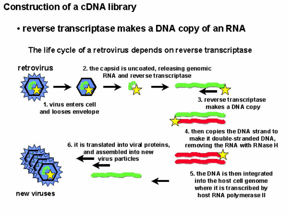
Southern Blots Probed with viral src Gene Revealed Cellular Origin of Oncogenes
Infected Infected Uninfected chicken
chicken #1 chicken #2 (Negative Control)
c-src
Proto-oncogene
SURPRISE!
v-src


Slow transforming retroviruses
Enhancer insertion
May be 5’ or 3’ in either orientation.
gag pol env LTR LTR
gag pol env LTR LTR
Promoter insertion
Proto-oncogene
Slow transforming retroviruses activate proto-oncogenes by
insertional mutagenesis.
Dysregulated expression occurs after insertion of strong
promoters or enhancers into the genetic loci.
cellular gene
provirus

An oncogene is:
Mutant or overactive form of a normal gene (normal gene is
referred to as a proto-oncogene)
A gene capable of inducing cancer.
Any gene which produces a “malignant phenotype”
when introduced into a “normal cell”.
A gene intimately associated with a particular
malignant disease such as a specific chimera in a particular
leukemia.

Oncogenes of Acutely Transforming
Retroviruses
src Rous sarcoma virus Chicken
myc Avian myelocytomatosis virus Chicken
erb A, erb B Avian erythroblastosis virus Chicken
myb Avian myeloblastosis virus Chicken
ets Avian erythroblastosis virus Chicken
rel Avian reticuloendotheliosis virus Turkey
H-ras Harvey rat sarcoma virus Rat
K-ras Kirsten murine sarcoma virus Mouse
abl Abelson murine leukemia virus Mouse
raf Murine sarcoma virus Mouse
fos Mouse osteosarcoma virus Mouse
fms Feline sarcoma virus Cat
fes Feline sarcoma virus Cat
sis Simian sarcoma virus Monkey
= Oncogenes of acutely transforming retroviruses important in human cancer


Robert Weinberg
Whitehead Institute-
MIT

Maestría en Biología Molecular Médica –
Dr. José Mordoh 2016

Discovery III. Identification of Oncogenes by
functional assays; *Transfection
DNA
Shear
mRNA cDNA
library
Transfect
Transfect
Transformed Phenotype
Tumor
Reporter Cell line


Some Oncogenes identified by Transfection
Weinberg- activated ras from bladder carcinoma.
Vande Woude- met oncogene which is hepatocyte growth factor
receptor from a chemically transformed cell line.
hst is a FGF-related gene identified from a human stomach
carcinoma.


LA GRAN SORPRESA !!!
V-ONC Y C-ONC SON IGUALES !!


FUNCION DE LOS PROTO-
ONCOGENES
• - Transductores de señales
• - Factores de transcripción
• - Receptores de factores de crecimiento
• - Factores de crecimiento
• - Reguladores de Apoptosis
• - Remodeladores de cromatina
• - miRNAs

NUCLEO
MEMBRANA CELULAR
Cáncer Normal


Mecanismos de Transformación de
Proto-oncogene a oncogene
1. Translocación
2. Amplificación
3. Inserción Viral
4. Mutagénesis
Maestría en Biología Molecular Médica –
Dr. José Mordoh 2016

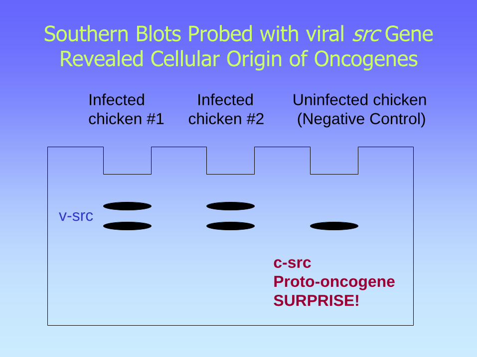
2- Traslocaciones cromosómicas: dos mecanismos - translocación que conduce a la sobreexpresión de un proto-oncogen: Ej: Linfoma de Burkitt c-myc de cromosoma 8 es traslocado al cromosoma 14 cerca del gen de cadena pesada de Ig, una región sujeta a gran actividad transcripcional, llevando a la sobreexpresión de la proteína myc normal. - Translocación y alteración genética de un proto-oncogen: Ej: Cromosoma Philadelphia en Leucemia Mieloide Crónica (CML) parte del gen abl (tirosin quinasa) en cromosoma 9 trasloca al cromosoma 22 para formar una proteína híbrida (quimera) con el gen bcr (breakpoint cluster region). La quimera
abl-bcr de 210 kDa tiene potente actividad tirosin quinasa constitutiva.
MECANISMOS DE ACTIVACION DE ONCOGENES

Mechanism of action: Oncogenes as signal
transducers
Growth Factors
v-sis (PDGF), int-1(WNT-1), int-2(FGF), hst, fgf-5
Growth Factors Receptors
v-erb-B (EGFR), v-fms (CSF-1R), v-kit (KIT)
Signal Transducers
v-ras, v-src, v-raf/mil, v-abl, v-mos, v-crk
Transcription Factors
v-ets, v-myc, v-myb, v-rel (NFkB), v-ski, v-erb-A (THR)
C
Y
T
O
P
L
A
S
M
EXTRACELLULAR
NUCLEUS


Enhancer cadena pesada Ig (Sen and Baltimore, Cell, 1986)


MYC River

MYC TRANSLOCADO RIO GANGES
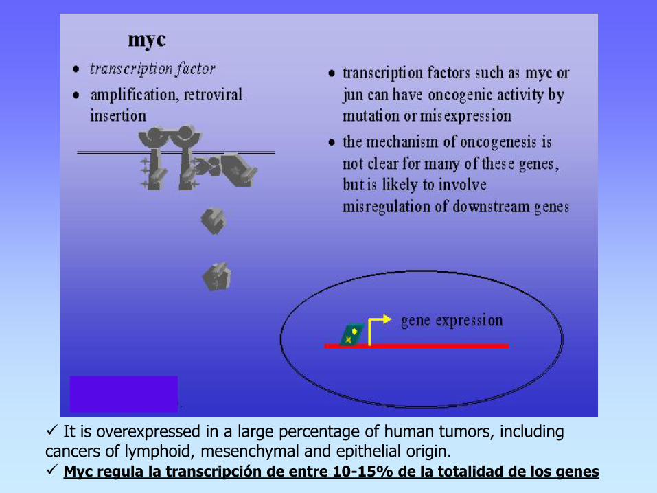
It is overexpressed in a large percentage of human tumors, including cancers of lymphoid, mesenchymal and epithelial origin. Myc regula la transcripción de entre 10-15% de la totalidad de los genes

Oncogenes and Signal Transduction: Transcription Factors-Myc
c-Myc plays a role in many human cancers; over-expression.
Translocations: c-myc and Ig genes
-Burkitt´s Lymphoma
-Low-grade follicular lymphomas (sometimes with BCL-2)
-Diffuse large cell lymphomas
Amplifications of c-myc
-Breast carcinoma
- Neuroblastoma (involves the related N-myc gene)
- Small cell lung cancer (involves the related L-myc gene)

c-Myc is an early response gene (Growth Factor Regulated)
GF
mRNA
Time 1 hr
Myc protein has very short
half-life <30 min.
Transcription regulates Myc
protein levels
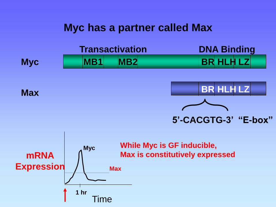
Myc has a partner called Max
BR HLH LZ MB1 MB2
BR HLH LZ
5’-CACGTG-3’ “E-box”
Transactivation DNA Binding
Myc
Max
mRNA
Expression
Time 1 hr
Myc
Max
While Myc is GF inducible,
Max is constitutively expressed


2- Traslocaciones cromosómicas: dos mecanismos - translocación que conduce a la sobreexpresión de un proto-oncogen: Ej: Linfoma de Burkitt c-myc de cromosoma 8 es traslocado al cromosoma 14 cerca del gen de cadena pesada de Ig, una región sujeta a gran actividad transcripcional, llevando a la sobreexpresión de la proteína myc normal. - Translocación y alteración genética de un proto-oncogen: Ej: Cromosoma Philadelphia en Leucemia Mieloide Crónica (CML) parte del gen abl (tirosin quinasa) en cromosoma 9 trasloca al cromosoma 22 para formar una proteína híbrida (quimera) con el gen bcr (breakpoint cluster region). La quimera
abl-bcr de 210 kDa tiene potente actividad tirosin quinasa constitutiva.
MECANISMOS DE ACTIVACION DE ONCOGENES

Identification of Oncogenes by mapping Chromosomal Rearrangements; description of
the philadelphia chromosome
1960: Nowell and Hungerford showed novel
chromosome in cells of CML patients.
Later termed the Philadelphia chromosome
(Ph1).
1973: Rowley identified the Ph1 chromosome as a t(9:22).
ID of oncogenes +
chomosomal mapping = ID of targets
(FISH) using unique-sequence double-fusion DNA probes for BCR (22q11.2) in red color
and c-abl (9q34) gene regions in green. The abnormal BCR/abl fusion present in positive
Philadelphia chromosome cells demonstrates the presence of yellow color (right panel)
compared to control (left panel) (used with permission, copyright, Emmanuel C. Besa, MD).


El protooncogen c-abl se fusiona con el gen bcr y el oncogen híbrido resultante bcr/c-abl es transcripcionalmente activo; el ciclo celular se desregula - se produce la leucemia mieloide crónica. Leucocitos únicos portadores del evento de translocación actuarían como origen
de la patología.
**-translocación recíproca entre crom. 9 y 22

Figure 1. The Translocation of t(9;22)(q34;q11) in CML. The Philadelphia (Ph) chromosome is a shortened
chromosome 22 that results from the translocation of 3' (toward the telomere) ABL segments on chromosome 9 to
5' BCR segments on chromosome 22. Breakpoints (arrowheads) on the ABL gene are located 5' (toward the
centromere) of exon a2 in most cases. Various breakpoint locations have been identified along the BCR gene on
chromosome 22. Depending on which breakpoints are involved, different-sized segments from BCR are fused with
the 3' sequences of the ABL gene. This results in fusion messenger RNA molecules (e1a2, b2a2, b3a2, and
e19a2) of different lengths that are transcribed into chimeric protein products (p190, p210, and p230) with variable
molecular weights and presumably variable function. The abbreviation m-bcr denotes minor breakpoint cluster
region, M-bcr major breakpoint cluster region, and µ-bcr a third breakpoint location in the BCR gene that is
downstream from the M-bcr region between exons e19 and e20.

Signaling pathways activated inBCR-ABL–positive cells. Note that this is a simplified diagram and that many more associations between Bcr-Abl and signaling proteins have been reported.
Deininger et al. BLOOD, 15 NOVEMBER 2000 z VOLUME 96, NUMBER 10

Mechanisms implicated in the pathogenesis of CML
Deininger et al. BLOOD, 15 NOVEMBER 2000 z VOLUME 96, NUMBER 10


MECANISMOS DE ACTIVACION DE ONCOGENES
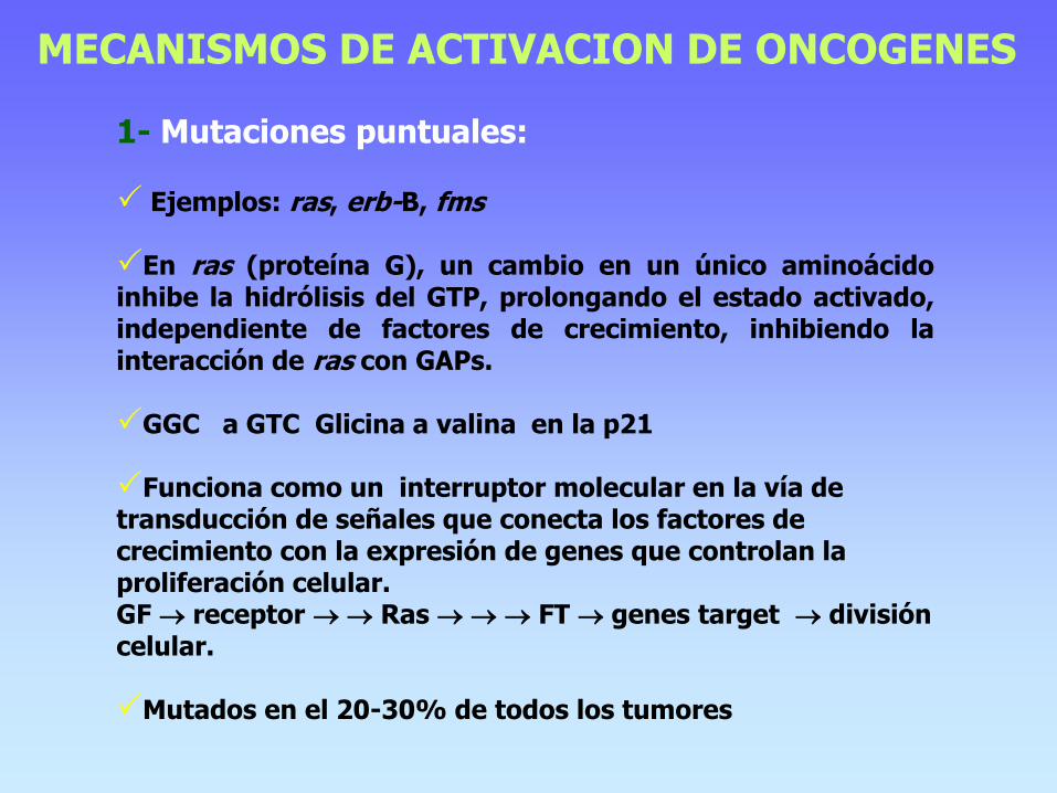
1- Mutaciones puntuales: Ejemplos: ras, erb-B, fms En ras (proteína G), un cambio en un único aminoácido inhibe la hidrólisis del GTP, prolongando el estado activado, independiente de factores de crecimiento, inhibiendo la interacción de ras con GAPs.
GGC a GTC Glicina a valina en la p21 Funciona como un interruptor molecular en la vía de transducción de señales que conecta los factores de crecimiento con la expresión de genes que controlan la proliferación celular. GF receptor Ras FT genes target división celular. Mutados en el 20-30% de todos los tumores

30% de todos los tumores, 90% de cáncer de páncreas, cáncer colorectal, pulmón, ALL

NSCLC Cáncer de pulmón de células no pequeñas, glioblastoma

BRAF oncogene

BRAF in melanoma The BRAF mutation was identified as an oncogene in melanoma in 2002. Scientists soon worked out the mechanics of the pathway and its key role in melanoma. BRAF is a version of RAF in the MAP kinase signaling pathway of RAS-RAF-MEK-ERK (see diagram). The early growth and survival of about half of all melanomas seems to depend upon a BRAF mutation that dials up the activity of the protein, pumping up activity at each next step, MEK and then ERK, which directs cell proliferation and survival, among other things. About 90 percent of BRAF mutations are in one spot: V600E, a substitution of one amino acid for another that renders BRAF deaf to the molecules that normally turn down its volume. However, in intact cells, vemurafenib only blocks MEK activation in cells that harbor the activating BRAF mutations. In BRAF wild-type cells, vemurafenib paradoxically increases MEK activation by stimulating the kinase activity of BRAF dimers. In the setting of activating mutations, BRAF can phosphorylate MEK as a monomer and its activity inhibited as the concentration of vemurafenib is increased. Only cancer cells that have activating BRAF mutations are growth-inhibited or undergo cell death upon vemurafenib exposure. However, increased MEK activation in normal cells appears to underlie some of the toxicities observed with vemurafenib treatment in patients. In healthy cells, BRAF is found in the testes, some hematopoietic precursors, and some brain cells (which develop from the same embryonic tissue as melanocytes). In contrast, BRAF’s better-known cousin CRAF is essential to the daily function of most other cells. Researchers hope highly selective inhibition of BRAF will translate to fewer debilitating toxicities for patients.

About half of all melanomas are “addicted” to an activating mutation in BRAF, which fuels cancer growth by constituently activating the kinases MEK and ERK. To overcome drug resistance to the selective BRAF inhibitors (RG7204/PLX4032, Roche) (GSK2118436, GlaxoSmithKline), researchers are testing the addition of a MEK inhibitor and are eyeing other targets in the same pathway and in the PI3K pathway. Courtesy of Keith Flaherty/Annals of Internal Medicine

Key components of the MAPK/ERK pathway. "P" represents phosphate, which communicates the signal. Top, epidermal growth factor (EGF) binds to the EGF receptor (EGFR) in the cell membrane, starting the cascade of signals. Further downstream, phosphate signal activates MAPK (also known as ERK). Bottom, signal enters the cell nucleus and causes transcription of DNA, which is then expressed as protein.
MAPK pathway

3- Activación por amplificación génica: Reduplicación de un proto-oncogen hasta varios cientos de veces en el mismo cromosoma Resulta en la aparición de regiones de tinción homogéneas (HSR’s) en los cromosomas, y /o la presencia de pequeñas porciones de DNA llamadas double minutes (DM’s) (minicromosomas sin centromero) La amplificación de un protooncogén resultará en incremento de la expresión de la proteína, predisponiendo a la transformación neoplásica. Ej: myc (neuroblastoma y cáncer de pulmón de células pequeñas) y neu (c-erb-B2) (cáncer de mama) El grado de amplificación incrementa la agresividad de los tumores y puede correlacionar con la sobrevida.
MECANISMOS DE ACTIVACION DE ONCOGENES


Cáncer de mama, osteosarcoma, glioblastoma, cáncer de vejiga

Carcinoma de esófago, cáncer de cabeza y cuello, cáncer de mama


1- Mutaciones en proto-oncogenes resultando en un estímulo proliferativo para la célula
Se pueden identificar 5 categorías: 1.1- Factores de crecimiento 1.2- Receptores de factores de crecimiento 1.3- Proteinas transductoras de señales (no receptores) con actividad kinasa 1.4- Proteínas G transductoras de señales 1.5- Reguladores de apoptosis 1.6- Factores reguladores nucleares

Factores de crecimiento
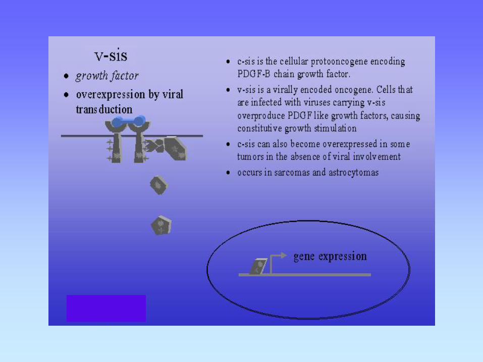
Por ejemplo PDGF, FGF .
Sobreexpresión de PDGF, exceso de secreción por la célula, resulta en proliferación celular por mecanismo de feed-back autócrino. Asociado con astrocitomas y osteosarcomas humanos.
Similarmente hst-1 y hst-2 sobreexpresan FGF. Asociado con cáncer de estómago, vejiga y mama y con melanoma.
Receptores de Factores de crecimiento Receptores para EGF y CSF-1 han sido implicados en neoplasia.
Estos receptores son normalmente receptores de transmembrana y poseen una kinasa en su cara citoplasmática

PROLIFERATIVE ONCOGENES

Mechanism of action: Growth Factors as Oncogenes
Growth Factors affect:
Proliferation- autocrine loop
c-sis (PDGF) and PDGFR in glioblastoma.
EGF and TGF-a and -EGFR in non-small cell lung
carcinoma.
Neovascularization
VEGF, FGF family members
Invasion
scatter factor/HGF (Met ligand)
Evasion of Immunosurveillance
TGF-b

Oncogenes as signal transducers
Growth Factors
v-sis (PDGF), int-1(WNT-1), int-2(FGF), hst, fgf-5
Growth Factors Receptors
v-erb-B (EGFR), v-fms (CSF-1R), c-kit (KIT)
Signal Transducers
v-ras, v-src, v-raf/mil, v-abl, v-mos, v-crk
Transcription Factors
v-ets, v-myc, v-myb, v-rel (NFkB), v-ski, v-erb-A (THR)
C
Y
T
O
P
L
A
S
M
EXTRACELLULAR
NUCLEUS

Examples of Receptor Tyrosine Kinases
Croce C. N Engl J Med 2008;358:502-511

Role of VEGF–VEGFR Interaction in Angiogenesis
Croce C. N Engl J Med 2008;358:502-511

HER family receptors are activated by ligand-induced dimerization, or receptor pairing.
Dimerization is a critical step in HER family-mediated signaling, and HER receptors are able to homodimerize or heterodimerize with other HER family members, allowing for multiple receptor combinations. The formation of dimers leads to activation of the intrinsic TK domain and subsequent phosphorylation on specific tyrosine residues, which serve as docking sites for a variety of molecules. Recruitment of these molecules leads to the activation of different downstream signaling cascades, including the MAPK proliferation pathway and/or the PI3K/Akt pro-survival pathway. Inappropriate signaling may occur as a result of receptor overexpression or dysregulation of receptor activation, which may lead to: Increased/uncontrolled cell proliferation Decreased apoptosis (programmed cell death) Enhanced cancer cell motility Angiogenesis
HER family receptors


Dysregulation of HER-mediated signaling pathways results in the growth and spread of cancer cells. The HER family consists of 4 structurally related receptors: HER1 (EGFR), HER2, HER3, and HER4.


Schematic of the signaling abnormalities resulting from HER2 overexpression that are felt to contribute to tumorigenesis. HER2 overexpression results in increased HER2 containing dimers of all kinds. Increased HER2-EGFR dimers drive proliferative and invasive functions. Increased HER2 homodimers disrupt cell polarity. Increased HER2-HER3 dimers drive proliferative, survival, invasive, and metabolic functions. Increased HER2 expression results in an increase in the rare ΔHER2 isoform with more potent signaling characteristics. Several transcription factors are induced in HER2 overexpressing cells resulting in a plethora of gene expression changes


Determination of HER2-protein overexpression 1- semiquantitative DAKO Hercep Test™
2- Fluorescence in situ hybridisation (FISH)
Paraffin section of breast tissue,
hybridisation with HER2-specific probe
showing HER2 gene amplification

HERCEPTIN BLOCKS HER2/neu protein

Proteínas G transductoras de señales

Mechanism of action: Oncogenes as signal
transducers
Growth Factors
v-sis (PDGF), int-1(WNT-1), int-2(FGF), hst, fgf-5
Growth Factors Receptors
v-erb-B (EGFR), v-fms (CSF-1R), v-kit (KIT)
Signal Transducers
v-ras, v-src, v-raf/mil, v-abl, v-mos, v-crk
Transcription Factors
v-ets, v-myc, v-myb, v-rel (NFkB), v-ski, v-erb-A (THR)
C
Y
T
O
P
L
A
S
M
EXTRACELLULAR
NUCLEUS


Ras Effectors

Oncogenes as Signal Transducers; Ras is altered in many human cancers
p21ras activating mutations lock Ras
in a GTP-bound state.
Activated in 90% of pancreatic
adenocarcinomas and
50% of colon adenocarcinomas
and leukemias.
p21ras refers to three closely related proteins
H-Ras (Harvey),
K-Ras (Kirsten)
N-Ras (neuronal).
** *** ** * p21ras
12&13 59&61
GTP/GDP
Binding
Switch
Region
GTP Exchange GTP Hydrolysis
GTP
GDP
Structures from
Sprang S.R.,
Annu. Rev. Biochem 1997. 66:639-78

Proteínas G transductoras de señales
Ligandos externos se unen a receptores de la superficie los cuales activan proteínas G (familias de proteínas intermediarias con la superficie celular, ej: ras) Las proteínas G se unen al GTP lo cual activa efectores específicos generando segundos mensajeros (ej: PLC o adenilato ciclasa) Segundos mensajeros (ej cAMP, cGMP, Ca++, IP, DG) Activación de quinasas Las proteínas G hidrolizan GTP a GDP desactivamdo la proteína G La proteína G cicla otra vez si un complejo ligando-receptor está todavía presente en la superficie celular. Proteínas activadoras de GTPasa (GAPs) aceleran la velocidad de la GTPasa (x 1000), actuando como frenos que evitan la actividad descon-trolada de ras.
Por lo tanto las GAPs normales son genes supresores de tumor


Apoptosis Regulators • The BCL2 gene, which is involved in the initiation of almost all follicular lymphomas and some diffuse large B-cell lymphomas encodes a cytoplasmic protein that localizes to mitochondria and increases cell survival by inhibiting apoptosis (anti-apoptótica). • BCL2 is also important in chronic lymphocytic leukemia and lung cancer.
•The BCL2 family members BCL-XL and BCL2 inhibit apoptosis and are up-regulated in many cancers (ONCOGEN, activado por translocación y sobre-expresión).
• Two main pathways lead to apoptosis: 1- the stress pathway: triggered by proteins that contain the BCL2 homology 3 domain; this domain inactivates BCL2 and BCL-XL (which normally inhibit apoptosis) and thereby activates the caspases that induce apoptosis. Drugs that mimic the BCL2 homology 3 domain and can bind to BCL-XL or BCL2 (peptides or small organic molecules that bind in a groove of these proteins) are under development. This approach has attracted considerable attention because many tumors overexpress BCL2 or related proteins
2- the death-receptor pathway: is activated by the binding of Fas ligand, TRAIL, and tumor necrosis factor α, to their corresponding (death) receptors on the cell surface. activation of death receptors activates caspases that cause cell death

Cancer Therapies That Target Oncogenic Proteins
Croce C. N Engl J Med 2008;358:502-511