Embed Size (px)
Citation preview
Significant contrast dose-reduction with Digital Variance Angiography
in carotid and cerebral X-ray angiography
Viktor Orias MD
Bács-Kiskun County Hospital, Kecskemét, Hungary
Heart and Vascular Center of Semmelweis University, Budapest, Hungary

Disclosure
Speaker name: Viktor Orias MD
.................................................................................
I have the following potential conflicts of interest to report:
Employment in industry: Kinepict Health Ltd.
Kinetic imaging (or DVA)
• New, patented x-ray image processing method
• Digital Variance Angiography (DVA) instead of subtraction(DSA)
• Significant image quality advantage over current reference-standard DSA in lower extremity arteriography with iodinated
contrast media (1) and carbon-dioxide (2)
• First aim: DVA feasibility in carotid angio setting
1: Gyano M, Gog I, Orias VI, et al. Kinetic Imaging in Lower Extremity Arteriography: Comparison to Digital Subtraction Angiography. Radiology. 2019;290(1):246-53
2: Orias VI, Gyano M, Szollosi D, et al. Digital Variance Angiography as a Paradigm Shift in Carbon-Dioxide Angiography, Investigative Radiology (forthcoming 2019)
Carotid and cerebral X-ray angiography
• Contrast medium
• Nephrotoxicity
• „Selective injections of hyperosmolar contrast material into thecommon and internal carotid arteries may...
• Cause pain, resulting in patient movement
• Decreased image quality
• Increased patient discomfort
• “May produce transient disruption of the blood-brain barrier with associated neurologic deficit or seizure” (3)
• Second aim: to reduce contrast dose without image quality loss
3: Bird et al, Am J of Neuroradiology, 1984
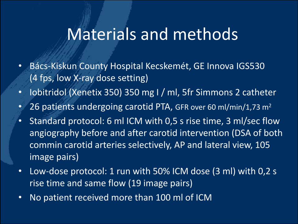
Materials and methods
• Bács-Kiskun County Hospital Kecskemét, GE Innova IGS530 (4 fps, low X-ray dose setting)
• Iobitridol (Xenetix 350) 350 mg I / ml, 5fr Simmons 2 catheter
• 26 patients undergoing carotid PTA, GFR over 60 ml/min/1,73 m2
• Standard protocol: 6 ml ICM with 0,5 s rise time, 3 ml/sec flow angiography before and after carotid intervention (DSA of bothcommin carotid arteries selectively, AP and lateral view, 105 image pairs)
• Low-dose protocol: 1 run with 50% ICM dose (3 ml) with 0,2 s rise time and same flow (19 image pairs)
• No patient received more than 100 ml of ICM
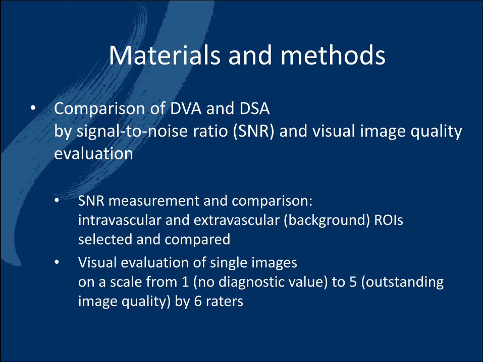
Materials and methods
• Comparison of DVA and DSA by signal-to-noise ratio (SNR) and visual image qualityevaluation
• SNR measurement and comparison: intravascular and extravascular (background) ROIs selected and compared
• Visual evaluation of single images on a scale from 1 (no diagnostic value) to 5 (outstanding image quality) by 6 raters
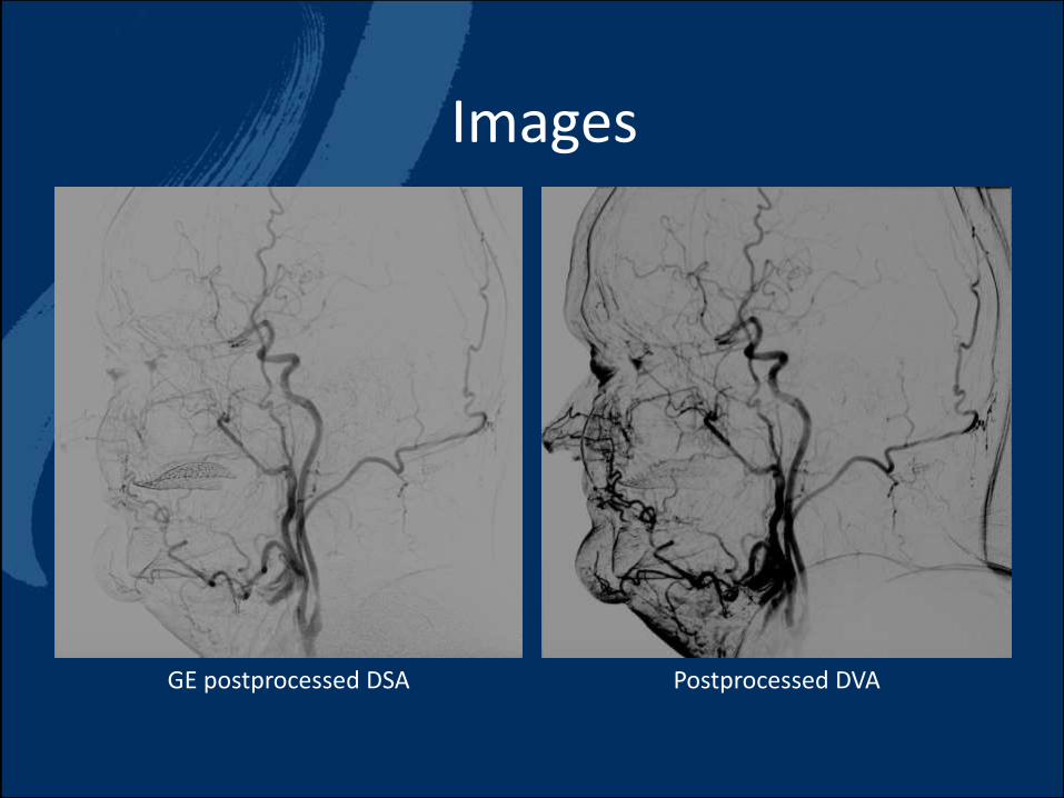
Images
GE postprocessed DSA Postprocessed DVA
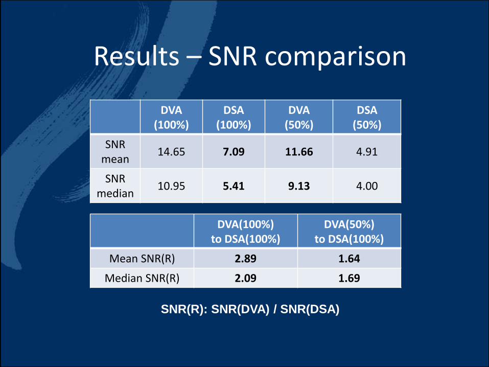
Results – SNR comparison
SNR(R): SNR(DVA) / SNR(DSA)
DVA (100%)
DSA(100%)
DVA (50%)
DSA (50%)
SNR mean
14.65 7.09 11.66 4.91
SNR median
10.95 5.41 9.13 4.00
DVA(100%) to DSA(100%)
DVA(50%) to DSA(100%)
Mean SNR(R) 2.89 1.64
Median SNR(R) 2.09 1.69
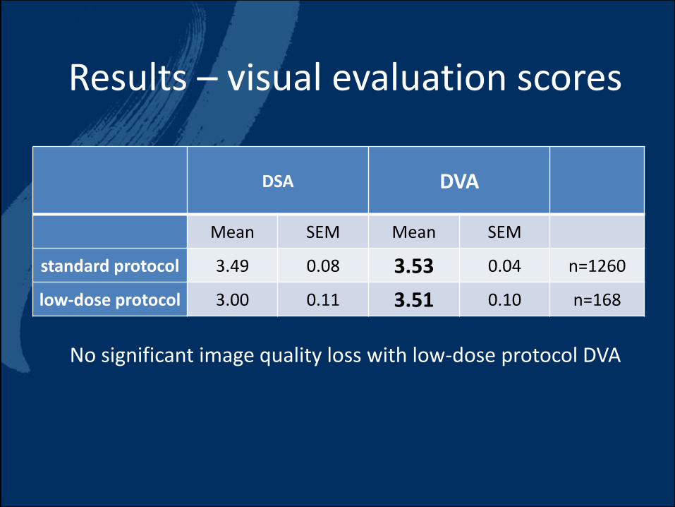
Results – visual evaluation scores
DSA DVA
Mean SEM Mean SEM
standard protocol 3.49 0.08 3.53 0.04 n=1260
low-dose protocol 3.00 0.11 3.51 0.10 n=168
No significant image quality loss with low-dose protocol DVA
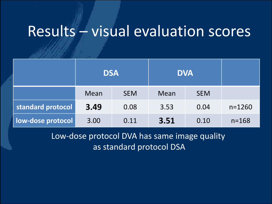
Results – visual evaluation scores
DSA DVA
Mean SEM Mean SEM
standard protocol 3.49 0.08 3.53 0.04 n=1260
low-dose protocol 3.00 0.11 3.51 0.10 n=168
Low-dose protocol DVA has same image quality as standard protocol DSA
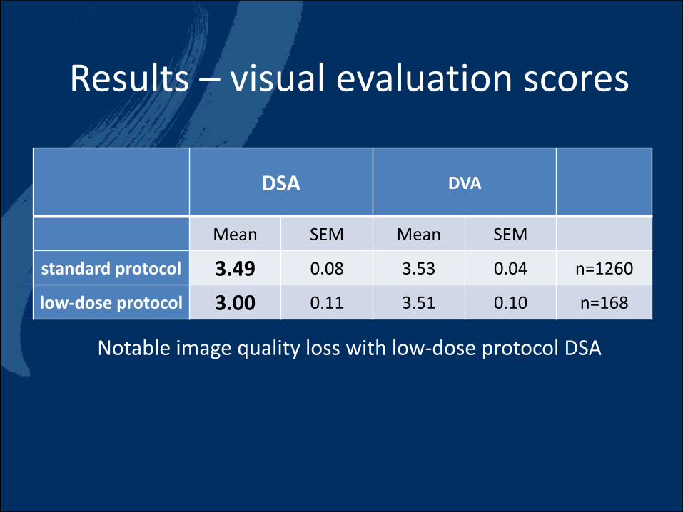
Results – visual evaluation scores
DSA DVA
Mean SEM Mean SEM
standard protocol 3.49 0.08 3.53 0.04 n=1260
low-dose protocol 3.00 0.11 3.51 0.10 n=168
Notable image quality loss with low-dose protocol DSA
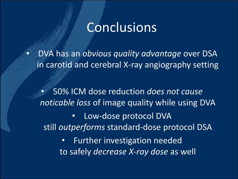
Conclusions
• DVA has an obvious quality advantage over DSA in carotid and cerebral X-ray angiography setting
• 50% ICM dose reduction does not cause noticable loss of image quality while using DVA
• Low-dose protocol DVA still outperforms standard-dose protocol DSA
• Further investigation neededto safely decrease X-ray dose as well

Thank you for yourattention!
Viktor Óriás [email protected]
Kinepict Health Ltd, Budapest, HUBács-Kiskun County Hospital, Kecskemét, HU
Heart and Vascular Center of Semmelweis University, Budapest, HU

Significant contrast dose-reduction with Digital Variance Angiography
in carotid and cerebral X-ray angiography
Viktor Orias MD
Bács-Kiskun County Hospital, Kecskemét, Hungary
Heart and Vascular Center of Semmelweis University, Budapest, Hungary