Embed Size (px)
DESCRIPTION
Contrast radiographic techniques in veterinary medicine with special reference to angiography.
Citation preview

Contrast Radiography With Special Reference to
Angiography
Amulya V.R.

Contrast Radiography
• Lack of contrast in soft tissue makes diagnosis by survey radiography
difficult.
• Various applications

Contrast Media
Positive
Contrast
Agents
BaSO4, I2
Negative
Contrast
Agents
Air, O2, CO2

Iodine Preparations
Water-Soluble agents• Triiodinated compounds
• Angiography, angiocardiography
Viscous/oily agents• Lymphography
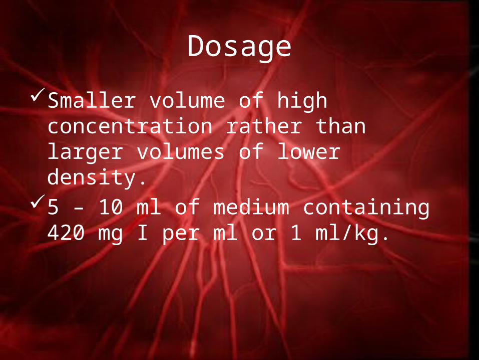
Dosage
Smaller volume of high concentration rather than larger volumes of lower density.
5 – 10 ml of medium containing 420 mg I per ml or 1 ml/kg.

Considerations
Contrast Medium
Hypersensitivity
Thromboemboli
sm
Technical
feasibility

TechniqueInsert catheter into access vessel of choice
Advance catheter (under fluoroscopic guidance) to desired point
Measure pressures, if applicable
Make and capture injection “sequence”
Treatment manoeuvres if applicable
Flush

Angiography
Radiographic demonstration of portions
of the vascular system by the injection of a water-soluble contrast agent either intra-venously or
intra-arterially.

Angiography contd.
• Single radiograph taken immediately at the termination of the injection
(portal venography)
• Films taken serially at intervals of 0.5 – 2 seconds may be necessary
(angiocardiography).

Angiocardiography
An intravenous radiographic contrast study evaluating the vascular system and
chambers of the heart
Congenital or acquired lesions

Arteriography
• Localised narrowing or obstruction of an artery
• Pathological circulation
• Tumour
• Treatment
• Time consuming : ½ hour – 2 hours
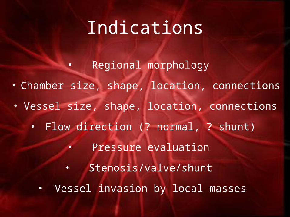
Indications
• Regional morphology
• Chamber size, shape, location, connections
• Vessel size, shape, location, connections
• Flow direction (? normal, ? shunt)
• Pressure evaluation
• Stenosis/valve/shunt
• Vessel invasion by local masses
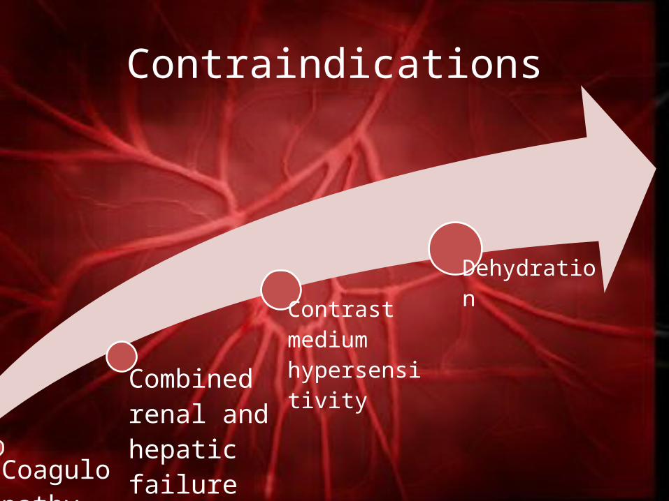
Contraindications
Coagulopathy or active anticoagulation
Combined renal and hepatic failure
Contrast medium hypersensitivity
Dehydration

Selective Angiography
Arterial & venous

Normal Left and Right Ventricles

VSD

PDA

Aortic Stenosis
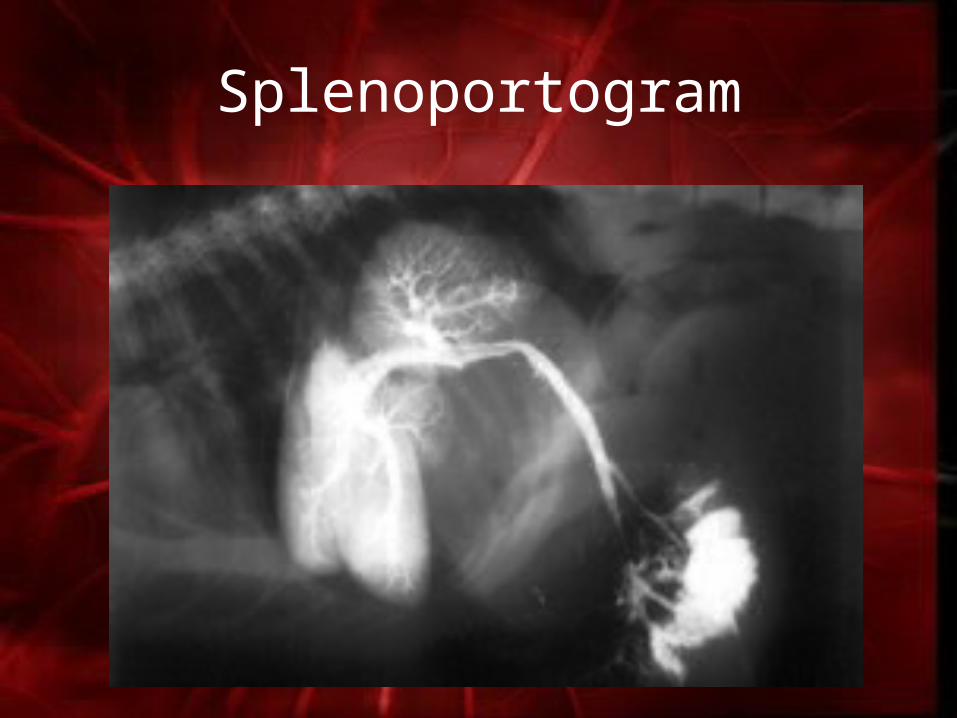
Splenoportogram

Non – Selective
AngiographyVenous only
Technique
• 2-5 seconds post injection for right ventricle
• 4-8 seconds post injection for left ventricle
• Technique not good for – L→R shunts– Distal arterial/limb evaluation
• Frequent re-injections
Limitations

Normal Canine Angiocardiogram
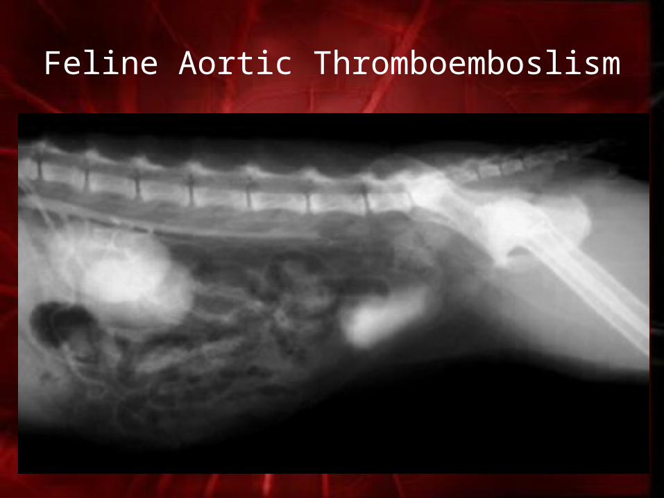
Feline Aortic Thromboemboslism
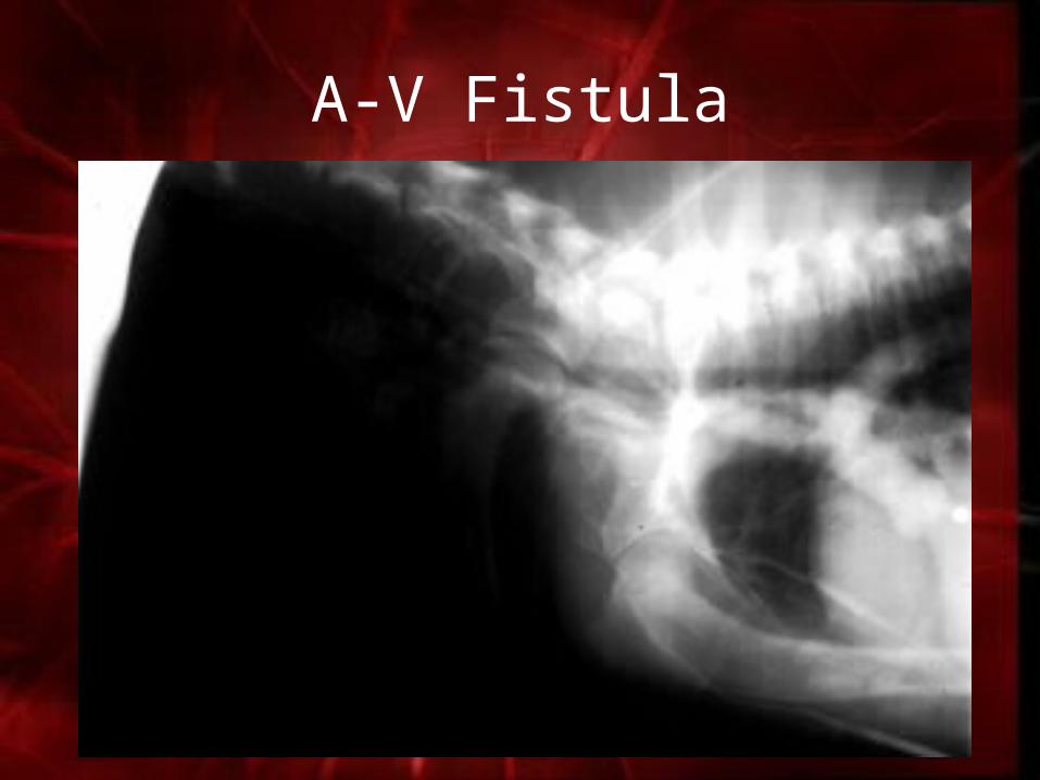
A-V Fistula
Direct Parenchymal
Injection

Indications
• Splenic venous drainage pattern• Trans-hepatic study• Lymph node morphology• Miscellaneous parenchymal organ
drainage• Cavity integrity (cavity injected)

Precautions
Bleeding
Organ injury
Pleurography/peritoneographyRoll patient to assure contrast distribution

Diaphragmatic Hernia

Digital Subtraction Angiography (DSA)
• Specialised electronic equipment, computing and radiographic hardware to
produce rapid sequential images.
• Contrast filled vessels free from the distraction of overlying structures
Lymphatic Imaging

Indications
• Drainage status of regional nodes
• Define internal node anatomy
• Find local lymphatic vessels :
(lymphangectasia/thoracic duct)

Normal Lymphangiogram

Urethral TCC

Sub-lumbar Lymph Nodes

Conclusion
• Limited use
• Much replaced by ultrasound, particularly colour-flow (Doppler imaging)
References• Feeney, D.A., 2003. Special Radiographic Procedures: Practical
Vascular Imaging. Available: www.academic-server.cvm.umn.edu/radiology/student/Student_Sp_09/Feeney/PDF/sp.%20Vascular.09.pdf. [13 Jan. 2015]
• Burke, R.L. and Feeney, D.A, 2003. Small Animal Radiology and Ultrasonography: A Diagnostic Atlas and Text. (3rd Ed.). Saunders Elsevier, Philadelphia, 740p.
• Lavin, L.M., 2005. Radiography in Veterinary Technology. (4th Ed.). Saunders Elsevier, Philadelphia, 400p.
• Trey. 2009. Special Procedures: Radiology. Available: www.kwilkerson.yolasite.com/resources/CH.%2018-Special%20Procedures.ppt. [13 Jan. 2015]
• Kealy, J.K., McAllister, H. and Graham, J.P., 2011. Diagnostic Radiology and Ultrasonography of the Dog and Cat. (5th Ed.). Saunders Elsevier, Philadelphia, 592p.

Thank You!