Embed Size (px)
Citation preview

Signal transduction during C. elegans vulvaldevelopment: a NeverEnding storyTobias Schmid and Alex Hajnal
Available online at www.sciencedirect.com
ScienceDirect
The Caenorhabditis elegans hermaphrodite vulva is one of the
best studied models for signal transduction and cell fate
determination during organogenesis. Systematic forward
genetic screens have identified a complex and highly
interconnected signaling network formed by the conserved
EGFR, NOTCH, and WNT signaling pathways that specifies an
invariant pattern of cell fates among the six vulval precursor
cells (VPCs). Multiple inhibitory interactions between the EGFR
and NOTCH pathways ensure the selection of a single 18 VPC
that is always flanked by two 28 VPCs thanks to lateral NOTCH
signaling. Building on this ‘central dogma’ of cell fate
specification, scientists have investigated a broad spectrum of
novel questions that are summarized in this review. For
example, vulval development is a unique model to study the
intracellular trafficking of signaling molecules, such as NOTCH
or EGFR, to investigate the interactions between the cell cycle
and cell fate specification pathways, and to observe epithelial
tube morphogenesis and cell invasion at single-cell resolution.
Finally, computer scientists have integrated the experimental
data into mathematical and state-based ‘in silico’ models of
vulval development, allowing them to test the completeness
and limits of our current understanding.
Addresses
University of Zurich, Institute of Molecular Life Sciences,
Winterthurerstrasse 190, CH-8057 Zurich, Switzerland
Corresponding author: Hajnal, Alex ([email protected])
Current Opinion in Genetics & Development 2015, 32:1–9
This review comes from a themed issue on Developmental
mechanisms, patterning and organogenesis
Edited by Deborah J Andrew and Deborah Yelon
http://dx.doi.org/10.1016/j.gde.2015.01.006
0959-437X/# 2015 Published by Elsevier Ltd.
A central dogma: the interplay of Wnt, EGFR,and NOTCH signaling determines the 1- and 2-vulval cell fatesFrom the P lineage to the vulval competence group: Wnt
and EGFR signaling maintain VPC competence
The C. elegans vulva originates from the ventral epidermal
P cells that divide during the first larval stage (L1) into
Pn.a and Pn.p daughter cells [1,2��]. The anterior Pn.a
www.sciencedirect.com
cells will later differentiate into ventral cord neurons,
whereas the posterior Pn.p cells form the epidermis. At
the end of the L1 stage, a Wnt signal from the posterior
body region selects six Pn.p cells (P3.p through P8.p) in
the mid-body region to become the vulval precursor cells
(VPCs) and form the vulval competence group
(Figure 1a) [3–5]. Canonical Wnt signaling maintains
the VPCs as polarized epithelial cells by inducing the
hox gene lin-39 [6–8]. Among other functions (see below),
lin-39 prevents the fusion of the VPCs with the surround-
ing syncytial epidermis (hyp7) by repressing the expres-
sion of the fusogen eff-1 [9–11]. The anterior (P1.p and
P2.p) and posterior (P9.p to P11.p) Pn.p cells fuse with
hyp7 and loose their potential to differentiate. An inter-
esting case is P3.p, the VPC at the anterior border of the
competence group; in around 50% of the animals, P3.p
looses its competence before the end of the L2 stage and
fuses with hyp7 [2��,12]. However, P3.p fusion can be
prevented by overexpression of the EGF growth factor
LIN-3, indicating that EGFR signaling acts redundantly
with the Wnt pathway to induce lin-39 expression [13,14].
Thus, the vulval equivalence group is specified by coop-
erative Wnt and EGFR signaling.
1- cell fate specification by the anchor cell
Beginning in the L2 stage, the anchor cell (AC) in the
somatic gonad secretes the LIN-3 protein, a member of
the epidermal growth factor family (Figure 1a) [15,16].
Even though LIN-3 is produced as a transmembrane
precursor similar to mammalian TGFa, LIN-3 is released
from the AC in a graded manner, and activates the LET-
23 EGFR in all VPCs [17–19]. However, when expressed
at a normal dosage LIN-3 is efficiently sequestered by the
VPC closest to the AC, P6.p, which presents the highest
levels of LET-23 on its basolateral membrane [20�,21].
Since P6.p receives most of the LIN-3 signal, it is the only
VPC that adopts the 18 vulval cell fate. Downstream of
the EGFR tyrosine kinase, a canonical RAS/MAPK path-
way transduces the signal into the nucleus. Conserved
components of the core pathway include the adaptor
protein SEM-5 (GRB2) [22], the guanine exchange factor
SOS-1 [23] and the RAS protein LET-60 [24], which
activates the LIN-45 RAF [25], MEK-2 MEK [26] and
MPK-1 MAP kinase cascade [27]. MPK-1 activation is
both necessary and sufficient to induce the 18 vulval cell
fate [28]. To date, the ETS family LIN-1 and forkheadLIN-31 transcription factors are the only known MPK-1
substrates [29–31]. In their unphosphorylated state, LIN-
1 and LIN-31 form a complex that inhibits vulval induc-
tion by repressing 18-specific transcription [32]. After
Current Opinion in Genetics & Development 2015, 32:1–9

2 Developmental mechanisms, patterning and organogenesis
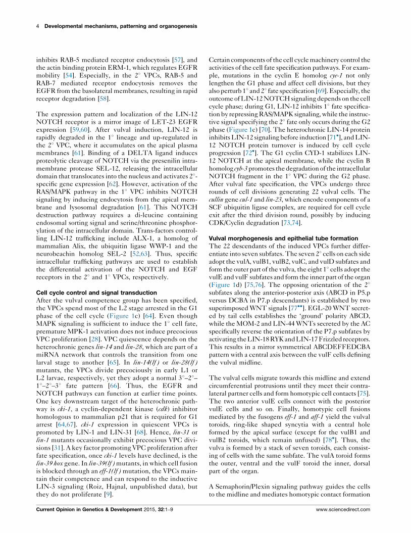
Figure 1
(a) Cell fate specification
Receptor Trafficking
Cell Cycle Control Anchor Cell Invasion
Computational Modeling
Tube Morphogenesis
(b)
Recycling
P3.p
P3.p
P4.p
P4.p
3° 3° 3°2°
2° 2°
1°
1° 2° Notch
EGF
1° fate2° fate 2° fate1° fate 1° fate
P5.p
P5.p
P6.p
P6.p
P7.p
P7.p
P8.p
P8.p
AC
AC
AC
Degradation
P7.pP6.pP5.pstage
G1
S
A A A AEEE F FF FEB1 B1B2 B2C C C CD D
G2/M
L2
L3
(c) (e)
(f)
(d)
migration
B2 B2B1 B1A AC C
D DE E
F
AC
AC
F F EEF
migrationextension
Current Opinion in Genetics & Development
Overview of vulval development. Different aspects of vulval development that have been investigated so far include (a) the mechanisms of cell fate
specification and pattern formation, (b) the intracellular trafficking of NOTCH and EGFR, (c) the interactions between cell cycle and cell fate
specification, (d) the morphogenesis of the vulval cells into an epithelial tube, (e) the invasion of the AC into the vulval epithelium, and (f) the
computational modeling of the cell fate specification pathways.
phosphorylation by MPK-1, the LIN-1/LIN-31 complex
dissociates, phospho-LIN-1 cannot repress anymore,
while phospho-LIN-31 is turned into an activator of 18gene expression [29]. Other transcription factors acting
downstream of the RAS/MAPK pathway include SUR-2
and LIN-25, which are both components of the transcrip-
tion mediator complex [33,34]. One key target of the
LIN-1/LIN-31 repressor complex is the lin-39 hox gene
[8,35]. lin-39 is required for 18 cell fate execution by
coordinating vulval cell proliferation and morphogenesis
[9,36–38]. Again, Wnt and RAS/MAPK signaling act in
parallel to induce lin-39 expression during fate specifica-
tion, as Wnt signaling can partially compensate for a
reduction in EGFR signaling [39]. Besides the core
components of the EGFR pathway, systematic genetic
screens have identified a number of modifiers and nega-
tive regulators of the EGFR pathway. For example, the
Current Opinion in Genetics & Development 2015, 32:1–9
tyrosine kinase ark-1 [40], the cbl homolog sli-1 encoding
an E3 ubiquitin ligase [41] or the receptor tyrosine
phosphatase dep-1, which de-phosphorylates LET-23
[42], all function as negative regulators of the EGFR.
2- cell fate specification: sequential induction by NOTCH
and graded LIN-3 EGF signaling
The 18 VPC P6.p up-regulates the expression of three
partially redundant ligands of the Delta/Serrate family,
lag-2, dsl-1, and apx-1 [43]. These ligands activate the
NOTCH receptor LIN-12 in the adjacent VPCs P5.p and
P7.p (Figure 1a). While lag-2 and apx-1 encode mem-
brane-bound ligands, dsl-1 encodes a secreted protein that
may activate NOTCH signaling without direct cell con-
tact [44�]. One outcome of NOTCH signaling is the
repression of the EGFR signaling pathway in P5.p and
P7.p, a classical example of lateral inhibition [45]. LIN-12
www.sciencedirect.com

C. elegans vulval development Schmid and Hajnal 3
NOTCH induces the transcription of several negative
regulators, such as the MAPK phosphatase lip-1, ark-1,
the adaptin homolog dpy-23, which may regulate EGFR
endocytosis, or the lst genes [46,47]. However, LIN-12
NOTCH signaling also plays an instructive role during
the subsequent 28 vulval fate specification, as a constitu-
tively active NOTCH receptor causes all VPCs to adopt
the 28 fate even in the absence of the inductive LIN-3
signal [16,48]. The distal VPCs P3.p, P4.p and P8.p that
receive neither inductive the LIN-3 nor the lateral
NOTCH signal adopt the 38, non-vulval cell fate [1].
These VPCs divide once and fuse with hyp7.
One hotly debated question has been the relative contri-
bution of EGFR and NOTCH signaling towards 28 fate
specification [18,49,50]. While let-23 mosaic experiments
demonstrated that VPCs lacking let-23 or other compo-
nents of the RAS/MAPK pathway can adopt the 28 fate, as
long as they are adjacent to a 18 VPC [49], lin-3 dosage
experiments indicated that an isolated VPC can adopt a 28fate if exposed to an intermediate LIN-3 concentration
[18]. Furthermore, it has been proposed that VPCs receiv-
ing intermediate levels of LIN-3 may adopt the 28 fate
through autocrine stimulation via the secreted NOTCH
ligand DSL-1 [44�]. However, Zand et al. [51��] identified a
RalGEF as an alternate RAS effector that antagonizes the
canonical RAF/MAPK pathway in P5.p and P7.p and
Figure 2
lin-7(If)
lin-7(If)
wild-type
wild-type
P6.p
P6.p
AC
LET-23 EGFRapical junctions
LET-23 EGFR localization. A tripartite LIN-2/LIN-7/LIN-10 localization comp
efficient receptor activation in the 18 VPC P6.p. In lin-7(lf), lin-2(lf) or lin-10(lf
cannot bind the LIN-3 growth factor ligand.
www.sciencedirect.com
inhibits 18 fate specification. Thus, the lateral NOTCH
signal together with a graded LIN-3 signal activating an
alternate RAS/RAL pathway ensure that a 18 VPC is always
flanked by two 28 VPCs.
The story goes on: new topics in vulvaldevelopmentReceptor localization and trafficking control signaling
Thanks to the transparent body, it is possible to observe
in live larvae the intracellular trafficking of signaling
molecules that determine the VPC fates (Figure 1b).
In particular, the two receptors LIN-12 NOTCH and
LET-23 EGFR show a dynamic expression pattern and
rapid protein turnover depending on the VPC fates [52–54]. lin-2, lin-7, and lin-10, which were among the first
lineage defective (lin) mutants identified, encode com-
ponents of a conserved protein localization complex that
retains the EGFR on the basolateral plasma membrane of
the 18 VPC (Figure 2) [53,55]. Basolateral EGFR locali-
zation is essential for receptor activation because only the
basolateral membrane compartment of the VPCs is ex-
posed to the LIN-3 EGF signal from the gonadal AC. On
the other hand, the small GTPase ARF together with its
exchange factor AGEF-1 and the AP1 component UNC-
101 antagonize basolateral EGFR localization via the
LIN-2/LIN-7/LIN-10 complex [56�]. Other factors regu-
lating EGFR trafficking include the EPS-8 protein, which
basal
apical
P6.p
P6.p
AC
LET-23 EGFR
LIN-3 EGF
LIN-2/LIN-7/LIN-10
apical junctions
basal laminae
LET-23 EGFR
apical junctions
Current Opinion in Genetics & Development
lex retains the LET-23 EGFR on the basolateral membrane, allowing
) mutants, the EGFR is mislocalized to the apical compartment and
Current Opinion in Genetics & Development 2015, 32:1–9
4 Developmental mechanisms, patterning and organogenesis
inhibits RAB-5 mediated receptor endocytosis [57], and
the actin binding protein ERM-1, which regulates EGFR
mobility [54]. Especially, in the 28 VPCs, RAB-5 and
RAB-7 mediated receptor endocytosis removes the
EGFR from the basolateral membranes, resulting in rapid
receptor degradation [58].
The expression pattern and localization of the LIN-12
NOTCH receptor is a mirror image of LET-23 EGFR
expression [59,60]. After vulval induction, LIN-12 is
rapidly degraded in the 18 lineage and up-regulated in
the 28 VPC, where it accumulates on the apical plasma
membranes [61]. Binding of a DELTA ligand induces
proteolytic cleavage of NOTCH via the presenilin intra-
membrane protease SEL-12, releasing the intracellular
domain that translocates into the nucleus and activates 28-specific gene expression [62]. However, activation of the
RAS/MAPK pathway in the 18 VPC inhibits NOTCH
signaling by inducing endocytosis from the apical mem-
brane and lysosomal degradation [61]. This NOTCH
destruction pathway requires a di-leucine containing
endosomal sorting signal and serine/threonine phosphor-
ylation of the intracellular domain. Trans-factors control-
ling LIN-12 trafficking include ALX-1, a homolog of
mammalian Alix, the ubiquitin ligase WWP-1 and the
neurobeachin homolog SEL-2 [52,63]. Thus, specific
intracellular trafficking pathways are used to establish
the differential activation of the NOTCH and EGF
receptors in the 28 and 18 VPCs, respectively.
Cell cycle control and signal transduction
After the vulval competence group has been specified,
the VPCs spend most of the L2 stage arrested in the G1
phase of the cell cycle (Figure 1c) [64]. Even though
MAPK signaling is sufficient to induce the 18 cell fate,
premature MPK-1 activation does not induce precocious
VPC proliferation [28]. VPC quiescence depends on the
heterochronic genes lin-14 and lin-28, which are part of a
miRNA network that controls the transition from one
larval stage to another [65]. In lin-14(lf ) or lin-28(lf )mutants, the VPCs divide precociously in early L1 or
L2 larvae, respectively, yet they adopt a normal 38–28–18–28–38 fate pattern [66]. Thus, the EGFR and
NOTCH pathways can function at earlier time points.
One key downstream target of the heterochronic path-
way is cki-1, a cyclin-dependent kinase (cdk) inhibitor
homologous to mammalian p21 that is required for G1
arrest [64,67]. cki-1 expression in quiescent VPCs is
promoted by LIN-1 and LIN-31 [68]. Hence, lin-31 or
lin-1 mutants occasionally exhibit precocious VPC divi-
sions [31]. A key factor promoting VPC proliferation after
fate specification, once cki-1 levels have declined, is the
lin-39 hox gene. In lin-39(lf ) mutants, in which cell fusion
is blocked through an eff-1(lf ) mutation, the VPCs main-
tain their competence and can respond to the inductive
LIN-3 signaling (Roiz, Hajnal, unpublished data), but
they do not proliferate [9].
Current Opinion in Genetics & Development 2015, 32:1–9
Certain components of the cell cycle machinery control the
activities of the cell fate specification pathways. For exam-
ple, mutations in the cyclin E homolog cye-1 not only
lengthen the G1 phase and affect cell divisions, but they
also perturb 18 and 28 fate specification [69]. Especially, the
outcome of LIN-12 NOTCH signaling depends on the cell
cycle phase; during G1, LIN-12 inhibits 18 fate specifica-
tion by repressing RAS/MAPK signaling, while the instruc-
tive signal specifying the 28 fate only occurs during the G2
phase (Figure 1c) [70]. The heterochronic LIN-14 protein
inhibits LIN-12 signaling before induction [71�], and LIN-
12 NOTCH protein turnover is induced by cell cycle
progression [72�]. The G1 cyclin CYD-1 stabilizes LIN-
12 NOTCH at the apical membrane, while the cyclin B
homolog cyb-3 promotes the degradation of the intracellular
NOTCH fragment in the 18 VPC during the G2 phase.
After vulval fate specification, the VPCs undergo three
rounds of cell divisions generating 22 vulval cells. The
cullin gene cul-1 and lin-23, which encode components of a
SCF ubiquitin ligase complex, are required for cell cycle
exit after the third division round, possibly by inducing
CDK/Cyclin degradation [73,74].
Vulval morphogenesis and epithelial tube formation
The 22 descendants of the induced VPCs further differ-
entiate into seven subfates. The seven 28 cells on each side
adopt the vulA, vulB1, vulB2, vulC, and vulD subfates and
form the outer part of the vulva, the eight 18 cells adopt the
vulE and vulF subfates and form the inner part of the organ
(Figure 1d) [75,76]. The opposing orientation of the 28subfates along the anterior-posterior axis (ABCD in P5.p
versus DCBA in P7.p descendants) is established by two
superimposed WNT signals [77��]. EGL-20 WNT secret-
ed by tail cells establishes the ‘ground’ polarity ABCD,
while the MOM-2 and LIN-44 WNTs secreted by the AC
specifically reverse the orientation of the P7.p subfates by
activating the LIN-18 RYK and LIN-17 Frizzled receptors.
This results in a mirror symmetrical ABCDEFFEDCBA
pattern with a central axis between the vulF cells defining
the vulval midline.
The vulval cells migrate towards this midline and extend
circumferential protrusions until they meet their contra-
lateral partner cells and form homotypic cell contacts [75].
The two anterior vulE cells connect with the posterior
vulE cells and so on. Finally, homotypic cell fusions
mediated by the fusogens eff-1 and aff-1 yield the vulval
toroids, ring-like shaped syncytia with a central hole
formed by the apical surface (except for the vulB1 and
vulB2 toroids, which remain unfused) [78�]. Thus, the
vulva is formed by a stack of seven toroids, each consist-
ing of cells with the same subfate. The vulA toroid forms
the outer, ventral and the vulF toroid the inner, dorsal
part of the organ.
A Semaphorin/Plexin signaling pathway guides the cells
to the midline and mediates homotypic contact formation
www.sciencedirect.com

C. elegans vulval development Schmid and Hajnal 5
[79,80]. SMP-1 Semaphorin, which is initially produced
by the AC, activates via the PLX-1 receptor a CED-10/
MIG-2 RAC signaling pathway. The signal then propa-
gates from the dorsal to the ventral vulval cells, as the
signal receiving vulF and vulE cells become signal pro-
ducing cells that activate PLX-1 signaling in the adjacent
vulD cells. Unlike Semaphorin signaling in other systems,
SMP-1 has an attractive rather than a repulsive effect on
the vulval cells [81]. Besides controlling cell proliferation,
the hox gene lin-39 is also involved in toroid formation [9].
LIN-39 induces the expression of the VAB-23 zinc finger
protein, which in turn activates smp-1 expression [38].
The cylindrical shape of the apical toroid lumen is estab-
lished through the sequential contraction of the ventral and
expansion of the dorsal toroids (Figure 3) [82�]. Ventral
contraction is mediated by the RHO kinase LET-502,
which is up-regulated by NOTCH signaling in 28 cells and
Figure 3
F
early L4 stage
E
DC
B2B1
A
actomyosin network ventral cont
AC
apical junctions
dorsal expancell boundaries
Lumen morphogenesis. The top panels show microscopic images of the ap
(left) and after (right) lumen contraction. Contraction of the ventral toroids (r
expansion of the dorsal toroids (blue arrows) by the invading AC shape the
black lines.
www.sciencedirect.com
induces the constriction of the circular actomyosin network
in the vulA and vulB1/2 toroids. Dorsal expansion, on the
other hand, requires the EGL-26 palmitoyltransferase in
vulE cells and the prior invasion of the AC into the dorsal
lumen (Figure 1e) [83,84]. At the same time, the secretion
of chondroitin and heparan sulfate carrying glycoproteins
into the apical lumen creates a hydrostatic pressure that
keeps the lumen expanded [85–88].
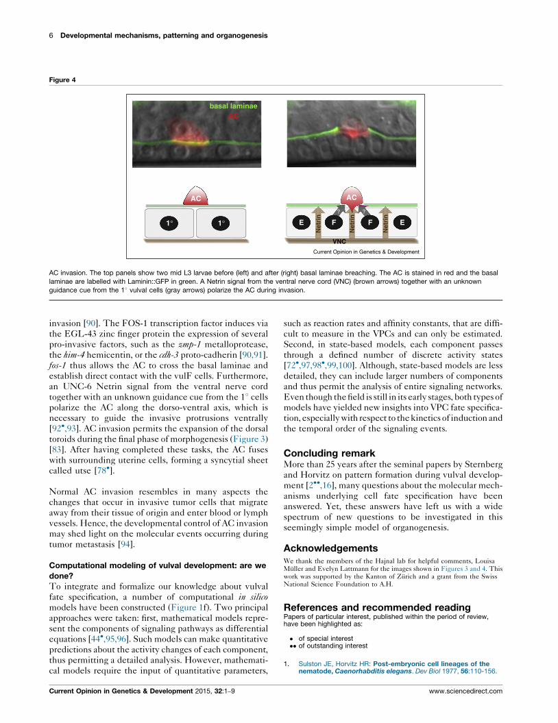
Anchor cell invasion during morphogenesis
A key event during vulval morphogenesis is the invasion
of the AC into the vulval tissue (Figure 1e) [89��]. Before
the last round of cell divisions, the AC undergoes an
epithelial to mesenchymal transition, breaches two basal
laminae that separate the somatic gonad from the ventral
epidermis and extends actin-rich filopodia into the 18vulval tissue (Figure 4). fos-1, a homolog of the mamma-
lian fos proto-oncogene, encodes a key regulator of AC
late L4 stage
raction
utse incl. ACapical junctions
utse incl. AC
sion
Current Opinion in Genetics & Development
ical toroid junctions stained in green and the AC stained in red before
ed arrows) via the actomyosin network (green lines) followed by
vulval lumen during the L4 stage. Cell boundaries are indicated with
Current Opinion in Genetics & Development 2015, 32:1–9
6 Developmental mechanisms, patterning and organogenesis
Figure 4
basal laminaeAC
AC
1° 1° E E
Net
rin
Net
rin
Net
rin
F
VNC
? ?
F
AC
Current Opinion in Genetics & Development
AC invasion. The top panels show two mid L3 larvae before (left) and after (right) basal laminae breaching. The AC is stained in red and the basal
laminae are labelled with Laminin::GFP in green. A Netrin signal from the ventral nerve cord (VNC) (brown arrows) together with an unknown
guidance cue from the 18 vulval cells (gray arrows) polarize the AC during invasion.
invasion [90]. The FOS-1 transcription factor induces via
the EGL-43 zinc finger protein the expression of several
pro-invasive factors, such as the zmp-1 metalloprotease,
the him-4 hemicentin, or the cdh-3 proto-cadherin [90,91].
fos-1 thus allows the AC to cross the basal laminae and
establish direct contact with the vulF cells. Furthermore,
an UNC-6 Netrin signal from the ventral nerve cord
together with an unknown guidance cue from the 18 cells
polarize the AC along the dorso-ventral axis, which is
necessary to guide the invasive protrusions ventrally
[92�,93]. AC invasion permits the expansion of the dorsal
toroids during the final phase of morphogenesis (Figure 3)
[83]. After having completed these tasks, the AC fuses
with surrounding uterine cells, forming a syncytial sheet
called utse [78�].
Normal AC invasion resembles in many aspects the
changes that occur in invasive tumor cells that migrate
away from their tissue of origin and enter blood or lymph
vessels. Hence, the developmental control of AC invasion
may shed light on the molecular events occurring during
tumor metastasis [94].
Computational modeling of vulval development: are we
done?
To integrate and formalize our knowledge about vulval
fate specification, a number of computational in silicomodels have been constructed (Figure 1f). Two principal
approaches were taken: first, mathematical models repre-
sent the components of signaling pathways as differential
equations [44�,95,96]. Such models can make quantitative
predictions about the activity changes of each component,
thus permitting a detailed analysis. However, mathemati-
cal models require the input of quantitative parameters,
Current Opinion in Genetics & Development 2015, 32:1–9
such as reaction rates and affinity constants, that are diffi-
cult to measure in the VPCs and can only be estimated.
Second, in state-based models, each component passes
through a defined number of discrete activity states
[72�,97,98�,99,100]. Although, state-based models are less
detailed, they can include larger numbers of components
and thus permit the analysis of entire signaling networks.
Even though the field is still in its early stages, both types of
models have yielded new insights into VPC fate specifica-
tion, especially with respect to the kinetics of induction and
the temporal order of the signaling events.
Concluding remarkMore than 25 years after the seminal papers by Sternberg
and Horvitz on pattern formation during vulval develop-
ment [2��,16], many questions about the molecular mech-
anisms underlying cell fate specification have been
answered. Yet, these answers have left us with a wide
spectrum of new questions to be investigated in this
seemingly simple model of organogenesis.
AcknowledgementsWe thank the members of the Hajnal lab for helpful comments, LouisaMuller and Evelyn Lattmann for the images shown in Figures 3 and 4. Thiswork was supported by the Kanton of Zurich and a grant from the SwissNational Science Foundation to A.H.
References and recommended readingPapers of particular interest, published within the period of review,have been highlighted as:
� of special interest�� of outstanding interest
1. Sulston JE, Horvitz HR: Post-embryonic cell lineages of thenematode, Caenorhabditis elegans. Dev Biol 1977, 56:110-156.
www.sciencedirect.com

C. elegans vulval development Schmid and Hajnal 7
2.��
Sternberg PW, Horvitz HR: Pattern formation during vulvaldevelopment in C. elegans. Cell 1986, 44:761-772.
A seminal paper that introduced vulval development as a model forpattern formation.
3. Eisenmann DM, Maloof JN, Simske JS, Kenyon C, Kim SK: Thebeta-catenin homolog BAR-1 and LET-60 Ras coordinatelyregulate the Hox gene lin-39 during Caenorhabditis elegansvulval development. Development 1998, 125:3667-3680.
4. Gleason JE, Szyleyko EA, Eisenmann DM: Multiple redundantWnt signaling components function in two processes duringC. elegans vulval development. Dev Biol 2006, 298:442-457.
5. Penigault J-B, Felix M-A: High sensitivity of C. elegans vulvalprecursor cells to the dose of posterior Wnts. Dev Biol 2011,357:428-438.
6. Salser SJ, Chang C, Ambros V, Loer CM, Kenyon C: MultipleHOM-C gene interactions specify cell fates in the nematodecentral nervous system. Genes Dev 1993, 7:1714-1724.
7. Clark S, Chisholm A, Horvitz H: Control of cell fates in the centralbody region of C. elegans by the homeobox gene lin-39. Cell1993, 74:43-55.
8. Wagmaister JA, Miley GR, Morris CA, Gleason JE, Miller LM,Kornfeld K, Eisenmann DM: Identification of cis-regulatoryelements from the C. elegans Hox gene lin-39 required forembryonic expression and for regulation by the transcriptionfactors LIN-1, LIN-31 and LIN-39. Dev Biol 2006, 297:550-565.
9. Shemer G, Podbilewicz B: LIN-39/Hox triggers cell division andrepresses EFF-1/fusogen-dependent vulval cell fusion. GenesDev 2002, 16:3136-3141.
10. Koh K, Peyrot SM, Wood CG, Wagmaister JA, Maduro MF,Eisenmann DM, Rothman JH: Cell fates and fusion in the C.elegans vulval primordium are regulated by the EGL-18 andELT-6 GATA factors — apparent direct targets of the LIN-39Hox protein. Development 2002, 129:5171-5180.
11. Mohler WA, Shemer G, del Campo JJ, Valansi C, Opoku-Serebuoh E, Scranton V, Assaf N, White JG, Podbilewicz B: Thetype I membrane protein EFF-1 is essential for developmentalcell fusion. Dev Cell 2002, 2:355-362.
12. Penigault J-B, Felix M-A: Evolution of a system sensitive tostochastic noise: P3.p cell fate in Caenorhabditis. Dev Biol2011, 357:419-427.
13. Myers TR, Greenwald I: Wnt signal from multiple tissues and lin-3/EGF signal from the gonad maintain vulval precursor cellcompetence in Caenorhabditis elegans. Proc Natl Acad Sci U SA 2007, 104:20368-20373.
14. Chen Z, Han M: C. elegans Rb, NuRD, and Ras regulate lin-39-mediated cell fusion during vulval fate specification. Curr Biol2001, 11:1874-1879.
15. Hill RJ, Sternberg PW: The gene lin-3 encodes an inductivesignal for vulval development in C. elegans. Nature 1992,358:470-476.
16. Sternberg PW, Horvitz HR: The combined action of twointercellular signaling pathways specifies three cell fatesduring vulval induction in C. elegans. Cell 1989, 58:679-693.
17. Dutt A, Canevascini S, Froehli-Hoier E, Hajnal A: EGF signalpropagation during C. elegans vulval development mediatedby ROM-1 rhomboid. PLoS Biol 2004, 2:e334.
18. Katz WS, Hill RJ, Clandinin TR, Sternberg PW: Different levels ofthe C. elegans growth factor LIN-3 promote distinct vulvalprecursor fates. Cell 1995, 82:297-307.
19. Aroian RV, Koga M, Mendel JE, Ohshima Y, Sternberg PW: Thelet-23 gene necessary for Caenorhabditis elegans vulvalinduction encodes a tyrosine kinase of the EGF receptorsubfamily. Nature 1990, 348:693-699.
20.�
Barkoulas M, van Zon JS, Milloz J, Van Oudenaarden A, Felix M-A:Robustness and epistasis in the C. elegans vulval signalingnetwork revealed by pathway dosage modulation. Dev Cell2013, 24:64-75.
By varying the dosage of the EGF and NOTCH signal the robustness ofthe vulval signaling network could be tested.
www.sciencedirect.com
21. Hajnal A, Whitfield CW, Kim SK: Inhibition of Caenorhabditiselegans vulval induction by gap-1 and by let-23 receptortyrosine kinase. Genes Dev 1997, 11:2715-2728.
22. Clark SG, Stern MJ, Horvitz HR: C. elegans cell-signaling genesem-5 encodes a protein with SH2 and SH3 domains. Nature1992, 356:340-344.
23. Chang C, Hopper NA, Sternberg PW: Caenorhabditis elegansSOS-1 is necessary for multiple RAS-mediated developmentalsignals. EMBO J 2000, 19:3283-3294.
24. Beitel GJ, Clark SG, Horvitz HR: Caenorhabditis elegans rasgene let-60 acts as a switch in the pathway of vulval induction.Nature 1990, 348:503-509.
25. Han M, Golden A, Han Y, Sternberg PW: C. elegans lin-45 rafgene participates in let-60 ras-stimulated vulvaldifferentiation. Nature 1993, 363:133-140.
26. Kornfeld K, Guan KL, Horvitz HR: The Caenorhabditis elegansgene mek-2 is required for vulval induction and encodes aprotein similar to the protein kinase MEK. Genes Dev 1995,9:756-768.
27. Lackner M, Kornfeld K, Miller L, Horvitz H, Kim S: A MAP kinasehomolog, mpk-1, is involved in ras-mediated induction ofvulval cell fates in Caenorhabditis elegans. Genes Dev 1994,8:160-173.
28. Lackner M, Kim S: Genetic analysis of the Caenorhabditiselegans MAP kinase gene mpk-1. Genetics 1998, 150:103-117.
29. Tan PB, Lackner MR, Kim SK: MAP kinase signaling specificitymediated by the LIN-1 Ets/LIN-31 WH transcription factorcomplex during C. elegans vulval induction. Cell 1998, 93:569-580.
30. Jacobs D, Beitel GJ, Clark SG, Horvitz HR, Kornfeld K: Gain-of-function mutations in the Caenorhabditis elegans lin-1 ETSgene identify a C-terminal regulatory domain phosphorylatedby ERK MAP kinase. genetics.org 1998, 149:1809.
31. Miller LM, Gallegos ME, Morisseau BA, Kim SK: lin-31, aCaenorhabditis elegans HNF-3/fork head transcription factorhomolog, specifies three alternative cell fates in vulvaldevelopment. Genes Dev 1993, 7:933-947.
32. Leight ER, Glossip D, Kornfeld K: Sumoylation of LIN-1promotes transcriptional repression and inhibition of vulvalcell fates. Development 2005, 132:1047-1056.
33. Nilsson L, Tiensuu T, Tuck S: Caenorhabditis elegans lin-25: astudy of its role in multiple cell fate specification eventsinvolving Ras and the identification and characterization ofevolutionarily conserved domains. Genetics 2000, 156:1083-1096.
34. Singh N, Han M: sur-2, a novel gene, functions late in the let-60ras-mediated signaling pathway during Caenorhabditiselegans vulval induction. Genes Dev 1995, 9:2251-2265.
35. Guerry F, Marti C-O, Zhang Y, Moroni PS, Jaquiery E, Muller F: TheMi-2 nucleosome-remodeling protein LET-418 is targeted viaLIN-1/ETS to the promoter of lin-39/Hox during vulvaldevelopment in C. elegans. Dev Biol 2007, 306:469-479.
36. Maloof J, Kenyon C: The Hox gene lin-39 is required during C.elegans vulval induction to select the outcome of Rassignaling. Development 1998, 125:181-190.
37. Clandinin TR, Hong Y, Katz WS, Roy R, Sternberg PW, Ambros V:Caenorhabditis elegans HOM-C genes regulate the responseof vulval precursor cells to inductive signal. Dev Biol 1997,182:150-161.
38. Pellegrino MW, Farooqui S, Frohli E, Rehrauer H, Kaeser-Pebernard S, Muller F, Gasser RB, Hajnal A: LIN-39 and theEGFR/RAS/MAPK pathway regulate C. elegans vulvalmorphogenesis via the VAB-23 zinc finger protein.Development 2011, 138:4649-4660.
39. Gleason JE, Korswagen HC, Eisenmann DM: Activation of Wntsignaling bypasses the requirement for RTK/Ras signalingduring C. elegans vulval induction. Genes Dev 2002, 16:1281-1290.
Current Opinion in Genetics & Development 2015, 32:1–9

8 Developmental mechanisms, patterning and organogenesis
40. Hopper NA, Lee J, Sternberg PW: ARK-1 inhibits EGFR signalingin C. elegans. Mol Cell 2000, 6:65-75.
41. Yoon CH, Lee J, Jongeward GD, Sternberg PW: Similarity of sli-1,a regulator of vulval development in C. elegans, to themammalian proto-oncogene c-cbl. Science 1995, 269:1102-1105.
42. Berset TA, Hoier EF, Hajnal A: The C. elegans homolog of themammalian tumor suppressor Dep-1/Scc1 inhibits EGFRsignaling to regulate binary cell fate decisions. Genes Dev2005, 19:1328-1340.
43. Chen N, Greenwald I: The lateral signal for LIN-12/Notch in C.elegans vulval development comprises redundant secretedand transmembrane DSL proteins. Dev Cell 2004, 6:183-192.
44.�
Hoyos E, Kim K, Milloz J, Barkoulas M, Penigault J-B, Munro E,Felix M-A: Quantitative variation in autocrine signaling andpathway crosstalk in the Caenorhabditis vulval network. CurrBiol 2011, 21:527-538.
By combining experimental data with computational modeling theauthors show that an autocrine Delta signal is essential for 28 fatespecification.
45. Sternberg PW: Lateral inhibition during vulval induction inCaenorhabditis elegans. Nature 1988, 335:551-554.
46. Berset T, Hoier EF, Battu G, Canevascini S, Hajnal A: Notchinhibition of RAS signaling through MAP kinase phosphataseLIP-1 during C. elegans vulval development. Science 2001,291:1055-1058.
47. Yoo AS, Bais C, Greenwald I: Crosstalk between the EGFR andLIN-12/Notch pathways in C. elegans vulval development.Science 2004, 303:663-666.
48. Greenwald I, Sternberg P, Horvitz H: The lin-12 locus specifiescell fates in Caenorhabditis elegans. Cell 1983, 34:435-444.
49. Simske JS, Kim SK: Sequential signalling duringCaenorhabditis elegans vulval induction. Nature 1995, 375:142-146.
50. Kenyon C: A perfect vulva every time: gradients and signalingcascades in C. elegans. Cell 1995, 82:171-174.
51.��
Zand TP, Reiner DJ, Der CJ: Ras effector switching promotesdivergent cell fates in C. elegans vulval patterning. Dev Cell2011, 20:84-96.
Combined RAS and NOTCH signaling promotes the 28 fate by activatingthe Ral GTPase rather than the canonical MAPK pathway.
52. Shaye DD, Greenwald I: LIN-12/Notch trafficking and regulationof DSL ligand activity during vulval induction inCaenorhabditis elegans. Development 2005, 132:5081-5092.
53. Kaech SM, Whitfield CW, Kim SK: The LIN-2/LIN-7/LIN-10complex mediates basolateral membrane localization of theC. elegans EGF receptor LET-23 in vulval epithelial cells. Cell1998, 94:761-771.
54. Haag A, Gutierrez P, Buhler A, Walser M, Yang Q, Langouet M,Kradolfer D, Frohli E, Herrmann CJ, Hajnal A et al.: An in vivo EGFreceptor localization screen in C. elegans identifies the Ezrinhomolog ERM-1 as a temporal regulator of signaling. PLoSGenet 2014, 10:e1004341.
55. Simske J, Kaech S, Harp S, Kim S: LET-23 receptor localizationby the cell junction protein LIN-7 during C. elegans vulvalinduction. Cell 1996, 85:195-204.
56.�
Skorobogata O, Escobar-Restrepo JM, Rocheleau CE: An AGEF-1/Arf GTPase/AP-1 ensemble antagonizes LET-23 EGFRbasolateral localization and signaling during C. elegans vulvainduction. PLoS Genet 2014, 10:e1004728.
Identification of novel regulators of the C. elegans EGFR by in vivoreceptor imaging.
57. Stetak A, Hoier EF, Croce A, Cassata G, Di Fiore PP, Hajnal A: Cellfate-specific regulation of EGF receptor trafficking duringCaenorhabditis elegans vulval development. EMBO J 2006,25:2347-2357.
58. Skorobogata O, Rocheleau CE: RAB-7 antagonizes LET-23EGFR signaling during vulva development in Caenorhabditiselegans. PLoS ONE 2012, 7:e36489.
Current Opinion in Genetics & Development 2015, 32:1–9
59. Levitan D, Greenwald I: LIN-12 protein expression andlocalization during vulval development in C. elegans.Development 1998, 125:3101-3109.
60. Whitfield CW, Benard C, Barnes T, Hekimi S, Kim SK: Basolaterallocalization of the Caenorhabditis elegans epidermal growthfactor receptor in epithelial cells by the PDZ protein LIN-10.Mol Biol Cell 1999, 10:2087-2100.
61. Shaye DD, Greenwald I: Endocytosis-mediated downregulationof LIN-12/Notch upon Ras activation in Caenorhabditiselegans. Nature 2002, 420:686-690.
62. Levitan D, Greenwald I: Effects of SEL-12 presenilin on LIN-12localization and function in Caenorhabditis elegans.Development 1998, 125:3599-3606.
63. de Souza N, Vallier LG, Fares H, Greenwald I: SEL-2, the C.elegans neurobeachin/LRBA homolog, is a negative regulatorof lin-12/Notch activity and affects endosomal traffic inpolarized epithelial cells. Development 2007, 134:691-702.
64. Hong Y, Roy R, Ambros V: Developmental regulation of a cyclin-dependent kinase inhibitor controls postembryonic cell cycleprogression in Caenorhabditis elegans. Development 1998,125:3585-3597.
65. Karp X, Ambros V: Developmental biology, encounteringmicroRNAs in cell fate signaling. Science 2005, 310:1288-1289.
66. Euling S, Hoyos E, Ambros V: Heterochronic genes control cellcycle progress and developmental competence of C. elegansvulva precursor cells. Cell 1996, 84:667-676.
67. Boxem M, van den Heuvel S: lin-35 Rb and cki-1 Cip/Kipcooperate in developmental regulation of G1 progression in C.elegans. Development 2001, 128:4349-4359.
68. Clayton JE, van den Heuvel SJL, Saito RM: Transcriptionalcontrol of cell-cycle quiescence during C. elegansdevelopment. Dev Biol 2008, 313:603-613.
69. Fay DS, Han M: Mutations in cye-1, a Caenorhabditis eleganscyclin E homolog, reveal coordination between cell-cyclecontrol and vulval development. Development 2000, 127:4049-4060.
70. Ambros V: Cell cycle-dependent sequencing of cell fatedecisions in Caenorhabditis elegans vulva precursor cells.Development 1999, 126:1947-1956.
71.�
Li J, Greenwald I: LIN-14 inhibition of LIN-12 contributes toprecision and timing of C. elegans vulval fate patterning. CurrBiol 2010, 20:1875-1879.
Heterochronic genes control the temporal activation of the NOCTHsignaling pathway.
72.�
Nusser-Stein S, Beyer A, Rimann I, Adamczyk M, Piterman N,Hajnal A, Fisher J: Cell-cycle regulation of NOTCH signalingduring C. elegans vulval development. Mol Syst Biol 2012,8:618-714.
A specific G2 cyclin terminates NOTCH signaling by inducing NICDdegradation.
73. Kipreos ET, Lander LE, Wing JP, He WW, Hedgecock EM: cul-1 isrequired for cell cycle exit in C. elegans and identifies a novelgene family. Cell 1996, 85:829-839.
74. Kipreos ET, Gohel SP, Hedgecock EM: The C. elegans F-box/WD-repeat protein LIN-23 functions to limit cell division duringdevelopment. Development 2000, 127:5071-5082.
75. Sharma-Kishore R, White JG, Southgate E, Podbilewicz B:Formation of the vulva in Caenorhabditis elegans: a paradigmfor organogenesis. Development 1999, 126:691-699.
76. Shemer G, Kishore R, Podbilewicz B: Ring formation drivesinvagination of the vulva in Caenorhabditis elegans: Ras, cellfusion, and cell migration determine structural fates. Dev Biol2000, 221:233-248.
77.��
Green JL, Inoue T, Sternberg PW: Opposing Wnt pathwaysorient cell polarity during organogenesis. Cell 2008, 134:646-656.
An elegant study demonstrating how the interplay between different Wntsignaling pathways controls cell polarity.
www.sciencedirect.com
C. elegans vulval development Schmid and Hajnal 9
78.�
Sapir A, Choi J, Leikina E, Avinoam O, Valansi C, Chernomordik LV,Newman AP, Podbilewicz B: AFF-1, a FOS-1-regulated fusogen,mediates fusion of the anchor cell in C. elegans. Dev Cell 2007,12:683-698.
Specific fusogenic proteins mediate the fusion of the vulval toroids andAC, respectively.
79. Dalpe G, Brown L, Culotti J: Vulva morphogenesis involvesattraction of plexin 1-expressing primordial vulva cells tosemaphorin 1a sequentially expressed at the vulva midline.Development 2005, 132:1387-1400.
80. Liu Z, Fujii T, Nukazuka A, Kurokawa R, Suzuki M: C. elegansPlexinA PLX-1 mediates a cell contact-dependent stop signalin vulval precursor cells. Dev Biol 2005, 282:138-151.
81. Dalpe G, Zhang LW, Zheng H, Culotti JG: Conversion of cellmovement responses to Semaphorin-1 and Plexin-1 fromattraction to repulsion by lowered levels of specific RACGTPases in C. elegans. Development 2004, 131:2073-2088.
82.�
Farooqui S, Pellegrino MW, Rimann I, Morf MK, Muller L, Frohli E,Hajnal A: Coordinated lumen contraction and expansion duringvulval tube morphogenesis in Caenorhabditis elegans. DevCell 2012, 23:494-506.
During vulval tube morphogenesis, NOTCH signaling induces the con-traction of the ventral toroids, while RAS signaling permits the expansionof dorsal toroids.
83. Estes KA, Hanna-Rose W: The anchor cell initiates dorsal lumenformation during C. elegans vulval tubulogenesis. Dev Biol2009, 328:297-304.
84. Hanna-Rose W, Han M: The Caenorhabditis elegans EGL-26protein mediates vulval cell morphogenesis. Dev Biol 2002,241:247-258.
85. Hwang H-Y, Horvitz HR: The Caenorhabditis elegans vulvalmorphogenesis gene sqv-4 encodes a UDP-glucosedehydrogenase that is temporally and spatially regulated. ProcNatl Acad Sci U S A 2002, 99:14224-14229.
86. Hwang H-Y, Horvitz HR: The SQV-1 UDP-glucuronic aciddecarboxylase and the SQV-7 nucleotide-sugar transportermay act in the Golgi apparatus to affect Caenorhabditiselegans vulval morphogenesis and embryonic development.Proc Natl Acad Sci U S A 2002, 99:14218-14223.
87. Bulik DA, Wei G, Toyoda H, Kinoshita-Toyoda A, Waldrip WR,Esko JD, Robbins PW, Selleck SB:: sqv-3, -7, and -8, a set ofgenes affecting morphogenesis in Caenorhabditis elegans,encode enzymes required for glycosaminoglycanbiosynthesis. Proc Natl Acad Sci U S A 2000, 97:10838-10843.
88. Herman T, Hartwieg E: Horvitz HR: sqv mutants ofCaenorhabditis elegans are defective in vulval epithelialinvagination. Proc Natl Acad Sci U S A 1999, 96:968-973.
www.sciencedirect.com
89.��
Sherwood DR, Sternberg PW: Anchor cell invasion into thevulval epithelium in C. elegans. Dev Cell 2003, 5:21-31.
The first paper to use vulval development as a model for developmentallyregulated cell invasion.
90. Sherwood DR, Butler JA, Kramer JM, Sternberg PW: FOS-1promotes basement-membrane removal during anchor-cellinvasion in C. elegans. Cell 2005, 121:951-962.
91. Rimann I, Hajnal A: Regulation of anchor cell invasion anduterine cell fates by the egl-43 Evi-1 proto-oncogene inCaenorhabditis elegans. Dev Biol 2007, 308:187-195.
92.�
Ziel JW, Hagedorn EJ, Audhya A, Sherwood DR: UNC-6 (netrin)orients the invasive membrane of the anchor cell in C. elegans.Nat Cell Biol 2009, 11:183-189.
Identification of NETRIN as a guidance signal during AC invasion.
93. Hagedorn EJ, Yashiro H, Ziel JW, Ihara S, Wang Z, Sherwood DR:Integrin acts upstream of netrin signaling to regulateformation of the anchor cell’s invasive membrane in C.elegans. Dev Cell 2009, 17:187-198.
94. Hagedorn EJ, Sherwood DR: Cell invasion through basementmembrane: the anchor cell breaches the barrier. Curr Opin CellBiol 2011, 23:589-596.
95. Sun X, Hong P: Computational modeling of Caenorhabditiselegans vulval induction. Bioinformatics 2007, 23:i499-i507.
96. Giurumescu CA, Sternberg PW, Asthagiri AR: Intercellularcoupling amplifies fate segregation during Caenorhabditiselegans vulval development. Proc Natl Acad Sci U S A 2006,103:1331-1336.
97. Fisher J, Piterman N, Hajnal A, Henzinger TA: Predictive modelingof signaling crosstalk during C. elegans vulval development.PLoS Comput Biol 2007, 3:e92.
98.�
Li C, Nagasaki M, Ueno K, Miyano S: Simulation-based modelchecking approach to cell fate specification duringCaenorhabditis elegans vulval development by hybridfunctional Petri net with extension. BMC Syst Biol 2009, 3:42.
A hybrid modeling approach was used to build and systematically test acomputer model of vulval cell fate specification.
99. Kam N, Kugler H, Marelly R, Appleby L, Fisher J, Pnueli A, Harel D,Stern MJ, Hubbard EJA: A scenario-based approach tomodeling development: a prototype model of C. elegans vulvalfate specification. Dev Biol 2008, 323:1-5.
100. Fisher J, Piterman N, Hubbard EJA, Stern MJ, Harel D:Computational insights into Caenorhabditis elegansvulval development. Proc Natl Acad Sci U S A 2005, 102:1951-1956.
Current Opinion in Genetics & Development 2015, 32:1–9