Embed Size (px)
Citation preview

1
WELCOMETO

Topic : Signal Transduction in cells
Submitted By : Indu Reg.No. : J4-001148-2013
Department : Genetics & Plant BreedingCollege of Agriculture , Junagadh.
Submitted to : Dr. Rehana NiyariaAssistant Research Scientist
Genetics & Plant Breeding Department,College of Agriculture , Junagadh.
2

3
Content :
1. Introduction
2. Receptor
3. Ligand
4. Mechanism
5. Pathways

1. Introduction :
Cell exhibits two critical features in relation to extracellular environment :
1). They prevent the entry of extracellular molecules or ions etc. and
Signal Transduction
2). Respond to such molecules by either sending a signal to cytosol
4

Signal Transduction :
It is a triggering of a response pathway in cytosol by a membrane-bound receptor in response to binding to it of a ligand on the outside of the cell.
• The response pathway itself is called cytosolic signal.
• There are receptor molecule available , which perceive stimuli and send signals elsewhere with in the cell.
• These signals are transmitted to the concerned gene with in the nucleus, through complex transduction pathways, so that the desired genes are expressed in response to the stimuli.
5
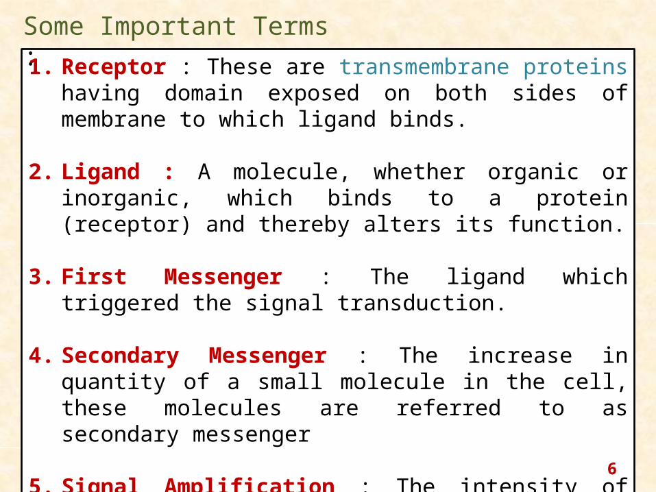
1. Receptor : These are transmembrane proteins having domain exposed on both sides of membrane to which ligand binds.
2. Ligand : A molecule, whether organic or inorganic, which binds to a protein (receptor) and thereby alters its function.
3. First Messenger : The ligand which triggered the signal transduction.
4. Secondary Messenger : The increase in quantity of a small molecule in the cell, these molecules are referred to as secondary messenger
5. Signal Amplification : The intensity of cytosolic signal is much greater than off extracellular signal, this phenomenon is called signal amplification.
Some Important Terms :
6
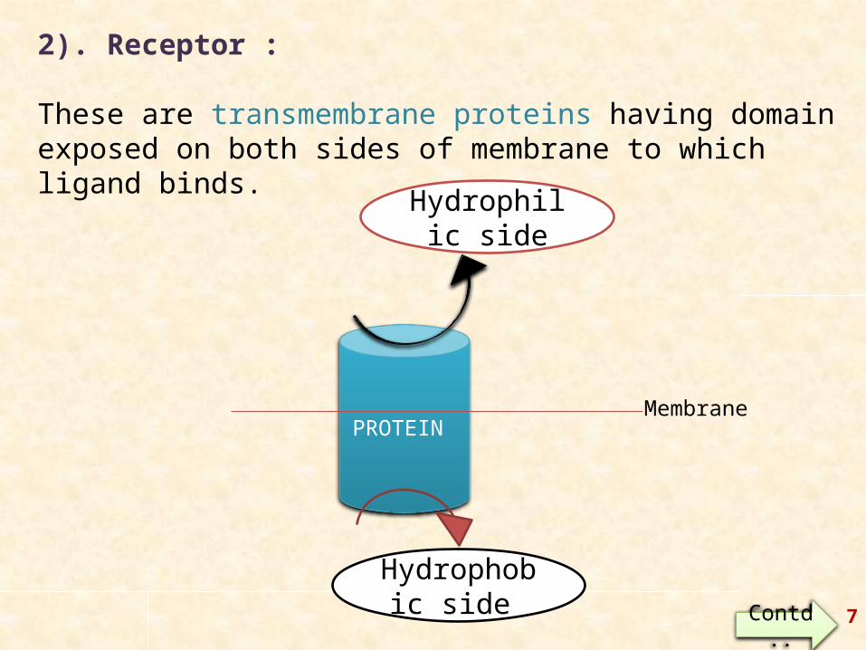
2). Receptor :
These are transmembrane proteins having domain exposed on both sides of membrane to which ligand binds.
PROTEIN
Hydrophilic side
Hydrophobic side
Membrane
Contd.. 7
• It consist of 21-26 hydrophobic amino acids coiled into an alpha-helix.
• These transmembrane proteins are of following two types :
1).One transmembrane Domain
2).Several transmembrane Domain
Contd..8

1).One Transmembrane Domain :
Incase of proteins having a single transmembrane domain, one end is located in cytosol while other is exposed outside the cell.Also categorized as : 1.1). Type 1 or Group 1 proteins :
N-Terminus - outside the CellC-Terminus - present in the cytosol
e.g., Tyrosine kinase receptors.
1.2). Type 2 or Group 2 proteins :
C-Terminus - outside the CellN-Terminus - present in the cytosol
Contd..9

Contd..10
2). Several Transmembrane Domain :
The Protein having two or more transmembrane domain pass through the membrane as many times.Also categorized into :
2.1) Even Number :
Incase of protein having even number of transmembrane domain. Both N-and C-Terminus lies in cytosol.
2.2). Odd Number :
Incase of protein having odd number of transmembrane domain. N-and C-Terminus are on opposite side of membrane.
Contd.. 11

Contd.. 12

FUNCTIONS :
1). Single Transmembrane Domain :
1. It has only a structural function of securing protein in membrane.2. Incase of receptors that bind lipophilic ligands,(ligands located
within the membranes).
2). Several Transmembrane Domain :
3. Polar residues are often present in these domain, these residues may interact with those of other domains to create hydrophilic passage within the hydrophobic membranes.
4. Many proteins have Subunits which may oligomerise within membrane, the transmembrane region of these subunits may interact with each other to trigger changes in the proteins leading to their activation.
13

Receptors are of two types:-
(1) Intracellular receptor :- When ligands are like steroid hormone, thyroid hormone, vitamin D, they diffuses across the plasma membrane of the target cells and bind to the intracellular receptor and activate them which regulate the transcription of specific genes.
(2) Cell surface receptor:- These are again classified into three groups as:-(a) G-protein linked receptor:- These receptor involved in activation of
another membrane bound target protein through a third protein called G-protein.
(b) Enzyme linked receptor :- These on activation either directly function as a enzyme or associated with enzyme mostly kinases causing phosphorylation.
(c) Ion channel linked receptor:- These are involved in neurotransmission through rapid signaling at synapse. 14
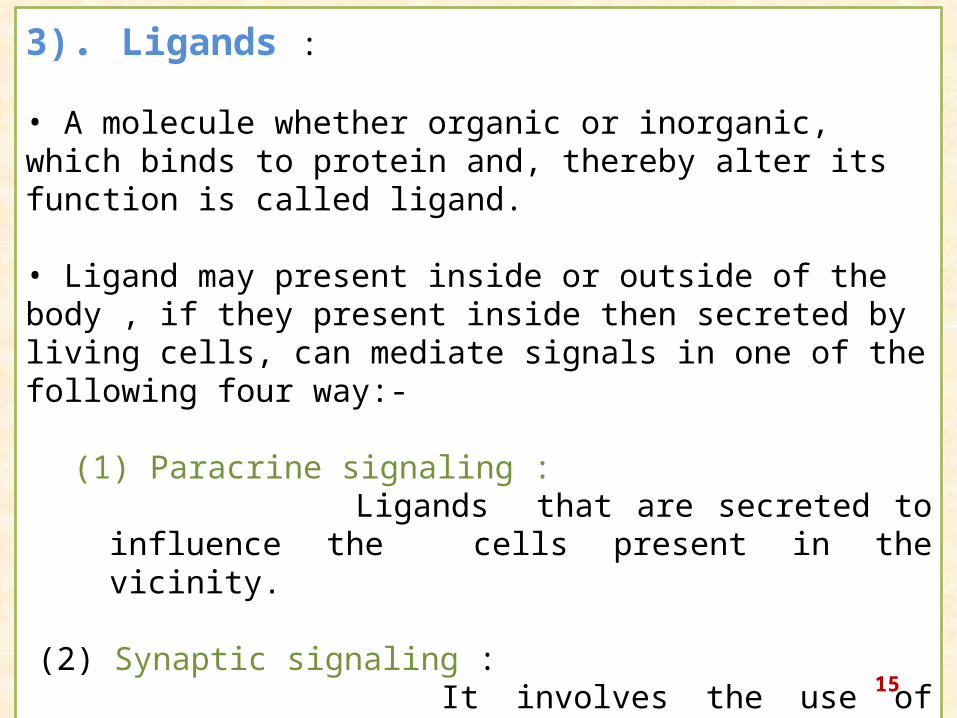
3). Ligands :
• A molecule whether organic or inorganic, which binds to protein and, thereby alter its function is called ligand.
• Ligand may present inside or outside of the body , if they present inside then secreted by living cells, can mediate signals in one of the following four way:-
(1) Paracrine signaling : Ligands that are secreted to influence the cells present in
the vicinity.
(2) Synaptic signaling : It involves the use of neurotransmitter, which transmit the
signal through specialized junction called synapse found between nerve cell.
15

4). MECHANISM :
Catabolic
activities
16

These Catabolic Activities may be grouped into following two basic types :
1). Phosphorylation Cascade.
2). G – Protein Activation.
17

1. Phosphorylation Cascades : When a ligand binds a receptor, the cytosolic domain of
receptor become active and initiates a chain of phosphorylation reactions, which are often termed as Phosphorylation cascade.
Protein kinases : They are receptors that transfer phosphate group to target proteins.
There are three types of protein kinases that catalyze the phosphorylation in cell :
1. Protein tyrosine kinases.2. Protein serine kinases.3. Dual specificity.
18
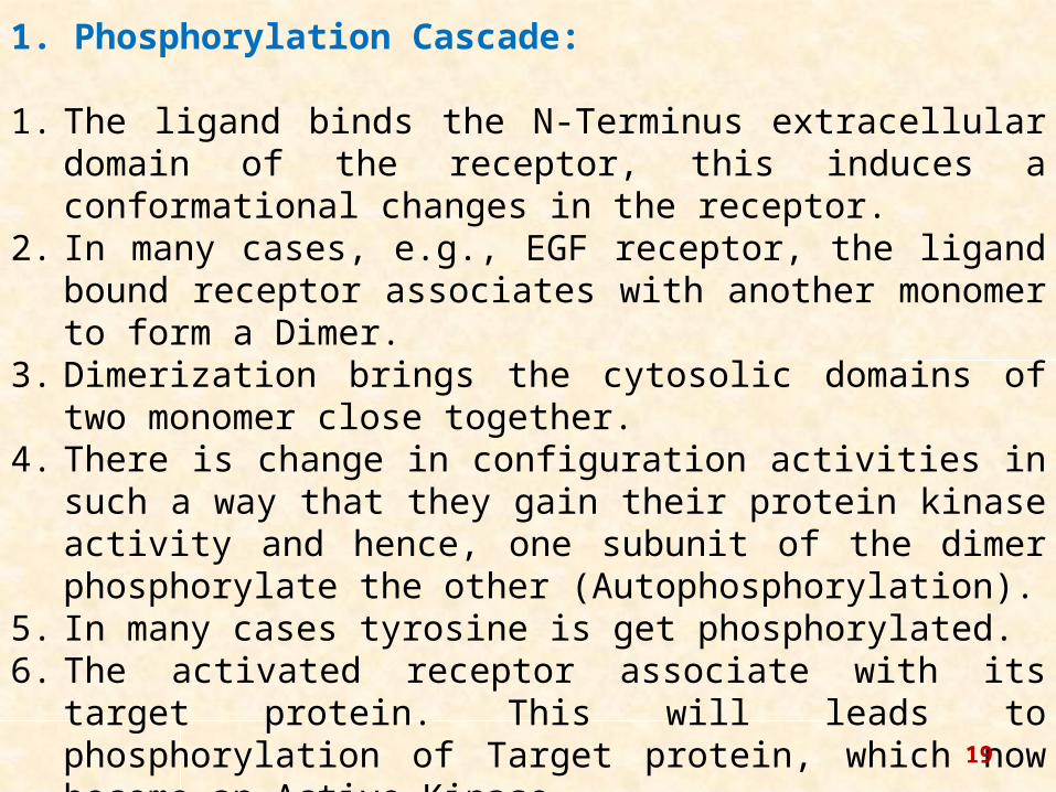
1. Phosphorylation Cascade:
1. The ligand binds the N-Terminus extracellular domain of the receptor, this induces a conformational changes in the receptor.
2. In many cases, e.g., EGF receptor, the ligand bound receptor associates with another monomer to form a Dimer.
3. Dimerization brings the cytosolic domains of two monomer close together.
4. There is change in configuration activities in such a way that they gain their protein kinase activity and hence, one subunit of the dimer phosphorylate the other (Autophosphorylation).
5. In many cases tyrosine is get phosphorylated.6. The activated receptor associate with its target protein. This will
leads to phosphorylation of Target protein, which now become an Active Kinase.
7. This active kinase now activate further kinase. Ultimately such target protein are activated by phosphorylation that affect transcription /cell function. 19

Events take place in phosphorylation cascade are :
Ligand
Ligand binds to receptor
Conformational changes in Receptors
Dimerization Autophosphorylation
Active kinase
Targeted protein
Active Kinase
Active Kinase
P
P PActive kinase
PP P
Contd.
20
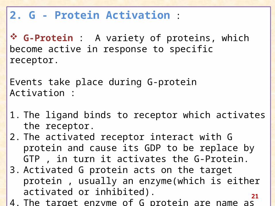
2. G - Protein Activation :
G-Protein : A variety of proteins, which become active in response to specific receptor.
Events take place during G-protein Activation :
1. The ligand binds to receptor which activates the receptor.2. The activated receptor interact with G protein and cause its GDP
to be replace by GTP , in turn it activates the G-Protein.3. Activated G protein acts on the target protein , usually an
enzyme(which is either activated or inhibited).4. The target enzyme of G protein are name as effector protein.5. The effector protein sometimes leads to production of secondary
messenger, which activates a variety of pathway.
21

5).Types of signal transduction pathways :
1. Signal transduction using “second messenger”.
2. Insulin Signaling .
3. Ras-Mediated signal transduction pathways.
4. JAK-STAT signal transduction pathway(Mediated by cytokine receptor).
5. Steroid-Mediated signal transduction.
22
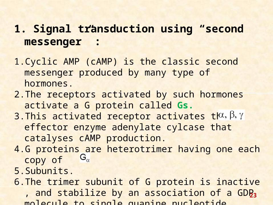
1. Signal transduction using “second messenger” :
1. Cyclic AMP (cAMP) is the classic second messenger produced by many type of hormones.
2. The receptors activated by such hormones activate a G protein called Gs.
3. This activated receptor activates the effector enzyme adenylate cylcase that catalyses cAMP production.
4. G proteins are heterotrimer having one each copy of 5. Subunits.6. The trimer subunit of G protein is inactive , and stabilize by an
association of a GDP molecule to single guanine nucleotide binding site of
23
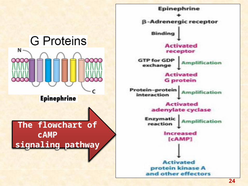
The flowchart of cAMP signaling pathway
24

Mechanism of cAMP signal transduction pathway.25
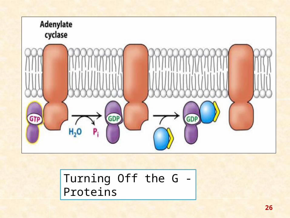
Turning Off the G - Proteins
26

CALCIUM
The release of calcium ions from the endoplasmic reticulum into the cytosol results in its binding to signaling proteins that are then activated; it is then sequestered in the smooth endoplasmic reticulum and the mitochondria. Calcium is used in many processes including muscle contraction, neurotransmitter release from nerve endings and cell migration.
LIPOPHILICS
Lipophilic second messenger molecules are derived from lipids residing in cellular membranes; enzymes stimulated by activated receptors activate the lipids by modifying them. Examples include diacylglycerol and ceramide, the former required for the activation of protein kinase C.
NITRIC
OXIDE
Nitric oxide (NO) acts as a second messenger because it is a free radical that can diffuse through the plasma membrane and affect nearby cells. It is toxic in high concentrations and causes damage during stroke , but is the cause of many other functions like relaxation of blood vessels etc.
Some Other Examples of Second Messenger :
27

2. Insulin Signaling Pathway :
This pathway is take place by Phosphorylation Cascade Mechanism.
3. Ras-Mediated Signal Transduction Pathways :
1. Among the proteins that are commonly recruited to activate tyrosine kinase receptors is the G-protein Ras.
2. Ras is a member of a large family of monomeric (single subunit) G-proteins.
3. Ras is active when bound to GTP and inactive when bound to GDP. It is also a GTPase and it hydrolyzes GTP to inactivate itself.
28

Regulation of Ras Activity 29

GROWTH FACTOR
GROWTH FACTOR RECEPTOR (Tyr Kinase)
RAS mechanism
Kinase Cascade (Ser/Thr Kinase)
Transcription Factor
The pathway for activation of phenotypic changes through Ras-mediated pathway.
30

4. JAK-STAT Signal Transduction Pathway (Mediated by cytokine receptor) :
Cellular responses to cytokines and growth factors are mediated by the evolutionarily conserved Janus kinase/signal transducers and activators of transcription (JAK/STAT) signaling pathway.
The binding of extracellular ligand leads to pathway activation via changes to the receptors that permit the intracellular JAKs associated with them to phosphorylate one another. (Douglas A. H.,2012).
JAK Janus Kinase STAT Signal Transducer and
Activator of transcription
31

The JAK-STAT system consists of three main components:
(1) a receptor (green), which penetrates the cell membrane
(2) Janus kinase (JAK) (yellow), which is bound to the receptor and
(3) Signal Transducer and Activator of Transcription (STAT) (blue), which carries the signal into the nucleus and DNA.
32

5. STEROID- Mediated Signal Transduction :
•Steroid receptors are a subclass of nuclear receptors, located primarily within the cytosol. In the absence of steroid hormone, the receptors cling together in a complex called aporeceptor complex, which also contains chaperone proteins (also known as heatshock proteins or Hsps).
•The Hsps are necessary to activate the receptor by assisting the protein to fold in a way such that the signal sequence which enables its passage into the nucleus is accessible.
•Steroid receptors can also have a repressive effect on gene expression, when their transactivation domain is hidden so it cannot activate transcription. Furthermore, steroid receptor activity can be enhanced by phosphorylation of serine residues at their N-terminal end, as a result of another signal transduction pathway, for example, a by a growth factor. This behaviour is called crosstalk. 33

34

1. B.D.Singh (2010). “Genetics” , Kalyani Publication ,New Dehli.
2. Douglas A. Harrison (2012), Cold Spring Harb Perspect Biol ; doi: 10.1101/cshperspect.a011205
3. Manning G, Whyte DB. et al. (2002). "The protein kinase complement of the human genome". Science 298 (5600): 1912–1934. doi:10.1126/science.1075762.PMID 12471243.
4. http://en.wikipedia.org/wiki/Steroid_hormone_receptor, accessed on 16th September,2014.
Refrences :
35

36
Thanks ….
![[VII]. Regulation of Gene Expression Via Signal Transduction Reading List VII: Signal transduction Signal transduction in biological systems](https://img.pdfslide.us/doc/110x75/56649e385503460f94b28319/vii-regulation-of-gene-expression-via-signal-transduction-reading-list-vii.jpg)