Embed Size (px)
Citation preview
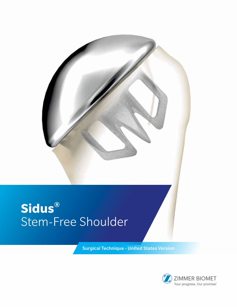
Sidus® Stem-Free Shoulder
Surgical Technique - United States Version

Table of Contents
Introduction .......................................................................................................4
Preoperative/Intraoperative Considerations .......................................................6
Surgical Technique Summary ..............................................................................7
Patient Positioning & Incision ..............................................................................8
Humeral Technique .............................................................................................9 Humeral Head Identification, Preparation and Resection ..................................... 9 Glenoid Options ................................................................................................ 17 Head Size Determination and Positioning .......................................................... 18 Anchor Size Determination, Preparation and Implantation ................................. 20 Head Preparation and Implantation ................................................................... 26
Revision/Intra-Operative Correction Surgical Steps ..........................................28
Postoperative Treatment ..................................................................................30
Appendix .......................................................................................................... 31 Technical Specifications ..................................................................................... 31 Indications & Contraindications ......................................................................... 32 Clinical Results .................................................................................................. 33 Adverse Events Reporting* ................................................................................ 34
* Please note that Adverse Events Reporting for the US Investigational Device Exemption clinical study, the Europe Post-Market Clinical Follow-up study and the Bigliani/Flatow historical control study are located in the Appendix.
4 | Sidus Stem-Free Shoulder Surgical Technique
IntroductionZimmer Biomet continues to lead the way in bone-preserving arthroplasty solutions with the Sidus Stem-Free Shoulder System. Sidus Shoulder was launched in Europe in 2012 and a subsequent Post-Market Clinical Follow-up (PMCF) study2 was conducted involving 152 patients. In addition, a clinical IDE (Investigational Device Exemption) study1 was initiated in the United States and Canada in 2015, that included another 95 patients. To date, both studies demonstrate strong clinical performance. Sidus Shoulder is a total shoulder arthroplasty solution for patients with good bone stock that have either osteoarthritis, posttraumatic arthrosis, focal avascular necrosis of the humeral head or who had previous surgeries of the shoulder that do not compromise the fixation. A full summary of the clinical data is provided in the Appendix.
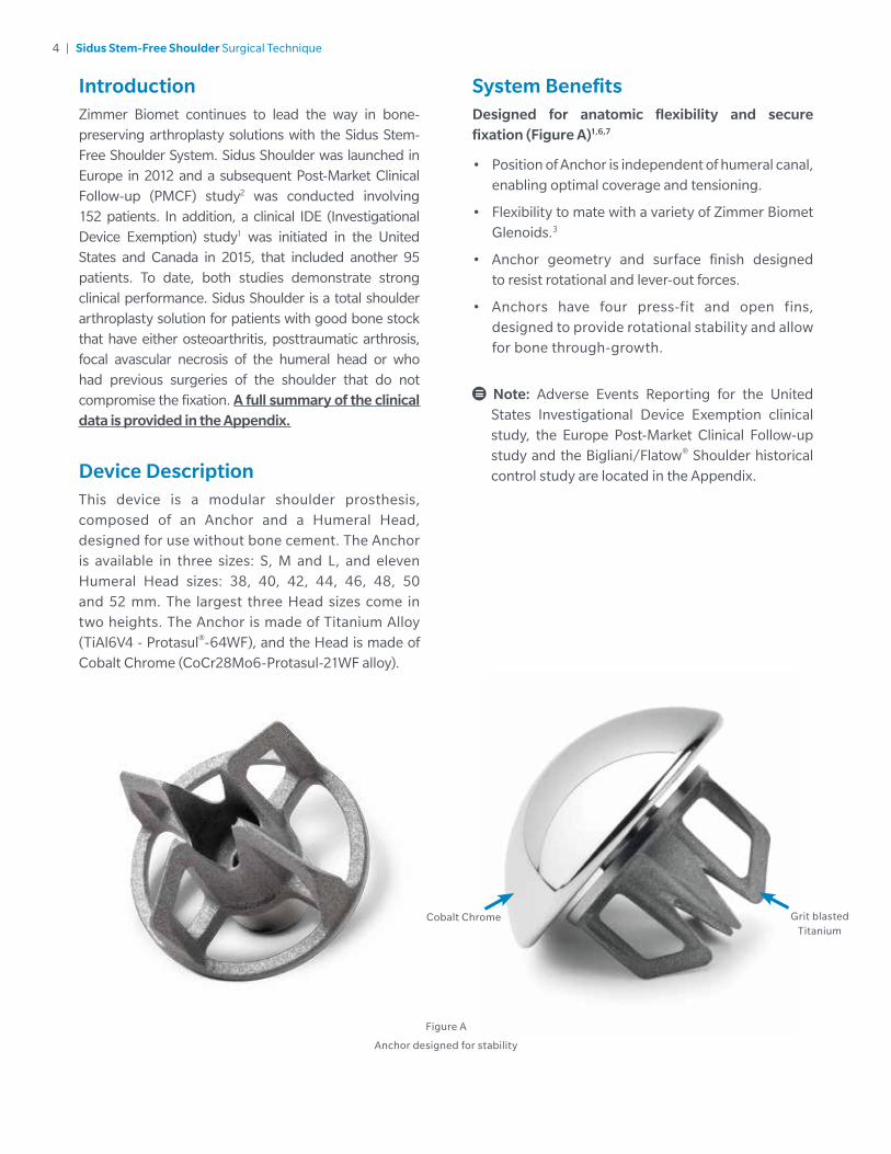
Device DescriptionThis device is a modular shoulder prosthesis, composed of an Anchor and a Humeral Head, designed for use without bone cement. The Anchor is available in three sizes: S, M and L, and eleven Humeral Head sizes: 38, 40, 42, 44, 46, 48, 50 and 52 mm. The largest three Head sizes come in two heights. The Anchor is made of Titanium Alloy (TiAl6V4 - Protasul®-64WF), and the Head is made of Cobalt Chrome (CoCr28Mo6-Protasul-21WF alloy).
System BenefitsDesigned for anatomic flexibility and secure fixation (Figure A)1,6,7
• Position of Anchor is independent of humeral canal, enabling optimal coverage and tensioning.
• Flexibility to mate with a variety of Zimmer Biomet Glenoids.3
• Anchor geometry and surface finish designed to resist rotational and lever-out forces.
• Anchors have four press-fit and open fins, designed to provide rotational stability and allow for bone through-growth.
Note: Adverse Events Reporting for the United States Investigational Device Exemption clinical study, the Europe Post-Market Clinical Follow-up study and the Bigliani/Flatow® Shoulder historical control study are located in the Appendix.
Figure A
Anchor designed for stability
Grit blasted Titanium
Cobalt Chrome
5 | Sidus Stem-Free Shoulder Surgical Technique
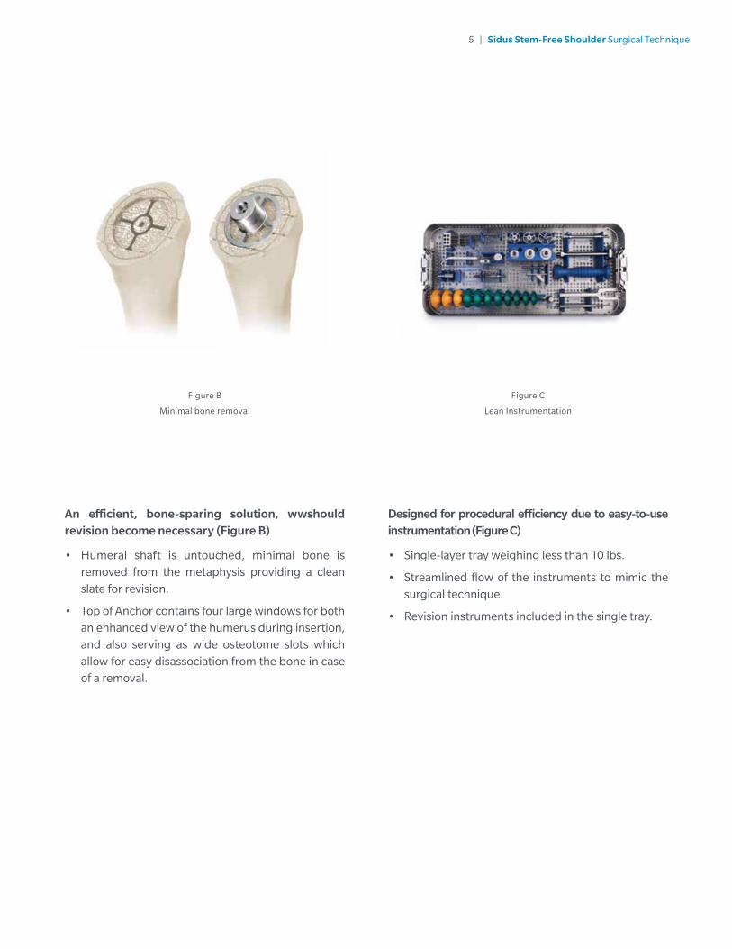
Designed for procedural efficiency due to easy-to-use instrumentation (Figure C)
• Single-layer tray weighing less than 10 lbs.
• Streamlined flow of the instruments to mimic the surgical technique.
• Revision instruments included in the single tray.
Figure B
Minimal bone removal
Figure C
Lean Instrumentation
An efficient, bone-sparing solution, wwshould revision become necessary (Figure B)
• Humeral shaft is untouched, minimal bone is removed from the metaphysis providing a clean slate for revision.
• Top of Anchor contains four large windows for both an enhanced view of the humerus during insertion, and also serving as wide osteotome slots which allow for easy disassociation from the bone in case of a removal.
6 | Sidus Stem-Free Shoulder Surgical Technique
Preoperative ConsiderationsTemplating
Preoperative evaluation of the Humerus using the Sidus Stem-Free Shoulder templates helps determine the size of the prosthesis and level of the head resection. The goal is to make a resection that matches the anatomy of the patient.
• Preoperative templating can be used to estimate the implant sizes prior to surgery (Figure D).
X-Ray Template
Figure D
Preoperative A/P x-rays with and without Sidus X-Ray Template
Intraoperative ConsiderationsBone Test
• To achieve a good outcome, the patient must have adequate bone stock to support the fixation of the implant (Figure E).
• Poor metaphyseal bone quality assessment is not possible until the humeral head has been resected. As a result, you should always be prepared with a back-up system.
Note: It is not advisable to perform a complete lesser tuberosity osteotomy.
Note: It is anticipated that up to 33% of total shoulder candidates lack sufficient bone stock to support a stemless device.4
Good Bone Quality
Soft/Weak bone quality
Figure E
Thumb Test
7 | Sidus Stem-Free Shoulder Surgical Technique
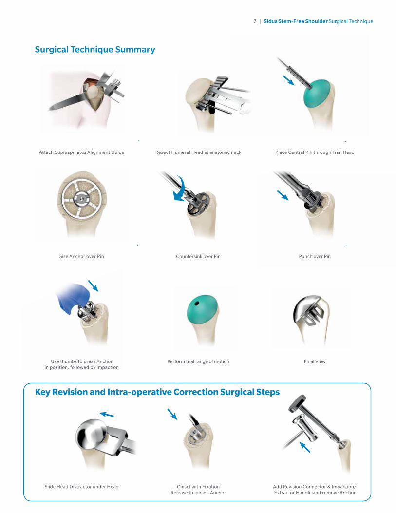
Attach Supraspinatus Alignment Guide Resect Humeral Head at anatomic neck
Punch over Pin
Final View
Add Revision Connector & Impaction/Extractor Handle and remove Anchor
Use thumbs to press Anchor in position, followed by impaction
Slide Head Distractor under Head
Perform trial range of motion
Chisel with Fixation Release to loosen Anchor
Place Central Pin through Trial Head
Size Anchor over Pin Countersink over Pin
Key Revision and Intra-operative Correction Surgical Steps
Surgical Technique Summary
8 | Sidus Stem-Free Shoulder Surgical Technique
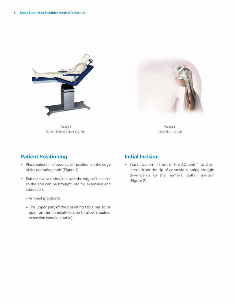
Initial Incision• Start incision in front of the AC joint 1 to 2 cm
lateral from the tip of coracoid running straight downwards to the humeral delta insertion (Figure 2).
Patient Positioning• Place patient in a beach chair position on the edge
of the operating table (Figure 1).
• Extend involved shoulder over the edge of the table so the arm can be brought into full extension and adduction.
– Armrest is optional.
– The upper part of the operating table has to be open on the homolateral side to allow shoulder extension (shoulder table).
Figure 1
Patient in beach chair position
Figure 2
Initial Skin incision
9 | Sidus Stem-Free Shoulder Surgical Technique
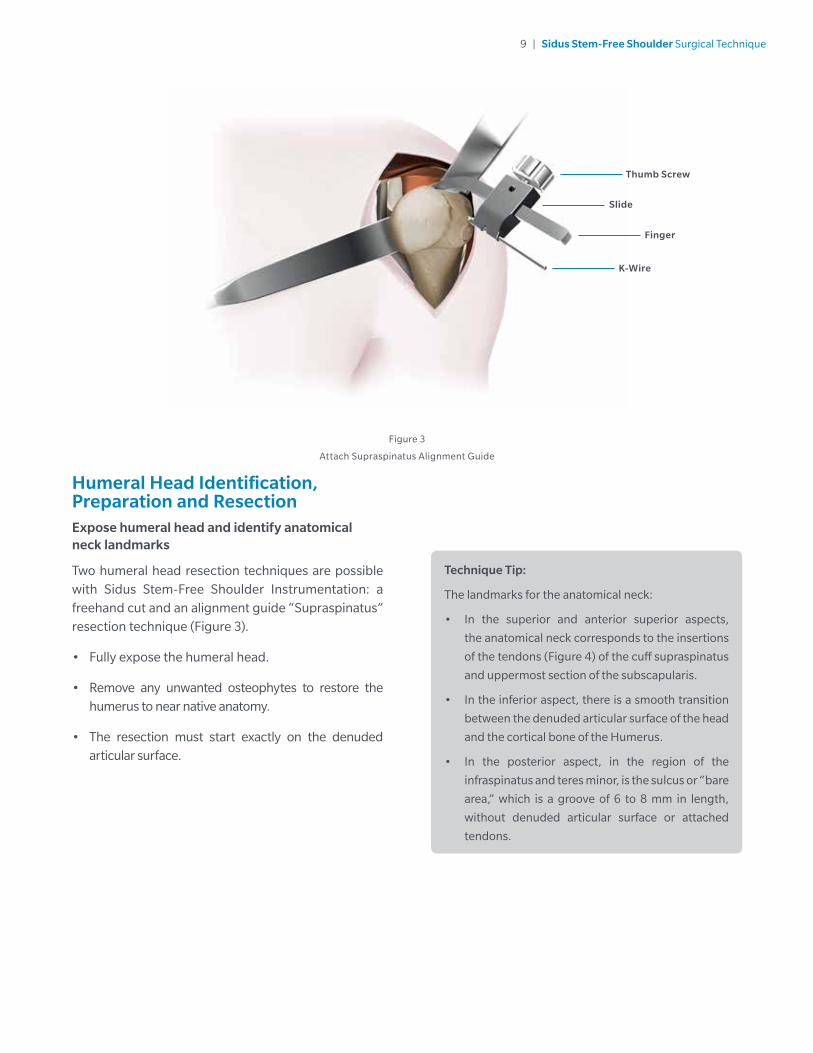
Humeral Head Identification, Preparation and ResectionExpose humeral head and identify anatomical neck landmarks
Two humeral head resection techniques are possible with Sidus Stem-Free Shoulder Instrumentation: a freehand cut and an alignment guide “Supraspinatus” resection technique (Figure 3).
• Fully expose the humeral head.
• Remove any unwanted osteophytes to restore the humerus to near native anatomy.
• The resection must start exactly on the denuded articular surface.
Technique Tip:
The landmarks for the anatomical neck:
• In the superior and anterior superior aspects,
the anatomical neck corresponds to the insertions
of the tendons (Figure 4) of the cuff supraspinatus
and uppermost section of the subscapularis.
• In the inferior aspect, there is a smooth transition
between the denuded articular surface of the head
and the cortical bone of the Humerus.
• In the posterior aspect, in the region of the
infraspinatus and teres minor, is the sulcus or “bare
area,” which is a groove of 6 to 8 mm in length,
without denuded articular surface or attached
tendons.
Figure 3
Attach Supraspinatus Alignment Guide
Thumb Screw
Finger
Slide
K-Wire
10 | Sidus Stem-Free Shoulder Surgical Technique
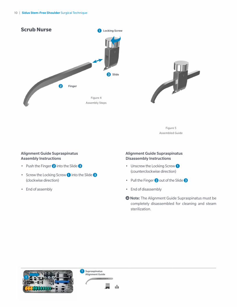
Alignment Guide Supraspinatus Disassembly Instructions
• Unscrew the Locking Screw 1 (counterclockwise direction)
• Pull the Finger 2 out of the Slide 3
• End of disassembly
Note: The Alignment Guide Supraspinatus must be completely disassembled for cleaning and steam sterilization.
Alignment Guide Supraspinatus Assembly Instructions
• Push the Finger 2 into the Slide 3
• Screw the Locking Screw 1 into the Slide 3 (clockwise direction)
• End of assembly
Figure 5
Assembled Guide
Figure 4
Assembly Steps
Finger
Slide
2
3
Supraspinatus Alignment Guide
1
1
Scrub Nurse Locking Screw 1
11 | Sidus Stem-Free Shoulder Surgical Technique
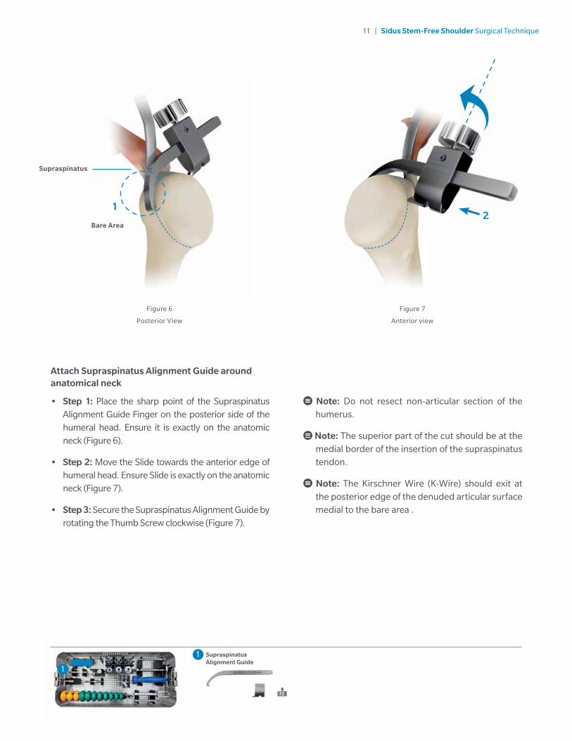
Supraspinatus
Bare Area
Attach Supraspinatus Alignment Guide around anatomical neck
• Step 1: Place the sharp point of the Supraspinatus Alignment Guide Finger on the posterior side of the humeral head. Ensure it is exactly on the anatomic neck (Figure 6).
• Step 2: Move the Slide towards the anterior edge of humeral head. Ensure Slide is exactly on the anatomic neck (Figure 7).
• Step 3: Secure the Supraspinatus Alignment Guide by rotating the Thumb Screw clockwise (Figure 7).
2
Figure 6
Posterior View
Figure 7
Anterior view
1
Note: Do not resect non-articular section of the humerus.
Note: The superior part of the cut should be at the medial border of the insertion of the supraspinatus tendon.
Note: The Kirschner Wire (K-Wire) should exit at the posterior edge of the denuded articular surface medial to the bare area .
Supraspinatus Alignment Guide
1
1
12 | Sidus Stem-Free Shoulder Surgical Technique
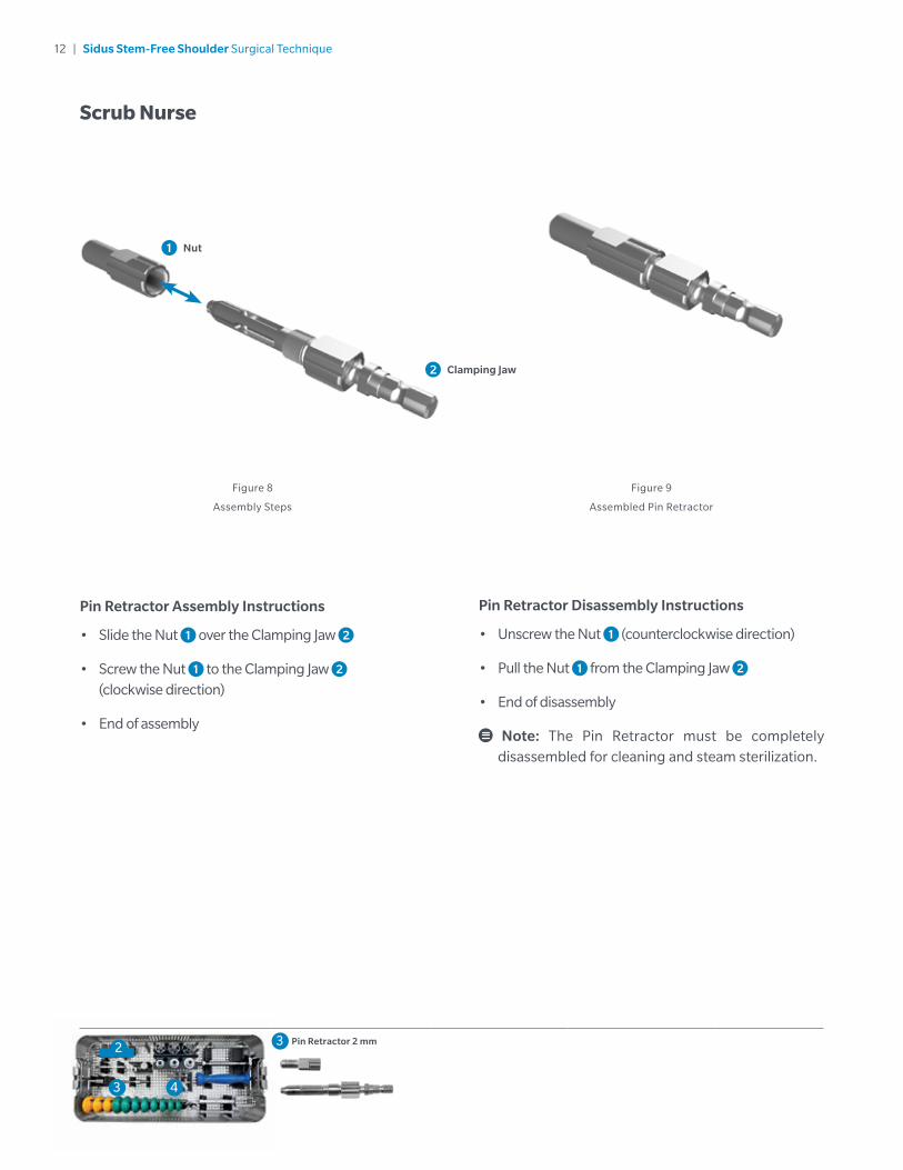
Pin Retractor Disassembly Instructions
• Unscrew the Nut 1 (counterclockwise direction)
• Pull the Nut 1 from the Clamping Jaw 2
• End of disassembly
Note: The Pin Retractor must be completely disassembled for cleaning and steam sterilization.
Pin Retractor Assembly Instructions
• Slide the Nut 1 over the Clamping Jaw 2
• Screw the Nut 1 to the Clamping Jaw 2 (clockwise direction)
• End of assembly
Figure 8
Assembly Steps
Figure 9
Assembled Pin Retractor
Nut
Clamping Jaw
1
2
4
2
3
3 Pin Retractor 2 mm
Scrub Nurse
13 | Sidus Stem-Free Shoulder Surgical Technique
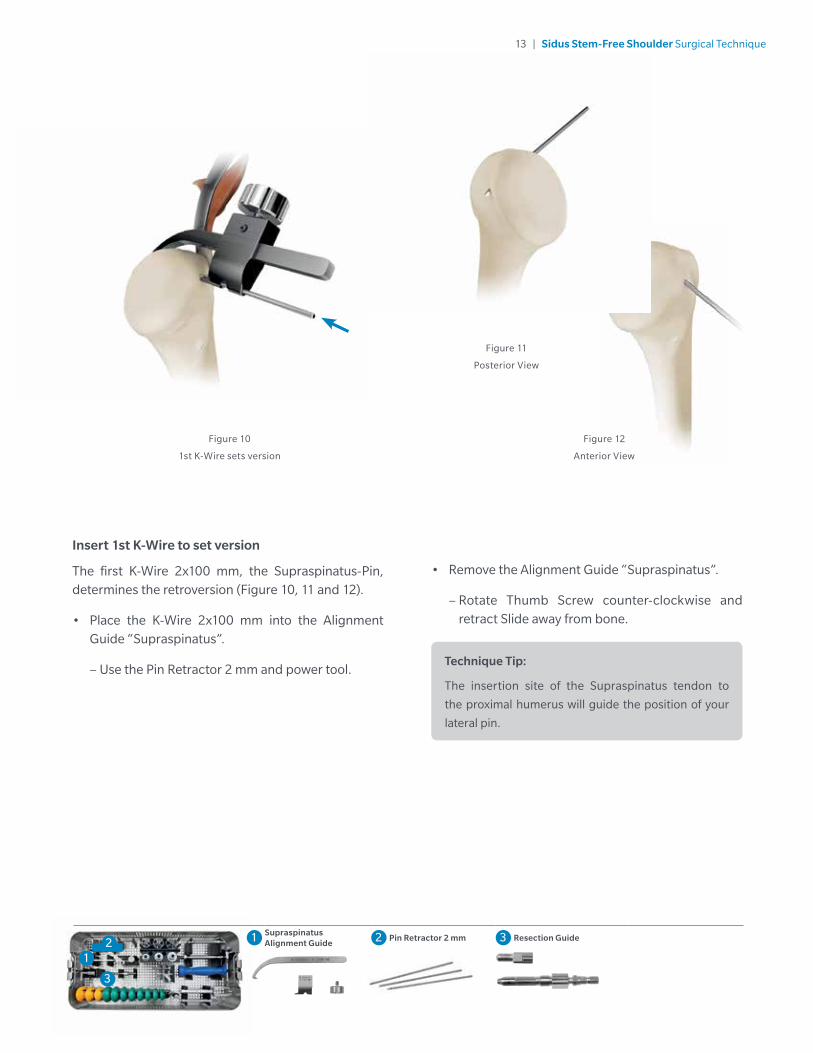
Insert 1st K-Wire to set version
The first K-Wire 2x100 mm, the Supraspinatus-Pin, determines the retroversion (Figure 10, 11 and 12).
• Place the K-Wire 2x100 mm into the Alignment Guide “Supraspinatus”.
– Use the Pin Retractor 2 mm and power tool.
Figure 12
Anterior View
Figure 10
1st K-Wire sets version
Figure 11
Posterior View
• Remove the Alignment Guide “Supraspinatus”.
– Rotate Thumb Screw counter-clockwise and retract Slide away from bone.
Technique Tip:
The insertion site of the Supraspinatus tendon to
the proximal humerus will guide the position of your
lateral pin.
2
3
1 Supraspinatus Alignment Guide 2 Pin Retractor 2 mm 3 Resection Guide
1
14 | Sidus Stem-Free Shoulder Surgical Technique
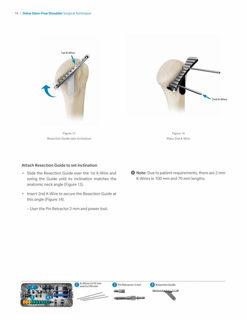
Attach Resection Guide to set inclination
• Slide the Resection Guide over the 1st K-Wire and swing the Guide until its inclination matches the anatomic neck angle (Figure 13).
• Insert 2nd K-Wire to secure the Resection Guide at this angle (Figure 14).
– User the Pin Retractor 2 mm and power tool.
Note: Due to patient requirements, there are 2 mm K-Wires in 100 mm and 70 mm lengths.
2nd K-Wire
Figure 13
Resection Guide sets inclination
Figure 14
Place 2nd K-Wire
1st K-Wire
4
2
3
2 K-Wires 2x70 mm and 2x100 mm 3 Pin Retractor 2 mm 4 Resection Guide
15 | Sidus Stem-Free Shoulder Surgical Technique
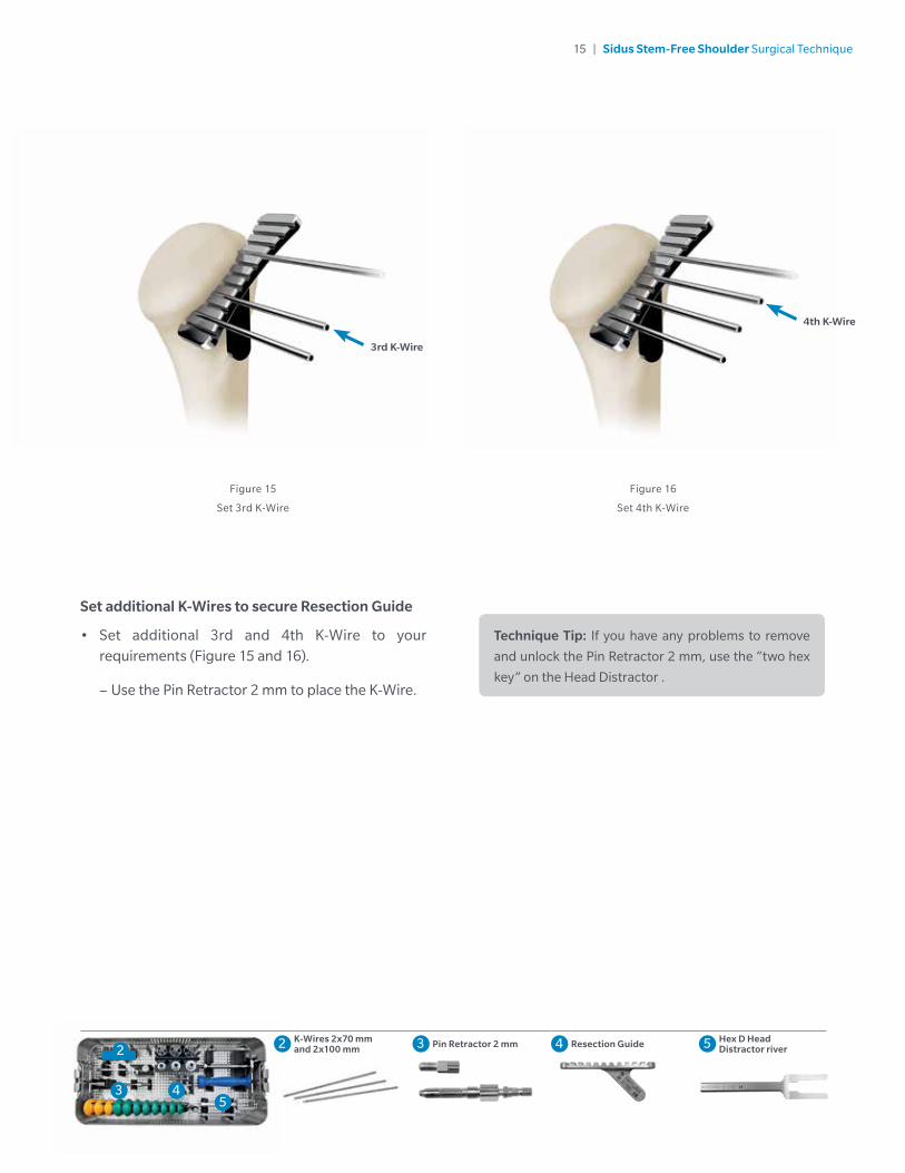
Set additional K-Wires to secure Resection Guide
• Set additional 3rd and 4th K-Wire to your requirements (Figure 15 and 16).
– Use the Pin Retractor 2 mm to place the K-Wire.
Figure 15
Set 3rd K-Wire
Figure 16
Set 4th K-Wire
Technique Tip: If you have any problems to remove
and unlock the Pin Retractor 2 mm, use the “two hex
key” on the Head Distractor .
4
2
35
3rd K-Wire
4th K-Wire
2 K-Wires 2x70 mm and 2x100 mm 3 Pin Retractor 2 mm 4 Resection Guide 5 Hex D Head
Distractor river
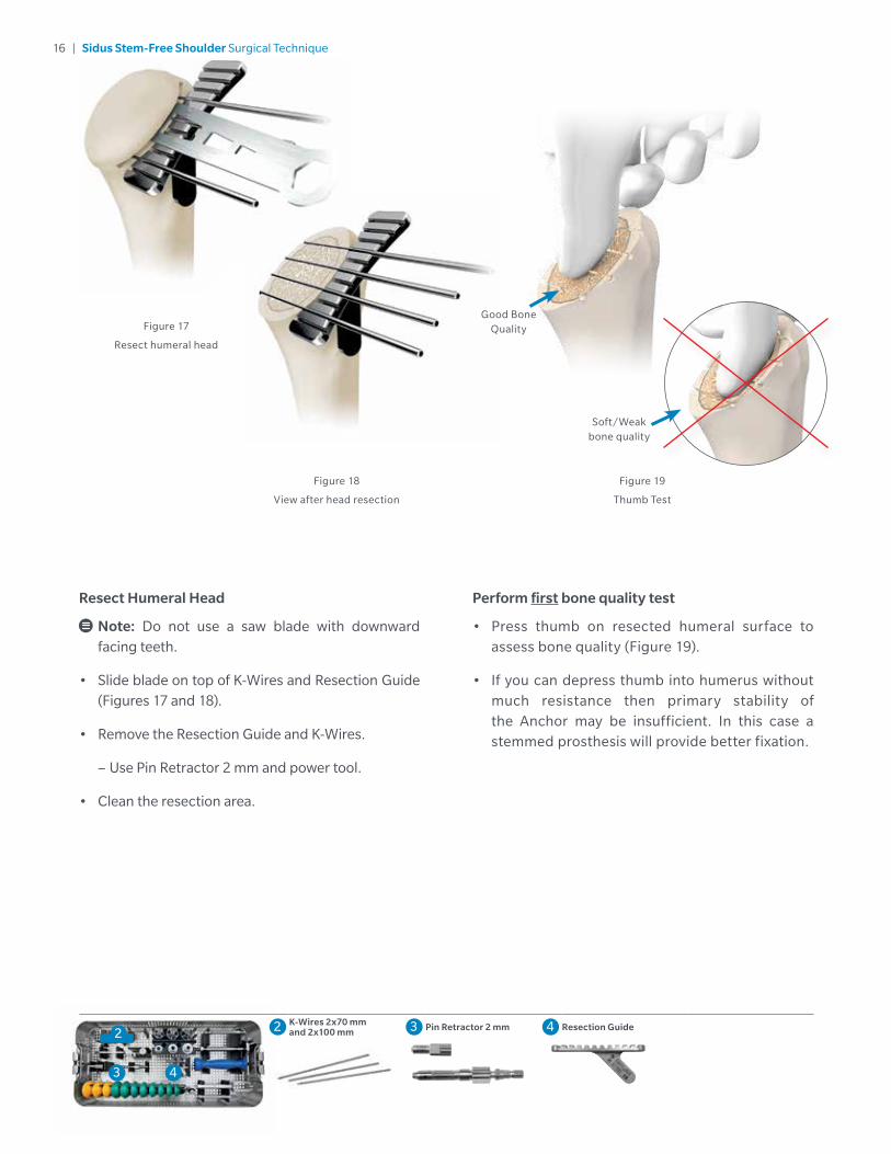
16 | Sidus Stem-Free Shoulder Surgical Technique
Figure 18
View after head resection
Figure 19
Thumb Test
4
2
3
Resect Humeral Head
Note: Do not use a saw blade with downward facing teeth.
• Slide blade on top of K-Wires and Resection Guide (Figures 17 and 18).
• Remove the Resection Guide and K-Wires.
– Use Pin Retractor 2 mm and power tool.
• Clean the resection area.
Figure 17
Resect humeral head
Perform first bone quality test
• Press thumb on resected humeral surface to assess bone quality (Figure 19).
• If you can depress thumb into humerus without much resistance then primary stability of the Anchor may be insufficient. In this case a stemmed prosthesis will provide better fixation.
2 K-Wires 2x70 mm and 2x100 mm 3 Pin Retractor 2 mm 4 Resection Guide
Good Bone Quality
Soft/Weak bone quality
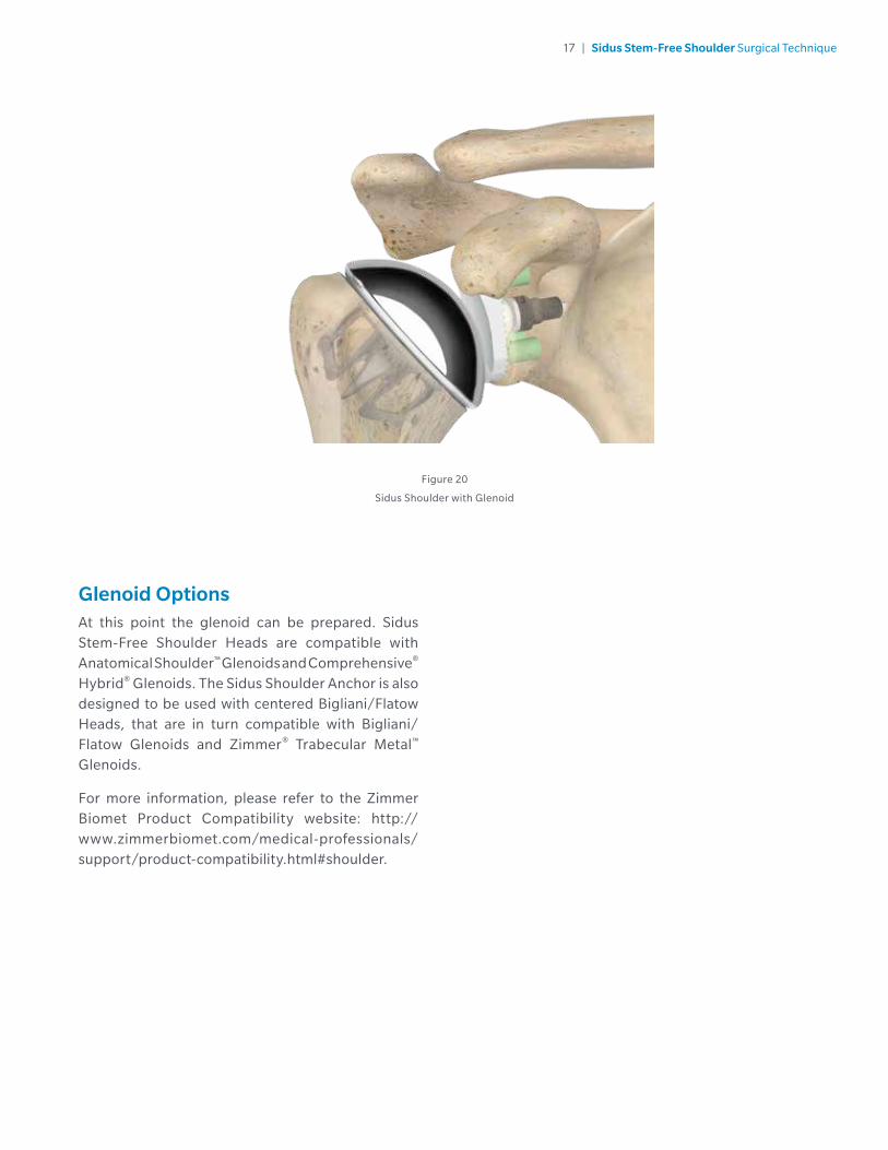
17 | Sidus Stem-Free Shoulder Surgical Technique
Glenoid OptionsAt this point the glenoid can be prepared. Sidus Stem-Free Shoulder Heads are compatible with Anatomical Shoulder™ Glenoids and Comprehensive® Hybrid® Glenoids. The Sidus Shoulder Anchor is also designed to be used with centered Bigliani/Flatow Heads, that are in turn compatible with Bigliani/Flatow Glenoids and Zimmer® Trabecular Metal™ Glenoids.
For more information, please refer to the Zimmer Biomet Product Compatibility website: http://www.zimmerbiomet.com/medical-professionals/support/product-compatibility.html#shoulder.
Figure 20
Sidus Shoulder with Glenoid
18 | Sidus Stem-Free Shoulder Surgical Technique
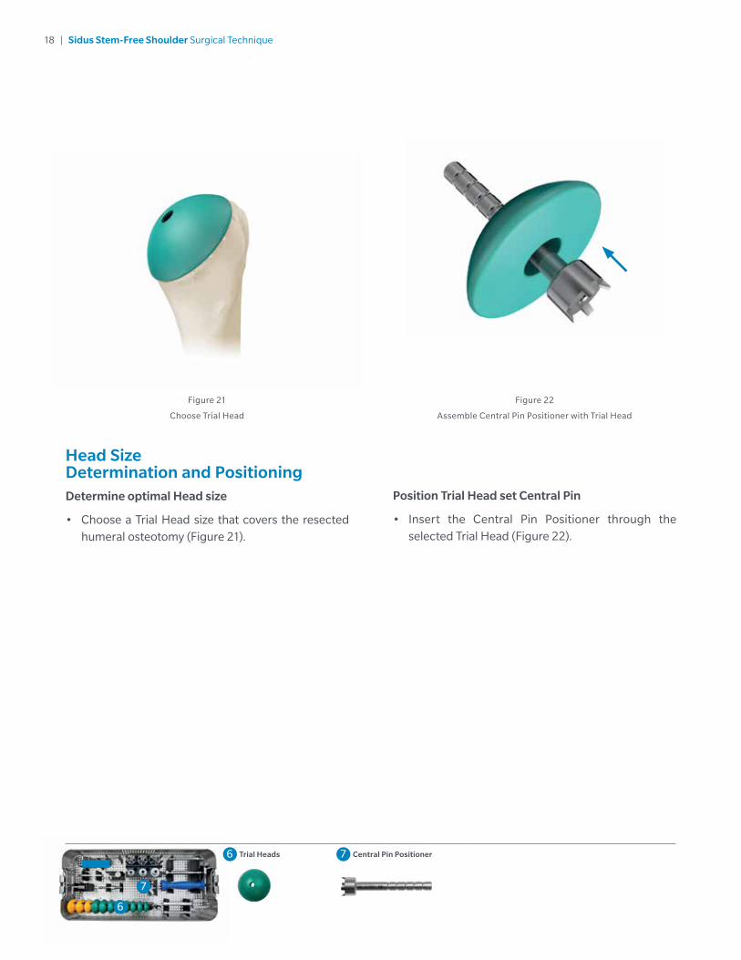
Position Trial Head set Central Pin
• Insert the Central Pin Positioner through the selected Trial Head (Figure 22).
Head Size Determination and PositioningDetermine optimal Head size
• Choose a Trial Head size that covers the resected humeral osteotomy (Figure 21).
Figure 21
Choose Trial Head
Figure 22
Assemble Central Pin Positioner with Trial Head
6
6 Trial Heads 7 Central Pin Positioner
7
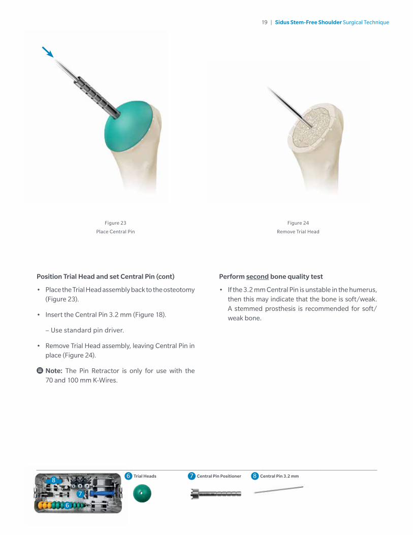
19 | Sidus Stem-Free Shoulder Surgical Technique
Position Trial Head and set Central Pin (cont)
• Place the Trial Head assembly back to the osteotomy (Figure 23).
• Insert the Central Pin 3.2 mm (Figure 18).
– Use standard pin driver.
• Remove Trial Head assembly, leaving Central Pin in place (Figure 24).
Note: The Pin Retractor is only for use with the 70 and 100 mm K-Wires.
Figure 23
Place Central Pin
Figure 24
Remove Trial Head
Perform second bone quality test
• If the 3.2 mm Central Pin is unstable in the humerus, then this may indicate that the bone is soft/weak. A stemmed prosthesis is recommended for soft/weak bone.
6
86 Trial Heads 7 Central Pin Positioner 8 Central Pin 3.2 mm
7
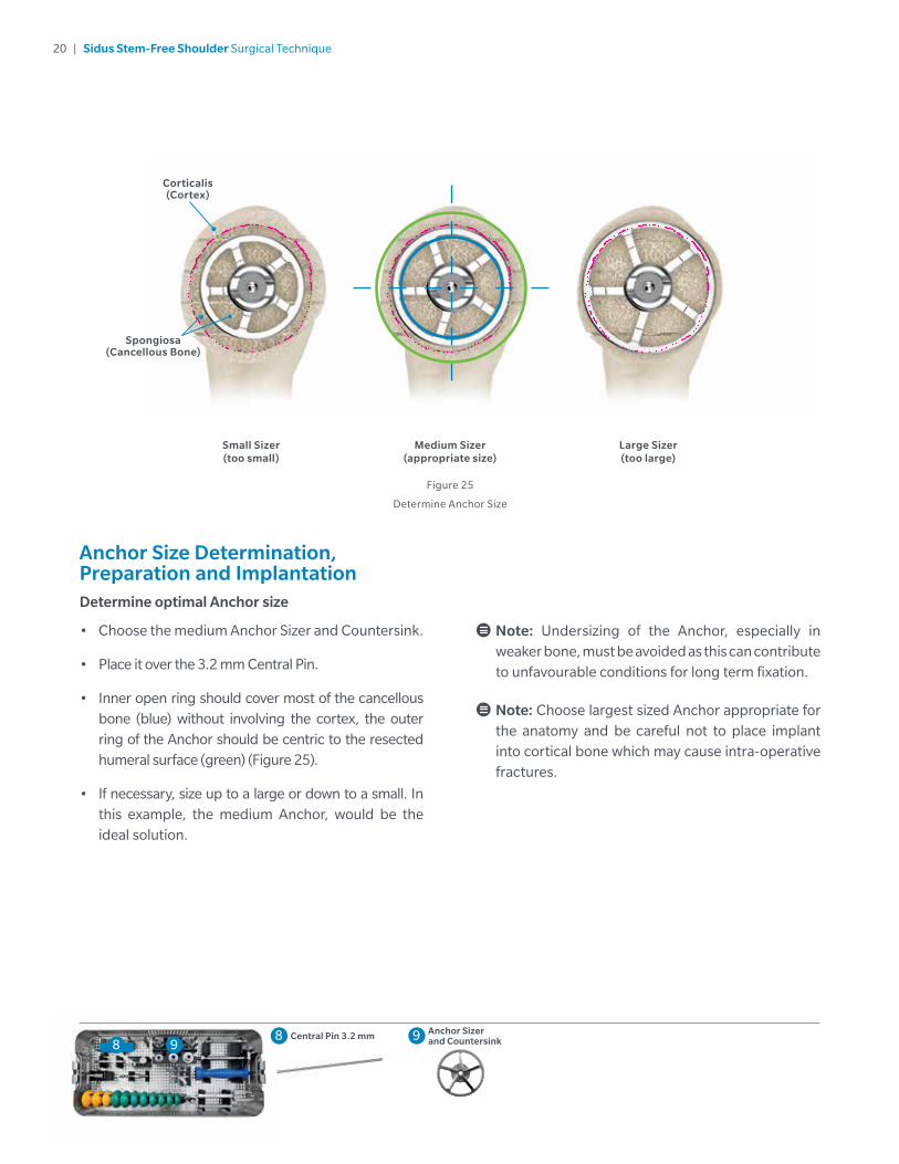
20 | Sidus Stem-Free Shoulder Surgical Technique
Anchor Size Determination, Preparation and ImplantationDetermine optimal Anchor size
• Choose the medium Anchor Sizer and Countersink.
• Place it over the 3.2 mm Central Pin.
• Inner open ring should cover most of the cancellous bone (blue) without involving the cortex, the outer ring of the Anchor should be centric to the resected humeral surface (green) (Figure 25).
• If necessary, size up to a large or down to a small. In this example, the medium Anchor, would be the ideal solution.
Small Sizer(too small)
Figure 25
Determine Anchor Size
Medium Sizer(appropriate size)
Large Sizer(too large)
Note: Undersizing of the Anchor, especially in weaker bone, must be avoided as this can contribute to unfavourable conditions for long term fixation.
Note: Choose largest sized Anchor appropriate for the anatomy and be careful not to place implant into cortical bone which may cause intra-operative fractures.
88 Central Pin 3.2 mm 9 Anchor Sizer
and Countersink 9
Corticalis (Cortex)
Spongiosa (Cancellous Bone)
21 | Sidus Stem-Free Shoulder Surgical Technique
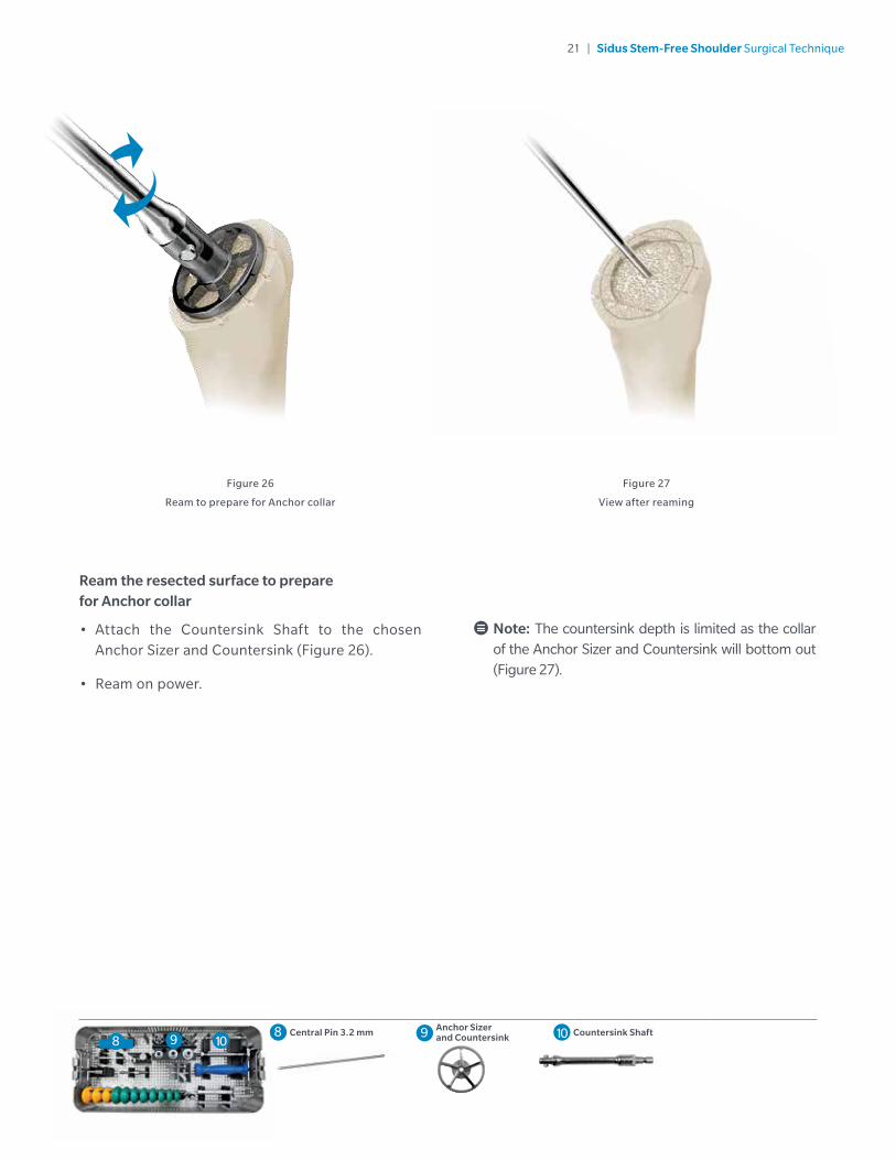
Ream the resected surface to prepare for Anchor collar
• Attach the Countersink Shaft to the chosen Anchor Sizer and Countersink (Figure 26).
• Ream on power.
Note: The countersink depth is limited as the collar of the Anchor Sizer and Countersink will bottom out (Figure 27).
Figure 26
Ream to prepare for Anchor collar
Figure 27
View after reaming
88 Central Pin 3.2 mm 9 Anchor Sizer
and Countersink 10 Countersink Shaft9 10
22 | Sidus Stem-Free Shoulder Surgical Technique
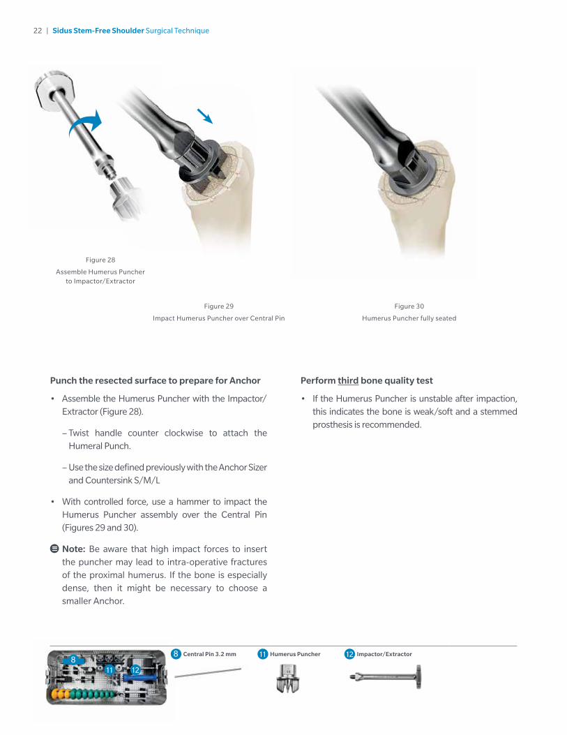
Perform third bone quality test
• If the Humerus Puncher is unstable after impaction, this indicates the bone is weak/soft and a stemmed prosthesis is recommended.
Punch the resected surface to prepare for Anchor
• Assemble the Humerus Puncher with the Impactor/Extractor (Figure 28).
– Twist handle counter clockwise to attach the Humeral Punch.
– Use the size defined previously with the Anchor Sizer and Countersink S/M/L
• With controlled force, use a hammer to impact the Humerus Puncher assembly over the Central Pin (Figures 29 and 30).
Note: Be aware that high impact forces to insert the puncher may lead to intra-operative fractures of the proximal humerus. If the bone is especially dense, then it might be necessary to choose a smaller Anchor.
Figure 29
Impact Humerus Puncher over Central Pin
Figure 28
Assemble Humerus Puncher to Impactor/Extractor
Figure 30
Humerus Puncher fully seated
88 Central Pin 3.2 mm 11 Humerus Puncher 12 Impactor/Extractor
11 12
23 | Sidus Stem-Free Shoulder Surgical Technique
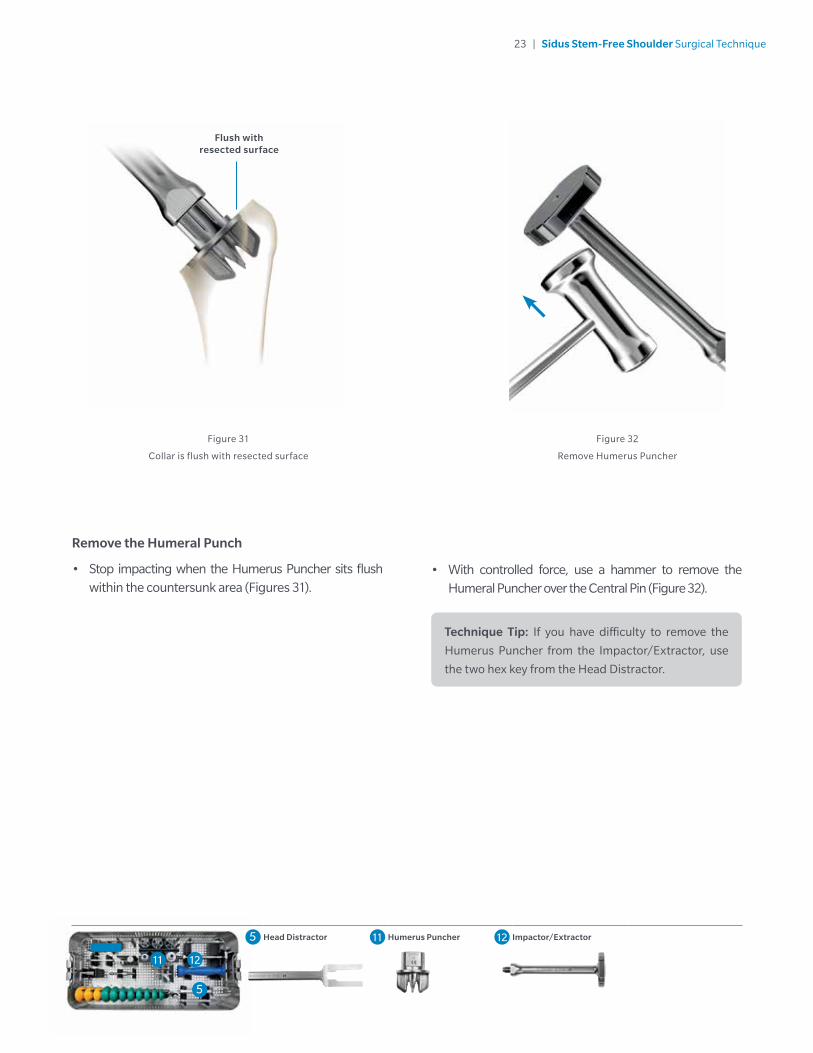
Remove the Humeral Punch
• Stop impacting when the Humerus Puncher sits flush within the countersunk area (Figures 31).
• With controlled force, use a hammer to remove the Humeral Puncher over the Central Pin (Figure 32).
Figure 32
Remove Humerus Puncher
Figure 31
Collar is flush with resected surface
Technique Tip: If you have difficulty to remove the
Humerus Puncher from the Impactor/Extractor, use
the two hex key from the Head Distractor.
5
Flush with resected surface
5 Head Distractor 11 Humerus Puncher 12 Impactor/Extractor
11 12
5
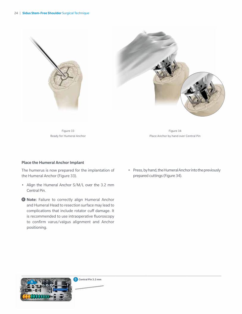
24 | Sidus Stem-Free Shoulder Surgical Technique
Place the Humeral Anchor Implant
The humerus is now prepared for the implantation of the Humeral Anchor (Figure 33).
• Align the Humeral Anchor S/M/L over the 3.2 mm Central Pin.
Note: Failure to correctly align Humeral Anchor and Humeral Head to resection surface may lead to complications that include rotator cuff damage. It is recommended to use intraoperative fluoroscopy to confirm varus/valgus alignment and Anchor positioning.
Figure 33
Ready for Humeral Anchor
Figure 34
Place Anchor by hand over Central Pin
• Press, by hand, the Humeral Anchor into the previously prepared cuttings (Figure 34).
88 Central Pin 3.2 mm
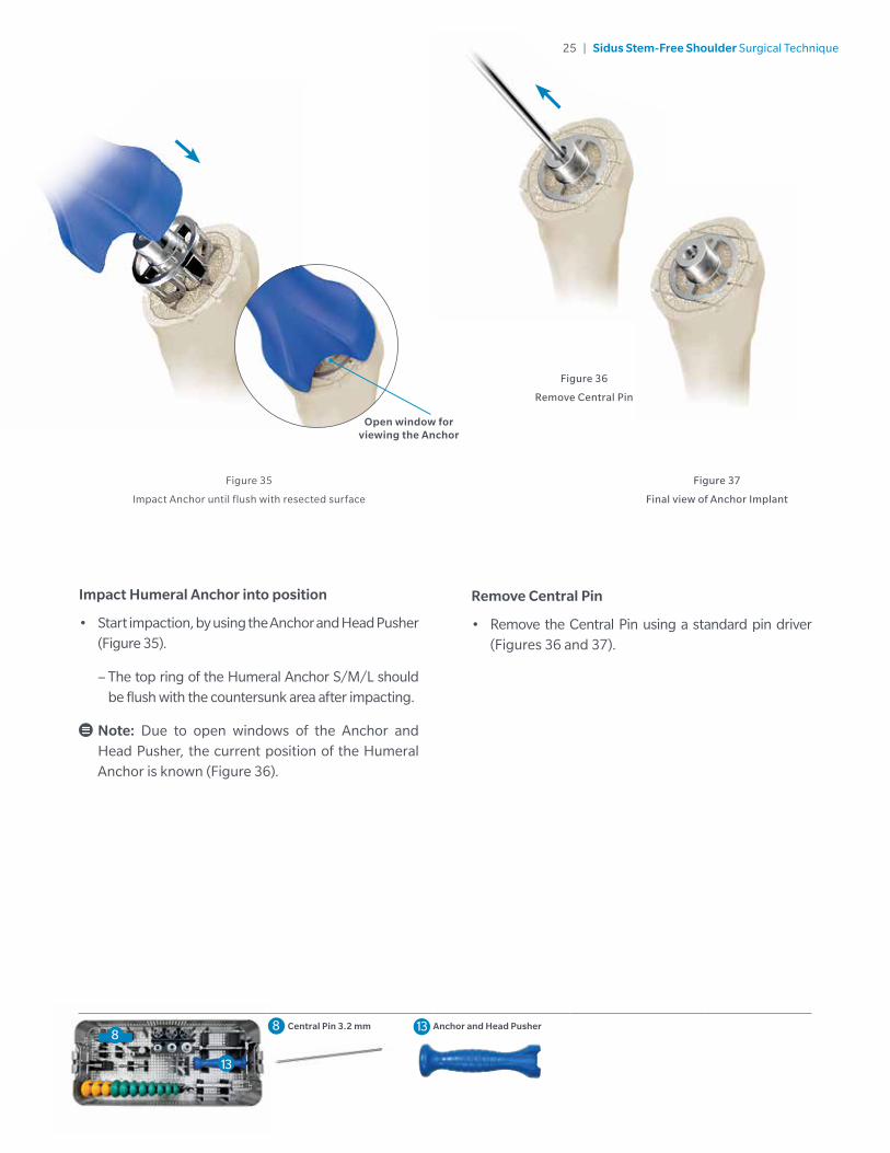
25 | Sidus Stem-Free Shoulder Surgical Technique
Figure 36
Remove Central Pin
Impact Humeral Anchor into position
• Start impaction, by using the Anchor and Head Pusher (Figure 35).
– The top ring of the Humeral Anchor S/M/L should be flush with the countersunk area after impacting.
Note: Due to open windows of the Anchor and Head Pusher, the current position of the Humeral Anchor is known (Figure 36).
Figure 35
Impact Anchor until flush with resected surface
Remove Central Pin
• Remove the Central Pin using a standard pin driver (Figures 36 and 37).
Figure 37
Final view of Anchor Implant
Open window for viewing the Anchor
88 Central Pin 3.2 mm 13 Anchor and Head Pusher
13
26 | Sidus Stem-Free Shoulder Surgical Technique
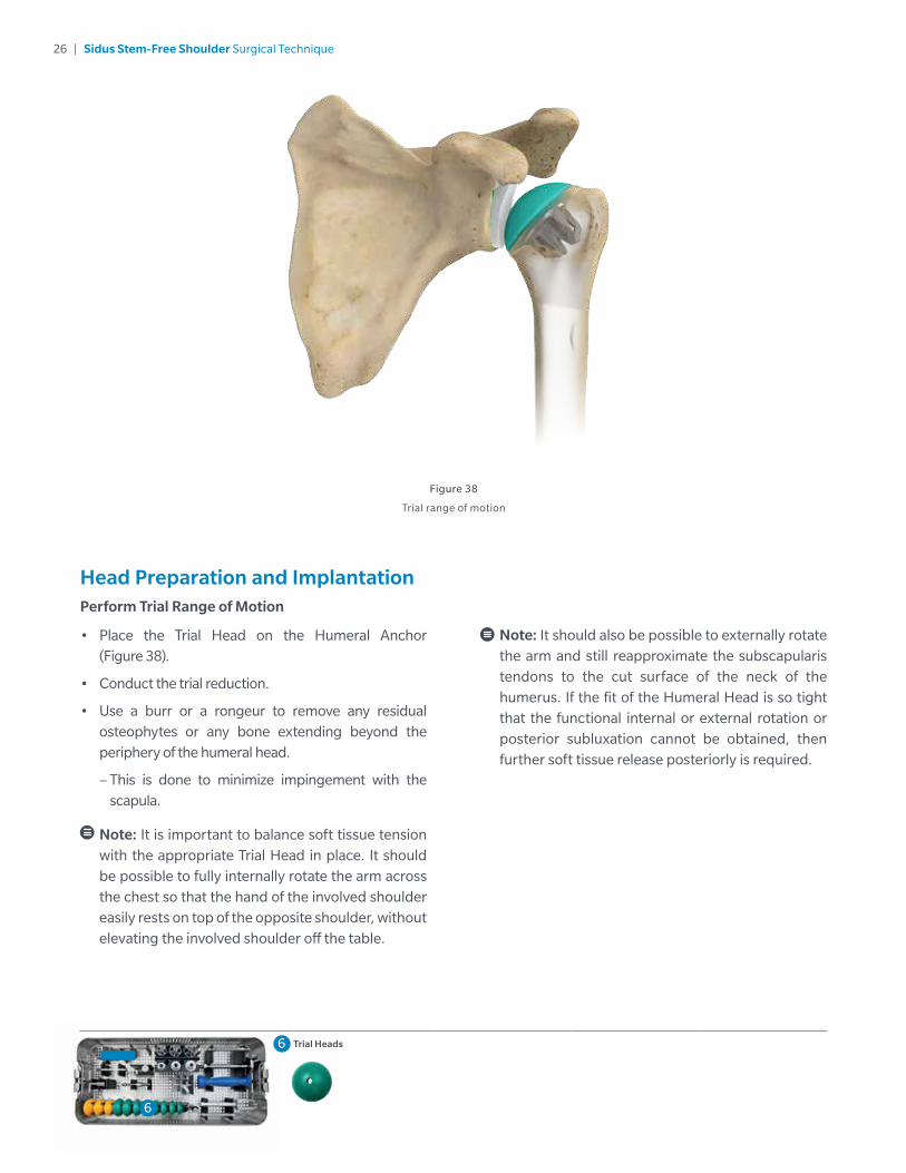
Head Preparation and ImplantationPerform Trial Range of Motion
• Place the Trial Head on the Humeral Anchor (Figure 38).
• Conduct the trial reduction.
• Use a burr or a rongeur to remove any residual osteophytes or any bone extending beyond the periphery of the humeral head.
– This is done to minimize impingement with the scapula.
Note: It is important to balance soft tissue tension with the appropriate Trial Head in place. It should be possible to fully internally rotate the arm across the chest so that the hand of the involved shoulder easily rests on top of the opposite shoulder, without elevating the involved shoulder off the table.
Note: It should also be possible to externally rotate the arm and still reapproximate the subscapularis tendons to the cut surface of the neck of the humerus. If the fit of the Humeral Head is so tight that the functional internal or external rotation or posterior subluxation cannot be obtained, then further soft tissue release posteriorly is required.
Figure 38
Trial range of motion
6
6 Trial Heads
27 | Sidus Stem-Free Shoulder Surgical Technique
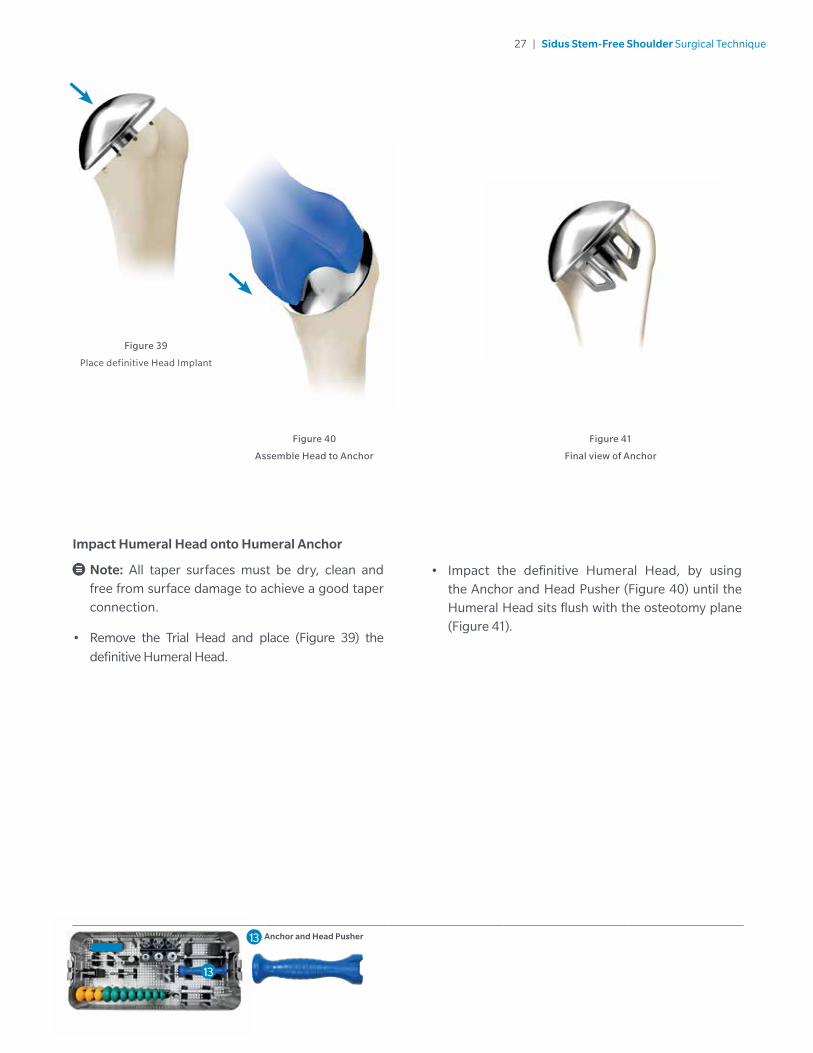
Figure 39
Place definitive Head Implant
Impact Humeral Head onto Humeral Anchor
Note: All taper surfaces must be dry, clean and free from surface damage to achieve a good taper connection.
• Remove the Trial Head and place (Figure 39) the
definitive Humeral Head.
Figure 40
Assemble Head to Anchor
Figure 41
Final view of Anchor
• Impact the definitive Humeral Head, by using the Anchor and Head Pusher (Figure 40) until the Humeral Head sits flush with the osteotomy plane (Figure 41).
13 Anchor and Head Pusher
13
28 | Sidus Stem-Free Shoulder Surgical Technique
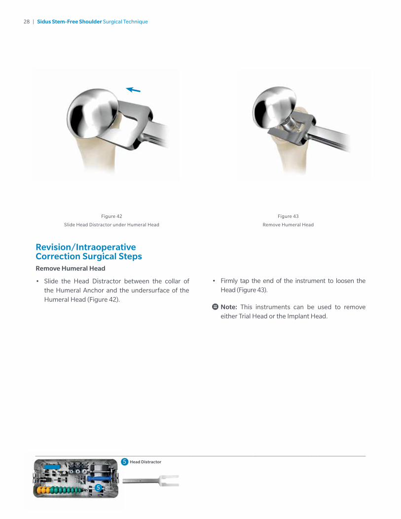
Revision/Intraoperative Correction Surgical StepsRemove Humeral Head
• Slide the Head Distractor between the collar of the Humeral Anchor and the undersurface of the Humeral Head (Figure 42).
• Firmly tap the end of the instrument to loosen the Head (Figure 43).
Note: This instruments can be used to remove either Trial Head or the Implant Head.
Figure 43
Remove Humeral Head
Figure 42
Slide Head Distractor under Humeral Head
5
5 Head Distractor
5
29 | Sidus Stem-Free Shoulder Surgical Technique
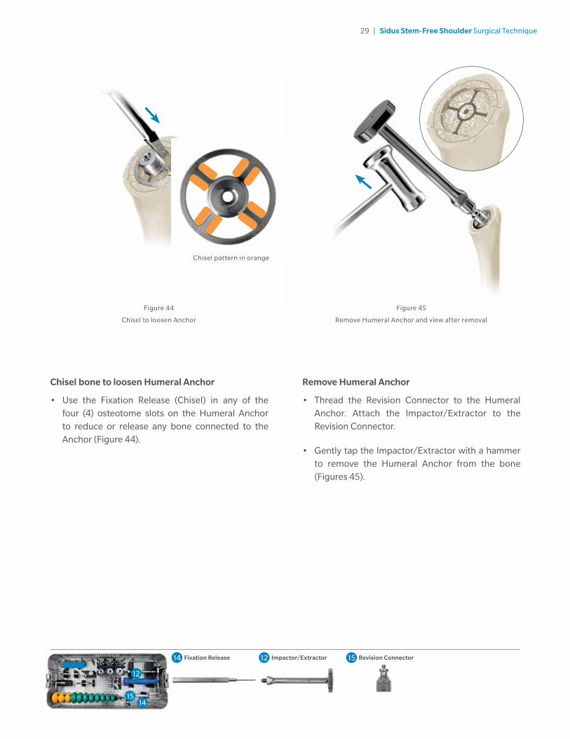
Chisel bone to loosen Humeral Anchor
• Use the Fixation Release (Chisel) in any of the four (4) osteotome slots on the Humeral Anchor to reduce or release any bone connected to the Anchor (Figure 44).
Figure 44
Chisel to loosen Anchor
Chisel pattern in orange
Figure 45
Remove Humeral Anchor and view after removal
Remove Humeral Anchor
• Thread the Revision Connector to the Humeral Anchor. Attach the Impactor/Extractor to the Revision Connector.
• Gently tap the Impactor/Extractor with a hammer to remove the Humeral Anchor from the bone (Figures 45).
14 Fixation Release 12 Impactor/Extractor 15 Revision Connector
12
1415
30 | Sidus Stem-Free Shoulder Surgical Technique
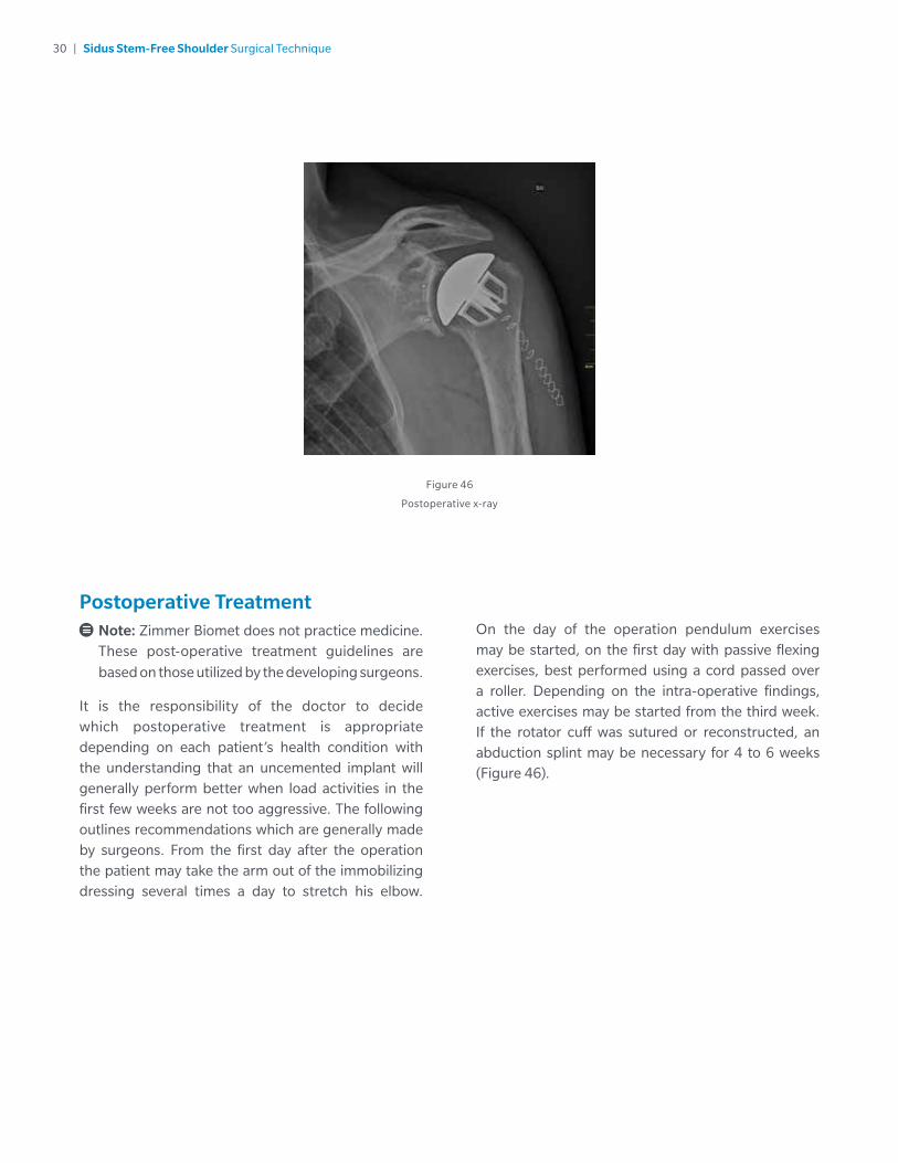
Figure 46
Postoperative x-ray
Postoperative Treatment Note: Zimmer Biomet does not practice medicine. These post-operative treatment guidelines are
based on those utilized by the developing surgeons.
It is the responsibility of the doctor to decide which postoperative treatment is appropriate depending on each patient’s health condition with the understanding that an uncemented implant will generally perform better when load activities in the first few weeks are not too aggressive. The following outlines recommendations which are generally made by surgeons. From the first day after the operation the patient may take the arm out of the immobilizing dressing several times a day to stretch his elbow.
On the day of the operation pendulum exercises may be started, on the first day with passive flexing exercises, best performed using a cord passed over a roller. Depending on the intra-operative findings, active exercises may be started from the third week. If the rotator cuff was sutured or reconstructed, an abduction splint may be necessary for 4 to 6 weeks (Figure 46).
31 | Sidus Stem-Free Shoulder Surgical Technique
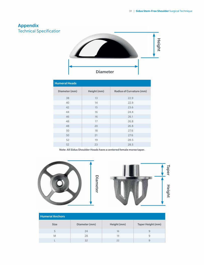
Appendix Technical Specifications
Humeral Heads
Diameter (mm) Height (mm) Radius of Curvature (mm)
38 13 22.9
40 14 22.9
42 15 23.6
44 16 24.4
46 16 26.1
48 17 26.8
48 20 26.8
50 18 27.6
50 21 27.6
52 19 28.5
52 23 28.5
Note: All Sidus Shoulder Heads have a centered female morse taper.
Heig
ht
Diameter
Humeral Anchors
Size Diameter (mm) Height (mm) Taper Height (mm)
S 24 16 9
M 28 19 9
L 32 22 9
Heig
ht
TaperD
iameter
32 | Sidus Stem-Free Shoulder Surgical Technique
Appendix Indications & Contraindications
Indications
• Osteoarthritis
• Posttraumatic arthrosis
• Focal Avascular Necrosis of the Humeral Head
• Previous surgeries of the Shoulder that do not compromise the fixation
Contraindications
• Soft or inadequate humeral bone (including Osteoporosis and extensive Avascular Necrosis or Rheumatoid arthritis) leading to poor implant fixation
• Metaphyseal bony defects (including large cysts)
• Post-traumatic tuberosity nonunion
• Signs of infection
• Irreparable cuff tear
• Revision from a failed stemmed prosthesis
• Charcot’s shoulder (neuroarthropathy)
The Sidus Stem-Free Shoulder is indicated for uncemented use in total shoulder arthroplasty, replacing the shoulder joint of patients suffering severe pain or disability. Patient conditions of non-inflammatory degenerative joint disease (NIDJD), such as avascular necrosis and osteoarthritis, provided there is an adequate bone stock to support the fixation of the implant.
33 | Sidus Stem-Free Shoulder Surgical Technique
Clinically-proven, bone-sparing alternative for Total Shoulder Arthroplasty1
All subjects enrolled in the Sidus Shoulder IDE were paired with Anatomical Shoulder™ Glenoid. 71 patients from the Sidus Shoulder IDE have reached two year follow-up and results show the following:
• 98.5% of patients have over a 30 point improvement on the ASES score from preoperative.
• Excellent survivorship of 95.8% at two years.
• 98.5% of patients completing two year visits successfully passed the radiographic success criteria with no progressive radiolucencies of the humeral component > 2 mm and no migration or subsidence of the humeral component.
When compared to a subset of 43 patients implanted with Sidus Shoulder and the Anatomical Shoulder Glenoid in the European Sidus PMCF, performance was similar at two years:
• 90.5% of patients have over a 30 point improvement on the ASES score from preoperative.
• Excellent survivorship of 97.7% at two years.
• 100% of patients successfully passed the radiographic success criteria with no progressive radiolucencies of the humeral component >2 mm and no migration or subsidence of the humeral component.
Clinical IDE Study demonstrated increased mobility and reduced pain compared to preoperative state1
• Statistically significant improvement in range of motion when compared to preoperative state.
• Significant improvement in functions of daily life.
• 92.6% of the patients in the Sidus IDE study were either very satisfied or satisfied at two years postoperative.
When compared to a subset of 43 patients implanted with Sidus Shoulder and the Anatomical Shoulder Glenoid in the European Sidus PMCF, performance was similar at two years:
• Statistically significant improvement in range of motion when compared to preoperative state.
• Significant improvement in functions of daily life.
• 92.8% of the patients were either very satisfied or satisfied at two years.
Performance of Sidus Shoulder has been benchmarked against the Bigliani/Flatow® Shoulder (B/F) historical control study of 48 patients. Performance was similar to Sidus Shoulder at two years:
• 91.7% of patients have over a 30 point improvement on the ASES from preoperative.
• 97.9% survivorship; one revision due to anterior shoulder pain.
• 100% of patients completing two year follow-up successfully passed the radiographic success criteria.
When benchmarked against the Bigliani/Flatow historical control study of 48 patients, performance was similar to Sidus Shoulder at two years:
• Significant improvement in range of motion when compared to preoperative state.
• Significant improvement in functions of daily life.
• Satisfaction data was not recorded for B/F.
Appendix Clinical Results
34 | Sidus Stem-Free Shoulder Surgical Technique
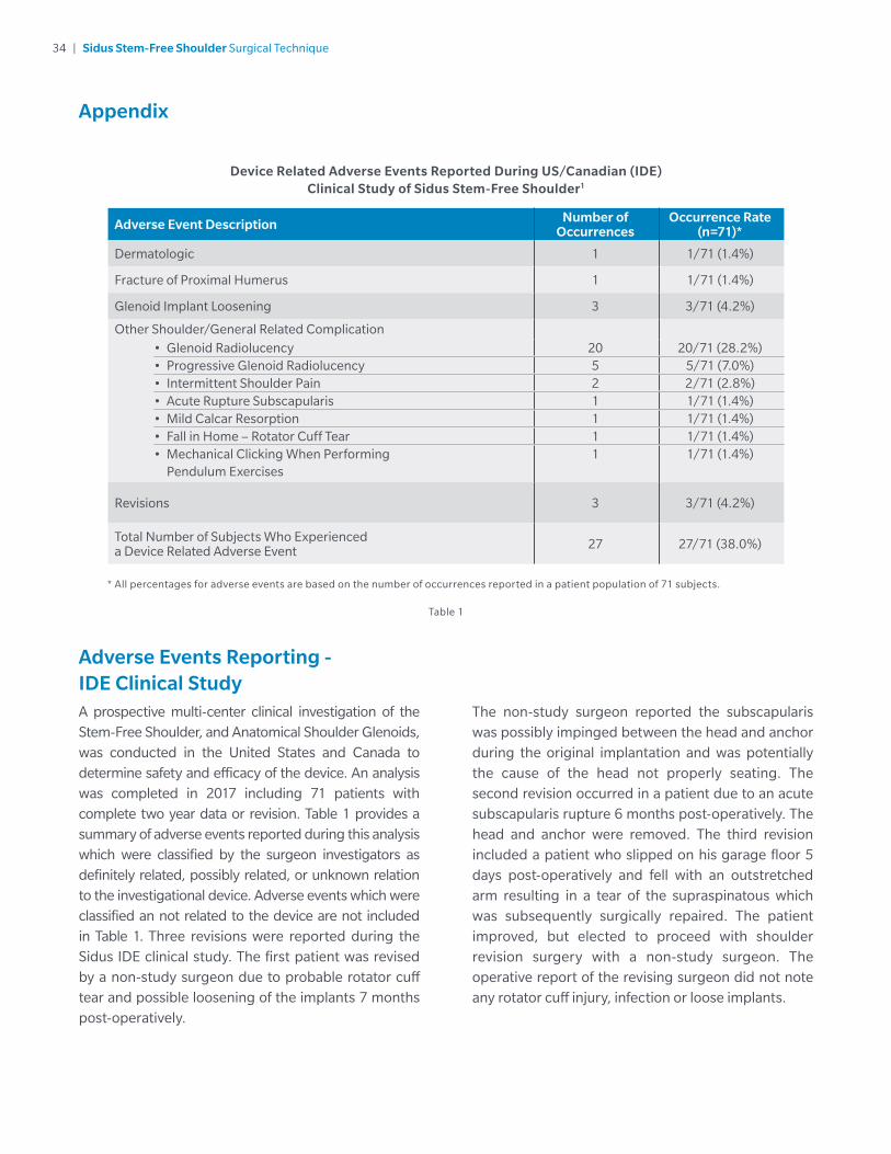
Adverse Events Reporting - IDE Clinical StudyA prospective multi-center clinical investigation of the Stem-Free Shoulder, and Anatomical Shoulder Glenoids, was conducted in the United States and Canada to determine safety and efficacy of the device. An analysis was completed in 2017 including 71 patients with complete two year data or revision. Table 1 provides a summary of adverse events reported during this analysis which were classified by the surgeon investigators as definitely related, possibly related, or unknown relation to the investigational device. Adverse events which were classified an not related to the device are not included in Table 1. Three revisions were reported during the Sidus IDE clinical study. The first patient was revised by a non-study surgeon due to probable rotator cuff tear and possible loosening of the implants 7 months post-operatively.
The non-study surgeon reported the subscapularis was possibly impinged between the head and anchor during the original implantation and was potentially the cause of the head not properly seating. The second revision occurred in a patient due to an acute subscapularis rupture 6 months post-operatively. The head and anchor were removed. The third revision included a patient who slipped on his garage floor 5 days post-operatively and fell with an outstretched arm resulting in a tear of the supraspinatous which was subsequently surgically repaired. The patient improved, but elected to proceed with shoulder revision surgery with a non-study surgeon. The operative report of the revising surgeon did not note any rotator cuff injury, infection or loose implants.
Table 1
Adverse Event Description Number of Occurrences
Occurrence Rate(n=71)*
Dermatologic 1 1/71 (1.4%)
Fracture of Proximal Humerus 1 1/71 (1.4%)
Glenoid Implant Loosening 3 3/71 (4.2%)
Other Shoulder/General Related Complication• Glenoid Radiolucency 20 20/71 (28.2%)• Progressive Glenoid Radiolucency 5 5/71 (7.0%)• Intermittent Shoulder Pain 2 2/71 (2.8%)• Acute Rupture Subscapularis 1 1/71 (1.4%)• Mild Calcar Resorption 1 1/71 (1.4%)• Fall in Home – Rotator Cuff Tear 1 1/71 (1.4%)• Mechanical Clicking When Performing
Pendulum Exercises1 1/71 (1.4%)
Revisions 3 3/71 (4.2%)
Total Number of Subjects Who Experienced a Device Related Adverse Event 27 27/71 (38.0%)
* All percentages for adverse events are based on the number of occurrences reported in a patient population of 71 subjects.
Device Related Adverse Events Reported During US/Canadian (IDE) Clinical Study of Sidus Stem-Free Shoulder1
Appendix
35 | Sidus Stem-Free Shoulder Surgical Technique
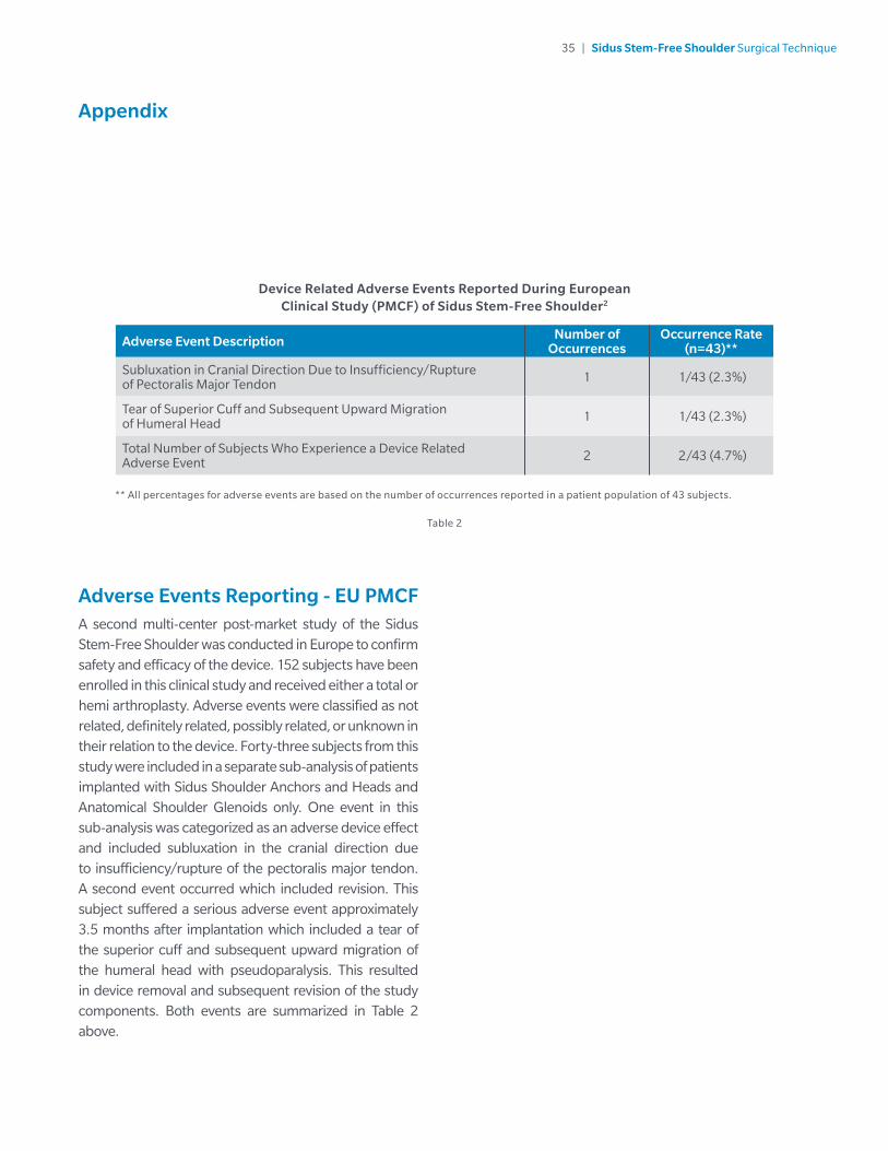
Adverse Events Reporting - EU PMCFA second multi-center post-market study of the Sidus Stem-Free Shoulder was conducted in Europe to confirm safety and efficacy of the device. 152 subjects have been enrolled in this clinical study and received either a total or hemi arthroplasty. Adverse events were classified as not related, definitely related, possibly related, or unknown in their relation to the device. Forty-three subjects from this study were included in a separate sub-analysis of patients implanted with Sidus Shoulder Anchors and Heads and Anatomical Shoulder Glenoids only. One event in this sub-analysis was categorized as an adverse device effect and included subluxation in the cranial direction due to insufficiency/rupture of the pectoralis major tendon. A second event occurred which included revision. This subject suffered a serious adverse event approximately 3.5 months after implantation which included a tear of the superior cuff and subsequent upward migration of the humeral head with pseudoparalysis. This resulted in device removal and subsequent revision of the study components. Both events are summarized in Table 2 above.
Table 2
Adverse Event Description Number of Occurrences
Occurrence Rate(n=43)**
Subluxation in Cranial Direction Due to Insufficiency/Rupture of Pectoralis Major Tendon 1 1/43 (2.3%)
Tear of Superior Cuff and Subsequent Upward Migration of Humeral Head 1 1/43 (2.3%)
Total Number of Subjects Who Experience a Device Related Adverse Event 2 2/43 (4.7%)
** All percentages for adverse events are based on the number of occurrences reported in a patient population of 43 subjects.
Device Related Adverse Events Reported During European Clinical Study (PMCF) of Sidus Stem-Free Shoulder2
Appendix
36 | Sidus Stem-Free Shoulder Surgical Technique
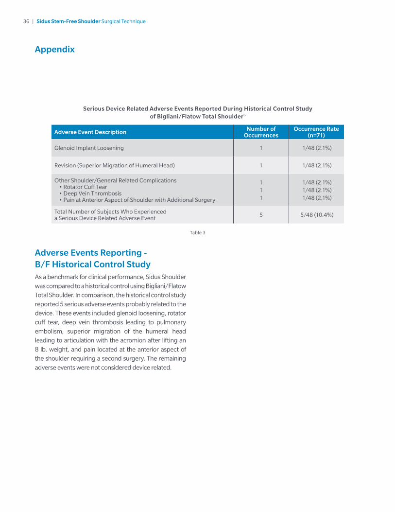
Adverse Events Reporting - B/F Historical Control StudyAs a benchmark for clinical performance, Sidus Shoulder was compared to a historical control using Bigliani/Flatow Total Shoulder. In comparison, the historical control study reported 5 serious adverse events probably related to the device. These events included glenoid loosening, rotator cuff tear, deep vein thrombosis leading to pulmonary embolism, superior migration of the humeral head leading to articulation with the acromion after lifting an 8 lb. weight, and pain located at the anterior aspect of the shoulder requiring a second surgery. The remaining adverse events were not considered device related.
Table 3
Adverse Event Description Number of Occurrences
Occurrence Rate(n=71)
Glenoid Implant Loosening 1 1/48 (2.1%)
Revision (Superior Migration of Humeral Head) 1 1/48 (2.1%)
Other Shoulder/General Related Complications• Rotator Cuff Tear• Deep Vein Thrombosis• Pain at Anterior Aspect of Shoulder with Additional Surgery
111
1/48 (2.1%)1/48 (2.1%)1/48 (2.1%)
Total Number of Subjects Who Experienced a Serious Device Related Adverse Event 5 5/48 (10.4%)
Serious Device Related Adverse Events Reported During Historical Control Study of Bigliani/Flatow Total Shoulder5
Appendix

Notes

Notes

References
1. Multicenter Trial of the Sidus Stem-Free Shoulder Arthroplasty System (Protocol CIU2012-12E/G130026, “IDE”).
2. Sidus Stem-Free Shoulder: A Multicenter, Prospective, Non-Controlled Post Market Clinical Follow-up Study (Clinical Investigation Plan CME2012-01E, “PMCF”).
3. Zimmer Biomet Compatibility Website: http://www.zimmerbiomet.com/medical-professionals/suppor t/product- compatibilit y.html#shoulder.
4. Churchill RS, Chuinard C, Wiater JM, Friedman R, Freehill M, Jacobson S, Spencer E, Holloway B, Wittstein J, Lassiter T, Smith M, Blaine T, Nicholson GP . Clinical and Radiographic Outcomes of the Simpliciti Canal-Sparing Shoulder Arthroplasty System. J Bone Joint Surg Am. 2016:552-560.
5. Litchfield RB, McKee MD, Balyk R, Mandel S, Holtby R, Hollinshead R, Drosdowech D, Wambolt SE, Griffin SH, McCormack R.: Cemented versus uncemented fixation of humeral components in total shoulder arthroplasty for osteoarthritis of the shoulder: a prospective, randomized, double-blind clinical trial-A JOINTs Canada Project. J Shoulder Elbow Surg. 2011 Jun;20(4):529-36.
6. Favre, Philippe, Henderson, Adam D.:Prediction of stemless humeral implant micromotion during upper limb activities. Clinical Biomechanics 36 (2016) 46-51.
7. Favre, Philippe, Seebeck, Jorn, Thistlethwaite, Paul A E, Obrist, Marc, Steffens, Jason G, Hopkins, Andrew R, Hulme, Paul A. In vitro initial stability of a stemless humeral implant. Clinical Biomechanics 32 (2016) 113-117.
This material is intended for health care professionals. Distribution to any other recipient is prohibited. For complete product information, including indications, contraindications, warnings, precautions, potential adverse effects and patient counseling information, see the package insert and http://www.zimmerbiomet.com.
Zimmer Biomet does not practice medicine. This technique was developed in conjunction with health care professionals. Each surgeon should exercise his or her own independent judgment in the diagnosis and treatment of an individual patient, and this information does not purport to replace the comprehensive training surgeons have received. As with all surgical procedures, the technique used in each case will depend on the surgeon’s medical judgment as the best treatment for each patient. Results will vary based on health, weight, activity and other variables. Not all patients are candidates for this product and/or procedure. Caution: Federal (USA) law restricts this device to sale by or on the order of a surgeon. Rx only.
All content herein is protected by copyright, trademarks and other intellectual property rights, as applicable, owned by or licensed to Zimmer Biomet or its affiliates unless otherwise indicated, and must not be redistributed, duplicated or disclosed, in whole or in part, without the express written consent of Zimmer Biomet.
©2018 Zimmer Biomet
1519.1-US-en-REV0118
Legal ManufacturerZimmer GmbH Sulzer Allee 8 CH-8404 Winterthur, Switzerland
Products compatible with this system are under the design control of various manufacturers.Refer to the product labelingof each device for the legalmanufacturer.