Embed Size (px)
DESCRIPTION
cause ,pathogenesis and presentation of sideroblastic anemia
Citation preview
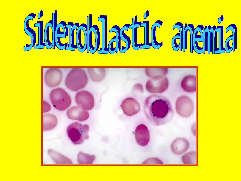
Sideroblastic anemia
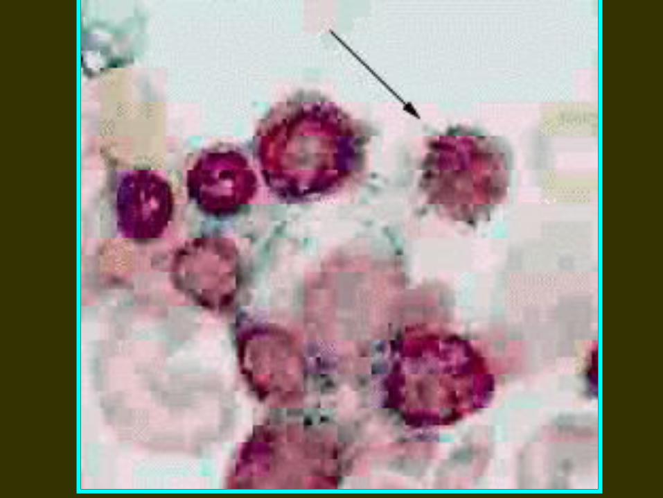
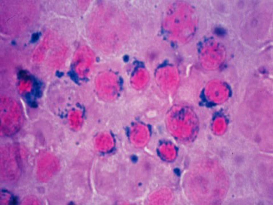
The Sideroblastic anemias comprise a group of disorders of diverse etiology in which the nucleated erythroid precursors in the bone marrow show characteristic ringed sideroblasts
Siderocytes; and sideroblasts are erythrocytes and normoblasts respectively, which contain cytoplasmic granules of iron.
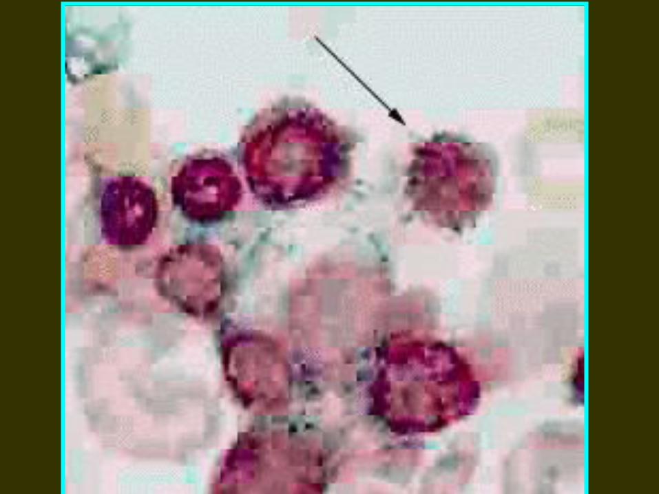
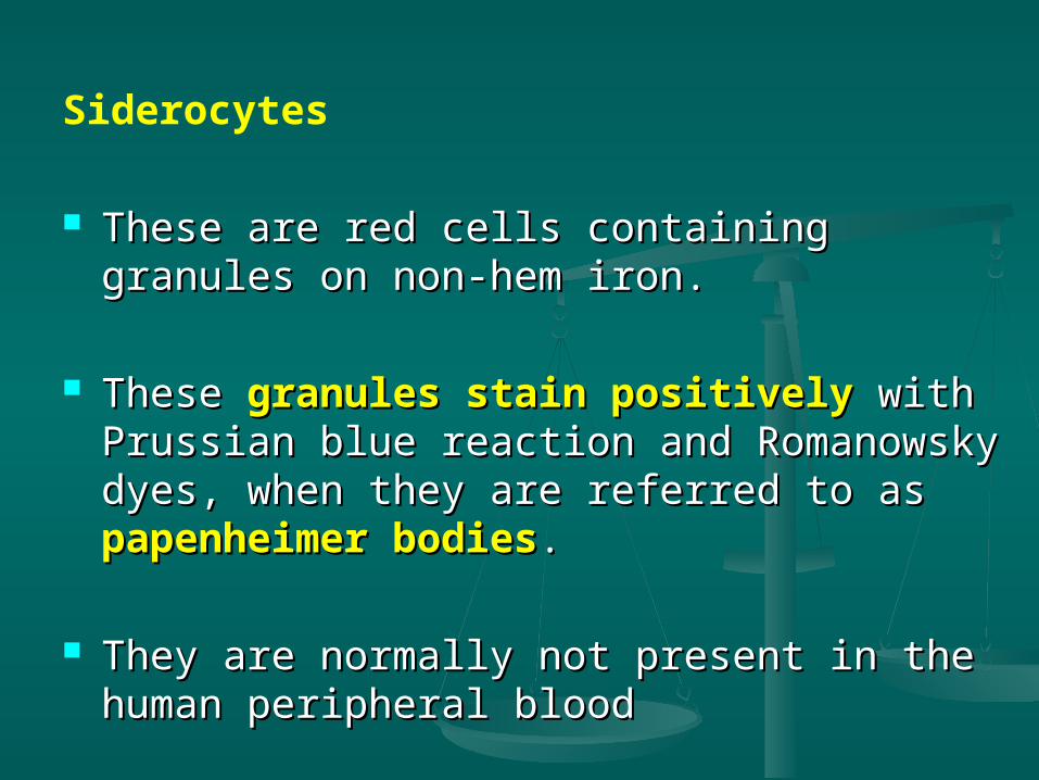
Siderocytes
These are red cells containing granules on These are red cells containing granules on non-hem iron. non-hem iron.
These These granules stain positivelygranules stain positively with with Prussian blue reaction and Romanowsky Prussian blue reaction and Romanowsky dyes, when they are referred to as dyes, when they are referred to as papenheimer bodiespapenheimer bodies. .
They are normally not present in the human They are normally not present in the human peripheral bloodperipheral blood
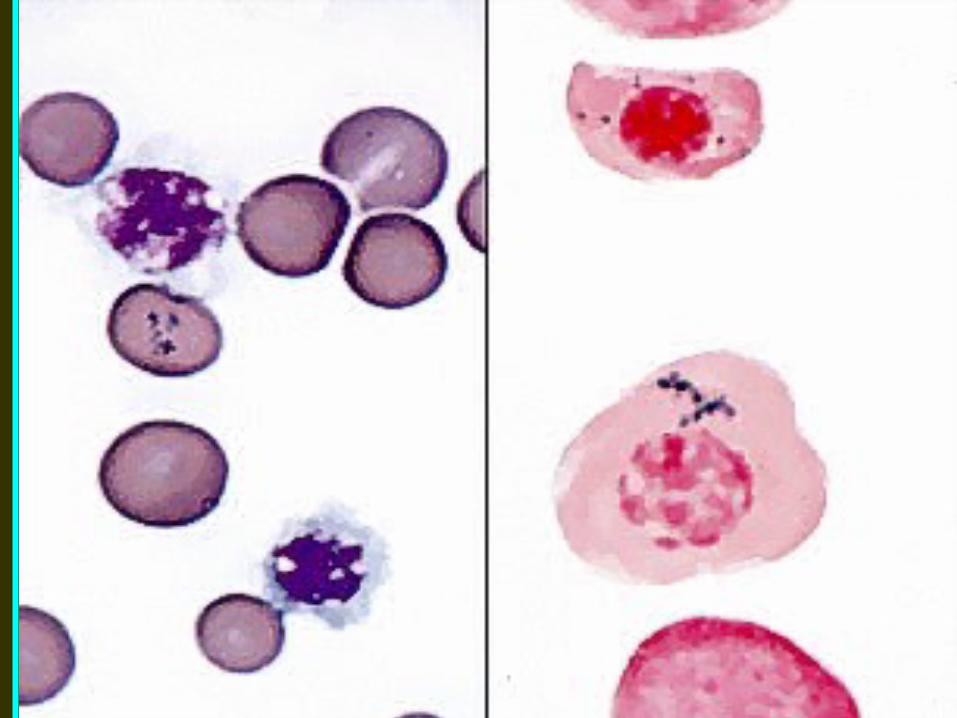
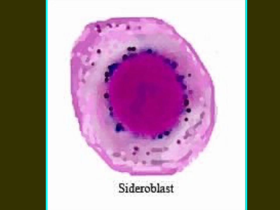
Sideroblasts These are nucleated red cells (normoblasts)
containing siderotic granules which stain positively with Prussian blue reaction .
Depending upon the number , size and distribution of siderotic granules, sideroblasts may be normal or abnormal
Sideroblastic anemia is diagnosed when 15% or more of marrow erythroblasts are ring sideroblasts
Types of Sideroblastic Anemias
Based on etiology, Sideroblastic anemia are classified into hereditary and acquired types.
1. Hereditary (Congenital) Sideroblastic anemia:
this is a rare x-linked disorder associated with defective enzyme activity of aminolevulinic acid (ALA) synthetase required for hem synthesis
2. Acquired Sideroblastic anemiaA. Primary (idiopathic, refractory) acquired
Sideroblastic anemia.B. Secondary acquired Sideroblastic
anemia: Drugs, chemicals and toxins e.g. isoniazid,
cycloserine, chloramphenicol, alcohol, lead Hematological disorders e.g. myelofibrosis,
polycythemia vera, acute leukemia, myeloma, lymphoma and hemolytic anemia
Miscellaneous e.g. carcinoma, Myxoedema, rheumatoid arthritis
Laboratory Findings
Sideroblastic anemia usually show the following hematological features
1. There is generally moderate to severe degree of anemia
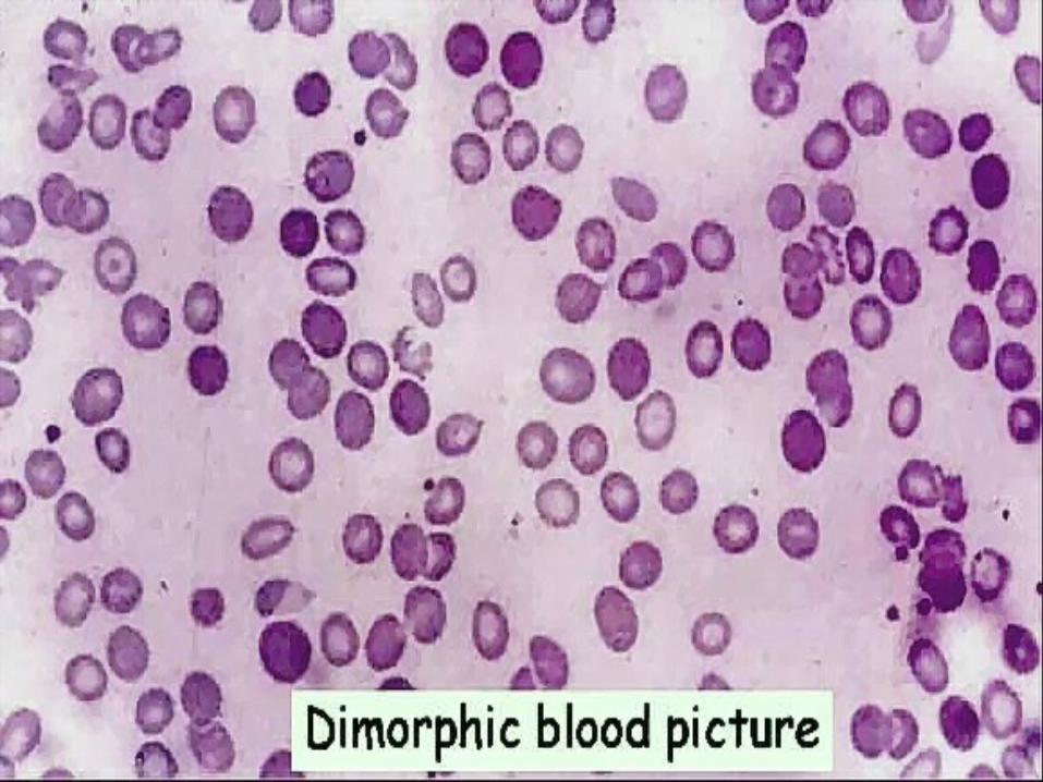
2. The blood picture shows hypochromic anemia which may be microcytic, or there may be some normocytic red cells as well (dimorphic)

3. Absolute values (MCV,MCH and MCHC) are reduced in hereditary type but MCV is often raised in acquired type.
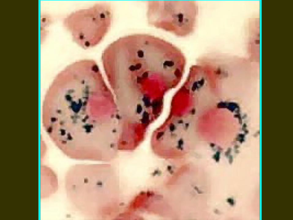
4. The bone marrow examination shows erythroid hyperplasia. Marrow iron store are raised and pathognomonic ring sideroblasts are present.
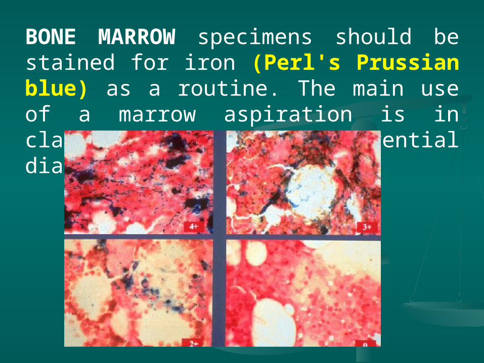
BONE MARROW specimens should be stained for iron (Perl's Prussian blue) as a routine. The main use of a marrow aspiration is in clarifying a differential diagnosis.

5. Serum ferritin levels are raised
6. Serum iron is usually raised with almost complete saturation of TIBC
7. There is increase iron deposition in the tissue