Embed Size (px)
Citation preview
SHOCK
Night Float CurriculumGeneral Pediatrics Hospitalist GroupLucile Packard Children’s Hospital
November, 2010
Learning Goals
Define shock Know the stages of shock Differentiate between
classifications of shock, generate differential dx of etiology
Know the initial management of shock
Review 2 cases of pediatric shock
What is shock?
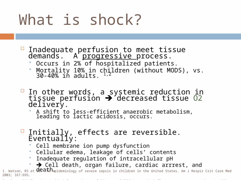
Inadequate perfusion to meet tissue demands. A progressive process. Occurs in 2% of hospitalized patients. Mortality 10% in children (without MODS), vs. 30-
40% in adults. 1,2
In other words, a systemic reduction in tissue perfusion decreased tissue O2 delivery. A shift to less-efficient anaerobic metabolism, leading to
lactic acidosis, occurs.
Initially, effects are reversible. Eventually: Cell membrane ion pump dysfunction Cellular edema, leakage of cells’ contents Inadequate regulation of intracellular pH Cell death, organ failure, cardiac arrrest, and death.
1. Watson, RS et al. The epidemiology of severe sepsis in children in the United States. Am J Respir Crit Care Med 2003; 167:695.
2. .Bone, RC. Toward an epidemiology and natural history of SIRS (systemic inflammatory response syndrome). JAMA 1992; 268:3452.
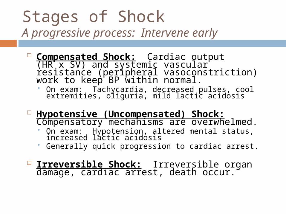
Stages of Shock A progressive process: Intervene early
Compensated Shock: Cardiac output (HR x SV) and systemic vascular resistance (peripheral vasoconstriction) work to keep BP within normal. On exam: Tachycardia, decreased pulses, cool
extremities, oliguria, mild lactic acidosis
Hypotensive (Uncompensated) Shock: Compensatory mechanisms are overwhelmed. On exam: Hypotension, altered mental status,
increased lactic acidosis Generally quick progression to cardiac arrest.
Irreversible Shock: Irreversible organ damage, cardiac arrest, death occur.
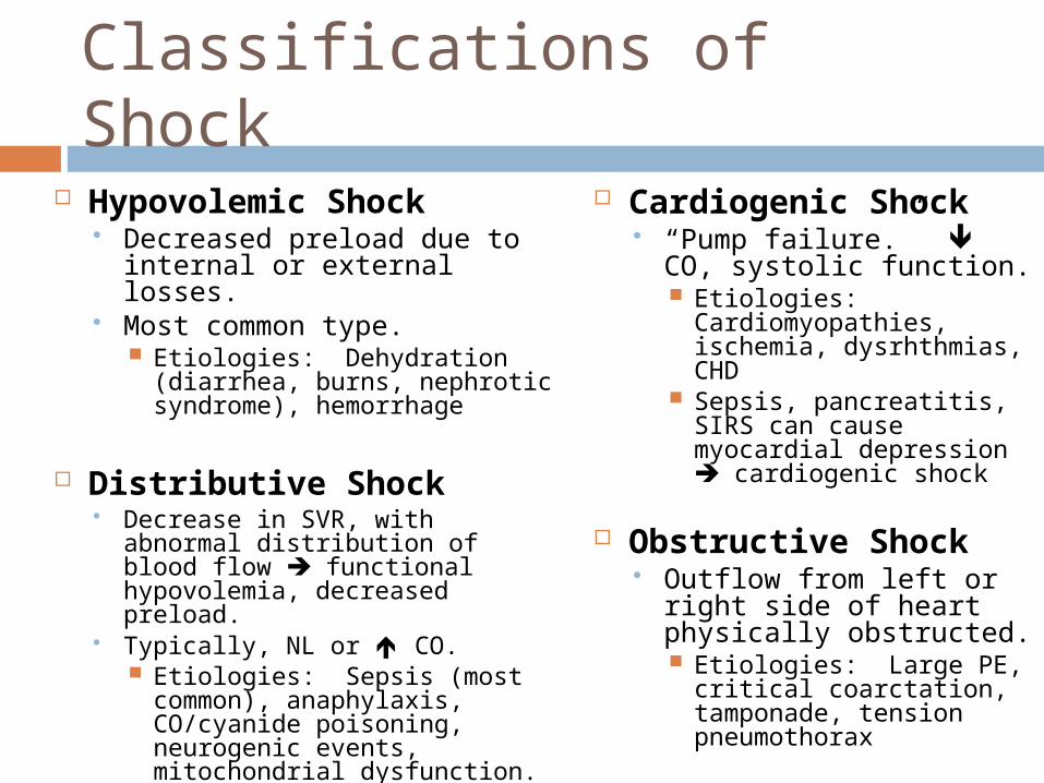
Hypovolemic Shock Decreased preload due to
internal or external losses. Most common type.
Etiologies: Dehydration (diarrhea, burns, nephrotic syndrome), hemorrhage
Distributive Shock Decrease in SVR, with abnormal
distribution of blood flow functional hypovolemia, decreased preload.
Typically, NL or CO. Etiologies: Sepsis (most
common), anaphylaxis, CO/cyanide poisoning, neurogenic events, mitochondrial dysfunction.
Cardiogenic Shock “Pump failure.” CO,
systolic function. Etiologies:
Cardiomyopathies, ischemia, dysrhthmias, CHD
Sepsis, pancreatitis, SIRS can cause myocardial depression cardiogenic shock
Obstructive Shock Outflow from left or right
side of heart physically obstructed. Etiologies: Large PE,
critical coarctation, tamponade, tension pneumothorax
Classifications of Shock
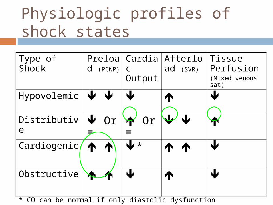
Physiologic profiles of shock states
Type of Shock Preload (PCWP)
Cardiac Output
Afterload (SVR)
Tissue Perfusion(Mixed venous sat)
Hypovolemic
Distributive Or =
Or =
Cardiogenic *
Obstructive
* CO can be normal if only diastolic dysfunction
Shock: General initial management
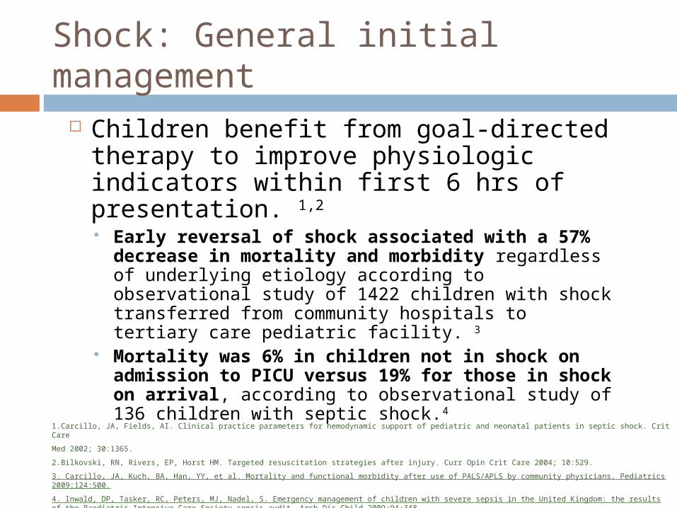
Children benefit from goal-directed therapy to improve physiologic indicators within first 6 hrs of presentation. 1,2
Early reversal of shock associated with a 57% decrease in mortality and morbidity regardless of underlying etiology according to observational study of 1422 children with shock transferred from community hospitals to tertiary care pediatric facility. 3
Mortality was 6% in children not in shock on admission to PICU versus 19% for those in shock on arrival, according to observational study of 136 children with septic shock.4
1.Carcillo, JA, Fields, AI. Clinical practice parameters for hemodynamic support of pediatric and neonatal patients in septic shock. Crit Care
Med 2002; 30:1365.
2.Bilkovski, RN, Rivers, EP, Horst HM. Targeted resuscitation strategies after injury. Curr Opin Crit Care 2004; 10:529.
3. Carcillo, JA, Kuch, BA, Han, YY, et al. Mortality and functional morbidity after use of PALS/APLS by community physicians. Pediatrics 2009;124:500.
4. Inwald, DP, Tasker, RC, Peters, MJ, Nadel, S. Emergency management of children with severe sepsis in the United Kingdom: the results of the Paediatric Intensive Care Society sepsis audit. Arch Dis Child 2009;94:348.
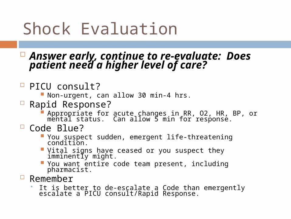
Shock Evaluation Answer early, continue to re-
evaluate: Does patient need a higher level of care?
PICU consult? Non-urgent, can allow 30 min-4 hrs.
Rapid Response? Appropriate for acute changes in RR, O2, HR, BP, or mental
status. Can allow 5 min for response. Code Blue?
You suspect sudden, emergent life-threatening condition. Vital signs have ceased or you suspect they imminently
might. You want entire code team present, including pharmacist.
Remember It is better to de-escalate a Code than emergently escalate a
PICU consult/Rapid Response.
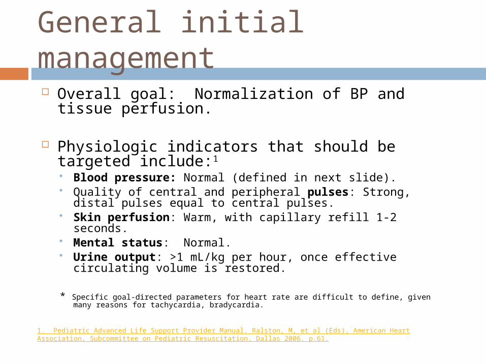
General initial management
Overall goal: Normalization of BP and tissue perfusion.
Physiologic indicators that should be targeted include:1
Blood pressure: Normal (defined in next slide). Quality of central and peripheral pulses: Strong, distal
pulses equal to central pulses. Skin perfusion: Warm, with capillary refill 1-2 seconds. Mental status: Normal. Urine output: >1 mL/kg per hour, once effective
circulating volume is restored.
* Specific goal-directed parameters for heart rate are difficult to define, given many reasons for tachycardia, bradycardia.
1. Pediatric Advanced Life Support Provider Manual. Ralston, M, et al (Eds), American Heart Association, Subcommittee on Pediatric Resuscitation, Dallas 2006. p.61.
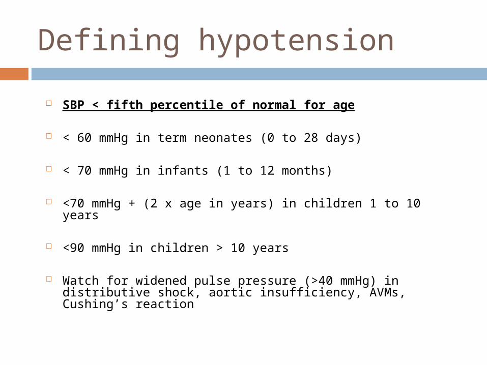
Defining hypotension
SBP < fifth percentile of normal for age
< 60 mmHg in term neonates (0 to 28 days)
< 70 mmHg in infants (1 to 12 months)
<70 mmHg + (2 x age in years) in children 1 to 10 years
<90 mmHg in children > 10 years
Watch for widened pulse pressure (>40 mmHg) in distributive shock, aortic insufficiency, AVMs, Cushing’s reaction
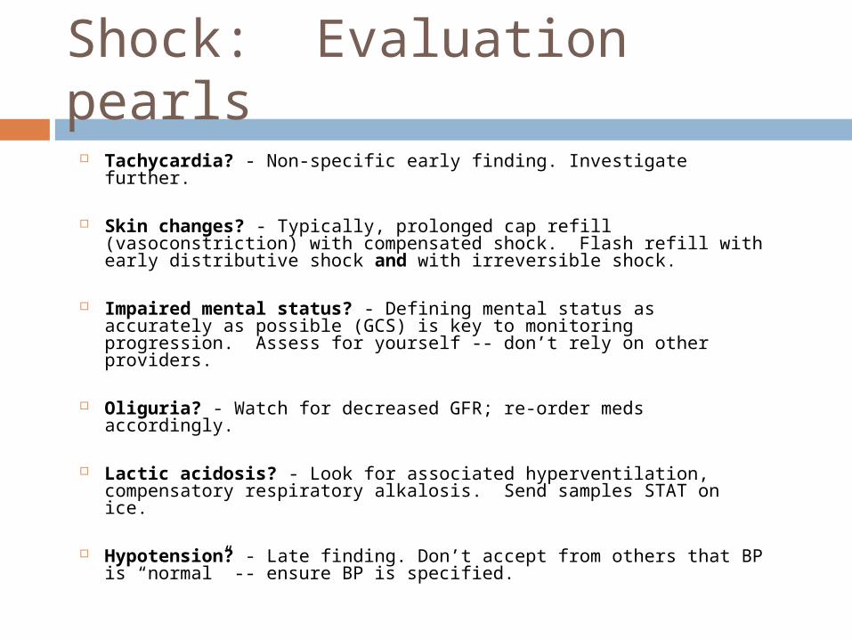
Shock: Evaluation pearls Tachycardia? - Non-specific early finding. Investigate further.
Skin changes? - Typically, prolonged cap refill (vasoconstriction) with compensated shock. Flash refill with early distributive shock and with irreversible shock.
Impaired mental status? - Defining mental status as accurately as possible (GCS) is key to monitoring progression. Assess for yourself -- don’t rely on other providers.
Oliguria? - Watch for decreased GFR; re-order meds accordingly.
Lactic acidosis? - Look for associated hyperventilation, compensatory respiratory alkalosis. Send samples STAT on ice.
Hypotension? - Late finding. Don’t accept from others that BP is “normal” -- ensure BP is specified.
Goal-directed algorithm 1,2
Based on consensus recommendations for septic shock in children, mostly extrapolated from adult data.
May initially be applied to any patient in shock, regardless of etiology.
Exact etiology must be sought to identify which patients may not respond, tailor therapy.
1. Carcillo, JA, Fields, AI. Clinical practice parameters for hemodynamic support of pediatric and neonatal patients in septic shock. Crit Care Med 2002; 30:1365.
2. Rivers, E, Nguyen, B, Havstad, S, et al. Early goal-directed therapy in the treatment of severe sepsis and septic shock. N Engl J Med 2001; 345:1368.
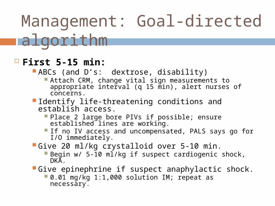
Management: Goal-directed algorithm
First 5-15 min: ABCs (and D’s: dextrose, disability)
Attach CRM, change vital sign measurements to appropriate interval (q 15 min), alert nurses of concerns.
Identify life-threatening conditions and establish access.
Place 2 large bore PIVs if possible; ensure established lines are working.
If no IV access and uncompensated, PALS says go for I/O immediately.
Give 20 ml/kg crystalloid over 5-10 min. Begin w/ 5-10 ml/kg if suspect cardiogenic shock,
DKA. Give epinephrine if suspect anaphylactic shock.
0.01 mg/kg 1:1,000 solution IM; repeat as necessary.
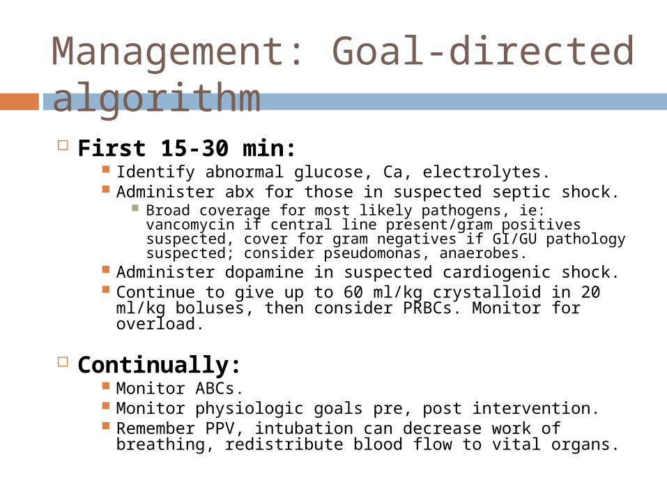
First 15-30 min: Identify abnormal glucose, Ca, electrolytes. Administer abx for those in suspected septic shock.
Broad coverage for most likely pathogens, ie: vancomycin if central line present/gram positives suspected, cover for gram negatives if GI/GU pathology suspected; consider pseudomonas, anaerobes.
Administer dopamine in suspected cardiogenic shock. Continue to give up to 60 ml/kg crystalloid in 20
ml/kg boluses, then consider PRBCs. Monitor for overload.
Continually: Monitor ABCs. Monitor physiologic goals pre, post intervention. Remember PPV, intubation can decrease work of
breathing, redistribute blood flow to vital organs.
Management: Goal-directed algorithm
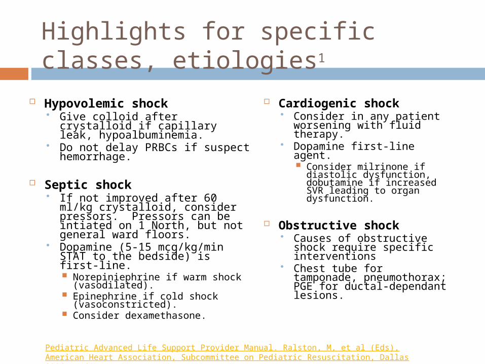
Highlights for specific classes, etiologies1
Hypovolemic shock Give colloid after crystalloid if
capillary leak, hypoalbuminemia.
Do not delay PRBCs if suspect hemorrhage.
Septic shock If not improved after 60 ml/kg
crystalloid, consider pressors. Pressors can be intiated on 1 North, but not general ward floors.
Dopamine (5-15 mcg/kg/min STAT to the bedside) is first-line. Norepiniephrine if warm shock
(vasodilated). Epinephrine if cold shock
(vasoconstricted). Consider dexamethasone.
Cardiogenic shock Consider in any patient
worsening with fluid therapy.
Dopamine first-line agent. Consider milrinone if
diastolic dysfunction, dobutamine if increased SVR leading to organ dysfunction.
Obstructive shock Causes of obstructive
shock require specific interventions
Chest tube for tamponade, pneumothorax; PGE for ductal-dependant lesions.
Pediatric Advanced Life Support Provider Manual. Ralston, M, et al (Eds), American Heart Association, Subcommittee on Pediatric Resuscitation, Dallas 2006. p.61.
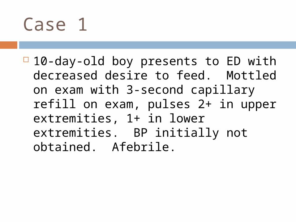
Case 1
10-day-old boy presents to ED with decreased desire to feed. Mottled on exam with 3-second capillary refill on exam, pulses 2+ in upper extremities, 1+ in lower extremities. BP initially not obtained. Afebrile.
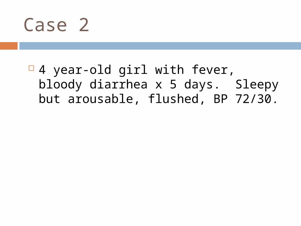
Case 2
4 year-old girl with fever, bloody diarrhea x 5 days. Sleepy but arousable, flushed, BP 72/30.
References
Bilkovski, RN, Rivers, EP, Horst HM. Targeted resuscitation strategies after injury. Curr Opin Crit Care 2004; 10:529.
Bone, RC. Toward an epidemiology and natural history of SIRS (systemic inflammatory response syndrome). JAMA 1992; 268:3452.
Carcillo, JA, Fields, AI. Clinical practice parameters for hemodynamic support of pediatric and neonatal patients in septic shock. Crit Care Med 2002; 30:1365.
Carcillo, JA, Kuch, BA, Han, YY, et al. Mortality and functional morbidity after use of PALS/APLS by community physicians. Pediatrics 2009;124:500.
Inwald, DP, Tasker, RC, Peters, MJ, Nadel, S. Emergency management of children with severe sepsis in the United Kingdom: the results of the Paediatric Intensive Care Society sepsis audit. Arch Dis Child 2009;94:348.
Fisher JD. Et al. Clinical spectrum of shock in the pediatric emergency department. Pediatr Emerg Care. 2010 Sep;26(9):622-5.
Kache, Saraswati; Frankel, Lorry Nelson’s Pediatrics: 18th edition, chapter 68.
Watson, RS, Carcillo, JA, Linde-Zwirble, WT, et al. The epidemiology of severe sepsis in children in the United States. Am J Respir Crit Care Med 2003; 167:695.
Pediatric Advanced Life Support Provider Manual. Ralston, M, et al (Eds), American Heart Association, Subcommittee on Pediatric Resuscitation, Dallas 2006. p.61.
Rivers, E, Nguyen, B, Havstad, S, et al. Early goal-directed therapy in the treatment of severe sepsis and septic shock. N Engl J Med 2001; 345:1368.
Up To Date, 2010. Initial management of shock in children; Physiology and classification of shock in children.