Embed Size (px)
Citation preview
Proc. Natl. Acad. Sci. USAVol. 84, pp. 6297-6301, September 1987Neurobiology
Shifter circuits: A computational strategy for dynamic aspects ofvisual processing
(stereopsis/attention/motion analysis/vision/striate cortex)
C. H. ANDERSON*t AND D. C. VAN ESSENt§*David Sarnoff Research Center, Princeton, NJ 08540; and tDivision of Biology, 216-76, California Institute of Technology, Pasadena, CA 91125
Communicated by John J. Hopfield, May 11, 1987 (receivedfor review March 17, 1987)
ABSTRACT We propose a general strategy for dynamiccontrol of information flow between arrays of neurons atdifferent levels of the visual pathway, starting in the lateralgeniculate nucleus and the geniculorecipient layers of corticalarea V1. This strategy can be used for resolving computationalproblems arising in the domains of stereopsis, directed visualattention, and the perception ofmoving images. In each ofthesesituations, some means of dynamically controlling how retinaloutputs map onto higher-level targets is desirable-in order toachieve binocular fusion, to allow shifts of the focus ofattention, and to prevent blurring of moving images. Theproposed solution involves what we term "shifter circuits,"which allow for dynamic shifts in the relative alignment ofinputand output arrays without loss of local spatial relationships.The shifts are produced in increments along a succession ofrelay stages that are linked by diverging excitatory inputs. Thedirection of shift is controlled at each stage by inhibitoryneurons that selectively suppress appropriate sets of ascendinginputs. The shifter hypothesis is consistent with availableanatomical and physiological evidence on the organization ofthe primate visual pathway, and it offers a sensible explanationfor a variety of otherwise puzzling facts, such as the plethoraof cells in the geniculorecipient layers of V1.
Information-processing systems, whether biological or elec-tronic, should be designed for efficient and timely routing ofthe signals used for computation. In a standard digitalcomputer, the routing of information to and from the centralprocessor is inherently dynamic. With each computationalcycle, the central processor selects its input data from a vastarray of memory addresses and likewise sends the output toany desired address. Are there analogous switching pro-cesses that regulate information flow in the nervous system?That is, can the inputs used by a neuron for its computationsbe dynamically switched, or are they rigidly fixed? Thepossibility of dynamic switching processes in the visualsystem has been suggested in relation to several functionallydistinct aspects of perception, including directed visualattention, stereopsis, and the compensation for motion blur(1-6).
In this report, we will show how a specific type ofinformation-routing strategy, implemented by what we call a"shifter circuit," may provide a common mechanism under-lying each ofthese seemingly disparate perceptual processes.The shifter circuit offers a general means of linking an arrayof lower-level neurons [such as retinal ganglion cells (RGCs)]to a higher-level processor in a manner that allows fordynamic shifts in the relative alignment of the two levelswithout the loss of local spatial relationships. We firstconsider the case of stereopsis, as it allows for the clearest
formulation of both the computational problem and ourproposed solution.The Registration Problem in Stereopsis. In stereopsis,
information about binocular disparities of images in the leftand right eyes is used to make inferences about the depth ofobjects in the visual field. This requires having the two eyesproperly converged, so that the two retinal images are inregister. However, this mechanical alignment process, me-diated by vergence eye movements, is imperfect; the mis-alignment is several minutes of arc under optimal conditionsand can be an order of magnitude larger under more realisticconditions where head movements are allowed (7). None-theless, images are readily fused binocularly and are per-ceived to be stable in depth, and relative disparities of onlya few seconds of arc are sufficient for stereoscopic depthdiscrimination (8). Thus, specialized neural mechanismsmust exist to provide for binocular fusion, perceptual stabil-ity, and stereo discrimination in the face of binocular verg-ence fluctuations. Poggio and his colleagues (4, 8, 9) havedemonstrated that cells in cortical area V1 (striate cortex) ofalert monkeys show disparity tuning that is considerablysharper than the measured variability in binocular alignment;they accordingly have suggested that a dynamic neuralalignment process occurs early in the primate visual pathway.An efficient way to compensate for misregistration of the
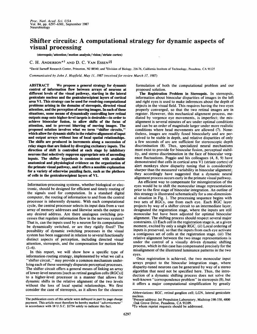
eyes would be to shift the monocular image representationsprior to the first stage of binocular integration. An outline ofthis strategy is illustrated schematically for a one-dimension-al situation in Fig. 1. The processing sequence begins withtwo sets of RGCs, one from each eye. Each RGC layerprojects by way of a shifter circuit to an intermediate layer,designated the registration stage, where the inputs are stillmonocular but have been adjusted for optimal binocularalignment. The shifting process should respect several majorconstraints. (i) Each cell in the registration stage is, at a givenmoment, excited by only a single RGC. (ii) Local ordering ofinputs is preserved, so that the inputs from each eye activatea contiguous set of cells at the registration stage. (iii) Therelative alignment between the two image representations isunder the control of a visually driven dynamic shiftingprocess, which in this case has compensated precisely for themisalignment of the illustrated luminance patterns in the twoeyes.Once registration is achieved, the two monocular input
arrays project to the binocular integration stage, wheredisparity-tuned neurons can be generated by way of a stereoalgorithm that need not be specified here. Thus, the intro-duction of a dynamic shifting process does not solve thewell-known "correspondence problem" in stereopsis (8), butit offers a major computational simplification by greatly
Abbreviations: RGC, retinal ganglion cell; LGN, lateral geniculatenucleus.tPresent address: Jet Propulsion Laboratory, Mailstop 198-330, 4800Oak Grove Drive, Pasadena, CA 91109.§To whom reprint requests should be addressed.
6297
The publication costs of this article were defrayed in part by page chargepayment. This article must therefore be hereby marked "advertisement"in accordance with 18 U.S.C. §1734 solely to indicate this fact.
6298 Neurobiology: Anderson and Van Essen
BINOCULARREGISTRATION BINOCULAR
INDEX INTEGRATION
REGISTRATION 0000000 0 004 0000000 RSTAGE - O O O O O O O O OOO O O O O O O O0 0 L
SHIFTCONTROL
RGC (
LUMINANCE / X.I'PATTERNS LEFT EYE RIGHT EYE
FIG. 1. Schematic diagram of how a shifting process could provide binocular registration at the cortical level (V1) despite misregistrationof luminance patterns for the two eyes. Note that the sharp luminance peak, which activates noncorresponding RGCs (hatched circles), mapsonto corresponding cells at the registration stage. R, right; L, left.
reducing the range over which false correspondences need tobe tested.A Basic Shifter Circuit. An elementary shifter circuit
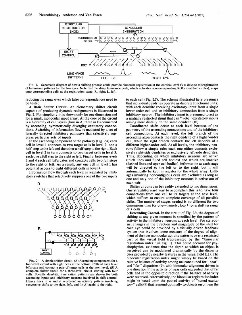
capable of producing dynamic realignments is illustrated inFig. 2. For simplicity, it is shown only for one dimension andfor a small, monocular input array. At the core of the circuitis a hierarchy of cell layers (four in A; three in B) connectedby ascending, symmetrically diverging excitatory connec-tions. Switching of information flow is mediated by a set oflaterally directed inhibitory pathways that selectively sup-press particular sets of inputs.
In the ascending component of the pathway (Fig. 2A) eachcell in level 1 connects to two target cells in level 2: one ahalf-step to the left and the other a half-step to the right. Eachcell in level 2 in turn connects to two target cells in level 3,each one a full step to the right or left. Finally, between levels3 and 4 each cell bifurcates and contacts cells two full stepsto the right or left. As a result, any one cell in level 1 haspotential access to eight different cells in level 4.
Information flow through each level is regulated by inhib-itory switches that selectively suppress one of the two inputs
A
B
FIG. 2. A simple shifter circuit. (A) Ascending components for afour-level circuit with eight cells at the bottom. Cells at each levelbifurcate and contact a pair of target cells at the next level. (B) Acomplete shifter circuit for a three-level circuit starting with fourcells. Specific dendritic innervation patterns are shown for bothascending inputs and inhibitory neurons involved in shift control.Heavy lines in A and B represent an activity pattern involvingsuccessive shifts to the right, left, and (in A) again to the right.
to each cell (Fig. 2B). The scheme illustrated here presumesthat individual dendrites operate as discrete functional units,with each dendrite receiving excitatory input from a singlelower-order cell and an inhibitory connection from a singleinhibitory neuron. The inhibitory input is presumed to act asa spatially restricted shunt that can "veto" excitatory inputsarising more distally on the same dendrite (10).
Coordinated shifts occur at each level because of thegeometry of the ascending connections and of the inhibitorycell connections. At each level, the left branch of theascending axon contacts the right dendrite of a higher-ordercell, while the right branch contacts the left dendrite of adifferent higher-order cell. At all levels, the inhibitory neu-rons follow a simple rule: each one either contacts exclu-sively right-side dendrites or exclusively left-side dendrites.Thus, depending on which inhibitory neurons are active(thick lines and filled cell bodies) and which are inactive(dashed lines and open cell bodies), information at each stagewill be directed to the left or to the right, but it willautomatically be kept in register for the whole array. Link-ages involving noncontiguous cells are excluded as long asone and only one of the inhibitory neurons is active out ofeach pair.
Shifter circuits can be readily extended to two dimensions.One straightforward way to accomplish this is to have fourconnections from one cell to its targets at the next level,which suffices to ensure complete coverage of all possibleshifts. The number of stages needed is no different for twodimensions than for one-namely, log2 k for a shifting rangeof k cells.
Descending Control. In the circuit of Fig. 2B, the degree ofshifting at any given moment is specified by the pattern ofactivity in the inhibitory neurons at each level. For stereop-sis, changes in the direction and magnitude of the shift foreach eye could be provided by a visually driven feedbacksystem that involves some measure of the degree of align-ment of the two monocular activity patterns over a restrictedpart of the visual field (represented by the "binocularregistration index" in Fig. 1). This could account for psy-chophysical evidence that the depth at which an object isperceived can be modulated dramatically by the disparitycues provided by nearby features in the visual field (11). Thebinocular registration index might simply be based on therelative balance of activity among neurons tuned for "near"and "far" disparities (9), with binocular alignment driven inone direction if the activity of near cells exceeded that of farcells and in the opposite direction if the balance of activitywere reversed. Alternatively, the binocular registration indexmight be based upon the pooled activity of "tuned excita-tory" cells (9) that respond optimally to objects on or near the
Proc. Natl. Acad. Sci. USA 84 (1987)
Proc. Nati. Acad. Sci. USA 84 (1987) 6299
perceptual horopter. In this case, the shifters could operateby keeping the binocular registration index maximized for agiven set of visual inputs. Simple versions of either type ofcontrol mechanisms should not be difficult to implement. Thecircuitry needed to attain and maintain binocular registrationin realistic operating circumstances is likely to be rathercomplex, though, since the system must be capable ofadjusting rapidly to a dynamic visual environment, mustperform smoothly over a range of spatial scales and over theentire visual field, and may also be linked to the motor systemfor vergence eye movements.
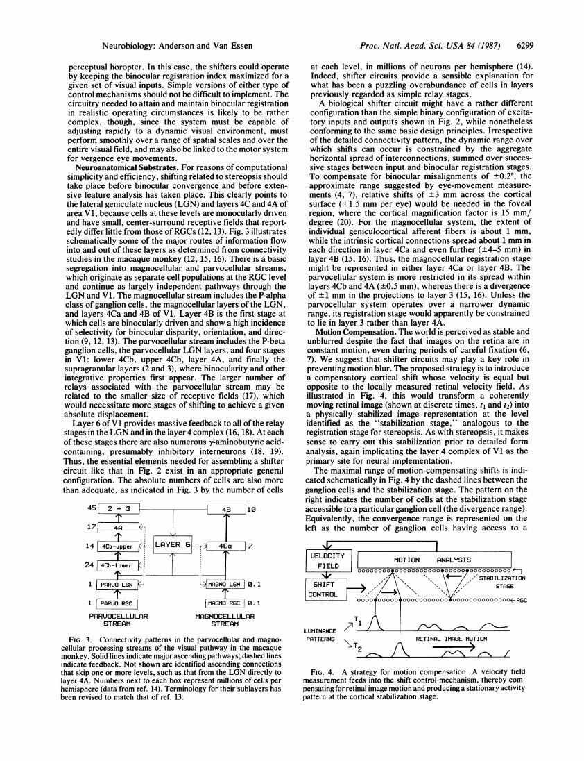
Neuroanatomical Substrates. For reasons of computationalsimplicity and efficiency, shifting related to stereopsis shouldtake place before binocular convergence and before exten-sive feature analysis has taken place. This clearly points tothe lateral geniculate nucleus (LGN) and layers 4C and 4A ofarea V1, because cells at these levels are monocularly drivenand have small, center-surround receptive fields that report-edly differ little from those ofRGCs (12, 13). Fig. 3 illustratesschematically some of the major routes of information flowinto and out of these layers as determined from connectivitystudies in the macaque monkey (12, 15, 16). There is a basicsegregation into magnocellular and parvocellular streams,which originate as separate cell populations at the RGC leveland continue as largely independent pathways through theLGN and V1. The magnocellular stream includes the P-alphaclass of ganglion cells, the magnocellular layers of the LGN,and layers 4Ca and 4B of V1. Layer 4B is the first stage atwhich cells are binocularly driven and show a high incidenceof selectivity for binocular disparity, orientation, and direc-tion (9, 12, 13). The parvocellular stream includes the P-betaganglion cells, the parvocellular LGN layers, and four stagesin V1: lower 4Cb, upper 4Cb, layer 4A, and finally thesupragranular layers (2 and 3), where binocularity and otherintegrative properties first appear. The larger number ofrelays associated with the parvocellular stream may berelated to the smaller size of receptive fields (17), whichwould necessitate more stages of shifting to achieve a givenabsolute displacement.Layer 6 ofV1 provides massive feedback to all of the relay
stages in the LGN and in the layer 4 complex (16, 18). At eachof these stages there are also numerous y-aminobutyric acid-containing, presumably inhibitory interneurons (18, 19).Thus, the essential elements needed for assembling a shiftercircuit like that in Fig. 2 exist in an appropriate generalconfiguration. The absolute numbers of cells are also morethan adequate, as indicated in Fig. 3 by the number of cells
45 2 + 3
I171 4A '
14 4Cb-upper (1
24 4Cb-lower -i
1 PARUO LGN -'
1 PARUO RGC
PARUOCELLULARSTREAM
L MAGNO LN . 1
MAGNO RGC 0. 1
MAGNOCELLULARSTREAM
FIG. 3. Connectivity patterns in the parvocellular and magno-cellular processing streams of the visual pathway in the macaquemonkey. Solid lines indicate major ascending pathways; dashed linesindicate feedback. Not shown are identified ascending connectionsthat skip one or more levels, such as that from the LGN directly tolayer 4A. Numbers next to each box represent millions of cells perhemisphere (data from ref. 14). Terminology for their sublayers hasbeen revised to match that of ref. 13.
at each level, in millions of neurons per hemisphere (14).Indeed, shifter circuits provide a sensible explanation forwhat has been a puzzling overabundance of cells in layerspreviously regarded as simple relay stages.A biological shifter circuit might have a rather different
configuration than the simple binary configuration of excita-tory inputs and outputs shown in Fig. 2, while nonethelessconforming to the same basic design principles. Irrespectiveof the detailed connectivity pattern, the dynamic range overwhich shifts can occur is constrained by the aggregatehorizontal spread of interconnections, summed over succes-sive stages between input and binocular registration stages.To compensate for binocular misalignments of ±0.20, theapproximate range suggested by eye-movement measure-ments (4, 7), relative shifts of ±3 mm across the corticalsurface (±1.5 mm per eye) would be needed in the fovealregion, where the cortical magnification factor is 15 mm/degree (20). For the magnocellular system, the extent ofindividual geniculocortical afferent fibers is about 1 mm,while the intrinsic cortical connections spread about 1 mm ineach direction in layer 4Ca and even further (±4-5 mm) inlayer 4B (15, 16). Thus, the magnocellular registration stagemight be represented in either layer 4Ca or layer 4B. Theparvocellular system is more restricted in its spread withinlayers 4Cb and 4A (±0.5 mm), whereas there is a divergenceof +1 mm in the projections to layer 3 (15, 16). Unless theparvocellular system operates over a narrower dynamicrange, its registration stage would apparently be constrainedto lie in layer 3 rather than layer 4A.Motion Compensation. The world is perceived as stable and
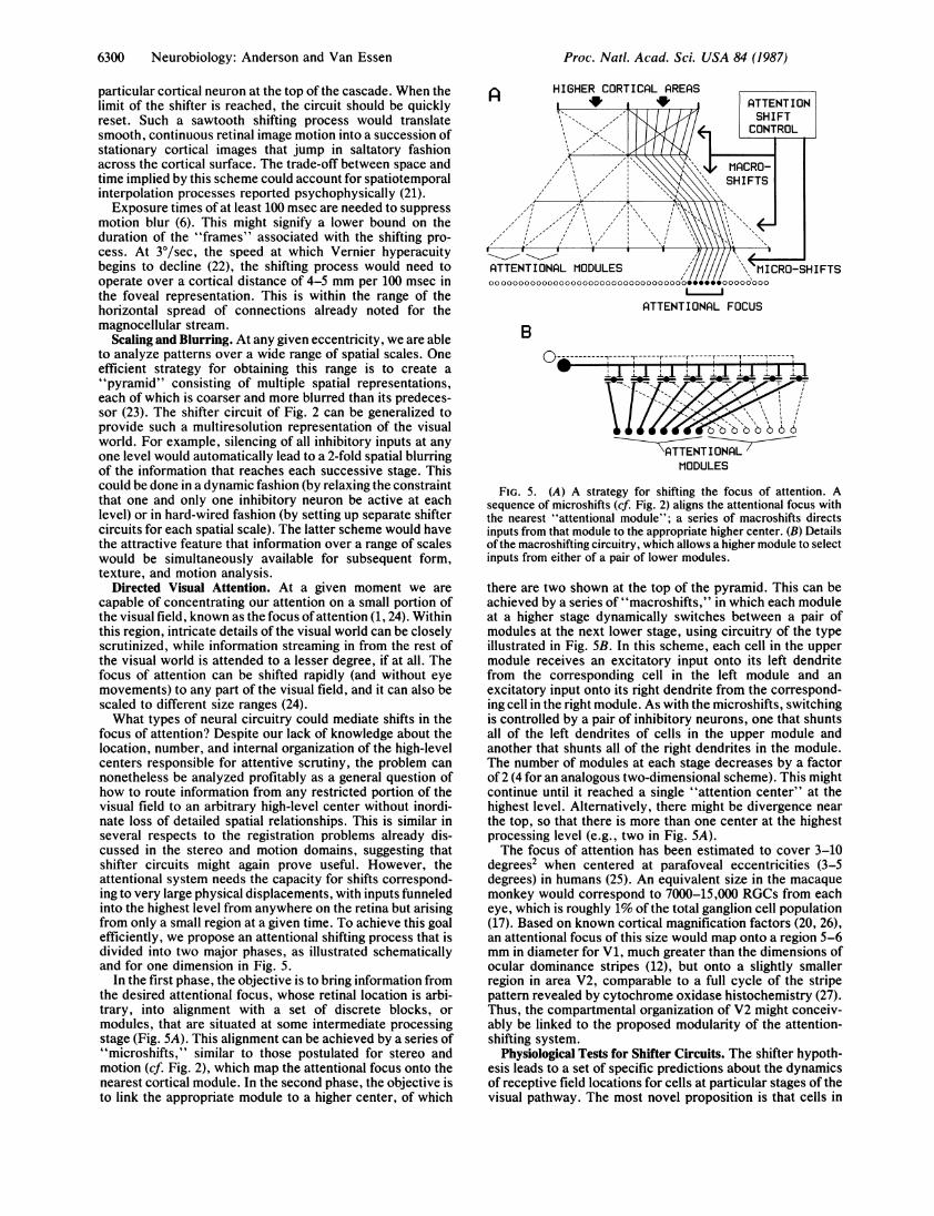
unblurred despite the fact that images on the retina are inconstant motion, even during periods of careful fixation (6,7). We suggest that shifter circuits may play a key role inpreventing motion blur. The proposed strategy is to introducea compensatory cortical shift whose velocity is equal butopposite to the locally measured retinal velocity field. Asillustrated in Fig. 4, this would transform a coherentlymoving retinal image (shown at discrete times, t1 and t2) intoa physically stabilized image representation at the levelidentified as the "stabilization stage," analogous to theregistration stage for stereopsis. As with stereopsis, it makessense to carry out this stabilization prior to detailed formanalysis, again implicating the layer 4 complex of V1 as theprimary site for neural implementation.The maximal range of motion-compensating shifts is indi-
cated schematically in Fig. 4 by the dashed lines between theganglion cells and the stabilization stage. The pattern on theright indicates the number of cells at the stabilization stageaccessible to a particular ganglion cell (the divergence range).Equivalently, the convergence range is represented on theleft as the number of ganglion cells having access to a
MOTION ANALYSIS00000000 00000000000 00000 0000000000
/'STABILIZATIONSTAGE
i oo t o o o o oooooooo0OOOOOO- RGC
71 ALUMINANCEPATTERNS
\T2 ilkRETINAL IMAGE MOTION
/
FIG. 4. A strategy for motion compensation. A velocity fieldmeasurement feeds into the shift control mechanism, thereby com-pensating for retinal image motion and producing a stationary activitypattern at the cortical stabilization stage.
Neurobiology: Anderson and Van Essen
I
6300 Neurobiology: Anderson and Van Essen
particular cortical neuron at the top of the cascade. When thelimit of the shifter is reached, the circuit should be quicklyreset. Such a sawtooth shifting process would translatesmooth, continuous retinal image motion into a succession ofstationary cortical images that jump in saltatory fashionacross the cortical surface. The trade-off between space andtime implied by this scheme could account for spatiotemporalinterpolation processes reported psychophysically (21).Exposure times of at least 100 msec are needed to suppress
motion blur (6). This might signify a lower bound on theduration of the "frames" associated with the shifting pro-cess. At 3Y/sec, the speed at which Vernier hyperacuitybegins to decline (22), the shifting process would need tooperate over a cortical distance of 4-5 mm per 100 msec inthe foveal representation. This is within the range of thehorizontal spread of connections already noted for themagnocellular stream.
Scaling and Blurring. At any given eccentricity, we are ableto analyze patterns over a wide range of spatial scales. Oneefficient strategy for obtaining this range is to create a"pyramid" consisting of multiple spatial representations,each of which is coarser and more blurred than its predeces-sor (23). The shifter circuit of Fig. 2 can be generalized toprovide such a multiresolution representation of the visualworld. For example, silencing of all inhibitory inputs at anyone level would automatically lead to a 2-fold spatial blurringof the information that reaches each successive stage. Thiscould be done in a dynamic fashion (by relaxing the constraintthat one and only one inhibitory neuron be active at eachlevel) or in hard-wired fashion (by setting up separate shiftercircuits for each spatial scale). The latter scheme would havethe attractive feature that information over a range of scaleswould be simultaneously available for subsequent form,texture, and motion analysis.
Directed Visual Attention. At a given moment we arecapable of concentrating our attention on a small portion ofthe visual field, known as the focus of attention (1, 24). Withinthis region, intricate details of the visual world can be closelyscrutinized, while information streaming in from the rest ofthe visual world is attended to a lesser degree, if at all. Thefocus of attention can be shifted rapidly (and without eyemovements) to any part of the visual field, and it can also bescaled to different size ranges (24).What types of neural circuitry could mediate shifts in the
focus of attention? Despite our lack of knowledge about thelocation, number, and internal organization of the high-levelcenters responsible for attentive scrutiny, the problem cannonetheless be analyzed profitably as a general question ofhow to route information from any restricted portion of thevisual field to an arbitrary high-level center without inordi-nate loss of detailed spatial relationships. This is similar inseveral respects to the registration problems already dis-cussed in the stereo and motion domains, suggesting thatshifter circuits might again prove useful. However, theattentional system needs the capacity for shifts correspond-ing to very large physical displacements, with inputs funneledinto the highest level from anywhere on the retina but arisingfrom only a small region at a given time. To achieve this goalefficiently, we propose an attentional shifting process that isdivided into two major phases, as illustrated schematicallyand for one dimension in Fig. 5.
In the first phase, the objective is to bring information fromthe desired attentional focus, whose retinal location is arbi-trary, into alignment with a set of discrete blocks, ormodules, that are situated at some intermediate processingstage (Fig. SA). This alignment can be achieved by a series of"microshifts," similar to those postulated for stereo andmotion (cf. Fig. 2), which map the attentional focus onto thenearest cortical module. In the second phase, the objective isto link the appropriate module to a higher center, of which
HIGHER CORTICAL AREAS
ATTENTIONAL FOCUS
B----------n------r---r--r-----r----- T--5----1
\ATTENT I ONALMODULES
FIG. 5. (A) A strategy for shifting the focus of attention. Asequence of microshifts (cf. Fig. 2) aligns the attentional focus withthe nearest "attentional module"; a series of macroshifts directsinputs from that module to the appropriate higher center. (B) Detailsof the macroshifting circuitry, which allows a higher module to selectinputs from either of a pair of lower modules.
there are two shown at the top of the pyramid. This can beachieved by a series of "macroshifts," in which each moduleat a higher stage dynamically switches between a pair ofmodules at the next lower stage, using circuitry of the typeillustrated in Fig. 5B. In this scheme, each cell in the uppermodule receives an excitatory input onto its left dendritefrom the corresponding cell in the left module and anexcitatory input onto its right dendrite from the correspond-ing cell in the right module. As with the microshifts, switchingis controlled by a pair of inhibitory neurons, one that shuntsall of the left dendrites of cells in the upper module andanother that shunts all of the right dendrites in the module.The number of modules at each stage decreases by a factorof 2 (4 for an analogous two-dimensional scheme). This mightcontinue until it reached a single "attention center" at thehighest level. Alternatively, there might be divergence nearthe top, so that there is more than one center at the highestprocessing level (e.g., two in Fig. SA).The focus of attention has been estimated to cover 3-10
degrees2 when centered at parafoveal eccentricities (3-5degrees) in humans (25). An equivalent size in the macaquemonkey would correspond to 7000-15,000 RGCs from eacheye, which is roughly 1% of the total ganglion cell population(17). Based on known cortical magnification factors (20, 26),an attentional focus of this size would map onto a region 5-6mm in diameter for V1, much greater than the dimensions ofocular dominance stripes (12), but onto a slightly smallerregion in area V2, comparable to a full cycle of the stripepattern revealed by cytochrome oxidase histochemistry (27).Thus, the compartmental organization of V2 might conceiv-ably be linked to the proposed modularity of the attention-shifting system.
Physiological Tests for Shifter Circuits. The shifter hypoth-esis leads to a set of specific predictions about the dynamicsof receptive field locations for cells at particular stages of thevisual pathway. The most novel proposition is that cells in
Proc. Natl. Acad. Sci. USA 84 (1987)
Proc. Natl. Acad. Sci. USA 84 (/987) 6301
layers 4C and 4A of V1, and possibly also in the LGN, shouldundergo outright receptive-field shifts when driven by stim-ulus patterns that provide an appropriate reference framebased on stereo, motion, or attentional cues. The range overwhich shifts occur should increase at successive stages;beyond layer 4C, the shifts should be many times the size ofRGC receptive fields.
Previous studies (28, 29) involving precise mapping of V1receptive fields in anesthetized, paralyzed monkeys indicatethat receptive fields are, under some circumstances, stable towithin a few minutes of arc. On the other hand, there are alsohints that dynamic receptive-field shifts may indeed occur(29, 35). We do not consider the existing evidence to becompelling, either for or against the shifter hypothesis. Inparticular, we note that anesthesia might seriously disruptcortical feedback control circuitry, thereby reducing oreliminating any dynamic shifting and possibly also increasingreceptive-field sizes, by way of the blurring process de-scribed in an earlier section. A more decisive test would beto record simultaneously from cells at different levels of theputative shifter circuit in alert animals, so that a low-level cellcould serve as a reference for eye position; the experimentshould also use a visual stimulation paradigm that wouldmodulate the shifting process over its full range and in acontrolled fashion.
If shifter circuits are indeed present in the early visualpathway, traditional notions of neuronal receptive-fieldboundaries as static entities must be substantially modified.Receptive fields are typically plotted using a simple stimulusmoving on a blank background. In the absence of a well-defined reference frame, the mapping stimulus itself mighttend to drive the shifting mechanism and thereby induce thereceptive field to move dynamically along with the stimulus.If so, the classical receptive field would be considerablylarger than the actual receptive-field size at any given instant(see Fig. 4).
In extrastriate cortex, evidence for dynamic modulation ofreceptive-field size by attentional cues has been presented forarea V4 (30) and for inferotemporal cortex (31) in monkeys.These findings are compatible with several possible mecha-nisms for selective attention (2, 3) and thus do not on theirown constitute strong evidence for the shifter hypothesis.Also, we have not dealt with the full complexity of attentivemechanisms, such as the ability to shift attention alongnonspatial dimensions (e.g., for color). Thus, the shifterhypothesis is arguably more speculative and less fully devel-oped for attention than for stereopsis and motion. Nonethe-less, it emphasizes that a relatively simple strategy may applyto a variety of dynamic routing problems in vision. Shiftercircuits might also be relevant to other systems and pro-cesses, such as the rapid adjustment of visual and auditorymaps in the primate superior colliculus (32) and the muchslower modulation of sensory maps in the somatosensorycortex after peripheral nerve lesions (33). More generally, theuse of shunting inhibition to modulate a restricted subset ofthe connections onto a given neuron offers a powerfulstrategy that can be exploited for tasks of computation per se(10, 34), as well as for the routing strategies discussed in thepresent report.
We thank Eric Mjolsness and many other colleagues for valuablediscussions, ideas, and suggestions. This work was supported in partby contract N00014-85K-0068 from the Office of Naval Research toD.C.V.E.
1. Julesz, B. (1984) in Dynamic Aspects of Neocortical Function(Wiley, New York), pp. 585-612.
2. Crick, F. (1984) Proc. Nati. Acad. Sci. USA 81, 4586-4590.3. Koch, C. & Ullman, S. (1985) Hum. Neurobiol. 4, 219-227.4. Motter, B. & Poggio, G. (1984) Exp. Brain Res. 54, 304-314.5. Barlow, H. B. (1981) Proc. R. Soc. London Ser. B 212, 1-36.6. Burr, D. C. & Ross, J. (1986) Trends NeuroSci. 9, 304-307.7. Steinman, R. M., Cushman, W. B. & Martins, A. J. (1982)
Hum. Neurobiol. 1, 97-109.8. Poggio, G. & Poggio, T. (1984) Annu. Rev. Neurosci. 7, 379-
412.9. Poggio, G. & Talbot, W. H. (1981) J. Physiol. 315, 469-492.
10. Koch, C., Poggio, T. & Torre, V. (1983) Proc. Natl. Acad. Sci.USA 80, 2799-2802.
11. Mitchison, G. J. & McKee, S. P. (1985) Nature (London) 315,402-404.
12. Hubel, D. H. & Wiesel, T. N. (1977) Proc. R. Soc. LondonSer. B 198, 1-59.
13. Blasdel, G. G. & Fitzpatrick, D. J. (1984) J. Neurosci. 4,880-895.
14. O'Kusky, J. & Colonnier, M. (1982) J. Comp. Neurol. 210,278-290.
15. Fitzpatrick, D. S., Lund, J. S. & Blasdel, G. G. (1985) J.Neurosci. 5, 3329-3349.
16. Blasdel, G. G., Lund, J. S. & Fitzpatrick, D. (1985) J. Neuro-sci. 5, 3350-3369.
17. Perry, N. H. & Cowey, A. (1985) Vision Res. 25, 1795-1810.18. Sherman, S. M. & Koch, C. (1986) Exp. Brain Res. 63, 1-20.19. Hendrickson, A. E., Hunt, S. P. & Wu, J.-Y. (1981) Nature
(London) 292, 605-607.20. Van Essen, D. C., Newsome, W. T. & Maunsell, J. H. R.
(1984) Vision Res. 24, 429-448.21. Barlow, H. B. (1979) Nature (London) 279, 189-190.22. Westheimer, G. & McKee, S. P. (1975) J. Opt. Soc. Am. 65,
847-850.23. Rosenfeld, A. (1984) Multiresolution Image Processing and
Analysis (Springer, New York).24. Bergen, J. R. & Julesz, B. (1983) Nature (London) 303, 696-
698.25. Sagi, D. & Julesz, B. (1985) Nature (London) 321, 693-695.26. Gattass, R., Gross, C. G. & Sandell, J. H. (1981) J. Comp.
Neurol. 201, 519-539.27. Tootell, R. B. H., Silverman, M. S., DeValois, R. G. & Ja-
cobs, G. H. (1983) Science 220, 737-739.28. DeValois, R. L., Albrecht, D. G. & Thorell, L. G. (1982)
Vision Res. 22, 545-559.29. Parker, A. & Hawken, M. (1985) J. Opt. Soc. Am. 2, 1101-
1114.30. Moran, J. & Desimone, R. (1985) Science 229, 782-784.31. Richmond, B. J., Wurtz, R. H. & Sato, T. (1983) J. Neuro-
physiol. 50, 1415-1432.32. Jay, M. F. & Sparks, D. L. (1984) Nature (London) 309,
345-347.33. Kaas, J. H., Merzenich, M. M. & Killackey, H. P. (1983)
Annu. Rev. Neurosci. 6, 325-350.34. Anderson, C. H. & Abrahams, E. (1987) Proceedings of the
First IEEE International Conference on Neural Networks, inpress.
35. Motter, B. C. & Poggio, G. F. (1982) Soc. Neurosci. Abstr. 8,707.
Neurobiology: Anderson and Van Essen


![Not for Distribution - Routledgecw.routledge.com/textbooks/9780415448789/5275-Parcak-Ch01.pdf · Not for Distribution [15:59 16/2/2009 5275-Parcak-Ch01.tex] Job No: 5275 PARCAK: Satellite](https://img.pdfslide.us/doc/110x75/5f06d9927e708231d41a0be7/not-for-distribution-not-for-distribution-1559-1622009-5275-parcak-ch01tex.jpg)


![5275-A4_EXS-1000_Remote_Terminal_Unit [PDF Search Engine]](https://img.pdfslide.us/doc/110x75/577d2f271a28ab4e1eb0f0b0/5275-a4exs-1000remoteterminalunit-pdf-search-engine.jpg)