Embed Size (px)
Citation preview

Anim. Reprod, v.7, n.3, p.187-196, Jul./Sept. 2010
_________________________________________
5Corresponding author: [email protected] Phone: +81(3)5463-0558; Fax: +81(3)5463-0558
Sexual plasticity of rainbow trout germ cells
G. Yoshizaki1,2,5, T. Okutsu3, M. Ichikawa1, M. Hayashi1, Y. Takeuchi4
1Department of Marine Biosciences, Tokyo University of Marine Science and Technology, Tokyo 108-8477, Japan 2SORST, Japan Science and Technology Agency, Saitama 332-0012, Japan
3Japan International Research Center for Agricultural Sciences, Ibaraki 305-8686, Japan 4Research Center for Advanced Science and Technology, Tokyo University of Marine Science and Technology,
Chiba 294-0308, Japan
Abstract
The sexual plasticity of fish gonads declines after the sex-differentiation period; however, the plasticity of the germ cells themselves after this stage remains poorly understood. We characterized the sexual plasticity of gonial germ cells by transplanting them into sexually undifferentiated embryonic gonads in rainbow trout (Oncorhynchus mykiss). Spermatogonia or oogonia isolated from the meiotic gonads of vasa-green fluorescent protein (Gfp) gene transgenic trout were transplanted into the peritoneal cavity of newly hatched embryos of both sexes, and the behavior of the GFP-labeled donor cells was observed. The transplanted spermatogonia and oogonia migrated towards the recipient gonadal anlagen, and were subsequently incorporated into them. We also confirmed that the donor-derived gonial germ cells resumed gametogenesis in the recipient somatic microenvironment synchronously with the endogenous germ cells. Surprisingly, the donor-derived spermatogonia started to proliferate and differentiate into oocytes in female recipients. At 2 years post-transplantation, the eggs from mature female recipients were artificially inseminated with sperm from intact male rainbow trout. Normal, live offspring with the donor-derived haplotype were obtained. In addition, oogonia-derived sperm were produced in the male recipients. These donor-derived sperm were shown to be fully functional, as live offspring carrying GFP-labeled germ cells with the donor haplotype were obtained in the first filial (F1) generation. These findings indicate that rainbow trout pre-meiotic germ cells, which are likely to be spermatogonial or oogonial stem cells, possess a high level of sexual plasticity, and that the sexual differentiation of germ cells is controlled solely by the somatic microenvironment, rather than being cell autonomous. Keywords: fish, germ cell transplantation, oogonia, sexual plasticity, spermatogonia, spermatogonial stem cell, xenotransplantation.
Introduction
The phenotypic sex of some species of fish is flexible and individuals can change gender during their
lifetime, which is known as hermaphroditism (Devlin and Nagahama, 2002). However, in most fish species, the phenotypic sexes are fixed (gonochorism) and are determined by genetic information. The sex-differentiation process in the medaka (Oryzias latipes) has been intensively studied for nearly half a century (Yamamoto, 1969; Saito and Tanaka, 2009). Although this species strictly follows the XY sex-determination system and is male heterogametic, the gender of individuals can be changed if they are exposed to androgens or estrogens during the sex-differentiation period (Yamamoto, 1969). Since the effects of environmental temperature on sex determination in the Atlantic silverside (Menidia menidia) were first reported by Conover and Kynard (1981), many studies have shown that sex-determination processes are affected by various environmental factors including temperature, rearing density, and growth rate (Baroiller et al., 2009). In gonochoristic species, however, exposure to exogenous sex steroids or environmental factors is not thought to induce sex reversal after the sex-differentiation period, which generally occurs around the hatching stage (Devlin and Nagahama, 2002). The sexual plasticity of fish gonads is therefore believed to decline after the sex-differentiation period. In a series of studies on germ-cell transplantation, however, we have revealed that the germ cells themselves retain a high level of sexual plasticity even after the sex-differentiation period. Here we review the principles and implications of our germ cell-transplantation studies, with an emphasis on the sex of germ cells.
Primordial germ-cell transplantation in fish
A spermatogonial transplantation system in rodents was established by Brinster and Zimmermann (1994). They delivered a testicular cell suspension into the seminiferous tubules of mice using a glass micro-capillary. The donor spermatogonia transplanted into the recipient seminiferous tubules formed colonies and produced large numbers of sperm throughout the lifetimes of recipient mice, suggesting that the colonizing spermatogonia behaved as spermatogonial stem cells (Brinster, 2002). Furthermore, the transplantation of donor spermatogonia into sterile recipients with genetic germ-cell defects was able to rescue their fertility (Brinster and Zimmermann, 1994;

Yoshizaki et al. Germ cell sex in fish.
188 Anim. Reprod, v.7, n.3, p.187-196, Jul./Sept. 2010
Ogawa et al., 2000). This provided further, strong evidence that the donor cells included spermatogonial stem cells, which appeared to have an unlimited ability to self-renew together with an ability to differentiate into sperm. Germ-cell transplantation is therefore a powerful tool to characterize these cells functionally.
In fish, however, work on gonial germ cells, such as spermatogonia and oogonia, has been largely limited to histological and endocrinological studies (Schulz et al., 2010); functional studies have remained relatively scarce until recently. In order to understand the stemness and sexual plasticity of gonial germ cells, we focused on germ-cell transplantation in fish. Before considering spermatogonia or oogonia, we carried out germ-cell transplantation using primordial germ cells (PGCs), which were isolated from sexually undifferentiated embryos.
When we began our PGC-transplantation experiments, there were no available techniques to transplant purified germ cells, although several groups had performed the transplantation of whole embryonic cells into recipient hatchlings in order to obtain germ-line chimeras in fish species including the zebrafish (Danio rerio; Lin et al., 1992), medaka (Wakamatsu et al., 1993), and rainbow trout (Oncorhynchus mykiss; Nilson and Cloud, 1993; Takeuchi et al., 2001). One obvious obstacle to germ-cell transplantation was the lack of available markers, such as antibodies against cell-surface antigens, to identify or trace live germ cells. Therefore, as a first step, we developed a method for labeling germ cells using a transgenic technique with the green fluorescent protein (Gfp) gene, in order to distinguish between germ and somatic cells. We chose the rainbow trout as a model for these transgenic studies, because the newly hatched embryos containing PGCs are much larger than those of most other fish species (the total length of a newly hatched rainbow trout embryo is ~15 mm), and they are suitable for dissection and PGC manipulation. The regulatory sequence used to drive the Gfp gene was the vasa gene, which is specifically expressed in the germ-cell lineages of various animal species (Raz, 2000), including the rainbow trout (Yoshizaki et al., 2000a). The vasa-Gfp construct was microinjected into the cytoplasm of fertilized eggs and used to establish stable, transgenic trout strains (Yoshizaki et al., 2000b). As expected, the transgenic trout carrying the vasa-Gfp construct showed green fluorescence specifically in the PGCs (Yoshizaki et al., 2000a; Takeuchi et al., 2002), spermatogonia (Okutsu et al., 2006b; Yano et al., 2008), oogonia, and oocytes (Yoshizaki et al., 2010a), which also expressed endogenous vasa. Notably, the inclusion of the 3-untranslated region of the trout vasa sequence was essential to obtain transgenic trout with GFP-labeled PGCs (Yoshizaki et al., 2000b). This was further confirmed by microinjection studies using chimeric RNA from the Gfp-coding region with the 3-untranslated region of the trout vasa gene (Yoshizaki et
al., 2005). Our study demonstrated that microinjection of the Gfp RNA ligated with the 3-untranslated region of the trout vasa gene into fertilized eggs resulted in the production of embryos with GFP-labeled PGCs. This suggested that the 3-untranslated region of the trout vasa gene plays an important role in stabilizing mRNA specifically in the PGCs. Similar findings were also reported in zebrafish (Knaut et al., 2002; Wolke et al., 2002), suggesting that this might be a universal phenomenon among teleosts.
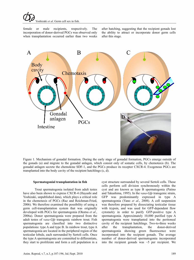
We next transplanted the PGCs using the vasa-Gfp transgenic rainbow trout (Takeuchi et al., 2003, 2004). In the absence of an immunodeficient strain - similar to the nude mouse - for fish, we chose newly hatched trout embryos as recipients, as their immature immune system is unable to reject exogenous cells (Manning and Nakanishi, 1996). Newly hatched rainbow trout embryos possess sexually immature gonadal anlagen that have recently coalesced with PGCs (Takashima et al., 1980). However, the gonadal anlagen were not large enough for us to deliver the donor PGCs using a micromanipulator. We therefore utilized the chemotaxis system used for PGC migration. During gonadal formation, the embryonic gonads initially consist only of somatic cells, and PGCs emerge in the extragonadal areas at this time (Fig. 1a; Patino and Takashima, 1995). The gonadal somatic cells then begin to secrete the chemokine stromal derived factor-1 (SDF-1), while the PGCs located outside of the gonadal anlagen express its receptor, CXC-chemokine receptor 4 (CXCR-4). The PGCs are attracted to SDF-1 and migrate to the gonadal anlagen using pseudopodia (Raz and Reichman-Fried, 2006; Fig. 1b). Based on these facts, we predicted that donor PGCs delivered into the vicinity of the recipient gonadal anlagen would migrate towards them, following the migration routes of endogenous PGCs. Our simple approach was therefore to deliver ~10 donor PGCs, isolated from the gonadal anlagen of trout hatchlings, into the peritoneal cavity of the recipient hatchlings under a dissection microscope (Fig. 1c, d). Twenty days after the transplantation, the donor-derived PGCs were found to be attached to the peritoneal wall adjacent to the recipient gonads with no sign of acute rejection. Moreover, many of the donor-derived PGCs had extended pseudopodia. Thirty days after the transplantation, the intraperitoneally transplanted PGCs had successfully migrated to the recipient gonads and had been incorporated into them. This migration process of the donor-derived PGCs took such a long time probably due to slow developmental speed of this species caused by low rearing temperature, which is around 10oC. Indeed, migration of endogenous PGCs also require nearly 2 weeks (Okutsu et al., 2006b). These donor-derived PGCs started gametogenesis in synchrony with the recipient germ cells and eventually produced functional gametes. In these experiments, the donor-derived PGCs were shown to undergo either oogenesis or spermatogenesis in
Yoshizaki et al. Germ cell sex in fish.
Anim. Reprod, v.7, n.3, p.187-196, Jul./Sept. 2010 189
female or male recipients, respectively. The incorporation of donor-derived PGCs was observed only when transplantation occurred earlier than two weeks
after hatching, suggesting that the recipient gonads lost the ability to attract or incorporate donor germ cells after this stage.
Figure 1. Mechanism of gonadal formation. During the early stage of gonadal formation, PGCs emerge outside of the gonads (a) and migrate to the gonadal anlagen, which consist only of somatic cells, by chemotaxis (b). The gonadal anlagen secrete the chemokine SDF-1, and the PGCs produce its receptor CXCR-4. Exogenous PGCs are transplanted into the body cavity of the recipient hatchlings (c, d).
Spermatogonial transplantation in fish
Trout spermatogonia isolated from adult testes have also been shown to express CXCR-4 (Hayashi and Yoshizaki, unpublished data), which plays a critical role in the chemotaxis of PGCs (Raz and Reichman-Fried, 2006). We therefore examined the possibility of using a germ cell-transplantation system that was originally developed with PGCs for spermatogonia (Okutsu et al., 2006a). Donor spermatogonia were prepared from the adult testes of vasa-Gfp transgenic rainbow trout. Fish spermatogonia are classified into two distinctive populations: type A and type B. In rainbow trout, type A spermatogonia are located in the peripheral region of the testicular lobule, each surrounded by Sertoli cells. Once the type A spermatogonia are committed to differentiate, they start to proliferate and form a cell population in a
cyst structure surrounded by several Sertoli cells. These cells perform cell division synchronously within the cyst and are known as type B spermatogonia (Patino and Takashima, 1995). In the vasa-Gfp transgenic strain, GFP was predominantly expressed in type A spermatogonia (Yano et al., 2008). A cell suspension was therefore prepared by dissociating testicular tissue with trypsin, and was used for GFP-dependent flow cytometry in order to purify GFP-positive type A spermatogonia. Approximately 10,000 purified type A spermatogonia were transplanted into the peritoneal cavity of the recipient hatchlings. Two-to-three weeks after the transplantation, the donor-derived spermatogonia showing green fluorescence were incorporated into the recipient gonads. The average number of donor-derived spermatogonia incorporated into the recipient gonads was ~5 per recipient. We

Yoshizaki et al. Germ cell sex in fish.
190 Anim. Reprod, v.7, n.3, p.187-196, Jul./Sept. 2010
further confirmed that the donor-derived spermatogonia proliferated rapidly and produced a large colony of GFP-positive donor-derived germ cells in the recipient testis. One-to-two years after the transplantation, several of the matured recipient trout were used for progeny tests. This study used vasa-Gfp transgenic orange-colored mutant rainbow trout as donors and wild-type non-transgenic rainbow trout as recipients. The milt obtained from the recipient trout was inseminated into the eggs obtained from wild-type non-transgenic female trout. As a result, orange-colored mutants carrying the vasa-Gfp gene with the donor-derived haplotype were observed among the wild-type non-transgenic offspring, which were recipient derived. The donor-derived offspring showed similar external morphology and maturation to the wild-type rainbow trout. This result indicated that the donor-derived spermatogonia could migrate to the recipient gonadal anlagen, become incorporated into them, resume spermatogenesis, and produce functional sperm in the recipient testes. Notably, the type A spermatogonia derived from the adult testes could interact well with somatic counterpart of embryonic gonadal anlagen. Furthermore, based on the number of sperm produced by the recipients (a mixture of donor-derived and recipient-derived sperm) and the frequency of offspring carrying the donor-derived haplotype in the first filial (F1) generation, the estimated number of sperm produced by one donor-derived spermatogonium was ~40 million. As differentiating spermatogonia can perform only seven cycles of mitosis before entering meiosis (in other words, one spermatogonium can produce only 512 spermatozoa; Loir, 1999), and the recipients were able to produce similar amounts of donor-derived sperm for at least 3 years, we concluded that the type A spermatogonia of rainbow trout contained a population of stem cells possessing an extremely high, or even unlimited, capacity to self-renew and differentiate into functional sperm.
Fish spermatogonia can produce functional eggs
We next examined the behavior of donor-
derived spermatogonia in female recipients (Okutsu et al., 2006a). Donor spermatogonia were prepared using GFP-dependent flow-cytometry from the adult testes of vasa-Gfp transgenic orange-colored mutants. As mentioned above, this cell sorting yielded only type A spermatogonia. Approximately 10,000 purified type A spermatogonia were microinjected into the body cavities of the recipient hatchlings. Two months after the transplantation, donor-derived spermatogonia were incorporated into the ovaries of the female recipients (Fig. 2a). Furthermore, the donor-derived spermatogonia had differentiated into peri-nucleolus-stage oocytes with a diameter of >100 m, in synchrony with the recipient-derived germ cells, 6 months after the transplantation (Fig. 2b). Rearing the female recipients for 2 years produced several mature individuals. We performed progeny tests by crossing eggs obtained from these recipients with sperm obtained from wild-type males. Several orange-colored offspring were detected among the wild-type F1 generation (Fig. 2c). These orange-colored mutants carried the vasa-Gfp gene, which was another indicator of the donor haplotype. Furthermore, they showed normal external morphology and maturation, similar to the wild-type rainbow trout. These data suggested that the type A spermatogonia transplanted into female recipients differentiated into female germ cells and eventually produced fully functional eggs. Furthermore, throughout this experiment, the obtained sex ratio was close to 1:1, and we did not detect any ovo-testes-like gonads, suggesting that spermatogonial transplantation did not induce sex reversal in the recipient fish at either the gonadal or the cellular level.
Figure 2. Intraperitoneally transplanted spermatogonia (GFP-positive) can be incorporated into the recipient ovary (a) and start to differentiate into oocytes (b). Female recipients carrying donor spermatogonia-derived eggs can produce F1 offspring showing the donor-derived phenotype (that is, orange body color; arrow heads; c).

Yoshizaki et al. Germ cell sex in fish.
Anim. Reprod, v.7, n.3, p.187-196, Jul./Sept. 2010 191
Although the X and Y chromosomes of rainbow trout cannot be distinguished based on their morphology, there is evidence that populations of gynogenetic diploids become all female, suggesting that their sex-determination follows the XY system (Chourrout and Quillet, 1982). This was further confirmed by progeny tests with sex-reversed individuals, such as XX males (phenotypically male and genetically female) and XY females (phenotypically female and genetically male), which were produced by sex-steroid treatment during the sex-differentiation period (Davidson et al., 2009). The production of eggs derived from spermatogonia in female recipients raised the question of whether spermatogonia carrying XY chromosomes could produce both X and Y eggs. To answer this, we performed progeny tests with female recipients, which received spermatogonia, and wild-type XY males (Okutsu and Yoshizaki, unpublished data). The resulting male-to-female sex ratio was nearly 3:1 (Fig. 3). If the female recipients produced functional Y eggs, and further YY individuals were viable males, the ratio between males and females would remain at 3:1 (1 XX: 2 XY: 1 YY). We therefore analyzed the sex ratio of the F2 generation, obtained by crossing F1 males (possibly both XY and YY males) and wild-type XX females. Approximately one-
third of the F1 males produced all-male F2 populations (Fig. 3), suggesting that these F1 males carried YY chromosomes. Thus, the female recipients produced Y eggs in addition to X eggs, and the resulting YY males were viable and fertile, suggesting that the X and Y chromosomes were at least functionally equivalent, except at the male-determining locus. These results are similar to previous observations made using sex-reversed XY females produced by estrogen treatment (Johnstone et al., 1979). Thus, we propose spermatogonial transplantation into female recipients as a novel non-pharmacological method to produce Y eggs in rainbow trout, which can be used to produce all-male populations for both basic biological and aquacultural purposes. Matsuda et al. (2007) reported that transgenic XX medaka fish carrying the DMY male-determining gene (Matsuda et al., 2002) developed into functional males with the ability to produce normal sperm. This also suggested that the X and Y chromosomes in fish are highly similar, except at the male-determining locus. This was in contrast to the results reported for a transgenic XX mouse carrying sry, which could not complete spermatogenesis (Koopman, 1995). The similarity of the X and Y chromosomes of fish might have made it possible to induce the sex reversal of spermatogonia in female recipients.
Figure 3. Behavior of sex chromosomes in progeny tests with female recipients of XY spermatogonia. As the recipient females produce both X and Y eggs, mating them with XY males creates a sex ratio in the F1 of three males to one female. Furthermore, one-third of the F1 males have YY sex chromosomes.

Yoshizaki et al. Germ cell sex in fish.
192 Anim. Reprod, v.7, n.3, p.187-196, Jul./Sept. 2010
Fish oogonia can produce functional sperm We also transplanted oogonia purified from
vasa-Gfp transgenic female rainbow trout into male recipient hatchlings using the same strategy employed for spermatogonial transplantation (Yoshizaki et al., 2010b). The donor-derived oogonia were incorporated into the male testes and started to proliferate. The number of donor-derived oogonia increased by more than 40-fold over the 130-day period after their incorporation into the recipient gonads. Two years after the transplantation, the donor-derived oogonia had differentiated into functional eggs in the female recipients. By contrast, they had differentiated into spermatogonia and started spermatogenesis in the male recipients. We could not find any significant differences in the frequencies of recipients with donor germ cells in their gonads, and those with colonies of proliferated donor germ cells, between the female and male recipients. To further confirm whether the donor-derived oogonia could differentiate into functional sperm in the male recipients, we transplanted oogonia into triploid male rainbow trout. Triploid males produce only transparent milt containing an extremely small amount of aneuploid sperm, which cannot fertilize eggs (Carrasco et al., 1998). Two years after the transplantation, some of the recipient triploid males produced normal white-colored milt. Progeny tests were therefore performed using the milt obtained from the recipient males and eggs obtained from wild-type females. We used donor trout that were heterozygous for the orange-colored mutant and hemizogous for the vasa-Gfp transgene; therefore, if the triploid recipients produced only donor-derived sperm (oogonia-derived sperm), one-half of the offspring would be expected to have orange body color and carry the vasa-Gfp transgene. As predicted, in all three mating experiments, ~50% of the F1 offspring showed the orange-colored phenotype and carried the vasa-Gfp transgene, suggesting that the oogonia that were intraperitoneally transplanted into the newly hatched embryos had differentiated into male germ cells and eventually produced functional sperm. In this experiment, one oogonium incorporated into the recipient testis produced ~1 billion sperm. As mentioned above, once spermatogonia are committed to differentiate, each can produce up to 512 sperm (Loir, 1999). The number of sperm produced from the donor oogonium far exceeded this value. Furthermore, the male recipients also produced donor oogonia-derived sperm during the following spawning season. These data suggested that
the oogonia that were incorporated into the recipient gonads differentiated into spermatogonial stem cells, which could differentiate into sperm and had a high, or even unlimited, capacity for self renewal.
If the oogonia carrying the XX sex chromosome produced sperm in the recipient males, then the F1 generation produced by the milt obtained from the recipient fish and the eggs obtained from wild-type females (XX) would be expected to be all female (XX). In order to examine the sex ratio, we investigated the sex of the F1 offspring. All those obtained from the three mating experiments (in total, 196 offspring) were female, further suggesting that the sperm produced by the recipient males was derived from oogonia carrying the XX sex chromosome. These oogonial transplantation studies always produced recipients with a sex ratio close to 1:1, and no ovo-testis-like morphology was observed, suggesting that the oogonial transplantation did not induce sex reversal in the recipient fish at either the gonadal or cellular level.
Conclusions and perspectives
Our series of studies showed that type A spermatogonia contain a cell population that can differentiate into functional eggs and oogonia also contain a cell population that can differentiate into functional sperm. Our results clearly indicate that both type A spermatogonia and oogonia, even those isolated from gonads after the sex-differentiation period, contain cell populations with a high level of sexual plasticity, and that the sex of fish germ cells is determined solely by the somatic microenvironment, rather than being cell autonomous. We also found that the spermatogonia could be transplanted into triploid sterile recipients of different species (xenotransplantation), and that the donor-derived germ cells completed gametogenesis if the genetic distance between the donors and recipients was small enough, as with rainbow trout donors and masu salmon (Oncorhynchus masou) recipients (in this case, the phylogenetic desistance between the donor and the recipient was approximately 8 million years; Okutsu et al., 2007, 2008; Fig. 4). The xenotransplantation of fish spermatogonia has great potential for a wide range of applications, including transgenesis by transfection into germ cells, cryobanking of germ cells for the conservation of the genetic diversity of wild fish populations (Fig. 5; Kobayashi et al., 2007; Yoshizaki et al., 2010a), and surrogate broodstock technology (Okutsu et al., 2006b, 2007, 2008). In the latter novel aquaculture system, the gametes of large-bodied fish

Yoshizaki et al. Germ cell sex in fish.
Anim. Reprod, v.7, n.3, p.187-196, Jul./Sept. 2010 193
with a long generation time, such as the bluefin tuna (Thunnus thynnus and Thunnus orientalis), are produced by a surrogate broodstock, which has a comparatively smaller body size and shorter generation time, such as chub mackerel (Scomber japonicus). This can be achieved by the xenotransplantation of spermatogonia from the target species into a suitable recipient species. This system is expected to reduce the rearing space, costs, and labor required for maintenance of the broodstock.
Research on fish germ-line stem cells has recently been gaining in popularity. A system for the direct transplantation of spermatogonia into the adult testis of recipient fish was reported in the tilapia (Oreochromis niloticus; Lacerda et al., 2006, 2008, 2010) and pejerrey (Odontesthes hatchery; Majhi et al., 2009). The localization and behavior of oogonial stem
cells were reported in the medaka ovary (Nakamura et al., 2010). Furthermore, methods for the in vitro culture of spermatogonia are being developed for several fish species, and could facilitate studies of the effects of various soluble factors on the survival, proliferation, and differentiation of gonial stem cells in vitro (Hong et al., 2004; Shikina et al., 2008; Shikina and Yoshizaki, 2010). We have also identified a cell-surface protein that is predominantly expressed in rainbow trout spermatogonia (Nagasawa et al., 2010). This could be a powerful tool for enriching spermatogonia from the testes of various fish species using specific antibody-mediated flow cytometry or magnetic cell sorting. We predict that these research efforts will lead to rapid advances in understanding of the cellular biology of fish germ cells, particularly spermatogonial stem cells and oogonial stem cells, in the near future.
Figure 4. Xenotransplantation of spermatogonia. When rainbow trout spermatogonia are transplanted into triploid sterile masu salmon hatchlings, the recipient salmon parents produce only rainbow trout gametes. Mating these masu salmon parents thus produces only rainbow trout offspring in the F1 generation.

Yoshizaki et al. Germ cell sex in fish.
194 Anim. Reprod, v.7, n.3, p.187-196, Jul./Sept. 2010
Figure 5. Application of spermatogonial transplantation to conservation of endangered fish species. By cryopreserving the spermatogonia of endangered fish species, their genetic resources might be preserved in the future. Even if the target species becomes extinct, it might be restored by transplanting the frozen spermatogonia into recipient hatchlings of a closely related species. When the recipients are mature, simply mating the female and male recipients could yield fertilized eggs of the extinct species.
References Baroiller JF, D'Cotta H, Saillant E. 2009. Environmental effects on fish sex determination and differentiation. Sex Dev, 3:118-135. Brinster RL, Zimmermann JW. 1994. Spermatogenesis following male germ-cell transplantation. Proc Natl Acad Sci USA, 91:11298-11302. Brinster RL. 2002. Germline stem cell transplantation and transgenesis. Science, 296:2174-2176. Carrasco LA, Doroshov S, Penman DJ, Bromage N. 1998. Long-term, quantitative analysis of gametogenesis in autotriploid rainbow trout, Oncorhynchus mykiss. J Reprod Fertil, 113:197-210. Chourrout D, Quillet E. 1982. Induced gynogenesis in the rainbow trout: Sex and survival of progenies production of all-triploid populations. Theor Appl Genet, 63:201-205. Conover DO, Kynard BE. 1981. Environmental sex determination: interaction of temperature and genotype in a fish. Science, 213:577-579. Davidson WS, Huang TK, Fujiki K, von Schalburg
KR, Koop BF. 2009. The sex determining loci and sex chromosomes in the family salmonidae. Sex Dev, 3:78-87. Devlin RH, Nagahama Y. 2002. Sex determination and sex differentiation in fish: an overview of genetic, physiological, and environmental influences. Aquaculture, 208:191-364. Hong Y, Liu T, Zhao H, Xu H, Wang W, Liu R, Chen T, Deng J, Gui J. 2004. Establishment of a normal medaka fish spermatogonial cell line capable of sperm production in vitro. Proc Natl Acad Sci USA, 101:8011-8016. Johnstone R, Simpson TH, Youngson AF, Whitehead C. 1979. Sex reversal in salmonid culture: part II. The progeny of sex-reversed rainbow trout. Aquaculture, 18:13-19. Knaut H, Steinbeisser H, Schwarz H, Nüsslein-Volhard C. 2002. An evolutionary conserved region in the vasa 3′UTR targets RNA translation to the germ cells in the zebrafish. Curr Biol, 12:454-466. Kobayashi T, Takeuchi Y, Takeuchi T, Yoshizaki G. 2007. Generation of viable fish from cryopreserved primordial germ cells. Mol Reprod Dev, 74:207-213.

Yoshizaki et al. Germ cell sex in fish.
Anim. Reprod, v.7, n.3, p.187-196, Jul./Sept. 2010 195
Koopman P. 1995. The molecular biology of SRY and its role in sex determination in mammals. Reprod Fertil Dev, 7:713-722. Lacerda SM, Batlouni SR, Silva SB, Homem CS, Franca LR. 2006. Germ cell transplantation in fish: the Nile-tilapia model. Anim Reprod, 3:146-159. Lacerda SM, Batlouni SR, Assis L, Resende F, Campos-Silva S, Campos-Silva R, Segatelli TM, Franca LR. 2008. Germ cell transplantation in tilapias (Oreochromis niloticus). Cybium, 32:115-118. Lacerda SM, Batlouni SR, Costa GM, Segatelli TM, Quirino BR, Queiroz BM, Kalapothakis E, França LR. 2010. A new and fast technique to generate offspring after germ cells transplantation in adult fish: the nile tilapia (Oreochromis niloticus) model. PLoS One, 5:e10740. Lin S, Long W, Chen J, Hopkins N. 1992. Production of germ-line chimeras in zebrafish by cell transplants from genetically pigmented to albino embryos. Proc Natl Acad Sci USA, 89:4519-4523. Loir M. 1999. Spermatogonia of rainbow trout: I. Morphological characterization, mitotic activity, and survival in primary cultures of testicular cells. Mol Reprod Dev, 53:422-433. Majhi SK, Hattori RS, Yokota M, Watanabe S, Strüssmann CA. 2009. Germ cell transplantation using sexually competent fish: an approach for rapid propagation of endangered and valuable germlines. PLoS One, 4:e6132. Matsuda M, Nagahama Y, Shinomiya A, Sato T, Matsuda C, Kobayashi T, Morrey CE, Shibata N, Asakawa S, Shimizu N, Hori H, Hamaguchi S, Sakaizumi M. 2002. DMY is a Y-specific DM-domain gene required for male development in the medaka fish. Nature, 417:559-563. Matsuda M, Shinomiya A, Kinoshita M, Suzuki A, Kobayashi T, Paul-Prasanth B, Lau EL, Hamaguchi S, Sakaizumi M, Nagahama Y. 2007. DMY gene induces male development in genetically female (XX) medaka fish. Proc Natl Acad Sci USA, 104:3865-3870. Manning MJ, Nakanishi T. 1996. The specific immune system: cellular defences. In: Iwama G, Nakanishi T (Ed.). The Fish Immune System. New York: Academic Press. pp. 159-205. Nagasawa K, Shikina S, Takeuchi Y, Yoshizaki G. 2010. Lymphocyte antigen 75 (Ly75/CD205) is a surface marker on mitotic germ cells in rainbow trout. Biol Reprod. doi: 10.1095/biolreprod.109.082081. Nakamura S, Kobayashi K, Nishimura T, Higashijima S, Tanaka M. 2010. Identification of germline stem cells in the ovary of the teleost medaka. Science, 328:1561-1563. Nilsson EE, Cloud JG. 1993. Extent of mosaicism in experimentally produced diploid/triploid chimeric trout. J Exp Zool, 266:47-50. Ogawa T, Dobrinski I, Avarbock MR, Brinster RL. 2000. Transplantation of male germ line stem cells restores fertility in infertile mice. Nat Med, 6:29-34.
Okutsu T, Suzuki K, Takeuchi Y, Takeuchi T, Yoshizaki G. 2006a. Testicular germ cells can colonize sexually undifferentiated embryonic gonad and produce functional eggs in fish. Proc Natl Acad Sci USA, 103:2725-2729. Okutsu T, Yano A, Nagasawa K, Shikina S, Kobayashi T, Takeuchi Y, Yoshizaki G. 2006b. Manipulation of fish germ cell: visualization, cryopreservation and transplantation. J Reprod Dev, 52:685-693. Okutsu T, Shikina S, Kanno M, Takeuchi Y, Yoshizaki G. 2007. Production of trout offspring from triploid salmon parents. Science, 317:1517. Okutsu T, Takeuchi Y, Yoshizaki G. 2008. Spermatogonial transplantation in fish: production of trout offspring from salmon parents. In: Tsukamoto K, Kawamura T, Takeuchi T, Beard Jr TD, Kaiser MJ (Ed.). Fisheries for Global Welfare and Environment. Tokyo: Terapub. pp. 209-219. Patiño R, Takashima F. 1995. Gonads. In: Takashima F, Hibiya T (Ed.). An Atlas of Fish Histology: Normal and Pathological Features. Tokyo: Kodansha. pp. 129-153. Raz E. 2000. The function and regulation of vasa-like genes in germ-cell development. Genome Biol, 1:reviews1017. Raz E, Reichman-Fried M. 2006. Attraction rules: germ cell migration in zebrafish. Curr Opin Genet Dev, 16:355-359. Saito D, Tanaka M. 2009. Comparative aspects of gonadal sex differentiation in medaka: a conserved role of developing oocytes in sexual canalization. Sex Dev, 3:99-107. Schulz RW, de França LR, Lareyre JJ, Le Gac F, Chiarini-Garcia H, Nobrega RH, Miura T. 2010. Spermatogenesis in fish. Gen Comp Endocrinol, 165:390-411. Shikina S, Ihara S, Yoshizaki G. 2008. Culture conditions for maintaining the survival and mitotic activity of rainbow trout transplantable type A spermatogonia. Mol Reprod Dev, 75:529-537. Shikina S, Yoshizaki G. 2010. Improved in vitro culture conditions to enhance the survival, mitotic activity, and transplantability of rainbow trout type A spermatogonia. Biol Reprod, 83:268-276. Takashima F, Patiño R, Nomura M. 1980. Histological studies on the sex differentiation in rainbow trout. Bull Japan Soc Sci Fish, 46:1317-1322. Takeuchi Y, Yoshizaki G, Takeuchi T. 2001. Production of germ-line chimeras in rainbow trout by blastomere transplantation. Mol Reprod Dev, 59:380-389. Takeuchi Y, Yoshizaki G, Kobayashi T, Takeuchi T. 2002. Mass isolation of primordial germ cells from transgenic rainbow trout carrying the green fluorescent protein gene driven by the vasa gene promoter. Biol Reprod, 67:1087-1092. Takeuchi Y, Yoshizaki G, Takeuchi T. 2003.

Yoshizaki et al. Germ cell sex in fish.
196 Anim. Reprod, v.7, n.3, p.187-196, Jul./Sept. 2010
Generation of live fry from intraperitoneally transplanted primordial germ cells in rainbow trout. Biol Reprod, 69:1142-1149. Takeuchi Y, Yoshizaki G, Takeuchi T. 2004. Surrogate broodstock produces salmonids. Nature, 430:629-630. Wakamatsu Y, Ozato K, Hashimoto H, Kinoshita M, Sakaguchi M, Iwamatsu T, Hyodo-Taguchi Y, Tomita H. 1993. Generation of germ-line chimeras in medaka (Oryzias latipes). Mol Mar Biol Biotechnol, 2:325-332. Yamamoto T. 1969. Sex differentiation. In: Hoar WS, Randall DJ (Ed.). Fish Physiology. New York: Academic Press. pp. 117-175. Yano A, Suzuki K, Yoshizaki G. 2008. Flow-cytometric isolation of testicular germ cells from rainbow trout (Oncorhynchus mykiss) carrying the green fluorescent protein gene driven by trout vasa regulatory regions. Biol Reprod, 78:151-158. Yoshizaki G, Sakatani S, Tominaga H, Takeuchi T. 2000a. Cloning and characterization of a vasa-like gene in rainbow trout and its expression in the germ cell lineage. Mol Reprod Dev, 55:364-371. Yoshizaki G, Takeuchi Y, Sakatani S, Takeuchi T.
2000b. Germ cell-specific expression of green fluorescent protein in transgenic rainbow trout under control of the rainbow trout vasa-like gene promoter. Int J Dev Biol, 44:323-326. Yoshizaki G, Tago Y, Takeuchi Y, Sawatari E, Kobayashi T, Takeuchi T. 2005. Green fluorescent protein labeling of primordial germ cells using a nontransgenic method and its application for germ cell transplantation in salmonidae. Biol Reprod, 73:88-93. Yoshizaki G, Fujinuma K, Iwasaki Y, Okutsu T, Shikina S, Yazawa R, Takeuchi Y. 2010a. Spermatogonial transplantation in fish: a novel method for the preservation of genetic resources. Comp Biochem Physiol Part D Genomics Proteomics. doi:10.1016/j.cbd.2010.05.003. Yoshizaki G, Ichikawa M, Hayashi M, Iwasaki Y, Miwa M, Shikina S, Okutsu T. 2010b. Sexual plasticity of ovarian germ cells in rainbow trout. Development, 137:1227-1230. Wolke U, Weidinger G, Köprunner M, Raz E. 2002. Multiple levels of posttranscriptional control lead to germ line-specific gene expression in the zebrafish. Curr Biol, 12:289-294.