Embed Size (px)
Citation preview
www.elsevier.com/locate/brainres
Brain Research 1014 (2004) 244–246
Short communication
Sex differences in systemic morphine analgesic tolerance following
intrathecal morphine injections
Eileen Hopkinsa, Grace Rossib, Benjamin Kesta,c,*
aNeuropsychology Doctoral Subprogram, Queens College, City University of New York, Flushing, NY 11367, USAbDepartment of Psychology, C.W. Post College, Long Island University, Brookville, NY 11548, USA
cDepartment of Psychology and Center for Developmental Neuroscience, College of Staten Island, City University of New York,
Staten Island, NY 10314, USA
Accepted 31 March 2004
Available online 28 May 2004
Abstract
Morphine analgesic potency following systemic administration was assessed in male and female mice undergoing prior and repeated
intrathecal morphine injections. Although morphine ED50 values were significantly increased in both sexes relative to their respective saline-
injected controls, the magnitude of tolerance was greater in females. Intrathecal injection alone had no effect on morphine analgesia. The data
suggest that spinal mechanisms contribute to sex differences in analgesic tolerance following systemic morphine administration.
D 2004 Elsevier B.V. All rights reserved.
Theme: Neurotransmitters, modulators, transporters, and receptors
Topic: Opioids: anatomy, physiology, and behavior
Keywords: Morphine; Tolerance; Sex difference; Spinal cord
The difference between males and females in analgesic
effect following morphine administration has deservedly
received notable attention [9]. Although the mechanism of
this sexual diergism remains unclear, the persistence of
analgesic sex differences after central morphine microin-
jection in mice [7] and rats [5] clearly implicates contri-
butions from sites within the CNS. Like morphine
analgesia, morphine analgesic tolerance is also subject to
sex differences in rats and mice [1,3,8 (see however Ref.
[2])]. In the mouse, we have previously reported that the
reduced analgesic potency of morphine is of greater
magnitude in female mice following 4 or 8 days of
repeated systemic injection [8]. Since morphine analgesia
can be elicited after either spinal [14] or supraspinal [5–7]
application, both sites can contribute to analgesia following
systemic morphine injection. Thus, although the mecha-
nism contributing to morphine tolerance has yet to be
0006-8993/$ - see front matter D 2004 Elsevier B.V. All rights reserved.
doi:10.1016/j.brainres.2004.03.056
* Corresponding author. Department of Psychology, and Center for
Developmental Neuroscience, City University of New York, College of
Staten Island, 2800 Victory Blvd., Staten Island, NY 10314, USA. Tel.: +1-
718-982-4070; fax: +1-718-982-4114.
E-mail address: [email protected] (B. Kest).
clearly elucidated, tolerance to the analgesic effect of
systemic morphine injection may result from alterations
at either, or both, CNS loci. Similarly, sex differences in
analgesic tolerance to morphine following its systemic
injection in mice may also reflect the differential develop-
ment of tolerance between males and females in morphine
analgesic mechanisms located spinally, supraspinally, or
both. We have previously studied the contribution of
supraspinal mechanisms to sex differences in tolerance
by comparing the morphine analgesia dose– response
curves of male and female mice following chronic intra-
cerebroventricular morphine injection. Although increased
ED50 values were observed for both sexes—demonstrative
of analgesic tolerance—there was no significant difference
between males and females [6].
The aim of the present study was to now assess the
contribution of spinal morphine analgesic mechanisms to
sex differences in systemic morphine analgesic tolerance.
Using the same CD-1 strain of mouse from previous studies,
cumulative dose–response curves for systemic morphine
analgesia were generated and derived ED50 values com-
pared for males and females after repeated intrathecal
morphine microinjection.
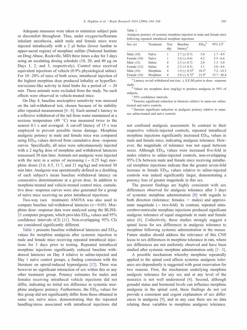
Table 1
Analgesic potency of systemic morphine injection in male and female mice
following repeated intrathecal morphine injections
Sex (n) Treatment Test
day
Baseline
latencyaED50
b 95% CIc
Male (10) Naı̈ve 1 2.7 (F 0.7) 3.0 1.7–4.9
Female (10) Naı̈ve 1 2.6 (F 0.6) 4.2 2.5–6.6
Male (12) Saline 4 2.5 (F 0.7) 2.0 1.3–3.0
Female (12) Saline 4 2.3 (F 0.3) 3.1 2.0–4.6
Male (16) Morphine 4 1.9 (F 0.5)d 10.3e 7.2–14.3
Female (16) Morphine 4 2.0 (F 0.7)d 21.9e 15.7–30.4
a Latency on tail withdrawal test (sec.F S.E.M) prior to dose– response
study.b Values are morphine dose (mg/kg) to produce analgesia in 50% of
subjects.c 95% confidence intervals.d Denotes significant reduction in latencies relative to same-sex saline-
treated and naı̈ve controls.e Denotes significant reduction in analgesic potency relative to same-
sex saline-treated and naı̈ve controls.
E. Hopkins et al. / Brain Research 1014 (2004) 244–246 245
Adequate measures were taken to minimize subject pain
or discomfort throughout. Thus, under oxygen/isoflourane
inhalant anesthesia, adult male and female mice were
injected intrathecally with a 2 Al bolus (lower lumbar to
upper-sacral region) of morphine sulfate (National Institute
on Drug Abuse, Rockville, MD) three times a day for 3 days
using an escalating dosing schedule (10, 20, and 40 Ag on
Days 1, 2, and 3, respectively). Control mice received
equivalent injections of vehicle (0.9% physiological saline).
For 10–20% of mice of both sexes, intrathecal injection of
the highest morphine dose produced lethality or hyperflex-
ion/seizure-like activity in hind limbs for a period of f 20
min. These animals were excluded from the study. No such
effects were observed in vehicle-treated mice.
On Day 4, baseline nociceptive sensitivity was assessed
on the tail-withdrawal test, chosen because of its stability
after repeated measurement [6–8]. Each animal’s latency to
a reflexive withdrawal of the tail from water maintained at a
noxious temperature (49 jC) was measured twice to the
nearest 0.1 s and averaged. A cut-off latency of 15 s was
employed to prevent possible tissue damage. Morphine
analgesic potency in male and female mice was compared
using ED50 values derived from cumulative dose–response
curves. Specifically, all mice were subcutaneously injected
with a 2 mg/kg dose of morphine and withdrawal latencies
reassessed 30 min later. Animals not analgesic were injected
with the next in a series of increasing (f 0.25 log) mor-
phine doses (3.6, 6.5, 11.7, and 21 mg/kg) and retested 30
min later. Analgesia was operationally defined as a doubling
of each subject’s mean baseline withdrawal latency on
consecutive determinations at a given dose. In addition to
morphine-treated and vehicle-treated control mice, cumula-
tive dose–response curves were also generated for a group
of naı̈ve mice receiving no prior intrathecal injections.
Two-way (sex� treatment) ANOVA was also used to
compare baseline tail-withdrawal latencies (a= 0.05). Mor-
phine dose–response data were analyzed using the BLISS-
21 computer program, which provides ED50 values and 95%
confidence intervals (CI) [11]. Non-overlapping 95% CIs
are considered significantly different.
Table 1 presents baseline withdrawal latencies and ED50
values for morphine analgesia after systemic injection in
male and female mice receiving repeated intrathecal injec-
tions for 3 days prior to testing. Repeated intrathecal
morphine injections significantly reduced baseline with-
drawal latencies on Day 4 relative to saline-injected and
Day 1 naı̈ve control groups, a finding consistent with the
literature on opioid-induced hyperalgesia [12]. There was
however no significant interaction of sex within this or any
other treatment group. Potency estimates for males and
females receiving intrathecal vehicle injections did not
differ, indicating no initial sex difference in systemic mor-
phine analgesic potency. Furthermore, the ED50 values for
this group did not significantly differ from those obtained in
same sex naı̈ve mice, demonstrating that the repeated
handling/stress associated with intrathecal injections did
not confound analgesic assessment. In contrast to their
respective vehicle-injected controls, repeated intrathecal
morphine injections significantly increased ED50 values in
male and female mice, indicating analgesic tolerance. How-
ever, the magnitude of tolerance was not equal between
sexes. Although ED50 values were increased five-fold in
males relative to saline-injected controls, non-overlapping
95% CIs between male and female mice receiving intrathe-
cal morphine injections indicate that the greater seven-fold
increase in female ED50 values relative to saline-injected
controls was indeed significantly larger, demonstrating a
potency loss of greater magnitude in this sex.
The present findings are highly consistent with sex
differences observed for analgesic tolerance after 3 days
of systemic morphine administration [8] with regards to
both direction (tolerance: females > males) and approxi-
mate magnitude (f two-fold). In contrast, repeated intra-
cerebroventricular morphine injections produced significant
analgesic tolerance of equal magnitude in male and female
mice [6]. Collectively, these studies strongly suggest a
spinal locus for sex differences in analgesic tolerance to
morphine following systemic administration in the mouse.
Future studies should address the relevance of this CNS
locus to sex differences in morphine tolerance in rats, where
sex differences are not uniformly observed and have been
studied after systemic morphine administration only [1–3].
A possible mechanism whereby morphine repeatedly
applied to the spinal cord affects systemic analgesic toler-
ance sex-dependently is suggested with great reservation for
two reasons. First, the mechanism underlying morphine
analgesic tolerance for any sex and at any level of the
neuraxis is not well understood [4]. Second, although
gonadal status and hormonal levels can influence morphine
analgesia in the spinal cord, these findings do not yet
provide a consistent and cogent explanation of sex differ-
ences in analgesia [9], and in any case there are no data
relating these variables to morphine analgesic tolerance.
E. Hopkins et al. / Brain Research 1014 (2004) 244–246246
Consequently, speculation about the role of sex hormones is
clearly beyond the scope of the present results. We are
intrigued, however, by the demonstration in mice [10,13]
and rats [15] that sub-analgesic doses of morphine co-
administered spinally and supraspinally act synergistically
in the production of analgesia. In addition, it has been
shown that the synergism between spinal and supraspinal
sites are reduced to additive effects when mice are rendered
tolerant to morphine following repeated systemic adminis-
tration [10]. If the synergistic relationship between spinal
and supraspinal sites is subject to sex differences, this would
not only account for sex differences in morphine analgesia,
but would be logically consistent with both the present
results and our previous observations on sex differences in
morphine analgesic tolerance after systemic administration
[8]. This possibility should be directly assessed in future
studies by directly comparing spinal and supraspinal mor-
phine analgesic synergy in male and female mice.
Acknowledgements
This work was supported by PSC/CUNY and CSI/IBR
Center for Developmental Neuroscience.
References
[1] D. Badillo-Martinez, A.L. Kirchegesner, P.D. Butler, R.J. Bodnar,
Monosodium glutamate and analgesia induced by morphine: test-spe-
cific effects, Neuropharmacology 23 (1984) 1141–1149.
[2] A.C. Barrett, C.D. Cook, J.M. Terner, R.M. Craft, M.J. Picker, Im-
portance of sex and relative efficacy at the mu opioid receptor in the
development of tolerance and cross-tolerance to the antinociceptive
effects of opioids, Psychopharmacology (Berl.) 158 (2001) 154–164.
[3] R.M. Craft, J.A. Stratmann, R.E. Bartok, T.I. Walpole, S.J. King, Sex
differences in development of morphine tolerance and dependence in
the rat, Psychopharmacology (Berl.) 143 (1999) 1–7.
[4] L.M. Harrison, A.J. Kastin, J.E. Zadina, Opiate tolerance and depen-
dence: receptors, G-proteins, and antiopiates, Peptides 19 (1998)
1603–1630.
[5] K.L. Kepler, B. Kest, J.M. Kiefel, M.L. Cooper, R.J. Bodnar, Roles of
gender, gonadectomy and estrous phase in the analgesic effects of
intracerebroventricular morphine in rats, Pharmacol. Biochem. Behav.
34 (1989) 119–127.
[6] B. Kest, E. Hopkins, Morphine tolerance after chronic intracerebro-
ventricular injection in male and female mice, Brain Res. 892 (2001)
208–210.
[7] B. Kest, S.G. Wilson, J.S. Mogil, Sex differences in supraspinal mor-
phine analgesia are dependent on genotype, J. Pharmacol. Exp. Ther.
289 (1999) 1370–1375.
[8] B. Kest, C.A. Palmese, E. Hopkins, A comparison of morphine an-
algesic tolerance in male and female mice, Brain Res. 879 (2000)
17–22.
[9] B. Kest, E. Sarton, A. Dahan, Gender differences in opioid-mediated
analgesia: animal and human studies, Anesthesiology 93 (2000)
539–547.
[10] S.C. Roerig, J.M. Fujimoto, Morphine antinociception in different
strains of mice: relationship of supraspinal-spinal multiplicative inter-
actions to tolerance, J. Pharmacol. Exp. Ther. 247 (1988) 603–608.
[11] J.G. Umans, C.E. Inturissi, Pharmacodynamics of subcutaneously
administered diacetylmorphine, 6-acetylmorphine, and morphine in
mice, J. Pharmacol. Exp. Ther. 219 (1981) 409–415.
[12] T.W. Vanderah, M.H. Ossipov, J. Lai, T.P. Malan Jr., F. Porreca,
Mechanisms of opioid-induced pain and antinociceptive tolerance:
descending facilitation and spinal dynorphin, Pain 92 (2001) 5–9.
[13] S. Wigdor, G.L. Wilcox, Central and systemic morphine-induced
antinociception in mice: comparison of descending serotonergic
and noradrenergic pathways, J. Pharmacol. Exp. Ther. 242 (1987)
90–95.
[14] T.L. Yaksh, T.A. Rudy, Analgesia mediated by a direct spinal action
of narcotics, Science 192 (1976) 1357–1358.
[15] J.C. Yeung, T.A. Rudy, Multiplicative interaction between narcotic
agonisms expressed at spinal and supraspinal sites of antinociceptive
action as revealed by concurrent intrathecal and intracerebroventric-
ular injection of morphine, J. Pharmacol. Exp. Ther. 215 (1980)
633–642.