Embed Size (px)
Citation preview
RESEARCH ARTICLE Open Access
Serum-free microcarrier based production ofreplication deficient Influenza vaccine candidatevirus lacking NS1 using Vero cellsAllen Chen1†, Swan Li Poh1†, Christian Dietzsch2, Elisabeth Roethl2, Mylene L Yan1 and Say Kong Ng1*
Abstract
Background: Influenza virus is a major health concern that has huge impacts on the human society, andvaccination remains as one of the most effective ways to mitigate this disease. Comparing the two types ofcommercially available Influenza vaccine, the live attenuated virus vaccine is more cross-reactive and easier toadminister than the traditional inactivated vaccines. One promising live attenuated Influenza vaccine that hascompleted Phase I clinical trial is deltaFLU, a deletion mutant lacking the viral Nonstructural Protein 1 (NS1) gene.As a consequence of this gene deletion, this mutant virus can only propagate effectively in cells with a deficientinterferon-mediated antiviral response. To demonstrate the manufacturability of this vaccine candidate, a batchbioreactor production process using adherent Vero cells on microcarriers in commercially available animal-component free, serum-free media is described.
Results: Five commercially available animal-component free, serum-free media (SFM) were evaluated for growth ofVero cells in agitated Cytodex 1 spinner flask microcarrier cultures. EX-CELL Vero SFM achieved the highest cellconcentration of 2.6 × 10^6 cells/ml, whereas other SFM achieved about 1.2 × 10^6 cells/ml. Time points forinfection between the late exponential and stationary phases of cell growth had no significant effect in the finalvirus titres. A virus yield of 7.6 Log10 TCID50/ml was achieved using trypsin concentration of 10 μg/ml and MOI of0.001. The Influenza vaccine production process was scaled up to a 3 liter controlled stirred tank bioreactor toachieve a cell density of 2.7 × 10^6 cells/ml and virus titre of 8.3 Log10 TCID50/ml. Finally, the bioreactor systemwas tested for the production of the corresponding wild type H1N1 Influenza virus, which is conventionally used inthe production of inactivated vaccine. High virus titres of up to 10 Log10 TCID50/ml were achieved.
Conclusions: We describe for the first time the production of Influenza viruses using Vero cells in commerciallyavailable animal-component free, serum-free medium. This work can be used as a basis for efficient production ofattenuated as well as wild type Influenza virus for research and vaccine production.
Keywords: Influenza, Vero, Microcarrier, NS1, Bioreactor
BackgroundInfluenza virus is a major health concern that has hugeimpacts on the human society. Historically responsiblefor millions of deaths in pandemics, the virus alsocauses seasonal outbreaks during colder months in tem-perate regions which annually result in up to 500,000
deaths worldwide [1]. Although antiviral drugs for acutetreatment are available in some countries, vaccinationremains as one of the most effective ways to mitigatethis disease.Both inactivated vaccine and the live attenuated Influ-
enza vaccines are commercially available. Although thelive attenuated virus vaccine has been used in Russiasince the 1960s [2], concerns regarding safety and possi-ble virus shedding have precluded it from use in therest of the world until recently: In 2003, a cold adapted,egg grown, live attenuated influenza virus vaccine by
* Correspondence: [email protected]† Contributed equally1Bioprocessing Technology Institute, Agency for Science, Technology andResearch (A*STAR), 20 Biopolis Way, #06-01, Centros, Singapore 138668,SingaporeFull list of author information is available at the end of the article
Chen et al. BMC Biotechnology 2011, 11:81http://www.biomedcentral.com/1472-6750/11/81
© 2011 Chen et al; licensee BioMed Central Ltd. This is an Open Access article distributed under the terms of the Creative CommonsAttribution License (http://creativecommons.org/licenses/by/2.0), which permits unrestricted use, distribution, and reproduction inany medium, provided the original work is properly cited.
MedImmune was licensed for use in the US [3,4]. Liveattenuated virus vaccines have the added advantage ofbeing more cross-reactive than traditional inactivatedvaccines [5-7]. This type of vaccine is also easier toadminister, since it is delivered in the form of nasalsprays, compared to injections for the traditional inacti-vated influenza vaccines.One promising live attenuated Influenza that has com-
pleted Phase I clinical trial is deltaFLU, a deletionmutant lacking the viral Nonstructural Protein 1 (NS1)gene developed by Avir Green Hills Biotechnology[8-12]. As NS1 is an interferon antagonist [13], the NS1deletion virus is replication defective in interferon com-petent host systems, enabling its use as a live attenuatedvaccine [9-11]. Another consequence of this gene dele-tion is that this virus vaccine can only propagate effec-tively in cells with a deficiency in the interferon-mediated antiviral response [8]. Vero (African GreenMonkey kidney) is one such cell line as the gene locusencoding the main Type I interferons, Interferon a andb, are missing from its genomic DNA [14,15]. Conse-quently, it has been previously demonstrated that theNS1 deletion Influenza virus grows efficiently in Verocells, but not in MDCK or mice [8,16,17]. This NS1deletion virus is also interesting because it may findapplications in cancer therapy [18,19] and other prophy-lactics [20].Regardless of vaccine type (inactivated or live attenu-
ated), virus vaccine production requires the initial stepof propagating the Influenza viruses carrying the haema-glutinin and neuraminidase antigens of the strains thatthe vaccine is providing prophylaxis for. These virusesare traditionally propagated in embryonated hen eggs.Two important limitations of this process are the inflex-ible supply of high quality specific pathogen free (SPF)eggs and possible low titres of emerging viruses, such asthe highly pathogenic Influenza A (H5N1) strain. Toprovide an alternative to egg-based vaccine production,mammalian cell culture based production has beendeveloped in recent years [21]. This provides a flexibleand scalable platform that can make use of existing bio-pharmaceutical infrastructure for Influenza vaccineproduction.Three cell lines commonly used for Influenza virus
production are the PER.C6 cells, MDCK (Madin-DarbyCanine Kidney) and Vero (African Green Monkey Kid-ney). All three cell lines can be grown in serum-freemedia. While PER.C6 and MDCK can be cultured insuspension [22,23], microcarriers are commonly usedfor culturing MDCK [22,24-30] and Vero cells [31-34]because these cell lines are typically anchorage depen-dent. The seasonal and pandemic Influenza vaccine pro-duced in MDCK cells by Novartis has gained variousregulatory approvals in 2007 and 2009 respectively,
while those produced in Vero cells by Baxter has alsogained approvals in 2010 and 2009 respectively.Although bioreactor production of Influenza virus has
been developed, serum-free production processesdescribed in literature commonly use proprietary in-house cell culture media [24,25,29,30,32]. To our knowl-edge, there are a few reports describing Influenza virusproduction using MDCK cells in commercially availableserum-free medium [21,26-28], while that using Verocells is described in only one recent report [35] althoughthe medium used contains animal components. Relatedliterature described serum-free media for Vero cells[36,37] and microcarrier bioreactor processes for theproduction of other viruses using Vero cells [37-46]. Itis important to bridge this gap to provide a scalable ani-mal-component free, serum-free platform for research-ers and academics to produce different Influenza virusesusing Vero cells.In this report, we describe for the first time, a scalable
bioreactor process for the production of Influenza Avirus lacking NS1 in Vero cells using commerciallyavailable animal-component free, serum-free media. Wechose to use Cytodex 1 microcarriers for our bioreactorcell culture, since this microcarrier has been previouslyreported for Vero cells [31,33,34,39-43,45,46]. We evalu-ated five commercially available animal-component free,serum-free media for Vero cells by comparing the cellyield in these media. The medium giving the highest celldensities was then used to develop the bioreactor pro-cess for Influenza virus production. This involved stu-dies of parameters that will affect the virus productionprocess, namely trypsin concentration, time-point ofinfection (TOI), and multiplicity of infection (MOI).These parameters were validated in classical stirred tankbioreactor processes. Finally, we also compared the pro-duction of the NS1 truncated Influenza A virus withthat of the corresponding wild type Influenza A virus.
Results and DiscussionGrowth kinetics of Vero cell microcarrier culture indifferent SFMsThe growth kinetics of Vero cells in the 5 commerciallyavailable animal-component free, serum-free media(SFM) were evaluated in 250 ml spinner flasks. Themedia evaluated were OptiPro SFM (Invitrogen), VP-SFM (Invitrogen), EX-CELL Vero SFM (SAFCBioscience), Provero-1 (Lonza) and HyQ SFM4MegaVir(HyClone). The results are presented in Figure 1A. Poorattachment of cells to microcarriers and poor cellgrowth was observed in HyQ SFM4MegaVir, which con-sequentially yielded a low cell concentration of 4.5 × 10⁵
cells/ml. OptiPro SFM, VP-SFM and Provero-1 SFMdisplayed similar cell growth profiles, yielding cell con-centrations of 1.2 × 10⁶ cells/ml with viability above
Chen et al. BMC Biotechnology 2011, 11:81http://www.biomedcentral.com/1472-6750/11/81
Page 2 of 16
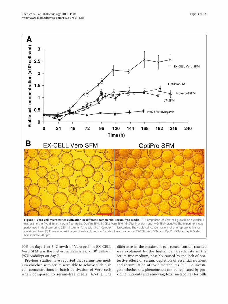
90% on days 4 or 5. Growth of Vero cells in EX-CELLVero SFM was the highest achieving 2.6 × 10⁶ cells/ml(97% viability) on day 7.Previous studies have reported that serum-free med-
ium enriched with serum were able to achieve such highcell concentrations in batch cultivation of Vero cellswhen compared to serum-free media [47-49]. The
difference in the maximum cell concentration reachedwas explained by the higher cell death rate in theserum-free medium, possibly caused by the lack of pro-tective effect of serum, depletion of essential nutrientand accumulation of toxic metabolites [50]. To investi-gate whether this phenomenon can be replicated by pro-viding nutrients and removing toxic metabolites for cells
0
0.5
1
1.5
2
2.5
3
0 24 48 72 96 120 144 168 192 216 240
Time (h)
Via
ble
cel
lco
nce
ntr
atio
n (x
106ce
lls/
ml)
A
B EX-CELL Vero SFM OptiPro SFM
Via
ble
cell
Figure 1 Vero cell microcarrier cultivation in different commercial serum-free media. (A) Comparison of Vero cell growth on Cytodex 1microcarriers in five different serum-free media, OptiPro SFM, EX-CELL Vero SFM, VP-SFM, Provero-1 and HyQ SFM4MegaVir. The experiment wasperformed in duplicate using 250 ml spinner flasks with 3 g/l Cytodex 1 microcarriers. The viable cell concentrations of one representative runare shown here. (B) Phase contrast images of cells cultured on Cytodex 1 microcarriers in EX-CELL Vero SFM and OptiPro SFM at day 8. Scalebars indicate 200 μm.
Chen et al. BMC Biotechnology 2011, 11:81http://www.biomedcentral.com/1472-6750/11/81
Page 3 of 16
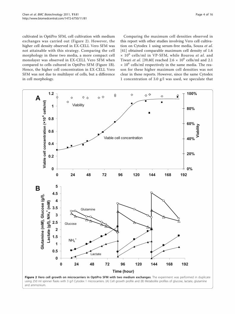
cultivated in OptiPro SFM, cell cultivation with mediumexchanges was carried out (Figure 2). However, thehigher cell density observed in EX-CELL Vero SFM wasnot attainable with this strategy. Comparing the cellmorphology in these two media, a more compact cellmonolayer was observed in EX-CELL Vero SFM whencompared to cells cultured in OptiPro SFM (Figure 1B).Hence, the higher cell concentration in EX-CELL VeroSFM was not due to multilayer of cells, but a differencein cell morphology.
Comparing the maximum cell densities observed inthis report with other studies involving Vero cell cultiva-tion on Cytodex 1 using serum-free media, Souza et al.[41] obtained comparable maximum cell density of 1.6× 10⁶ cells/ml in VP-SFM, while Rourou et al. andTiwari et al. [39,40] reached 2.6 × 10⁶ cells/ml and 2.1× 10⁶ cells/ml respectively in the same media. The rea-son for these higher maximum cell densities was notclear in these reports. However, since the same Cytodex1 concentration of 3.0 g/l was used, we speculate that
0
0.5
1
1.5
2
2.5
3
3.5
4
4.5
5
0 24 48 72 96 120 144 168 192
Time (hour)
Glu
tam
ine
(mM
), G
luco
se (g
/l),
Lact
ate
(g/l)
, NH
4+ (mM
)
Glucose
Lactate
NH4+
Glutamine
B
0
0.2
0.4
0.6
0.8
1
1.2
0 24 48 72 96 120 144 168 1920%
20%
40%
60%
80%
100%
Viab
ility
Viab
le c
ell c
once
ntra
tion
(×10
6 cel
ls/m
l)AViability
Viable cell concentration
Figure 2 Vero cell growth on microcarriers in OptiPro SFM with two medium exchanges. The experiment was performed in duplicateusing 250 ml spinner flasks with 3 g/l Cytodex 1 microcarriers. (A) Cell growth profile and (B) Metabolite profiles of glucose, lactate, glutamineand ammonium.
Chen et al. BMC Biotechnology 2011, 11:81http://www.biomedcentral.com/1472-6750/11/81
Page 4 of 16
these may also be due to a more compact cell mono-layer, similar to our observation in EX-CELL Vero SFMcultures. The difference in cell morphology may begreater in the former study [42] because an even highermaximum cell density of 5 × 10⁶ cells/ml on 3.0 g/lCytodex 1 was reported in perfusion mode. In contrast,Silva et al. [42] reported a maximum cell density of 1 ×10⁶ cells/ml for Vero cells cultivated in EX-CELL VeroSFM using the same microcarrier concentration. Thismay be due to a lack of adaptation from serum contain-ing medium since the cells were directly seeded intoEX-CELL Vero SFM for infection 24 h later. Otherreports of Vero cultivation in different SFM typicallyachieved less than 2 × 10⁶ cells/ml [35,43,44,46], exceptone using a proprietary medium [45].As higher maximum cell densities were observed in
other studies using VP-SFM [39,40], one possible expla-nation for the observed change in cell morphology maybe differences in cell handling during adaptation toSFM. As such, relevant characteristics such as tumori-genicity of the cells should be investigated before thesecells are used to produce clinical materials. Another per-spective to investigate this phenomenon is to look at theavailable information on the components and formula-tion of these media: We observed higher starting glu-cose and amino acid contents in EX-CELL Vero SFMand VP-SFM compared to OptiPro SFM, as well asundefined plant hydrolysates and recombinant proteins.We speculate that these differences may also play anintegral role in enabling the higher cell concentrationsin EX-CELL Vero SFM and VP-SFM cultures.Since viable cell yield in EX-CELL Vero SFM was
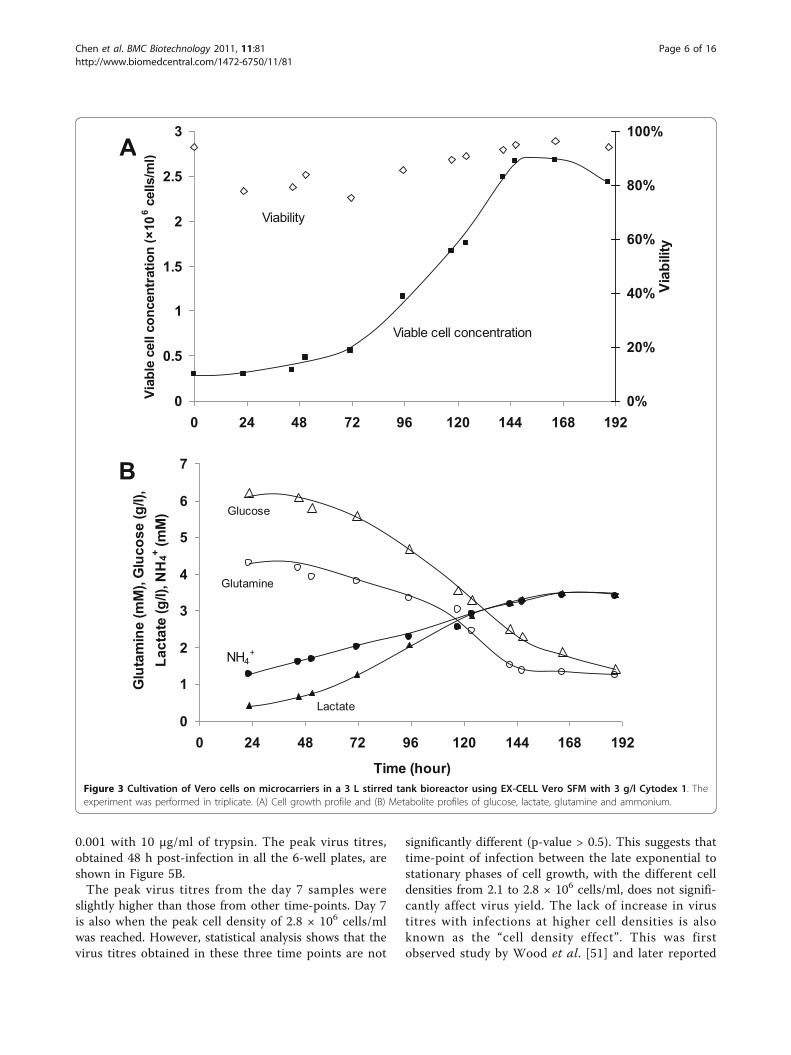
highest, it was chosen for our subsequent studies. Verocell cultivation in EX-CELL Vero SFM was scaled up ina 3 L stirred tank bioreactor for validation. The resultsare presented in Figure 3. Despite the longer lag phase,cell yield of 2.7 × 10⁶ cells/ml (93% viability) were com-parable to those achieved in spinner flask (Figure 1). 3.9g/l of glucose and 2.9 mM of glutamine were consumedand 2.9 g/l lactate and 2.2 mM ammonium were pro-duced by day 6 (Figure 3B) when peak cell density wasreached. The maximal specific growth rate was calcu-lated to be 0.019 h-1, which is similar to that of ourspinner flask culture (0.017 h-1) and those from previousstudies with other SFM (0.026 h-1 [48,50], 0.023-0.033 h-1 [39]).
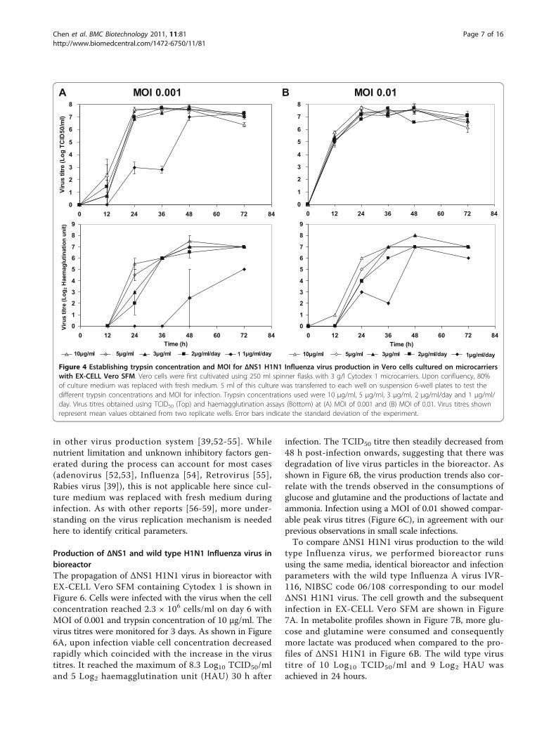
Parameters for Influenza infection: trypsin concentration,multiplicity of infection (MOI) and time-point of infection(TOI)Trypsin is essential for the replication of some Influenzavirus strains. To assess the effect of trypsin concentra-tion on the amplification of ΔNS1 H1N1, we performedsmall scale infections of microcarrier cultures in 6-well
suspension culture plates. Vero cells were first cultivatedin 250 ml spinner flask in EX-CELL Vero SFM. Whenthe culture reached 2 × 10⁶ cells/ml, cells were trans-ferred into 6-well suspension culture plates for infec-tions. The wells were supplemented with differenttrypsin concentrations of 3 μg/ml, 5 μg/ml and 10 μg/ml in duplicates. To investigate the possibility of usinglower trypsin concentrations by daily feeding, 2 sets ofwells were supplemented with trypsin at 1 μg/ml/dayand 2 μg/ml/day respectively. MOI of 0.01 and 0.001were used in this experiment to concurrently assess theeffect of this parameter on virus amplification. Sampleswere harvested 12 hourly and virus titres were deter-mined by haemagglutination (HA) and TCID50 assays(Figure 4).Virus production with 1 μg/ml/day of trypsin yielded
lower HA titres for both MOI tested, although peakTCID50 titres were similar albeit at a later time-pointfor MOI of 0.001. This suggests that ΔNS1 H1N1 virusproduction was limited by trypsin at a concentration of1 μg/ml. On the other hand, virus production using 3μg/ml, 5 μg/ml and 10 μg/ml trypsin, as well as dailytrypsin feed at 2 μg/ml/day, yielded high peak virustitres between 7.5 and 8.0 Log10 TCID50/ml for bothMOI tested. Since the assay has a standard deviation of0.4 Log10 TCID50/ml, the peak virus titres with theabove conditions were not significantly different. How-ever, infection using 10 μg/ml trypsin resulted in highervirus titres at the 24 h time point for both MOI, imply-ing a faster virus amplification process. Similar observa-tions were also described in literature [27,35]. As live(TCID50) virus titres were reported to decrease withtime [27,35], a faster virus amplification process with 10μg/ml trypsin is beneficial for the production of liveattenuated virus vaccines such as ΔNS1 H1N1. Hence10 μg/ml trypsin and MOI of 0.001 (for lower amountsof virus inoculums during vaccine production) wereused in the subsequent experiments.For cell-based Influenza virus production, culture
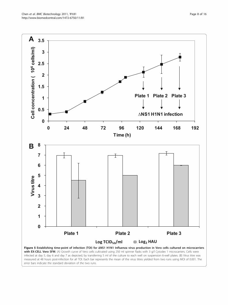
infection is typically performed at a time-point close towhen the peak cell density is reached without time-point of infection (TOI) studies [26-28,32,35]. However,varying TOI has been shown to increase titres of otherviruses [38,41]. In addition to a difference in cell densi-ties at the different TOI, the state of the cells at the dif-ferent phases of cell culture may also affect virusproduction. To determine the effect of TOI for ΔNS1H1N1 infection, 3 time-points at the late exponential tostationary phase were tested. Vero cells were first culti-vated on microcarriers in spinner flask using the samecondition as previously described. Figure 5A shows theaverage cell concentration measured from two spinnerflasks. At days 5, 6, and 7, cells were transferred to 6-well suspension culture plates and infected at MOI
Chen et al. BMC Biotechnology 2011, 11:81http://www.biomedcentral.com/1472-6750/11/81
Page 5 of 16
0.001 with 10 μg/ml of trypsin. The peak virus titres,obtained 48 h post-infection in all the 6-well plates, areshown in Figure 5B.The peak virus titres from the day 7 samples were
slightly higher than those from other time-points. Day 7is also when the peak cell density of 2.8 × 10⁶ cells/mlwas reached. However, statistical analysis shows that thevirus titres obtained in these three time points are not
significantly different (p-value > 0.5). This suggests thattime-point of infection between the late exponential tostationary phases of cell growth, with the different celldensities from 2.1 to 2.8 × 10⁶ cells/ml, does not signifi-cantly affect virus yield. The lack of increase in virustitres with infections at higher cell densities is alsoknown as the “cell density effect”. This was firstobserved study by Wood et al. [51] and later reported
0
1
2
3
4
5
6
7
0 24 48 72 96 120 144 168 192
Time (hour)
Glu
tam
ine
(mM
), G
luco
se (g
/l),
Lact
ate
(g/l)
, NH
4+ (mM
) Glucose
Lactate
NH4+
Glutamine
B
0
0.5
1
1.5
2
2.5
3
0 24 48 72 96 120 144 168 1920%
20%
40%
60%
80%
100%
Viab
ility
Viab
le c
ell c
once
ntra
tion
(×10
6 cel
ls/m
l)A
Viability
Viable cell concentration
Figure 3 Cultivation of Vero cells on microcarriers in a 3 L stirred tank bioreactor using EX-CELL Vero SFM with 3 g/l Cytodex 1. Theexperiment was performed in triplicate. (A) Cell growth profile and (B) Metabolite profiles of glucose, lactate, glutamine and ammonium.
Chen et al. BMC Biotechnology 2011, 11:81http://www.biomedcentral.com/1472-6750/11/81
Page 6 of 16
in other virus production system [39,52-55]. Whilenutrient limitation and unknown inhibitory factors gen-erated during the process can account for most cases(adenovirus [52,53], Influenza [54], Retrovirus [55],Rabies virus [39]), this is not applicable here since cul-ture medium was replaced with fresh medium duringinfection. As with other reports [56-59], more under-standing on the virus replication mechanism is neededhere to identify critical parameters.
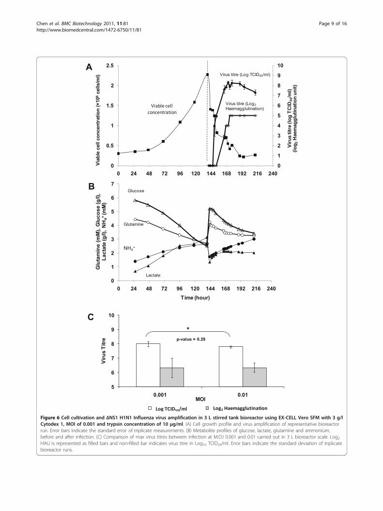
Production of ΔNS1 and wild type H1N1 Influenza virus inbioreactorThe propagation of ΔNS1 H1N1 virus in bioreactor withEX-CELL Vero SFM containing Cytodex 1 is shown inFigure 6. Cells were infected with the virus when the cellconcentration reached 2.3 × 10⁶ cells/ml on day 6 withMOI of 0.001 and trypsin concentration of 10 μg/ml. Thevirus titres were monitored for 3 days. As shown in Figure6A, upon infection viable cell concentration decreasedrapidly which coincided with the increase in the virustitres. It reached the maximum of 8.3 Log10 TCID50/mland 5 Log2 haemagglutination unit (HAU) 30 h after
infection. The TCID50 titre then steadily decreased from48 h post-infection onwards, suggesting that there wasdegradation of live virus particles in the bioreactor. Asshown in Figure 6B, the virus production trends also cor-relate with the trends observed in the consumptions ofglucose and glutamine and the productions of lactate andammonia. Infection using a MOI of 0.01 showed compar-able peak virus titres (Figure 6C), in agreement with ourprevious observations in small scale infections.To compare ΔNS1 H1N1 virus production to the wild
type Influenza virus, we performed bioreactor runsusing the same media, identical bioreactor and infectionparameters with the wild type Influenza A virus IVR-116, NIBSC code 06/108 corresponding to our modelΔNS1 H1N1 virus. The cell growth and the subsequentinfection in EX-CELL Vero SFM are shown in Figure7A. In metabolite profiles shown in Figure 7B, more glu-cose and glutamine were consumed and consequentlymore lactate was produced when compared to the pro-files of ΔNS1 H1N1 in Figure 6B. The wild type virustitre of 10 Log10 TCID50/ml and 9 Log2 HAU wasachieved in 24 hours.
0
1
2
3
4
5
6
7
8
0 12 24 36 48 60 72 84
Viru
s tit
re (L
og T
CID
50/m
l)
A MOI 0.001
0
1
2
3
4
5
6
7
8
0 12 24 36 48 60 72 84
B MOI 0.01
0
1
2
3
4
5
6
7
8
9
0 12 24 36 48 60 72 84Time (h)
Viru
s tit
re (L
og2 H
aem
aglu
tinat
ion
unit)
Series5 Series4 Series3 2ug/ml/day 1ug/ml/day10 g/ml 5 g/ml 1 g/ml/day2 g/ml/day3 g/ml
0
1
2
3
4
5
6
7
8
9
0 12 24 36 48 60 72 84Time (h)
10 ug/ml 5ug/ml 3ug/ml 2ug/ml/day 1ug/ml/day10 g/ml 5 g/ml 1 g/ml/day2 g/ml/day3 g/ml
Figure 4 Establishing trypsin concentration and MOI for ΔNS1 H1N1 Influenza virus production in Vero cells cultured on microcarrierswith EX-CELL Vero SFM. Vero cells were first cultivated using 250 ml spinner flasks with 3 g/l Cytodex 1 microcarriers. Upon confluency, 80%of culture medium was replaced with fresh medium. 5 ml of this culture was transferred to each well on suspension 6-well plates to test thedifferent trypsin concentrations and MOI for infection. Trypsin concentrations used were 10 μg/ml, 5 μg/ml, 3 μg/ml, 2 μg/ml/day and 1 μg/ml/day. Virus titres obtained using TCID50 (Top) and haemagglutination assays (Bottom) at (A) MOI of 0.001 and (B) MOI of 0.01. Virus titres shownrepresent mean values obtained from two replicate wells. Error bars indicate the standard deviation of the experiment.
Chen et al. BMC Biotechnology 2011, 11:81http://www.biomedcentral.com/1472-6750/11/81
Page 7 of 16
0
0.5
1
1.5
2
2.5
3
3.5
0 24 48 72 96 120 144 168 192Time (h)
Cel
l con
cent
ratio
n (
106
cells
/ml)
Plate 1 Plate 3Plate 2
NS1 H1N1 infection
A
0
1
2
3
4
5
6
7
8
Plate 1 Plate 2 Plate 3
Series1 Series2
Viru
s tit
re
Log TCID50/ml Log2 HAU
B
Figure 5 Establishing time-point of infection (TOI) for ΔNS1 H1N1 Influenza virus production in Vero cells cultured on microcarrierswith EX-CELL Vero SFM. (A) Growth curve of Vero cells cultivated using 250 ml spinner flasks with 3 g/l Cytodex 1 microcarriers. Cells wereinfected at day 5, day 6 and day 7 as depicted, by transferring 5 ml of the culture to each well on suspension 6-well plates. (B) Virus titre wasmeasured at 48 hours post-infection for all TOI. Each bar represents the mean of the virus titres yielded from two runs using MOI of 0.001. Theerror bars indicate the standard deviation of the two runs.
Chen et al. BMC Biotechnology 2011, 11:81http://www.biomedcentral.com/1472-6750/11/81
Page 8 of 16
0
1
2
3
4
5
6
7
8
9
10
0
0.5
1
1.5
2
2.5
0 24 48 72 96 120 144 168 192 216 240
Viab
le c
ell c
once
ntra
tion
(×10
6ce
lls/m
l)
Viru
s tit
re (l
og T
CID
50/m
l)(lo
g 2H
aem
aggl
utin
atio
n un
it)
Virus titre (Log TCID50/ml)A
Virus titre (Log2Haemagglutination)
0
1
2
3
4
5
6
7
0 24 48 72 96 120 144 168 192 216 240Time (hour)
Glu
tam
ine
(mM
), G
luco
se (g
/l),
Lact
ate
(g/l)
, NH
4+(m
M)
Glucose
Lactate
NH4+
Glutamine
B
5
6
7
8
9
10
0.001 0.01
Viru
s Ti
tre
MOI
Peak TCID50 titres Series2
C*
p-value = 0.29
Log TCID50/ml Log2 Haemagglutination
Viable cell concentration
Figure 6 Cell cultivation and ΔNS1 H1N1 Influenza virus amplification in 3 L stirred tank bioreactor using EX-CELL Vero SFM with 3 g/lCytodex 1, MOI of 0.001 and trypsin concentration of 10 μg/ml. (A) Cell growth profile and virus amplification of representative bioreactorrun. Error bars indicate the standard error of triplicate measurements. (B) Metabolite profiles of glucose, lactate, glutamine and ammonium,before and after infection. (C) Comparison of max virus titres between infection at M.O.I 0.001 and 0.01 carried out in 3 L bioreactor scale. Log2HAU is represented as filled bars and non-filled bar indicates virus titre in Log10 TCID50/ml. Error bars indicate the standard deviation of triplicatebioreactor runs.
Chen et al. BMC Biotechnology 2011, 11:81http://www.biomedcentral.com/1472-6750/11/81
Page 9 of 16
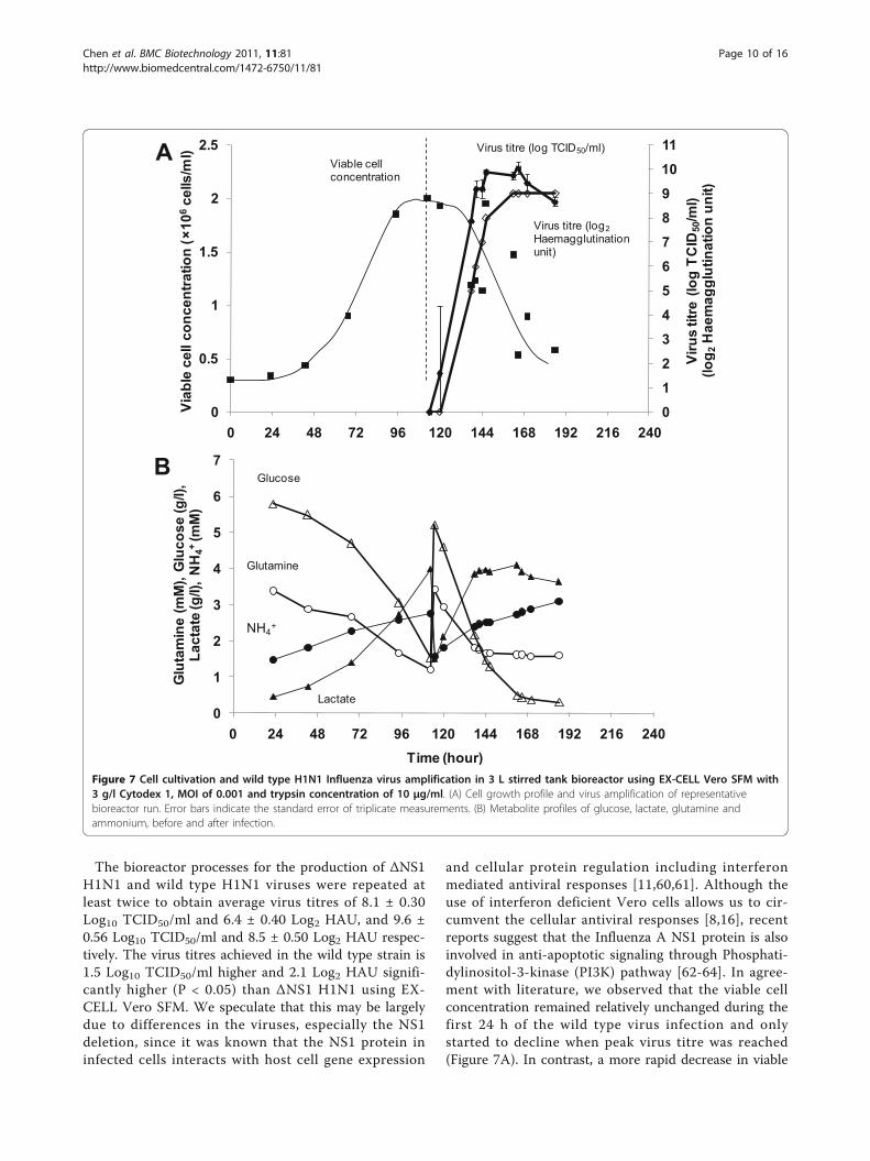
The bioreactor processes for the production of ΔNS1H1N1 and wild type H1N1 viruses were repeated atleast twice to obtain average virus titres of 8.1 ± 0.30Log10 TCID50/ml and 6.4 ± 0.40 Log2 HAU, and 9.6 ±0.56 Log10 TCID50/ml and 8.5 ± 0.50 Log2 HAU respec-tively. The virus titres achieved in the wild type strain is1.5 Log10 TCID50/ml higher and 2.1 Log2 HAU signifi-cantly higher (P < 0.05) than ΔNS1 H1N1 using EX-CELL Vero SFM. We speculate that this may be largelydue to differences in the viruses, especially the NS1deletion, since it was known that the NS1 protein ininfected cells interacts with host cell gene expression
and cellular protein regulation including interferonmediated antiviral responses [11,60,61]. Although theuse of interferon deficient Vero cells allows us to cir-cumvent the cellular antiviral responses [8,16], recentreports suggest that the Influenza A NS1 protein is alsoinvolved in anti-apoptotic signaling through Phosphati-dylinositol-3-kinase (PI3K) pathway [62-64]. In agree-ment with literature, we observed that the viable cellconcentration remained relatively unchanged during thefirst 24 h of the wild type virus infection and onlystarted to decline when peak virus titre was reached(Figure 7A). In contrast, a more rapid decrease in viable
0
12
3
45
67
8
910
11
0
0.5
1
1.5
2
2.5
0 24 48 72 96 120 144 168 192 216 240
Viab
le c
ell c
once
ntra
tion
(×10
6ce
lls/m
l)
Viru
s tit
re (l
og T
CID
50/m
l)(lo
g 2H
aem
aggl
utin
atio
n un
it)
Viable cell concentration
Virus titre (log TCID50/ml)
Virus titre (log2Haemagglutination unit)
A
0
1
2
3
4
5
6
7
0 24 48 72 96 120 144 168 192 216 240Time (hour)
Glu
tam
ine
(mM
), G
luco
se (g
/l),
Lact
ate
(g/l)
, NH
4+(m
M)
Glucose
Lactate
NH4+
Glutamine
B
Figure 7 Cell cultivation and wild type H1N1 Influenza virus amplification in 3 L stirred tank bioreactor using EX-CELL Vero SFM with3 g/l Cytodex 1, MOI of 0.001 and trypsin concentration of 10 μg/ml. (A) Cell growth profile and virus amplification of representativebioreactor run. Error bars indicate the standard error of triplicate measurements. (B) Metabolite profiles of glucose, lactate, glutamine andammonium, before and after infection.
Chen et al. BMC Biotechnology 2011, 11:81http://www.biomedcentral.com/1472-6750/11/81
Page 10 of 16
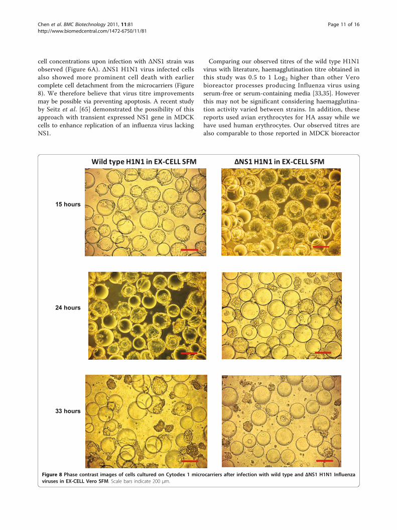
cell concentrations upon infection with ΔNS1 strain wasobserved (Figure 6A). ΔNS1 H1N1 virus infected cellsalso showed more prominent cell death with earliercomplete cell detachment from the microcarriers (Figure8). We therefore believe that virus titre improvementsmay be possible via preventing apoptosis. A recent studyby Seitz et al. [65] demonstrated the possibility of thisapproach with transient expressed NS1 gene in MDCKcells to enhance replication of an influenza virus lackingNS1.
Comparing our observed titres of the wild type H1N1virus with literature, haemagglutination titre obtained inthis study was 0.5 to 1 Log2 higher than other Verobioreactor processes producing Influenza virus usingserum-free or serum-containing media [33,35]. Howeverthis may not be significant considering haemagglutina-tion activity varied between strains. In addition, thesereports used avian erythrocytes for HA assay while wehave used human erythrocytes. Our observed titres arealso comparable to those reported in MDCK bioreactor
15 hours
24 hours
33 hours
Wild type H1N1 in EX-CELL SFM NS1 H1N1 in EX-CELL SFM
Figure 8 Phase contrast images of cells cultured on Cytodex 1 microcarriers after infection with wild type and ΔNS1 H1N1 Influenzaviruses in EX-CELL Vero SFM. Scale bars indicate 200 μm.
Chen et al. BMC Biotechnology 2011, 11:81http://www.biomedcentral.com/1472-6750/11/81
Page 11 of 16
processes producing Influenza viruses (2.4 to 3.3 Log10HA/100 μl [26,27,35], 7.7 Log10 TCID50/ml [27], and 8.5to 10 Log10 EID50/ml [28]).
Comparison of ΔNS1 and wild type H1N1 Influenza virusproduction using EX-CELL Vero SFM and OptiPro SFMAs the ΔNS1 and wild type H1N1 Influenza virus titreswere significantly different in EX-CELL Vero SFM, wewanted to find out whether this is also true in otherSFM. In addition, we wanted to investigate whether the“cell density effect” can be observed when we compareEX-CELL Vero SFM with another serum-free mediumthat gives a lower maximum cell density. We thus per-formed the bioreactor virus production using OptiProSFM for this comparison.Using OptiPro SFM, the average virus titres of ΔNS1
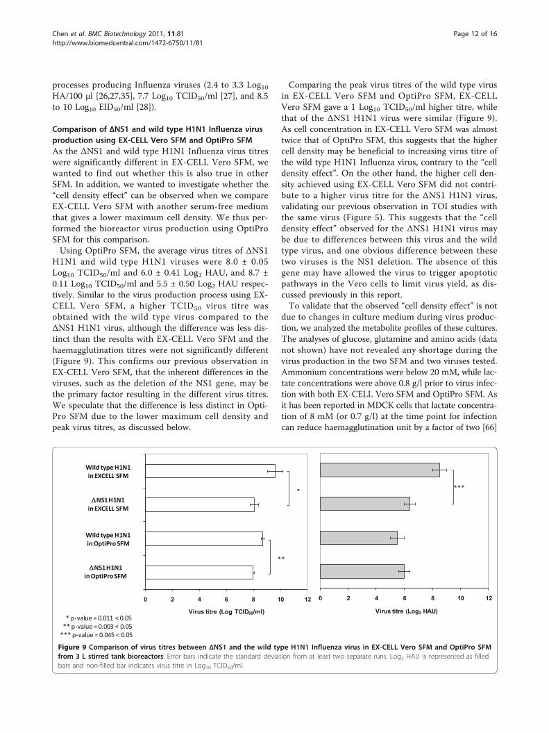
H1N1 and wild type H1N1 viruses were 8.0 ± 0.05Log10 TCID50/ml and 6.0 ± 0.41 Log2 HAU, and 8.7 ±0.11 Log10 TCID50/ml and 5.5 ± 0.50 Log2 HAU respec-tively. Similar to the virus production process using EX-CELL Vero SFM, a higher TCID50 virus titre wasobtained with the wild type virus compared to theΔNS1 H1N1 virus, although the difference was less dis-tinct than the results with EX-CELL Vero SFM and thehaemagglutination titres were not significantly different(Figure 9). This confirms our previous observation inEX-CELL Vero SFM, that the inherent differences in theviruses, such as the deletion of the NS1 gene, may bethe primary factor resulting in the different virus titres.We speculate that the difference is less distinct in Opti-Pro SFM due to the lower maximum cell density andpeak virus titres, as discussed below.
Comparing the peak virus titres of the wild type virusin EX-CELL Vero SFM and OptiPro SFM, EX-CELLVero SFM gave a 1 Log10 TCID50/ml higher titre, whilethat of the ΔNS1 H1N1 virus were similar (Figure 9).As cell concentration in EX-CELL Vero SFM was almosttwice that of OptiPro SFM, this suggests that the highercell density may be beneficial to increasing virus titre ofthe wild type H1N1 Influenza virus, contrary to the “celldensity effect”. On the other hand, the higher cell den-sity achieved using EX-CELL Vero SFM did not contri-bute to a higher virus titre for the ΔNS1 H1N1 virus,validating our previous observation in TOI studies withthe same virus (Figure 5). This suggests that the “celldensity effect” observed for the ΔNS1 H1N1 virus maybe due to differences between this virus and the wildtype virus, and one obvious difference between thesetwo viruses is the NS1 deletion. The absence of thisgene may have allowed the virus to trigger apoptoticpathways in the Vero cells to limit virus yield, as dis-cussed previously in this report.To validate that the observed “cell density effect” is not
due to changes in culture medium during virus produc-tion, we analyzed the metabolite profiles of these cultures.The analyses of glucose, glutamine and amino acids (datanot shown) have not revealed any shortage during thevirus production in the two SFM and two viruses tested.Ammonium concentrations were below 20 mM, while lac-tate concentrations were above 0.8 g/l prior to virus infec-tion with both EX-CELL Vero SFM and OptiPro SFM. Asit has been reported in MDCK cells that lactate concentra-tion of 8 mM (or 0.7 g/l) at the time point for infectioncan reduce haemagglutination unit by a factor of two [66]
0 2 4 6 8 10 12
Virus titre (Log2 HAU)
***
0 2 4 6 8 10 12
NS1 H1N1 OptiPro SFM
H1N1 in OptiPro SFM
NS1 H1N1 in EXCELL SFM
H1N1 in EXCELL SFM
Virus titre (Log TCID50/ml)
*
**
* p-value = 0.011 < 0.05** p-value = 0.003 < 0.05
*** p-value = 0.045 < 0.05
Wild type H1N1 in EXCELL SFM
NS1 H1N1 in EXCELL SFM
Wild type H1N1 in OptiPro SFM
NS1 H1N1 in OptiPro SFM
Figure 9 Comparison of virus titres between ΔNS1 and the wild type H1N1 Influenza virus in EX-CELL Vero SFM and OptiPro SFMfrom 3 L stirred tank bioreactors. Error bars indicate the standard deviation from at least two separate runs. Log2 HAU is represented as filledbars and non-filled bar indicates virus titre in Log10 TCID50/ml.
Chen et al. BMC Biotechnology 2011, 11:81http://www.biomedcentral.com/1472-6750/11/81
Page 12 of 16
and the addition of ammonium chloride to 20 mM wasreported to block infection [67-69], the high lactate levelwe observed may have some influences to limit the maxi-mum virus titres achieved with these medium. On theother hand, a higher lactate level was observed in the wildtype virus production with 3.9 g/l generated 24 h post-infection. Hence it is unclear to what extent the virus pro-duction using Vero cells can be affected by lactate accu-mulation without further experiments.
ConclusionsWe have compared five commercially available SFM forthe microcarrier based cultivation of Vero cells. In addi-tion, we described for the first time the production ofInfluenza viruses using Vero cells in commercially avail-able animal component-free, serum-free medium, and apotentially scalable stirred tank bioreactor process forthe production of ΔNS1 H1N1 virus. Comparing theproduction of the ΔNS1 H1N1 virus to that of the cor-responding wild-type strain, we showed that titres ofΔNS1 H1N1 virus were lower than that of the wild-type, and postulated that this may be a result of earlierVero host cell death due to the NS1 deletion.
MethodsPreparation of cell line, virus strain, trypsin stock andmicrocarriersVero cells (ATCC CCL-81) from a master cell bank atpassage 126 was thawed into DMEM (Invitrogen, GrandIsland, NY) + 10% (v/v) fetal bovine serum (FBS) (Invi-trogen) and passaged three times before stepwise adap-tation to different serum-free media (SFM) to formworking cell banks. The SFM used were OptiPro SFM(Invitrogen, Grand Island, NY, Cat. No. 12309-019), VP-SFM (Invitrogen, Cat. No. 11681-020), EX-CELL VeroSFM (SAFC Biosciences, Lenexa, KS, Cat. No. 14585),Provero-1 (Lonza, Belgium, Cat. No. BE-02-030Q) andHyQ SFM4MegaVir (HyClone, Logan, UT, Cat. No. SH-30522.01), supplemented according to manufacturers’instructions. Cells were subsequently thawed from theseworking cell banks and passaged in tissue culture flasks(T-flasks) in their respective media for 2 or more pas-sages prior to use. Passage numbers of cells used forexperiments were less than 150. Cells cultures wereincubated in 37°C/5% CO2 humidified incubators(Sanyo, Japan).Influenza A/New Caledonia/20/99(H1N1)-like virus,
with NS1 deletion (ΔNS1) was provided by Avir GreenHills Biotechnology. The corresponding wild type Influ-enza A virus was obtained from NIBSC (Influenza IVR-116, NIBSC code 06/108). Working banks of the viruseswere created by amplifying the virus using Vero cellscultivated in OptiPro SFM (Invitrogen) in T-flasks orCell Factories (Nunc, Denmark). While the ΔNS1 H1N1
virus was already adapted to Vero cells, the wild typevirus was propagated using Vero cells for 6 passagesprior to the creation of the working bank. Virus titres ofthe ΔNS1 and wild type H1N1 working banks were 7.1to 7.3 Log10 TCID50/ml and 4 to 5 Log2 HAU, and 9.0Log10 TCID50/ml and 6.5 Log2 HAU respectively, quan-tified as described below.Porcine trypsin used for Influenza virus activation
(Sigma-Aldrich, St. Louis, MO, Cat. No. T5266, 1500BAEE unit/mg) was dissolved in deionized water andsterile filtered to make a 5 mg/ml stock solution. Thisstock solution was aliquoted and stored in -20°C freezer.Trypsin aliquots were thawed once for experiments.Cytodex 1 microcarriers (GE Healthcare, Sweden, Cat.
No. 17-0448-01) were hydrated and sterilized in a glassbottle pre-coated with Sigmacote® (Sigma-Aldrich, Cat.No. SL2) according to manufacturers’ instructions.
Quantification of virus titres and monitoring of cellcultivation processVirus titres were quantified using the haemagglutinationassay [70] and tissue culture infectious dose (TCID50)assay [53]. For the haemagglutination assay, 4% humanerythrocytes (Siemens Healthcare Diagnostics, Germany)were diluted 8-fold in Dulbecco’s phosphate buffer solu-tion (PBS, Invitrogen, Cat. No. 14190-250) to obtain a0.5% cell suspension. 50 μl of this 0.5% cell suspensionwas then added to an equal volume of virus and controlsamples in 2-fold serial dilutions. Haemagglutinationunit (HAU) of a virus sample was read as the highestdilution in which haemagglutination was observed.TCID50 assay was performed by adding 50 μl of 10-
fold serially diluted virus samples to Vero cells culti-vated on 96 well plates using OptiPro SFM (Invitrogen)supplemented with 5 μg/ml porcine trypsin (Sigma-Aldrich). The assay was carried out in triplicate sets,each consisting of 6 wells per diluted virus sample.TCID50 titres were calculated according to the formulaof Reed and Muench [71]. As the dose for attenuatedinfluenza virus vaccine in clinical studies is measuredbased on TCID50 assay [72,73], the comparisons of virustitres in our report were predominantly based onTCID50 instead of haemagglutination assay.Cell cultivation process was monitored by measuring
total cell density, viable cell density and key metaboliteconcentrations. Total cell density was determined withcrystal violet nuclei staining. Briefly, a 500 μl aliquot ofcell culture sample was treated with an equal volume of0.01% (w/v) crystal violet (Sigma-Aldrich) in 0.1M citricacid solution and incubated for 30-45 minutes at 37°C.The released nuclei from the cells were then countedusing a hemacytometer. To determine viable cell den-sity, we first observed that cells attached to the micro-carriers were mostly viable with cell viability greater
Chen et al. BMC Biotechnology 2011, 11:81http://www.biomedcentral.com/1472-6750/11/81
Page 13 of 16
than 90% regardless of the stage of cell cultivation (datanot shown). Based on this observation, Trypan blue cellexclusion method was used to obtain the density ofnon-viable cells that were not attached to microcarriers.Viable cell density was then estimated by deducting thisfrom the total cell density measured with crystal violetnuclei staining.Concentrations of key metabolites (glutamine, glucose,
lactate and ammonium) in cell culture supernatantswere analyzed using BioProfile 100 Plus (Nova Biomedi-cal, Waltham, MA) as per manufacturer’s instructions.Virus-containing samples were deactivated by heating ina 60°C water bath for 30 minutes prior to analysis.
Cell cultivation and virus infection in spinner flasks250 ml spinner flasks (Bellco, Vineland, NJ) were coatedwith Sigmacote (Sigma-Aldrich) according to manufac-turer’s instructions. Vero cells were seeded into thespinner flasks at 6 × 10⁵ cells/ml in 125 ml mediumcontaining 6 mg/ml of hydrated Cytodex 1 microcar-riers. Mixing in the spinner flasks was performed usinga magnetic stirrer platform (Cellgro Type 45600, Ther-molyne, Dubuque, IA) which rotates the magneticmicrocarrier paddle impeller (Bellco, Cat No. 1965-30100) inside the flasks. The spinner flasks were stirredat 40 rpm inside a 37°C/5% CO2 humidified incubator.Surface aeration was allowed by loosening the cap onone arm of the spinner flask. After 24 h, the stirringspeed was increased to 60 rpm, and the culture volumewas increased to 250 ml by addition of fresh medium.This results in cell and microcarrier concentrations of 3× 10⁵ cells/ml and 3 mg/ml respectively.Prior to influenza virus infection, approximate 80% of
the culture medium was exchanged. Influenza virus atpredetermined MOI and trypsin were added to the spin-ner flask together with fresh medium. Stirring speed wasthen maintained at 60 rpm.To determine the conditions for infection, cells were
first cultivated on microcarriers in the spinner flasks asdescribed above. At various stages of cultivation, thecells were transferred to suspension 6-well plates (Grei-ner Bio-One, Cat no. 657102) with 5 ml culture volumeper well. The well was incubated in 37°C/5% CO2 humi-dified incubators (Sanyo, Japan) on orbital shaker agi-tated at speed of 100 rpm. Different virus infectionconditions, namely time of infection, trypsin concentra-tion, and MOI, were tested in this format.
Cell cultivation and virus infection in 3 L bioreactor3 L bioreactor (Applikon, Netherlands) was coatedwith Sigmacote® (Sigma-Aldrich) and sterilized accord-ing to manufacturer’s instructions. The bioreactor set-tings for experiments were as follows: culturetemperature was set at 37°C; maximum flow rate for
O2 and CO2 were set at 10 ml/min; dissolved oxygen(DO) was set at 40% saturated air concentration; initialstirring speed was 60 rpm; initial pH were 7.1 and 7.3for EX-CELL Vero SFM and OptiPro SFM respectively.Vero cells were seeded at 6 × 10⁵ cells/ml in 750 mlmedium with a microcarrier density of 6 mg/ml. At 6h post-seeding, the pH and stirring speed were chan-ged to 7.2 and 100 rpm respectively, as most cells wereattached to the microcarriers. At 24 h post-seeding,the culture volume was increased to 1.5 L by addingfresh medium, to result in cell and microcarrier con-centrations of 3 × 10⁵ cells/ml and 3 mg/mlrespectively.Influenza virus infection in bioreactor is similar to that
in spinner flasks. 80% of the culture medium was firstexchanged with fresh medium. Influenza virus at prede-termined MOI and trypsin were then added to the bior-eactor together with fresh medium. For infection inOptiPro SFM, 5 μg/ml trypsin was used, and the cellswere infected at MOI 0.001 and 0.01 at cell concentra-tions of 1.2-1.4 × 10⁶ cells/ml. The bioreactor settingsremained unchanged.
Statistical analysisStatistical analysis was carried out using two-tailed stu-dent’s t-tests assuming equal variance with replications.P values less than 0.05 were considered significant.
AcknowledgementsThe authors thank Dr Kim Do Yun for his critical review of this paper. Thiswork was supported by the Biomedical Research Council of A*STAR (Agencyfor Science, Technology and Research), Singapore, and Avir Green HillsBiotechnology, Austria.
Author details1Bioprocessing Technology Institute, Agency for Science, Technology andResearch (A*STAR), 20 Biopolis Way, #06-01, Centros, Singapore 138668,Singapore. 2Avir Green Hills Biotechnology, Forsthausgasse 11, 1200 Vienna,Austria.
Authors’ contributionsAC participated in the design of the study, performed the statistical analysisand drafted the manuscript. SLP carried out the virus quantifications and cellculture studies, and participated in equipment set up and materialpreparations. CD participated in the design of the study, in equipment setup and in parallel experiment verification (not presented). ER participated inthe design of the study, in equipment set up and material preparations. MLYparticipated in the virus quantifications and cell culture studies, and inequipment set up and material preparations. SKN conceived the study,participated in its design and coordination, and drafted the manuscript. Allauthors read and approved this final manuscript.
Received: 9 May 2011 Accepted: 11 August 2011Published: 11 August 2011
References1. Poland GA, Jacobson RM, Targonski PV: Avian and pandemic influenza: An
overview. Vaccine 2007, 25:3057-3061.2. Kendal AP, Maassab HF, Alexandrova GI, Ghendon YZ: Development of
cold-adapted recombinant live, attenuated influenza A vaccines in theU.S.A. and U.S.S.R. Antivir Res 1982, 1(6):339-365.
Chen et al. BMC Biotechnology 2011, 11:81http://www.biomedcentral.com/1472-6750/11/81
Page 14 of 16
3. ADIS: Influenza Virus Vaccine Live Intranasal - MedImmune Vaccines:CAIV-T, Influenza Vaccine Live Intranasal. Drugs R D 2003, 4(5):312-319.
4. Maassab HF, Bryant ML: The development of live attenuated cold-adapted influenza virus vaccine for humans. Rev Med Virol 1999,9:237-244.
5. Belshe R, Lee M-S, Walker RE, Stoddard J, Mendelman PM: Safety,immunogenicity and efficacy of intranasal, live attenuated influenzavaccine. Expert Rev Vaccines 2004, 3(6):643-654.
6. Richt JA, García-Sastre A: Attenuated influenza virus vaccines withmodified NS1 proteins. Curr Top Microbiol Immunol 2009, 333:177-195.
7. Treanor JJ, Kotloff K, Betts RF, Belshe R, Newman F, Iacuzio D, Wittes J,Bryant M: Evaluation of trivalent, live, cold-adapted (CAIV-T) andinactivated (TIV) influenza vaccines in prevention of virus infection andillness following challenge of adults with wild-type influenza A (H1N1),A (H3N2), and B viruses. Vaccine 1999, 18(9-10):899-906.
8. García-Sastre A, Egorov A, Matassov D, Brandt S, Levy DE, Durbin JE,Palese P, Muster T: Influenza A Virus Lacking the NS1 Gene Replicates inInterferon-Deficient Systems. Virology 1998, 252(2):324-330.
9. Hai R, Martínez-Sobrido L, Fraser KA, Ayllon J, García-Sastre A, Palese P:Influenza B Virus NS1-Truncated Mutants: Live-Attenuated VaccineApproach. J Virol 2008, 82(21):10580-10590.
10. Steel J, Lowen AC, Pena L, Angel M, Solórzano A, Albrecht R, Perez DR,García-Sastre A, Palese P: Live attenuated influenza viruses containingNS1 truncations as vaccine candidates against H5N1 highly pathogenicavian influenza. J Virol 2009, 83(4):1742-1753.
11. Talon J, Salvatore M, O’Neill RE, Nakaya Y, Zheng H, Muster T, García-Sastre A, Palese P: Influenza A and B viruses expressing altered NS1proteins: A vaccine approach. Proc Natl Acad Sci USA 2000,97(8):4309-4314.
12. Wacheck V, Egorov A, Groiss F, Pfeiffer A, Fuereder T, Hoeflmayer D,Kundi M, Popow-Kraupp T, Redlberger-Fritz M, Mueller CA, et al: A NovelType of Influenza Vaccine: Safety and Immunogenicity of Replication-Deficient Influenza Virus Created by Deletion of the InterferonAntagonist NS1. J Infect Dis 2010, 201:354-362.
13. Kochs G, García-Sastre A, Martínez-Sobrido L: Multiple anti-interferonactions of the influenza A virus NS1 protein. J Virol 2007,81(13):7011-7021.
14. Diaz MO, Ziemin S, Le Beau MM, Pitha P, Smith SD, Chilcote RR, Rowley JD:Homozygous deletion of the alpha- and beta 1-interferon genes inhuman leukemia and derived cell lines. Proc Natl Acad Sci USA 1988,85(14):5259-5263.
15. Mosca JD, Pitha PM: Transcriptional and posttranscriptional regulation ofexogenous human beta interferon gene in simian cells defective ininterferon synthesis. Mol Cell Biol 1986, 6(6):2279-2283.
16. Egorov A, Brandt S, Sereinig S, Romanova J, Ferko B, Katinger D,Grassauer A, Alexandrova G, Katinger H, Muster T: Transfectant Influenza AViruses with Long Deletions in the NS1 Protein Grow Efficiently in VeroCells. J Virol 1998, 72(8):6437-6441.
17. Wressnigg N, Shurygina AP, Wolff T, Redlberger-Fritz M, Popow-Kraupp T,Muster T, Egorov A, Kittel C: Influenza B mutant viruses with truncatedNS1 proteins grow efficiently in Vero cells and are immunogenic inmice. J Gen Virol 2009, 90:366-374.
18. Efferson CL, Schickli J, Ko BK, Kawano K, Mouzi S, Palese P, García-Sastre A,Ioannides CG: Activation of tumor antigen-specific cytotoxic Tlymphocytes (CTLs) by human dendritic cells infected with anattenuated influenza A virus expressing a CTL epitope derived from theHER-2/neu proto-oncogene. J Virol 2003, 77(13):7411-7424.
19. Efferson CL, Tsuda N, Kawano K, Nistal-Villán E, Sellappan S, Yu D, Murray JL,García-Sastre A, Ioannides CG: Prostate Tumor Cells Infected with aRecombinant Influenza Virus Expressing a Truncated NS1 ProteinActivate Cytolytic CD8+ Cells To Recognize Noninfected Tumor Cells. JVirol 2006, 80(1):383-394.
20. He Q, Martinez-Sobrido L, Eko FO, Palese P, Garcia-Sastre A, Lyn D,Okenu D, Bandea C, Ananaba GA, Black CM, et al: Live-attenuatedinfluenza viruses as delivery vectors for Chlamydia vaccines. Immunology2007, 122(1):28-37.
21. Genzel Y, Reichl U: Continuous cell lines as a production system forinfluenza vaccines. Expert Rev Vaccines 2009, 8(12):1681-1692.
22. Hu AY-C, Weng T-C, Tseng Y-F, Chen Y-S, Wu C-H, Hsiao S, Chou A-H,Chao H-J, Gu A, Wu S-C, et al: Microcarrier-based MDCK cell culture
system for the production of influenza H5N1 vaccines. Vaccine 2008,26(45):5736-5740.
23. Pau MG, Ophorst C, Koldijk MH, Schouten G, Mehtali M, Uytdehaag F: Thehuman cell line PER.C6 provides a new manufacturing system for theproduction of influenza vaccines. Vaccine 2001, 19:2716-2721.
24. Ambrozaitis A, Groth N, Bugarini R, Sparacio V, Podda A, Lattanzi M: Anovel mammalian cell-culture technique for consistent production of awell-tolerated and immunogenic trivalent subunit influenza vaccine.Vaccine 2009, 27(43):6022-6029.
25. Doroshenko A, Halperin SA: Trivalent MDCK cell culture-derived influenzavaccine Optaflu (Novartis Vaccines). Expert Rev Vaccines 2009, 8(6):679-688.
26. Genzel Y, Fischer M, Reichl U: Serum-free influenza virus productionavoiding washing steps and medium exchange in large-scalemicrocarrier culture. Vaccine 2006, 24(16):3261-3272.
27. Genzel Y, Olmer RM, Schäfer B, Reichl U: Wave microcarrier cultivation ofMDCK cells for influenza virus production in serum containing andserum-free media. Vaccine 2006, 24(35-36):6074-6087.
28. Ghendon YZ, Markushin SG, Akopova II, Koptiaeva IB, Nechaeva EA,Mazurkova LA, Radaeva IF, Kolokoltseva TD: Development of cell culture(MDCK) live cold-adapted (CA) attenuated influenza vaccine. Vaccine2005, 23(38):4678-4684.
29. Liu J, Mani S, Schwartz R, Richman L, Tabor DE: Cloning and assessment oftumorigenicity and oncogenicity of a Madin-Darby canine kidney(MDCK) cell line for influenza vaccine production. Vaccine 2010,28(5):1285-1293.
30. Liu J, Shi X, Schwartz R, Kemble G: Use of MDCK cells for production oflive attenuated influenza vaccine. Vaccine 2009, 27(46):6460-6463.
31. Hu W-S, Giard DJ, Wang DIC: Serial Propagation of Mammalian Cells onMicrocarriers. Biotechnol Bioeng 1985, 27(10):1466-1476.
32. Kistner O, Barrett PN, Mundt W, Reiter M, Schober-Bendixen S, Dorner F:Development of a mammalian cell (Vero) derived candidate influenzavirus vaccine. Vaccine 1998, 16(9-10):960-968.
33. Ng Y-C, Berry JM, Butler M: Optimization of physical parameters for cellattachment and growth on macroporous microcarriers. Biotechnol Bioeng1996, 50(6):627-635.
34. Yokomizo AY, Antoniazzi MM, Galdino PL, A N Jr, Jorge SAC, Pereira CA:Rabies virus production in high vero cell density cultures onmacroporous microcarriers. Biotechnol Bioeng 2004, 85(5):506-515.
35. Genzel Y, Dietzsch C, Rapp E, Schwarzer J, Reichl U: MDCK and Vero cellsfor influenza virus vaccine production: a one-to-one comparison up tolab-scale bioreactor cultivation. Appl Microbiol Biotechnol 2010,88(2):461-475.
36. Cinatl JJ, Cinatl J, Rabenau H, Rapp J, Kornhuber B, Doerr H: Protein-freeculture of Vero cells: A substrate for replication of pathogenic viruses.Cell Biol Int 1993, 17(9):885-895.
37. Merten O-W, Kierulff JV, Castignolles N, Perrin P: Evaluation of the newserum free medium (MDSS2) for the production of different biologicals:use of various cell lines. Cytotechnology 1994, 14(1):47-59.
38. Yuk IH, Lin GB, Ju H, Sifi I, Lam Y, Cortez A, Liebertz D, Berry JM,Schwartz RM: A serum-free Vero production platform for a chimeric virusvaccine candidate. Cytotechnology 2006, 51:183-192.
39. Rourou S, van der Ark A, van der Velden T, Kallel H: A microcarrier cellculture process for propagating rabies virus in Vero cells grown in astirred bioreactor under fully animal component free conditions. Vaccine2007, 25(19):3879-3889.
40. Tiwari M, Parida M, Santhosh SR, Khan M, Dash PK, Rao PVL: Assessment ofimmunogenic potential of Vero adapted formalin inactivated vaccinederived from novel ECSA genotype of Chikungunya virus. Vaccine 2009,27(18):2513-2522.
41. Souza MCO, Freire MS, Schulze EA, Gaspar LP, Castilho LR: Production ofyellow fever virus in microcarrier-based vero cell cultures. Vaccine 2009,27(46):6420-6423.
42. Silva AC, Delgado I, Sousa MFQ, Carrondo MJT, Alves PM: Scalable culturesystems using different cell lines for the production of Peste des Petitsruminants vaccine. Vaccine 2008, 26:3305-3311.
43. Liu C-C, Lian W-C, Butler M, Wu S-C: High immunogenic enterovirus 71strain and its production using serum-free microcarrier Vero cell culture.Vaccine 2007, 25(1):19-24.
44. Butler M, Burgener A, Patrick M, Berry M, Moffatt D, Huzel N, Barnabé N,Coombs K: Application of a Serum-Free Medium for the Growth of VeroCells and the Production of Reovirus. Biotechnol Prog 2000, 16(5):854-858.
Chen et al. BMC Biotechnology 2011, 11:81http://www.biomedcentral.com/1472-6750/11/81
Page 15 of 16
45. Rourou S, van der Ark A, Majoul S, Trabelsi K, van der Velden T, Kallel H: Anovel animal-component-free medium for rabies virus production inVero cells grown on Cytodex 1 microcarriers in a stirred bioreactor. ApplMicrobiol Biotechnol 2009, 85(1):53-63.
46. Petiot E, Fournier F, Gény C, Pinton H, Marc A: Rapid Screening of Serum-Free Media for the Growth of Adherent Vero Cells by Using a Small-Scale and Non-invasive Tool. Appl Biochem Biotechnol 2010,160(6):1600-1615.
47. Frazzati-Gallina NM, Paoli RL, Mourão-Fuches RM, Jorge SAC, Pereira CA:Higher production of rabies virus in serum-free medium cell cultures onmicrocarriers. J Biotechnol 2001, 92:67-72.
48. Quesney S, Marvel J, Gerdil C, Meignier B: Characterization of Vero cellgrowth and death in bioreactor with serum-containing and serum-freemedia. Cytotechnology 2001, 35:115-125.
49. Souza MCdO, Freire MdS, Castilho LdR: Influence of Culture Conditions onVero Cell Propagation on Non-Porous Microcarriers. Braz Arch Biol Technol2005, 48:71-77.
50. Quesney Sb, Marc A, Gerdil C, Gimenez C, Marvel J, Richard Y, Meignier B:Kinetics and metabolic specificities of Vero cells in bioreactor cultureswith serum-free medium. Cytotechnology 2003, 42:1-11.
51. Wood HA, Johnston LB, Burand JP: Inhibition of Autographa californicaNuclear Polyhedrosis Virus Replication in High-Density Trichoplusia niCell Culture. Virology 1982, 119:245-254.
52. Kamen A, Henry O: Development and optimization of an adenovirusproduction process. J Gene Med 2004, 6:S184-S192.
53. Yuk IHY, Olsen MM, Geyer S, Forestell SP: Perfusion cultures of humantumor cells: a scalable production platform for oncolytic adenoviralvectors. Biotechnol Bioeng 2004, 86(6):637-642.
54. Bock A, Schulze-Horsel J, Rapp E, Genzel Y, Reichl U: High-DensityMicrocarrier Cell Cultures for Influenza Virus Production. Biotechnol Prog2011, 27(1):241-250.
55. Ghani K, Garnier A, Coelho H, Transfiguracion J, Trudel P, Kamen A:Retroviral vector production using suspension-adapted 293GPG cells ina 3L acoustic filter-based perfusion bioreactor. Biotechnol Bioeng 2006,95(4):653-660.
56. Merten O-W: State-of-the-art of the production of retroviral vectors. JGene Med 2004, 6:S105-S124.
57. Beer C, Meyer A, Müller K, Wirth M: The temperature stability of mouseretroviruses depends on the cholesterol levels of viral lipid shell andcellular plasma membrane. Virology 2003, 308(1):137-146.
58. Le Rub A, Jacob D, Transfiguracion J, Ansorge S, Henry O, Kamen AA:Scalable production of influenza virus in HEK-293 cells for efficientvaccine manufacturing. Vaccine 2010, 28(21):3661-3671.
59. Barber GN: Host defense, viruses and apoptosis. Cell Death Differ 2001,8(2):113-126.
60. Bergmann M, Garcia-Sastre A, Carnero E, Pehamberger H, Wolff K, Palese P,Muster T: Influenza Virus NS1 Protein Counteracts PKR-MediatedInhibition of Replication. J Virol 2000, 74(13):6203-6206.
61. Hayman A, Comely S, Lackenby A, Hartgroves LCS, Goodbourn S,McCauley JW, Barclay WS: NS1 proteins of avian influenza A viruses canact as antagonists of the human alpha/beta interferon response. J Virol2007, 81(5):2318-2327.
62. Ehrhardt C, Ludwig S: A new player in a deadly game: influenza virusesand the PI3K/Akt signaling pathway. Cell Microbiol 2009, 11(6):863-871.
63. Zhirnov OP, Konakova TE, Wolff T, Klenk HD: NS1 Protein of Influenza AVirus Down-Regulates Apoptosis. J Virol 2002, 76(4):1617-1625.
64. Ehrhardt C, Wolff T, Ludwig S: Activation of phosphatidylinositol 3-kinasesignaling by the nonstructural NS1 protein is not conserved among typeA and B influenza viruses. J Virol 2007, 81(21):12097-12100.
65. Seitz C, Frensing T, Höper D, Kochs G, Reichl U: High yields of Influenza Avirus in MDCK cells are promoted by an insufficient IFN-inducedantiviral state. J Gen Virol 2010, 91(7):1754-1763.
66. Genzel Y, Behrendt I, Konig S, Sann H, Reichl U: Metabolism of MDCK cellsduring cell growth and influenza virus production in large-scalemicrocarrier culture. Vaccine 2004, 22:2202-2208.
67. Matlin KS: Ammonium Chloride Slows Transport of the Influenza VirusHemagglutinin but Does Not Cause Mis-sorting in a Polarized EpthelialCell Line. J Biol Chem 1986, 261(32):15172-15178.
68. Morris SJ, Price GE, Barnett JM, Hiscox SA, Smith H, Sweet C: Role ofneuraminidase in influenza virus-induced apoptosis. J Gen Virol 1999,80:137-146.
69. Whittaker G, Bui M, Helenius A: The role of nuclear import and export ininfluenza virus infection. Trends Cell Biol 1996, 6:67-71.
70. Cox N: WHO manual on animal influenza diagnosis and surveillance. InWHO Animal Influenza Manual. Edited by: Webster RG, Krauss S. Geneva:World Health Organization; 2002:1-97.
71. Reed LJ, Muench H: A simple method of estimating fifty per centendpoints. The American Journal of Hygiene 1938, 27(3):493-497.
72. Karrona RA, Talaat K, Luke C, Callahan K, Thumar B, DiLorenzo S, McAuliffe J,Schappell E, Suguitan A, Mills K, et al: Evaluation of two live attenuatedcold-adapted H5N1 influenza virus vaccines in healthy adults. Vaccine2009, 27:4953-4960.
73. Talaat KR, Karrona RA, Callahan KA, Luke CJ, DiLorenzo SC, Chen GL,Lamirande EW, Jin H, Coelingh KL, Murphy BR, et al: A live attenuatedH7N3 influenza virus vaccine is well tolerated and immunogenic in aPhase I trial in healthy adults. Vaccine 2009, 27:3744-3753.
doi:10.1186/1472-6750-11-81Cite this article as: Chen et al.: Serum-free microcarrier basedproduction of replication deficient Influenza vaccine candidate viruslacking NS1 using Vero cells. BMC Biotechnology 2011 11:81.
Submit your next manuscript to BioMed Centraland take full advantage of:
• Convenient online submission
• Thorough peer review
• No space constraints or color figure charges
• Immediate publication on acceptance
• Inclusion in PubMed, CAS, Scopus and Google Scholar
• Research which is freely available for redistribution
Submit your manuscript at www.biomedcentral.com/submit
Chen et al. BMC Biotechnology 2011, 11:81http://www.biomedcentral.com/1472-6750/11/81
Page 16 of 16