Embed Size (px)
Citation preview
4Microbiology Monographs
Series Editor: Alexander Steinbüchel
Predatory ProkaryotesBiology, Ecology and Evolution
Volume Editor: Edouard Jurkevitch
With 53 Figures, 1 in Color
123
Volume Editor:
Dr. Edouard JurkevitchDepartment of Plant Pathology and MicrobiologyFaculty of Agricultural, Food and EnvironmentalQuality Sciences76100 RehovotIsraele-mail: [email protected]
Series Editor:
Professor Dr. Alexander SteinbüchelInstitut für Molekulare Mikrobiologie und BiotechnologieWestfälische Wilhelms-UniversitätCorrensstraße 348149 MünsterGermanye-mail: [email protected]
Library of Congress Control Number: 2006931569
ISSN 1862-5576ISBN-10 3-540-38577-0 Springer Berlin Heidelberg New YorkISBN-13 978-3-540-38577-6 Springer Berlin Heidelberg New YorkDOI 10.1007/978-3-540-38582-0
This work is subject to copyright. All rights are reserved, whether the whole or part of the materialis concerned, specifically the rights of translation, reprinting, reuse of illustrations, recitation, broad-casting, reproduction on microfilm or in any other way, and storage in data banks. Duplication ofthis publication or parts thereof is permitted only under the provisions of the German Copyright Lawof September 9, 1965, in its current version, and permission for use must always be obtained fromSpringer. Violations are liable for prosecution under the German Copyright Law.
Springer is a part of Springer Science+Business Media
springer.com
c© Springer-Verlag Berlin Heidelberg 2007
The use of registered names, trademarks, etc. in this publication does not imply, even in the absenceof a specific statement, that such names are exempt from the relevant protective laws and regulationsand therefore free for general use.
Editor: Dr. Christina Eckey, HeidelbergDesk Editor: Dr. Jutta Lindenborn, HeidelbergCover Design: WMXDesign GmbH, HeidelbergTypesetting and Production: LE-TEX Jelonek, Schmidt & Vöckler GbR, Leipzig
Printed on acid-free paper 02/3100 YL – 5 4 3 2 1 0
Preface
Few biologists, including microbiologists, are aware of the existence of preda-tory bacteria. However, the reaction of these scientists when they learn aboutthem, whether through casual talks or through exposure at meetings, is invari-ably: “cool!!” In modern dialect, this is really a good score.
The aim of this monograph is to increase the awareness of the biologistsat large about the great possibilities that predation between microbes offerfor research and teaching. One can also view this aim within a trend we fullysupport and think should be strengthened: microbial systems are excellentmodels for studying and discussing basic ecological and general biologicalconcepts. We won’t deliberate over the reasons, as excellent and up-to-datereviews are available, but we hope that a short description of the chapterscontained in this book will convince the reader that bacterial predators, theirecology, and their biology at large form an especially appealing field.
While the variety of known predatory bacteria may already appear to besubstantial, we are convinced that only the surface of this diversity has beenscratched. Moreover, one should not take the term “known” at face value asmost of these bacterial predators have only been anecdotally described. Bacte-rial predators may even be much more widespread than presently accounted forowing to the fact that only a tiny fraction of bacteria occurring in natural habi-tats can be cultivated in the laboratory. The discovery of new bacterial predatorscannot be performed solely by relying upon culture-independent approachesas no one particular molecular signature will account for all predatory pheno-types. The exploration of the diversity of the voracious smalls should also bebased on observation, enrichment or isolation whenever possible. The studyof this diversity and of its phylogenetic roots can bring forward evolution-ary insights that are pertinent to seemingly unrelated fields such as biologicalpolymers degradation and the evolution of the eukaryotic cell. This is treatedin the chapter by Edouard Jurkevitch and Yaacov Davidov.
The dynamics of trophic interactions between bacteria is very much a blackbox. However, they can be addressed in a systematic way, thanks to the inher-ent possibilities of manipulating variables in microbial systems in an efficient,precise, and reproducible manner. Such studies can bear a large impact on ourunderstanding of the central role of predation in ecology. Microbial preda-tory systems can be utilized to test essential ecological questions pertaining
VI Preface
to predation, such as the role of spatial structure, the presence of decoy orof multiple species (prey or predator) on predator-prey interactions, and howpredation may lead to speciation. In order to address such questions, math-ematical frameworks can be developed to define the variables to be tested.Michael Wilkinson presents in his chapter such models in a clear and readilyunderstandable manner.
Klaus Jürgens’s chapter provides a thorough background on predation inthe microbial world at large and emerging “rules” of bacterial predation. Hischapter addresses the impact of predation in microbial systems at variousscales, from bacteriophages to metazoans. Bacterial predation is further ana-lyzed in the larger context of trophic cascades and ecological networks. Protistsare treated in depth and the larger body of knowledge in this domain is usedas a background against which the lesser understanding of predation in theprokaryotic realm is evaluated. This knowledge and understanding is an im-portant resource to draw upon for comparison and inspiration.
Laboratory work with Bdellovibrio and like organisms (BALOs), the bac-teria most studied for their predatory behavior, presents challenges to theinvestigator. As wild type strains can only grow in the presence of a host,isolation and enumeration of BALOs from natural samples are not straight-forward tasks. Susan Koval addresses these issues as well as other importantsubjects related to the analysis of isolates in her chapter. The chapter also cov-ers culture-independent approaches, such as fluorescent in situ hybridizationas they are applied to BALOs. This chapter should prove of great help to themicrobiologist wishing to engage in work with predatory bacteria as well as tothe more experienced “BALOlogist.”
BALOs are ubiquitously found in nature or at least in most of the environ-ments in which they were looked for. Since oceans cover about 70% of theEarth’s surface, the marine milieu is the largest of all environments and alsothe one that Henry Williams has been studying for three decades. Togetherwith Silvia Piñeiro, he presents a very comprehensive and in-depth review ofthe ecology of BALOs in aquatic as well as in terrestrial habitats. They ask whatmay be the central, yet unanswered questions in BALO ecology, i.e. what is theimpact of BALOs in bacterial mortality, which bacterial groups constitue preyorganisms and how are environmental processes affected. While these are truechallenges for the microbial ecologist, the authors suggest that the tools beingdeveloped and implemented in BALO research will greatly enhance our abilityto answer these basic questions.
Among the most powerful instruments in the biologist’s toolbox are ge-nomics and bioinformatics. John Tudor and Michael McCann reexamined thefirst published BALO genome, that of Bdellovibrio bacteriovorus type strain100, a terrestrial bacterium. They present us new analyses of chemotactic,regulatory, and sensory circuits of the predator, as well as a reevaluation ofthe amino acid biosynthetic capabilities of this organism. They also providethe first comparative analysis of different BALO genomes using the published
Preface VII
genome data available on the marine BALO Bacteriovorax marinus SJ andon the cyst-forming Bdellovibrio sp. strain W. These, along with data fromproteomic studies are examined within the frame of the unusual life cycle ofBALOs, shedding new light on the predators’ developmental phases.
Eckard Strauch, Sebastian Beck, and Bernd Appel describe the peculiarbiochemistry of BALO cell walls with the presence of sphingolipids in Bac-teriovorax stolpii, a sugar-linked LPS, and a new family of outer membraneproteins. They address the importance of chemotaxis, locomotion, and attach-ment appendages in the predatory process and review the literature pertainingto intracellular regulatory signals. They link BALO biochemistry to the lifecycle and to the ecology of the predators and examine the potential uses ofBALOs or derived compounds as therapeutic agents, weighing the potentialuses against the potential hurdles.
We hope the readers of this book will marvel at the intricacies of the biologyof microbial predators and at how much of the natural sciences are “packed”in these small cells. We expect it will keep in them the feeling that these tinypredators are indeed “really cool.”
We would like to again thank the contributing authors for their dedicationto this project and for their enthusiasm from its very beginning.
Finally, I (Edouard Jurkevitch) would like to thank my wife Einat and mychildren Yaniv, Yoav and Maya for the smiles on their faces when I “talkbdellovibrio” with them, and my mother and late father for the sweet gift ofeducation.
Rehovot and Münster, June 2006 Edouard JurkevitchAlexander Steinbüchel
Contents
A Brief History of Short Bacteria:A Chronicle of Bdellovibrio (and Like Organisms) ResearchE. Jurkevitch . . . . . . . . . . . . . . . . . . . . . . . . . . . . . . . . 1
Phylogenetic Diversity and Evolution of Predatory ProkaryotesE. Jurkevitch · Y. Davidov . . . . . . . . . . . . . . . . . . . . . . . . . . 11
Predation on Bacteria and Bacterial Resistance Mechanisms:Comparative Aspects Among Different Predator Groupsin Aquatic SystemsK. Jürgens . . . . . . . . . . . . . . . . . . . . . . . . . . . . . . . . . . 57
Mathematical Modelling of Predatory ProkaryotesM. H. F. Wilkinson . . . . . . . . . . . . . . . . . . . . . . . . . . . . . 93
Bdellovibrio and Like Organisms:Potential Sources for New Biochemicals and Therapeutic Agents?E. Strauch · S. Beck · B. Appel . . . . . . . . . . . . . . . . . . . . . . . 131
Genomic Analysis and Molecular Biology of Predatory ProkaryotesJ. J. Tudor · M. P. McCann . . . . . . . . . . . . . . . . . . . . . . . . . . 153
The Search for Hunters:Culture-Dependent and -Independent Methodsfor Analysis of Bdellovibrio and Like OrganismsS. F. Koval . . . . . . . . . . . . . . . . . . . . . . . . . . . . . . . . . . 191
Ecology of the Predatory Bdellovibrio and Like OrganismsH. N. Williams · S. Piñeiro . . . . . . . . . . . . . . . . . . . . . . . . . 213
Subject Index . . . . . . . . . . . . . . . . . . . . . . . . . . . . . . . . 249
Microbiol Monogr (4)E. Jurkevitch: Predatory ProkaryotesDOI 10.1007/7171_051/Published online: 11 October 2006© Springer-Verlag Berlin Heidelberg 2006
A Brief History of Short Bacteria:A Chronicle of Bdellovibrio (and Like Organisms) Research
Edouard Jurkevitch
Department of Plant Pathology and Microbiology,and the Otto Warburg Center for Agricultural Biotechnology,Faculty of Agricultural, Food and Environmental Quality Sciences,The Hebrew University of Jerusalem, 76100 Rehovot, [email protected]
1 Introduction . . . . . . . . . . . . . . . . . . . . . . . . . . . . . . . . . . . 1
2 Historical Perspective . . . . . . . . . . . . . . . . . . . . . . . . . . . . . . 2
References . . . . . . . . . . . . . . . . . . . . . . . . . . . . . . . . . . . . . . . 8
Abstract Like many good things in science (and in life at large, starting with evolution-ary processes), the obligate predatory bacteria Bdellovibrio and like organisms (BALOs)were discovered by chance. These fascinating creatures have since been studied by (nottoo) many great scientists. As the community studying these organisms has never beentoo large, small changes in its number of scientists have had a large impact on the ad-vancement of this field. A historical perspective of BALO research is presented here.
1Introduction
Predation is pervasive at all levels of life and maybe as old as life, or cel-lular life, itself (Maynard Smith and Szathmáry 1995; Bengston 2002). Fromthe tiniest viruses that parasitize and finally lyze their bacterial hosts to thelargest of the sharks, it is found in all walks of life, and possibly in all envi-ronments.
Predators kill their prey. Predation is a significant cause of mortality, animportant evolutionary force, driving the selection of escape strategies inprey and of effectiveness in predators. Predation is such a basic tenet of lifethat it is strongly embedded in the human psyche; it has been used to describeeconomic processes (predatory pricing), asocial, criminal behaviors (sexuallyviolent predator), or political developments (predatory democracy).
This central role played by predation in nature is reflected in the greatinterest it is generating among the scientific community. The scientific liter-ature abounds with articles and books dealing with this matter but a simplesearch through a few databases reveals that the amount of work performed onpredation in the various fields of ecology differ greatly.
2 E. Jurkevitch
Table 1 Numbers of entries in the PubMed and GoogleScholar databases relevant to pre-dation in predator and prey systems of vertebrates, arthropods, and microorganisms
Search Keywordsengine Predation Mammals Predation Arthropods Predation Predation Bacteria
+ mammals + arthropods + Bacteria + Bacteria(-protozoa,protozoan)
PubMed 1127 11 328 519 952 165 401 207 969 292
Google 21 100 451 000 7370 59 200 10 500 261 1 170 000Scholar
As seen from Table 1, no rule can be formulated as to the relationship ofthe number of studies centered on predation and the size of the subject or-ganisms, be they predator or prey. However, one thing appears to be clear:a dearth of work on predatory interactions within the prokaryotic realm.
Predation between bacteria has been known for a long time (Beebe 1941,and probably earlier) but the described interactions were of a facultative na-ture. Mostly, myxobacterial systems have served as “role models” for thistype of interaction. However, because of the peculiar and fascinating socialbehavior exhibited by these bacteria, predation usually took the back seatof research priorities in these systems. Nevertheless, the lytic activities ofmyxobacteria and other facultative predators have been thoroughly investi-gated and some data pertaining to the ecological significance of predation bythese organisms is available (see chapter by Jurkevitch and Davidov in thisvolume).
Another class of predatory bacteria are the obligate predators. Althoughthis book is not solely dedicated to these organisms, they form its centraltheme. I shall therefore present a short history of the discovery and devel-opment of the research centered on Bdellovibrio, or according to presentdesignation, the Bdellovibrio and like organisms (BALOs).
2Historical Perspective
In 1963, Moshe Shilo, from the Hebrew University of Jerusalem was spendinga sabbatical in Berkeley at Roger Stanier’s laboratory, working on endotoxicproperties of bacterial lipopolysaccharides. During the very same year, HeinzStolp was also in California as a postdoctoral fellow, staying with MortimerStarr in UC Davis.
A year earlier, Stolp had described small, fast-swimming gram negativebacteria, obligate predators of other gram negative cells (Stolp and Petzold1962). At that time, Heinz Stolp (today a professor Emeritus of the Uni-
A Brief History of Short Bacteria 3
versity of Bayreuth) was working in Berlin at the Institut für Bakteriologie,developing lyzotyping methods for pseudomonads. In a particular experi-ment designed to isolate bacteriophages of the phytopathogen Pseudomonassyringae pv. phaseolicola from a soil suspension, he ran short of filters, andinstead used sintered glass filters. The following day, no lytic plaques wereapparent in the top agar, so the plates should have been discarded. However,they were not, and when reexamined two days later, plaques had developed(also see Stolp 1973). Then, “just because the belated generation of the plaquespoke against the existence of phage activity, the cause of this lysis was furtherinspected” (Stolp 1968). What Heinz Stolp saw were rapidly moving, tiny bac-teria that attached to the substrate cell, and finally, lyzed them. Hence, theywere named Bdellovibrio bacteriovorus, the name describing the morphologyand the supposed way of life of the bacteria; they were curved and seemed tostick to their prey and to absorb the prey cell content, reminiscent of a leech(“bdella” in Greek). The term was coined by Robert E. Buchanan, a notedtaxonomist and Professor at Iowa State College of Agriculture and MechanicArt. Had the required filters been available, their cut-off size (0.2 µm) wouldnot have enabled the Bdellovibrio cells (0.25 – 0.5 × 0.75–2 µm) to pass, butthe sintered glass (1.35 µm) allowed their passage. Moreover, had the nega-tive plates been discarded ...Dans les champs de l’observation le hasard nefavorise que les esprits préparés (In the fields of observation, chance only fa-vors the prepared mind – Louis Pasteur, lecturing in at the Université de Lille,December 7, 1854).
Back to Davis, 1963. Stolp and Starr thoroughly investigated the newly dis-covered organism, describing its morphology, providing first insights into thedynamics of predation and isolating saprophytic host-independent mutants.They remarked that isolates vary in prey (always gram negative) range, thatprey bacteria surviving predation do not appear to be mutants, and that sinceBdellovibrio could be recovered in many natural habitats, it probably wasan integral component of the microbial flora (Stolp and Starr 1963). Theseresults are as pertinent today as when they were first published. However,Bdellovibrio was thought to remain extracellular and was therefore called anectoparasite. The term ectoparasite rather than exoparasite was used to dis-tinguish it from a parasite that does not require a continuous contact withthe prey. Starr and Baigent (1966) later described host penetration and theintraperiplasmic nature of the predator.
Moshe Shilo went on a visit to Davis and “met the bdellovibrios”. Fromhis correspondence, he seems to have been fascinated, and rapidly started towork on the subject along with Barbara Bruff, a student in Stanier’s lab. Bythe summer of 1963, they had developed an efficient protocol for the recov-ery of host-independent mutants, which were then used to demonstrate thepresence of enzymatic activities able to lyze dead prey cells (Shilo and Bruff1965). Excited about these new and peculiar bacteria, Shilo wrote from Berke-ley to Mazal Varon, who had just terminated her M.Sc., and proposed that she
4 E. Jurkevitch
takes up this project and study Bdellovibrio as a part of her Ph.D program inhis laboratory in Jerusalem. Although microbial ecology was his main field ofresearch, Shilo had an all-encompassing interest in microbiology, as reflectedin his work with Bdellovibrio. Within the next 15 years or so, Shilo and Varon(first as a student, and then as a researcher), together and independently, con-tributed enormously to the field. They, and this is not a exhaustive listing,studied the attachment and penetration of Bdellovibrio to its prey (Varon andShilo 1968, 1969a), followed the dynamics of the predatory interaction anddeduced mathematical models (Varon and Shilo 1969b; Varon and Ziegler1978; Varon et al. 1984), and examined various aspects of the physiology of in-vaded prey cells and of host-independent mutants (Varon and Seijffers 1975;Eksztejn and Varon 1977). They isolated phages active against bdellovibrios(Varon and Levisohn 1972), developed protocols for isolation of the preda-tor from the environment (Varon and Shilo 1970), evaluated the impact ofpollutants on predation (Varon and Shilo 1981), addressed the peculiar re-quirements of marine bdellovibrios (Marbach et al. 1976), and showed thatpredation can select for a resistant, slower growing prey, leading to the coex-istence of the wild type and the mutant in the presence of the predator (Varon1979). Work on Bdellovibrio at the Hebrew University came to a stop whenVaron moved to Tel-Aviv University in 1982. I feel lucky that since 1998, I havebeen able to revive this line of research at the Hebrew University.
In 1969, Sydney Rittenberg, from the department of Microbiology atUCLA, came to Shilo’s laboratory for a sabbatical and was infected by Shilo’senthusiasm for Bdellovibrio. He spent a year in Jerusalem (Rittenberg andShilo 1970) and initiated a long and fruitful research on bdellovibrio, almostexclusively publishing on this subject until his retirement in the mid-1980s.The bounds between the Shilo and the Rittenberg groups were tight, with co-operation enduring for years, as Shilo went for a sabbatical at Rittenberg’s labin 1975 and Varon in 1979. Rittenberg and his students (A. Matin, R. Hespell,M. Thomashow, to mention only a few) made landmark discoveries, reveal-ing the extraordinary physiological adaptations of Bdellovibrio bacteriovorusas a predator able to built itself an intracellular “cozy niche” within its prey,in which it could devour the latter with amazing efficiency. After leaving Rit-tenberg’s group, Robert (Bob) Hespell continued studying the metabolismof intraperiplasmically growing bdellovibrios and of starving attack cells.Rittenberg’s and Hespell’s findings are still the reference for understandingthe latest data originating from genome and biochemical analyses (for an indepth description see chapters by Tudor and McCann, and by Strauss et al inthis volume).
In parallel, Samuel Conti at the University of Kentucky was also interestedin Bdellovibrio. His group provided the first detailed ultrastructural analysisof the interaction between the micropredator and its prey, showing by elec-tron microscopy how the outer membrane is breached and a penetration poreformed (Burnham et al. 1968). More outstanding electron microscopy was
A Brief History of Short Bacteria 5
performed by Dinah Abram and colleagues, who described the possible exis-tence of pilus-like structures at the proximal, penetrating pole of the invadingcell, and its association with the cytoplasmic membrane of the prey (Abramet al. 1974). Conti’s interest in Bdellovibrio was profound and his group inves-tigated the role of chemotaxis in predation (Lamarre et al. 1977; Straley andConti 1977; Straley et al. 1979), isolated the first bacteriophages of Bdellovib-rio (Hashimoto et al. 1970), and discovered the presence of sphingolipids inthe cell wall of B. stolpii (Steiner et al. 1973). With John Tudor, they investi-gated the peculiar Bdellovibrio strain W that becomes encysted within its prey(Tudor and Conti 1977a,b, 1978). Tudor went on studying strain W (Tudor1980; Tudor and Bende 1986; Tudor and DiGiuseppe 1988) and today he takespart in the ongoing analysis of its genome (see chapter by Tudor and McCannin this volume). Tudor’s never ending curiosity about BALOs started in themid-1960s when, in his words “I first became enamored with the bdellovib-rios when I was in graduate school for my master’s degree. As part of anelectron microscopy course, I chose to do a project using B. bacteriovorus.I have been fascinated by these smallest of creatures ever since, and choseto work with S.F. Conti at the University of Kentucky for my doctoral stud-ies mainly because he had one of the few established labs working on thebdellovibrios”. The field was rapidly moving forward, with dedicated sessions(such as a roundtable at the ASM 1970 general meeting) and “it was alwayssuch fun in those early days to interact at national conferences with grad-uate students and post-docs from the Rittenberg lab” and “people like SamConti and Syd Rittenberg had a profound influence on the direction of my ca-reer, and encouraged me greatly in my pursuits with the bdellovibrios... Now,nearly 40 years since I first peered into the microscope to see these most inter-esting critters, I find myself drawn to them as much as ever, looking forwardwith great anticipation to being able to understand them a little better”.
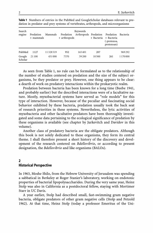
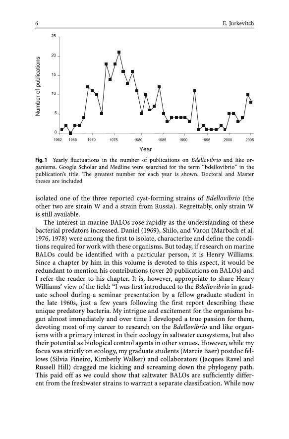
The contribution of scientists in the former Soviet Union is noteworthy. Ofabout 300 publications (Fig. 1) on BALOs, 75 emanated from Soviet groups,50 appearing between 1970 and 1981. Two scientists, Albina Afigenova and V.Lambina worked diligently on the subject, leading a long-term research pro-gram from the 1970s into the 1990s and contributing about 35 publications.Sadly, this project was terminated, and since then few works have emanatedfrom Russia. The various groups studied the distribution, taxonomy, preda-tion dynamics, and other ecological subjects pertaining to BALOs, as well astheir physiology and biochemistry. They isolated Micavibrio (Lambina et al.1982, 1983), micropredators that while resembling Bdellovibrio, were recentlyshown to be phylogenetically unrelated to these bacteria (see chapter by Ju-rkevitch and Davidov in this volume). Unfortunately, only a fraction of thesepublications are available in English.
During the late 1960s and early 1970s, Antonina Guélin, from the Sta-tion Biologique in Roscoff, France also actively pursued research on (mainlymarine) bdellovibrios, partly in collaboration with the Russian group. She
6 E. Jurkevitch
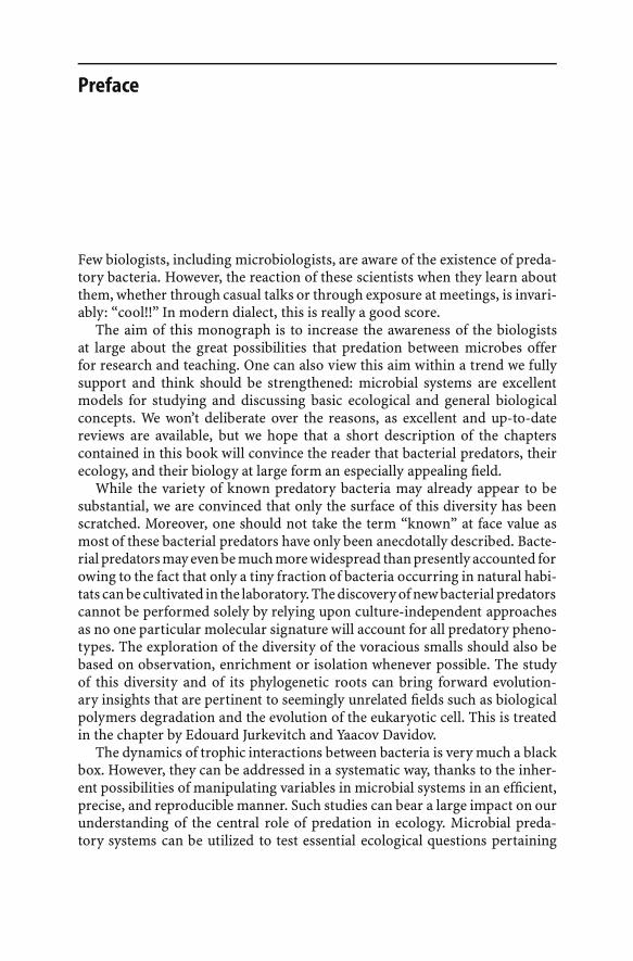
Fig. 1 Yearly fluctuations in the number of publications on Bdellovibrio and like or-ganisms. Google Scholar and Medline were searched for the term “bdellovibrio” in thepublication’s title. The greatest number for each year is shown. Doctoral and Mastertheses are included
isolated one of the three reported cyst-forming strains of Bdellovibrio (theother two are strain W and a strain from Russia). Regrettably, only strain Wis still available.
The interest in marine BALOs rose rapidly as the understanding of thesebacterial predators increased. Daniel (1969), Shilo, and Varon (Marbach et al.1976, 1978) were among the first to isolate, characterize and define the condi-tions required for work with these organisms. But today, if research on marineBALOs could be identified with a particular person, it is Henry Williams.Since a chapter by him in this volume is devoted to this aspect, it would beredundant to mention his contributions (over 20 publications on BALOs) andI refer the reader to his chapter. It is, however, appropriate to share HenryWilliams’ view of the field: “I was first introduced to the Bdellovibrio in grad-uate school during a seminar presentation by a fellow graduate student inthe late 1960s, just a few years following the first report describing theseunique predatory bacteria. My intrigue and excitement for the organisms be-gan almost immediately and over time I developed a true passion for them,devoting most of my career to research on the Bdellovibrio and like organ-isms with a primary interest in their ecology in saltwater ecosystems, but alsotheir potential as biological control agents in other venues. However, while myfocus was strictly on ecology, my graduate students (Marcie Baer) postdoc fel-lows (Silvia Pineiro, Kimberly Walker) and collaborators (Jacques Ravel andRussell Hill) dragged me kicking and screaming down the phylogeny path.This paid off as we could show that saltwater BALOs are sufficiently differ-ent from the freshwater strains to warrant a separate classification. While now
A Brief History of Short Bacteria 7
at Florida A&M, my efforts to maintain support to study the BALOs over theyears were made difficult in the early years because I was on the faculty ata dental school. Reviewers would question why this research was being donein a dental school and did not think serious work on aquatic organism couldbe done in such an environment. I would however use the justification withtongue in cheek that a toothbrush was used to brush oyster shells to removesurface biofilms in our studies on the association of BALOs with surfaces innatural ecosystems...”.
Williams also mentions that “funding to support research on the Bdellovib-rio has always been difficult to come by” because “program managers atfunding agencies and reviewers of proposals would question the significanceof research on the BALOs since such few investigators were submitting re-search proposals for the study of the organisms”.
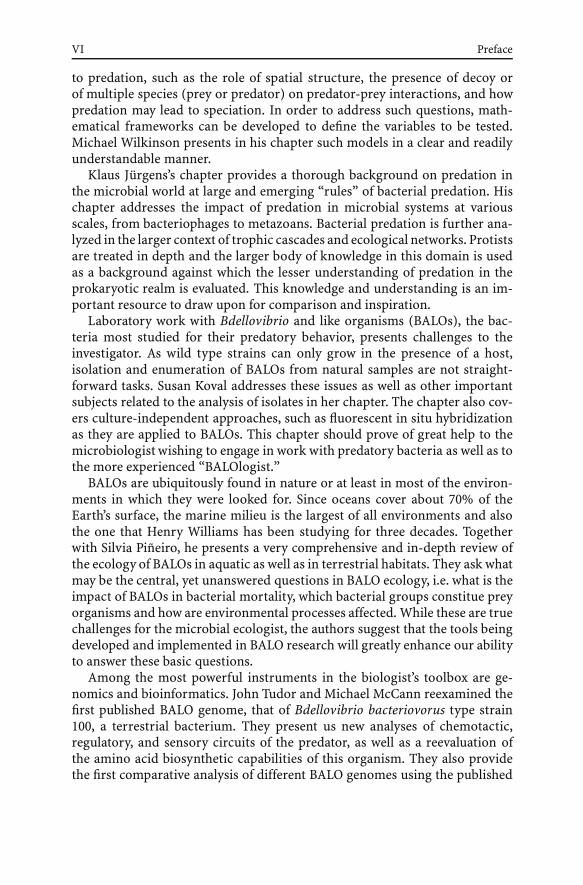
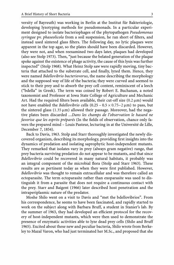
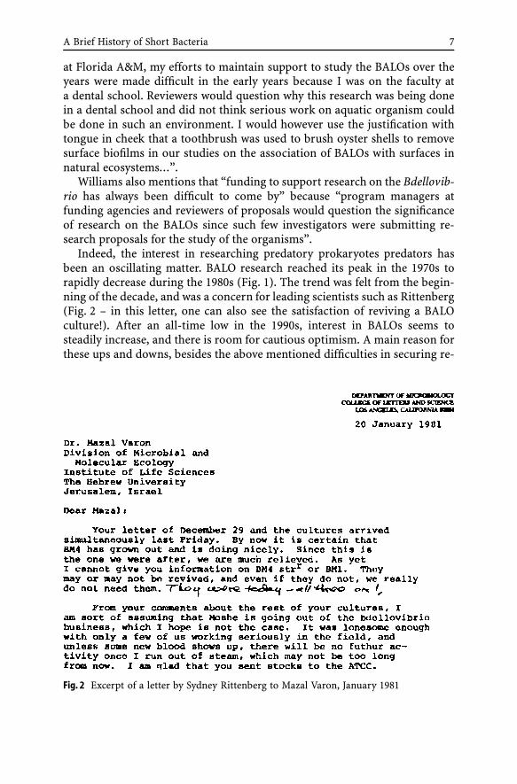
Indeed, the interest in researching predatory prokaryotes predators hasbeen an oscillating matter. BALO research reached its peak in the 1970s torapidly decrease during the 1980s (Fig. 1). The trend was felt from the begin-ning of the decade, and was a concern for leading scientists such as Rittenberg(Fig. 2 – in this letter, one can also see the satisfaction of reviving a BALOculture!). After an all-time low in the 1990s, interest in BALOs seems tosteadily increase, and there is room for cautious optimism. A main reason forthese ups and downs, besides the above mentioned difficulties in securing re-
Fig. 2 Excerpt of a letter by Sydney Rittenberg to Mazal Varon, January 1981
8 E. Jurkevitch
search funds, was, as both Varon and Williams pointed out to me, becauseof the inherent complexity involved in working with two-membered culturesof predator and prey. It is somehow ironic that the interest in the field dwin-dled as molecular biological tools, which can help overcome some of the basicproblems of mixed cultures, were becoming available and were even appliedto the study of BALOs (Cotter and Thomashow 1992a,b). Fortunately, one cansense that this trend is reversing, and research on predatory bacteria is ris-ing again as new groups are now entering the field, and genome projects havebeen and are being pursued. This book may also stand witness to this trend: itsummarizes the latest developments in the field and hopefully, will contributeto its strengthening.
Acknowledgements My deepest gratitude goes to Mazal Varon, John Tudor, and HenryWilliams for their help in gathering information and for their personal insights. I wouldlike to warmly thank Heinz Stolp for providing unique material. I am grateful to RafaelSpringmann for translations from German.
References
Abram D, Castro e Melo J, Chou D (1974) Penetration of Bdellovibrio bacteriovorus intohost cells. J Bacteriol 118:663–680
Beebe JM (1941) Studies on the myxobacteria. 2. The role of myxobacteria as bacterialparasites. Iowa State J Sci 15:319–337
Bengston S (2002) Origins and early evolution of predation. Paleontol Soc P 8:289–318Burnham JC, Hashimoto T, Conti SF (1968) Electron microscopic observations on the
penetration of Bdellovibrio bacteriovorus into gram-negative bacterial hosts. J Bacte-riol 96:1366–1381
Cotter T, Thomashow MF (1992a) Identification of a Bdellovibrio bacteriovorus geneticlocus, hit, associated with the host-independent phenotype. J Bacteriol 174:6018–6024
Cotter TW, Thomashow MF (1992b) Identification of a Bdellovibrio bacteriovorus geneticlocus, hit, associated with the host-independent phenotype. J Bacteriol 174:6018–6024
Daniel S (1969) Etude de l’influence de Bdellovibrio bacteriovorus dans l’auto-épurationmarine. Rev Int Océanogr 15–16:61–102
Ekstejn M, Varon M (1977) Elongation and cell division in Bdellovibrio bacteriovorus.Arch Microbiol 114:175–181
Hashimoto T, Diedrich DL, Conti SF (1970) Isolation of a bacteriophage for Bdellovibriobacteriovorus. J Virol 5:97–98
LaMarre AG, Straley SC, Conti SF (1977) Chemotaxis toward amino acids by Bdellovibriobacteriovorus. J Bacteriol 131:201–207
Lambina VA, Afinogenova AV, Romai Penabad S, Konovalona SM, Pushkareva AP (1982)Micavibrio admirandus gen. et sp. nov. Mikrobiologiya 51:114–117
Lambina VA, Afinogenova AV, Romay Penabad S, Konovalona SM, Andreev LV (1983)A new species of exoparasitic bacteria from the genus Micavibrio. Mikrobiologiya53:777–780
Marbach A, Varon M, Shilo M (1976) Properties of marine bdellovibrio. Microb Ecol2:284–295
Maynard-Smith J, Szathamary E (1995) The major transitions in evolution. Freeman, NewYork
A Brief History of Short Bacteria 9
Rittenberg SC, Shilo M (1970) Early host damage in the infection cycle of Bdellovibriobacteriovorus. J Bacteriol 102:149–160
Steiner S, Conti SF, Lester RL (1973) Occurrence of phosphonosphingolipids in Bdellovib-rio bacteriovorus strain UKi2. J Bacteriol 1199–1211
Stolp H, Pertzold H (1962) Untersuchungen uber einen obligat parasitischen Mikroorgan-ismus mit lytischer Aktivitat fur Pseudomonas-Bakterien. Phytopathol Z 45:364–390
Stolp H, Starr MP (1963) Bdellovibrio bacteriovorus gen. et sp., a predatory, ectoparasitic,and bacteriolytic microorganism. Antonie van Leeuwenhoek 29:217–248
Stolp H (1969) Am 11. Dezember 1968 jährte sich zum 125. Male der Geburtstag vonRobert Koch 843–1910. Bundesgesundheitsblatt 2:S17–21
Stolp H (1973) The bdellovibrios: bacterial parasites of bacteria. Ann Rev Phytopathol11:53–76
Starr MP, Baigent NL (1966) Parasitic interaction of Bdellovibrio bacteriovorus with otherbacteria. J Bacteriol 9:2006–2017
Shilo M, Bruff B (1965) Lysis of Gram negative bacteria by host-independent ectoparasiticBdellovibrio bacteriovorus isolates. J Gen Microbiol 40:317–328
Straley SC, Conti SF (1977) Chemotaxis of Bdellovibrio bacteriovorus toward prey. J Bac-teriol 132:628–640
Straley SC, LaMarre AG, Lawrence LJ, Conti SF (1979) Chemotaxis of Bdellovibrio bacte-riovorus toward pure compounds. J Bacteriol 140:634–642
Tudor JJ (1980) Chemical analysis of the outer cyst wall and inclusion material ofBdellovibrio bdellocysts. Curr Microbiol 4:251–256
Tudor JJ, Bende S (1986) The outer cyst wall of bdellocysts is made de novo and not frompre-formed units from the prey wall. Curr Microbiol 13:185–189
Tudor JJ, Conti SF (1977) Characterization of bdellocysts of Bdellovibrio sp. J Bacteriol131:314–322
Tudor JJ, Conti SF (1977b) Ultrastructural changes during encystment and germinationof Bdellovibrio sp. J Bacteriol 131:323–330
Tudor JJ, Conti SF (1978) Characterization of germination and activation of Bdellovibriobdellocysts. J Bacteriol 133:130–138
Tudor JJ, DiGiuseppe JA (1988) The effect of penicillin on encystment of Bdellovibrio sp.strain W. Arch Microbiol 149:422–426
Varon M (1979) Selection of predation-resistant bacteria in continuous culture. Nature277:386–388
Varon M, Fine M, Stein A (1984) The maintenance of Bdellovibrio at low prey density.Microb Ecol 10:95–98
Varon M, Levinsohn R (1972) Three-membered parasitic system: a bacteriophage,Bdellovibrio bacteriovorus, and Escherichia coli. J Virol 9:519–525
Varon M, Seijffers J (1975) Symbiosis-independent and symbiosis-incompetent mutantsof Bdellovibrio bacteriovorus 109J. J Bacteriol 124:1191–1197
Varon M, Shilo M (1968) Interaction of Bdellovibrio bacteriovorus and host bacteria: I.Kinetic studies of attachment and invasion of Escherichia coli B by Bdellovibrio bac-teriovorus. J Bacteriol 95:744–753
Varon M, Shilo M (1969a) Attachment of Bdellovibrio bacteriovorus to cell wall mutantsof Salmonella spp and Escherichia coli. J Bacteriol 97:977–979
Varon M, Shilo M (1970) Methods for separation of Bdellovibrio from mixed bacterialpopulation by filtration through Millipore filters or by gradient differential centrifu-gation. Rev Intern Océanogr Med 18–19:145–152
Varon M, Shilo M (1981) Inhibition of the predatory activity of Bdellovibrio by variousenvironmental pollutants. Microb Ecol 7:107–111
Microbiol Monogr (4)E. Jurkevitch: Predatory ProkaryotesDOI 10.1007/7171_052/Published online: 27 October 2006© Springer-Verlag Berlin Heidelberg 2006
Phylogenetic Diversity and Evolutionof Predatory Prokaryotes
Edouard Jurkevitch (�) · Yaacov Davidov
Department of Plant Pathology and Microbiology,and the Otto Warburg Center for Agricultural Biotechnology,Faculty of Agricultural, Food and Environmental Quality Sciences,The Hebrew University of Jerusalem, 76100 Rehovot, [email protected]
1 Introduction . . . . . . . . . . . . . . . . . . . . . . . . . . . . . . . . . . . 12
2 Predatory Bacteria: a Phylogenetic Perspective . . . . . . . . . . . . . . . . 142.1 Alpha Proteobacteria . . . . . . . . . . . . . . . . . . . . . . . . . . . . . . 142.2 Beta Proteobacteria . . . . . . . . . . . . . . . . . . . . . . . . . . . . . . . 172.3 Gamma Proteobacteria . . . . . . . . . . . . . . . . . . . . . . . . . . . . . 182.4 Delta Proteobacteria . . . . . . . . . . . . . . . . . . . . . . . . . . . . . . . 212.5 Chloroflexi . . . . . . . . . . . . . . . . . . . . . . . . . . . . . . . . . . . . 312.6 Cytophagaceae . . . . . . . . . . . . . . . . . . . . . . . . . . . . . . . . . . 312.7 Gram Positives . . . . . . . . . . . . . . . . . . . . . . . . . . . . . . . . . . 332.8 Archea . . . . . . . . . . . . . . . . . . . . . . . . . . . . . . . . . . . . . . 332.9 Phylogenetically Undefined Predators . . . . . . . . . . . . . . . . . . . . . 34
3 A Word on Predatory Strategies . . . . . . . . . . . . . . . . . . . . . . . . 38
4 Predation Between Prokaryotes: An Evolutionary Perspective . . . . . . . 394.1 Origins . . . . . . . . . . . . . . . . . . . . . . . . . . . . . . . . . . . . . . 394.2 The Predatory Hypothesis to the Origin of Mitochondria . . . . . . . . . . 43
5 Conclusions . . . . . . . . . . . . . . . . . . . . . . . . . . . . . . . . . . . 45
References . . . . . . . . . . . . . . . . . . . . . . . . . . . . . . . . . . . . . . . 47
Abstract Predation is one of the commonest types of interaction in the living world. Itsroots appear to be ancient, and it may have first occurred early in the evolution of lifeforms. Predators have evolved many times in the animal realm, and this also seems tobe the case within the prokaryotes. Although still rather limited, our knowledge of obli-gate and non-obligate bacterial predators suggest that they are common in many bacterialphyla, as well as in the environment. In this work, we survey and describe the knownbacterial predators according to their phylogenetic affiliation. A hallmark of many bacte-rial predators is their ability to degrade the polymeric structures of their bacterial preys.An additional characteristic of known obligate predators is a small cell size. We use suchdistinguishing features to put forward hypotheses relating to the origin of predation inprokaryotes and to the impact of predation on the evolution of the eukaryotic cell.
12 E. Jurkevitch · Y. Davidov
1Introduction
Predation is a common mode of interaction between organisms across thescales of the realm of life. If bacteriophages are included, a continuumof predators exists from submicron to (tens) of meters in size, spanningabout eight orders of magnitude. At the lower end of the scale—viruses andbacteria—predators are usually smaller than their prey, while at the other lev-els, the opposite is the rule. The boundary stands at the prokaryote–eukaryotedivide, as protozoa are usually larger than their bacterial prey. The rea-son underlying this difference in predatory modes between eukaryotes andprokaryotes may stem from the ability of the former, and the inability of thelatter to engulf particles. Obviously, a cell that is unable to phagocytose is notable to “swallow” an object, let alone if this object happens to be larger than it.Although size matters, strategies and mechanisms are crucial to the outcomeof the interaction, as described below.
In fact, an escape strategy to protozoal predatory pressure is observed asthe size distribution of bacterial cells exposed to grazing is altered (see Jur-gens, 2006, in this volume). As it is advantageous for a swallowing predatorto be larger than its prey, a positive feedback loop is created (Bengston 2002).Therefore, predatory eukaryotes would most necessarily prey on cells smallerthan themselves. In contrast, for cells unable to phagocytose such as prokary-otes, efficient predation would almost dictate the opposite: a small cell sizeand therefore, predation upon larger prey cells. A small predatory cell couldmore efficiently adhere to the surface of a large prey. Moreover, and pos-sibly more significant for an obligate predator, it could be advantageous toprey upon a cell larger than your own as this could provide enough sup-plies for replication and growth at once. A relatively large prey could providean ample nutrient and energy source for each successful predatory interac-tion, yielding a number of progeny cells. Moreover, a small-sized predatorcould more easily gain access to the interior of its host. Another interest-ing feature of some prokaryotic predators is their high motility. If motilityis a plus for scavengers, it is obviously an advantage for predators. It seemsthat a small cell size could enable rapid and very active motility (Starr andSeidler 1971). However, larger organisms are usually faster than smaller ones(Bonner 1993), but paradoxically small bacterial predators, such as Bdellovib-rio and like organisms (BALOs) are also the fastest swimmers (Stolp 1967).Therefore, opting for a small size would seem a likely strategy for predatorybacteria, at least for obligate predators.
It has been argued that prokaryotes are limited to small sizes because theydo not possess a real cytoskeleton (Zlatanova 1997) (although its origin maybe traced to bacteria (van den Ent et al. 2001; Doolittle and York 2002)),they cannot fuel the metabolic demands of a large cell as diffusion limitsthe uptake of nutrients and their internal distribution as well as waste dis-
Phylogenetic Diversity and Evolution of Predatory Prokaryotes 13
posal (Koch 1996). While these may place constraints on the evolution of largeprokaryotic cells, the discovery of “gigantic” prokaryotes such as Thiomar-garita namibiensus (Schulz 1999) and Epulopiscium fishelsoni (Angert et al.1993; Clements and Bullivant 1991) demonstrates that prokaryotes may reachsizes well above the “common” bacterial size. Theoretical considerationshows that in fact, diffusion may sustain cells even larger than the largestknown prokaryotes (Koch 1996). While prokaryotic cells do not grow to beas large as most eukaryotic cells, one may ask why aren’t large bacteria morecommon. A frequently invoked advantage for a large cell size is protectionfrom protozoan predation. While a large body would protect from predationby protozoa, could it make the organism more exposed to bacterial preda-tion? Could that be a reason for the dearth of such large bacteria? Or hasour sampling been biased by our inability to culture most prokaryotes (Rappéand Giovannoni 2003) and a propensity to mainly sample accessible habitats?T. namibiensus and E. fishelsoni have been found in sulfurous marine sedi-ments in the sea-floor off the coast of Namibia, and in the intestinal tract ofsurgeonfish, respectively. It would be interesting to test for the presence ofbacterial and protozoan predators in the environments supporting these largeprokaryotes.
Predatory bacteria are phylogenetically diverse and ubiquitous in terres-trial and aquatic environments and appear to form a part of their microbialfabric (Baer et al. 2000; Snyder et al. 2002; Davidov and Jurkevitch 2004, andthis work). Moreover, culture-based and culture-independent analyses of ex-treme or “exotic” environments reveal that predatory bacteria are also tobe found there: 16S rDNA sequences or isolates related to Bdellovibrio andlike organisms (BALOs) were retrieved from arsenite-oxidizing biofilms, fromhot-spring travertine depositions, arctic marine sediments and hyper-salinewaters (Davidov and Jurkevitch 2004; Pineiro et al. 2004), and predatory in-teractions between bacteria have been documented in anaerobic layers insulfurous lakes (Esteve et al. 1983; Guerrero et al. 1986). Predatory bacteriaexhibit very different phenotypes and often amazing physiological adapta-tions. They also probably make use of different predatory strategies. Theyrepresent an untapped resource for the microbial ecologist as well as for mi-crobiologists at large.
Predation is a major ecological force, shaping the structure of commu-nities, driving diversity and evolution of life histories (Stanley 1973; Dayet al. 2002), and as such is a central subject for ecological research. Microbialmodels are very useful for testing basic questions of ecological importanceas they can be controlled, tracked, manipulated and replicated much moreeasily than most biological systems (Jessup 2004), enabling experimental ver-ification that otherwise may be very difficult to achieve. For example, under-standing the evolution of a predator–prey interaction requires a descriptionof the potential dynamics of one or more traits in one or both species throughtime (Abrams 2000). This may be difficult to implement experimentally but
14 E. Jurkevitch · Y. Davidov
microbial models can offer great opportunities to test such systems (Yoshidaet al. 2003). Other theoretical features of predator–prey interactions, can betested such as (among many others) the role of spatial structure for explain-ing coexistence (Schrag and Mittler 1996; Bohannan et al. 2002), fitness costsof resistance to predation (Lenski 1988; Bohannan et al. 2002) and the link be-tween productivity and food chain length (Kaunzinger 1998). However, fewstudies have used purely bacterial components as predator and prey to testtheoretical hypotheses (Varon 1978, 1979).
In this work, we shall try to show that while the data on predatory bacteriais not immense, predation is a common mode of feeding within prokaryotes,and that by investigating it, hypotheses pertinent to the evolution of certainextant life forms can be proposed.
2Predatory Bacteria: a Phylogenetic Perspective
As our primary focus in this work is the description of bacterial predators inan evolutionary perspective, micropredators will be mostly treated on a phy-logenetic basis. We shall start with a description of phylogenetically definedbacterial predators along taxonomic lines. We shall focus on bacterial species,in which predatory behavior has been demonstrated, i.e. the ability to growon prey cells as the sole source of nutrients, including both obligate and fac-ultative predators.
Until recently, the obligate predators described as Bdellovibrio and like or-ganisms (BALOs), while forming different families, were only found withinthe δ-proteobacteria and were historically treated together. However, newfindings indicate that obligate predators can be found in different proteobac-terial classes: Micavibrio spp. belong to the α-proteobacteria. Since this work’sorganization is based on a phylogenetic classification and the term BALO hashitherto only been used to designate δ-proteobacteria predators, Micavibriowill be treated here under the α-proteobacteria. However, we propose thatobligate predators such as Micavibrio, the mode of action, morphology andbehavior of which resemble the hitherto describes BALOs should also be cov-ered by this general term. Furthermore, we propose to add a prefix to the termBALO that would indicate the phylogenetic affiliation of the organism. Mi-cavibrio would therefore be a-BALOs, and the other known obligate bacteriapredators, d-BALOs.
2.1Alpha Proteobacteria
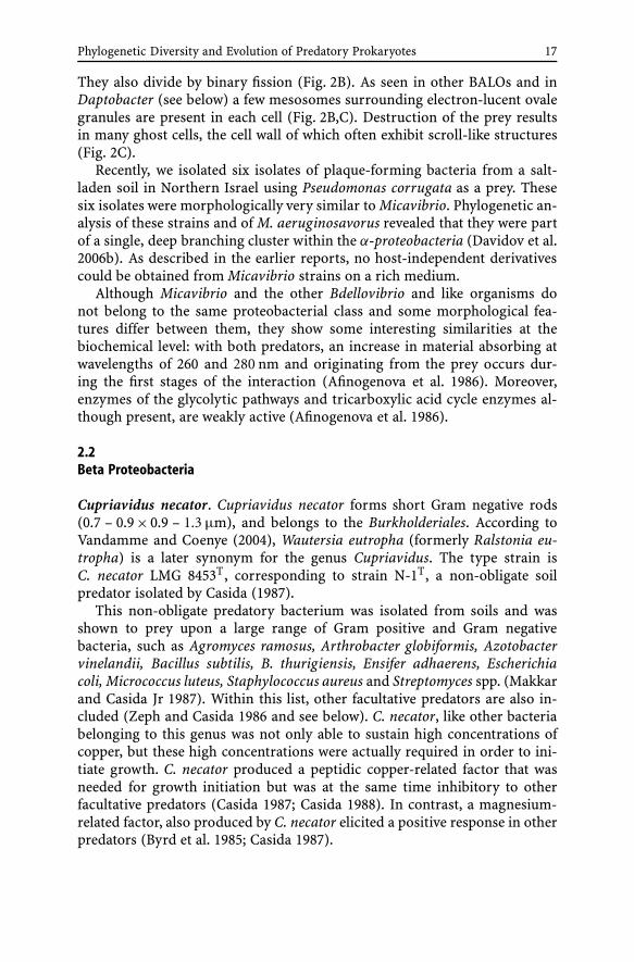
Ensifer adhaerens. Ensifer adhaerens is an aerobic rod-like Gram-negativesoil bacterium (0.7–1.1×1.0–1.9 µm) occurring singly or in pairs. It attaches
Phylogenetic Diversity and Evolution of Predatory Prokaryotes 15
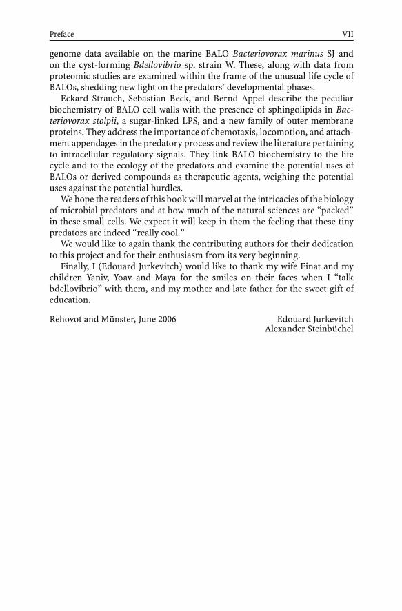
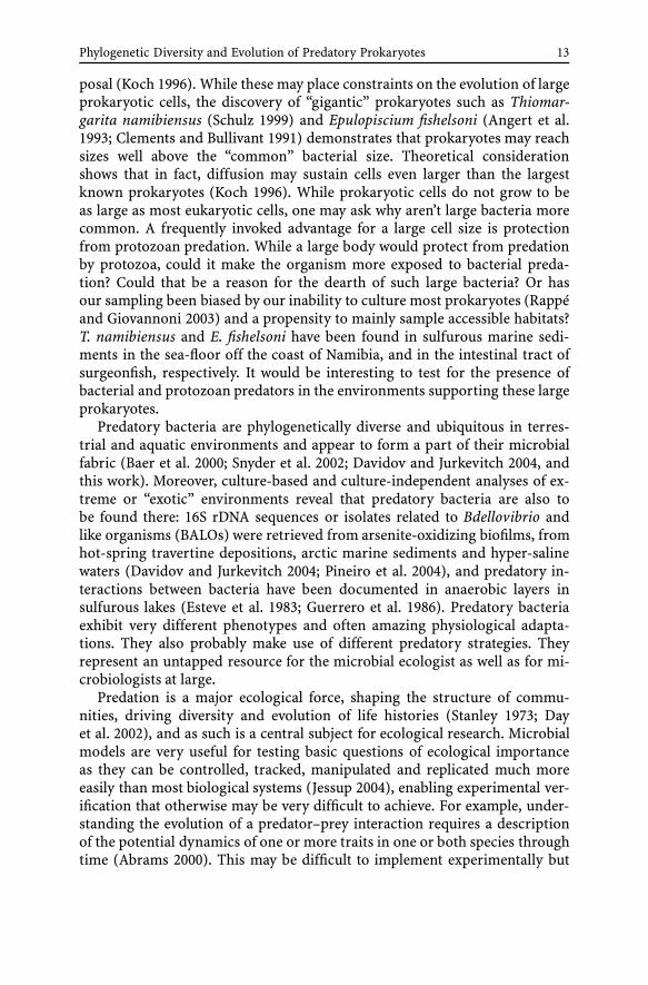
endwise, in a picket fence-like fashion to its prey (Fig. 1A) which are vari-ous living Gram-positive and Gram-negative bacteria but it is not an obligatepredator (Casida 1982). The prey and the predator are connected through anelectron dense material that seems to emanate from the predator (Fig. 1B).Prey lysis occurs in the absence of other available resources (Casida 1982).E. adhaerens divides by budding at one cell pole followed by asymmetri-cal polar growth and binary fission (Casida 1982; Fig. 1C). E. adhaerensexhibiting a predatory behavior towards Micrococcus luteus and Gram nega-tive cells as well were found in the four soils in which they were searchedfor (Casida 1980). In soils enriched with M. luteus, both the population ofE. adhaerens and of a Streptoverticillium-like predatory bacterium increasedsimultaneously as they preyed upon the added bacteria. A Myxococcus-likepredator also able to utilize M. luteus, developed later (Casida 1980). E. ad-haerens was able to lyze both these predators in soil (Casida 1980; Germida1983). However, under laboratory conditions, E. adhaerens could not lyze theStreptoverticillium-like strain and was itself destroyed by the Myxococcus-likepredator (Germida 1983).
Recently, polyphasic analyses including genetic and phenetic charactersshowed that strains of non-symbiotic rhizobia, some isolated from nodules,were closely related to the type strain E. adherens 7A (Willems 2003). Unfor-tunately, it is not known whether these other strains are capable of predatorybehavior. On the basis of this relatedness, it was proposed that the nomencla-ture be changed to Sinorhizobium adhearens in spite of the fact that Ensifershould take prevalence (Young 2003).
When a symbiotic plasmid from Rhizobium tropici was introduced intoa predatory E. adhaerens strain, the latter was able to form nitrogen-fixing nodules in Phaseolus vulgaris (bean) and Leucaena leucocephala (Ro-gel 2001). Similarly, introduction of a plasmid from Rhizobium phaseoliinto Agrobacterium tumefaciens endowed the plant pathogen with nodule-forming and nitrogen-fixing capacities in P. vulgaris and L. leucocephala
Fig. 1 A Attachment of Ensifer adhaerens strain A cells (arrow) onto Micrococcus luteusprey in a picket fence manner (×3123). B Electron dense connective material appearsbetween an E. adhaerens cell and the darkly stained Micrococcus luteus prey (arrow)(×50 900). C Bud formation and growth E. adhaerens strain A (×31 450). (Casida LE 1982,Int J Syst Bacteriol 32:339–345, by permission)
16 E. Jurkevitch · Y. Davidov
(Martinez et al. 1987). A genome comparison between A. tumefaciens andSinorhizobium meliloti suggested a recent evolutionary divergence (Woodet al. 2001). Conceptually, it is tempting to link predation with symbio-sis and pathogenesis because these behaviors share common needs such asrecognition of a host, attachment, and eventually penetration. E. adhaerens,a little-studied organism can serve as a good model, and the taxonomic iden-tity of E. adhaerens with S. meliloti calls for further investigations of thislink: Is E. adhaerens ancestral, are predatory activities outstanding and solelyfound in a few non-symbiotic strains or are they expressed in symbiotic rhi-zobia? We can suggest that a closer look at this species, the determination ofthe presence or the absence of predatory capabilities in E. adhaerens (S. ad-hearens) strains and other rhizobia, and genome-wide comparison betweenstrains could lead to interesting and surprising insights into rhizobial ecology.
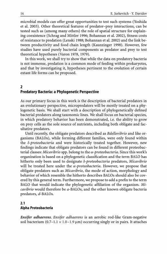
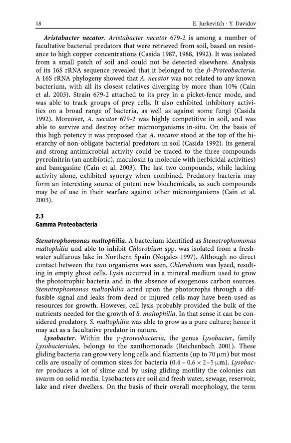
Micavibrio. Micavibrio are obligate predatory bacteria. Two species weredescribed, M. admirantus (Lambina et al. 1982), isolated using Stenotropho-monas maltophila as a prey and M. aeruginosavorus (Lambina et al. 1983),isolated on Pseudomonas aeruginosa. Both were isolated from sewage works.They were not able to prey upon any of the 55 other prey cell types tested, al-though they did utilize almost all of the different strains of the species thatwere used for isolating them. These bacteria are small (0.25–0.4 × 0.5–1 µm),they possess a single, non-sheathed flagellum of 15 nm in diameter, witha regular wavelength (in opposition to other d-BALOs that have a sheath flag-ellum with a damping wavelength form, see below) (Fig. 2A). Micavibrio preyin an epibiotic manner and do not penetrate the prey’s inner compartments.
Fig. 2 A A Micavibrio sp. predator. The scale bar represents 1 µm. B A dividing Micav-ibrio cell attached to a Pseudomonas corrugata prey. The scale bar represents 0.5 µm.C Micavibrio cells (m) and an empty prey (p). Note the electron-lucent zones in the preda-tor and the scroll-like structures in the prey. The scale bar represents 1 µm. (Pictures bySusan Koval)
Phylogenetic Diversity and Evolution of Predatory Prokaryotes 17
They also divide by binary fission (Fig. 2B). As seen in other BALOs and inDaptobacter (see below) a few mesosomes surrounding electron-lucent ovalegranules are present in each cell (Fig. 2B,C). Destruction of the prey resultsin many ghost cells, the cell wall of which often exhibit scroll-like structures(Fig. 2C).
Recently, we isolated six isolates of plaque-forming bacteria from a salt-laden soil in Northern Israel using Pseudomonas corrugata as a prey. Thesesix isolates were morphologically very similar to Micavibrio. Phylogenetic an-alysis of these strains and of M. aeruginosavorus revealed that they were partof a single, deep branching cluster within the α-proteobacteria (Davidov et al.2006b). As described in the earlier reports, no host-independent derivativescould be obtained from Micavibrio strains on a rich medium.
Although Micavibrio and the other Bdellovibrio and like organisms donot belong to the same proteobacterial class and some morphological fea-tures differ between them, they show some interesting similarities at thebiochemical level: with both predators, an increase in material absorbing atwavelengths of 260 and 280 nm and originating from the prey occurs dur-ing the first stages of the interaction (Afinogenova et al. 1986). Moreover,enzymes of the glycolytic pathways and tricarboxylic acid cycle enzymes al-though present, are weakly active (Afinogenova et al. 1986).
2.2Beta Proteobacteria
Cupriavidus necator. Cupriavidus necator forms short Gram negative rods(0.7 – 0.9 × 0.9 – 1.3 µm), and belongs to the Burkholderiales. According toVandamme and Coenye (2004), Wautersia eutropha (formerly Ralstonia eu-tropha) is a later synonym for the genus Cupriavidus. The type strain isC. necator LMG 8453T, corresponding to strain N-1T, a non-obligate soilpredator isolated by Casida (1987).
This non-obligate predatory bacterium was isolated from soils and wasshown to prey upon a large range of Gram positive and Gram negativebacteria, such as Agromyces ramosus, Arthrobacter globiformis, Azotobactervinelandii, Bacillus subtilis, B. thurigiensis, Ensifer adhaerens, Escherichiacoli, Micrococcus luteus, Staphylococcus aureus and Streptomyces spp. (Makkarand Casida Jr 1987). Within this list, other facultative predators are also in-cluded (Zeph and Casida 1986 and see below). C. necator, like other bacteriabelonging to this genus was not only able to sustain high concentrations ofcopper, but these high concentrations were actually required in order to ini-tiate growth. C. necator produced a peptidic copper-related factor that wasneeded for growth initiation but was at the same time inhibitory to otherfacultative predators (Casida 1987; Casida 1988). In contrast, a magnesium-related factor, also produced by C. necator elicited a positive response in otherpredators (Byrd et al. 1985; Casida 1987).
18 E. Jurkevitch · Y. Davidov
Aristabacter necator. Aristabacter necator 679-2 is among a number offacultative bacterial predators that were retrieved from soil, based on resist-ance to high copper concentrations (Casida 1987, 1988, 1992). It was isolatedfrom a small patch of soil and could not be detected elsewhere. Analysisof its 16S rRNA sequence revealed that it belonged to the β-Proteobacteria.A 16S rRNA phylogeny showed that A. necator was not related to any knownbacterium, with all its closest relatives diverging by more than 10% (Cainet al. 2003). Strain 679-2 attached to its prey in a picket-fence mode, andwas able to track groups of prey cells. It also exhibited inhibitory activi-ties on a broad range of bacteria, as well as against some fungi (Casida1992). Moreover, A. necator 679-2 was highly competitive in soil, and wasable to survive and destroy other microorganisms in-situ. On the basis ofthis high potency it was proposed that A. necator stood at the top of the hi-erarchy of non-obligate bacterial predators in soil (Casida 1992). Its generaland strong antimicrobial activity could be traced to the three compoundspyrrolnitrin (an antibiotic), maculosin (a molecule with herbicidal activities)and banegasine (Cain et al. 2003). The last two compounds, while lackingactivity alone, exhibited synergy when combined. Predatory bacteria mayform an interesting source of potent new biochemicals, as such compoundsmay be of use in their warfare against other microorganisms (Cain et al.2003).
2.3Gamma Proteobacteria
Stenotrophomonas maltophilia. A bacterium identified as Stenotrophomonasmaltophilia and able to inhibit Chlorobium spp. was isolated from a fresh-water sulfurous lake in Northern Spain (Nogales 1997). Although no directcontact between the two organisms was seen, Chlorobium was lyzed, result-ing in empty ghost cells. Lysis occurred in a mineral medium used to growthe phototrophic bacteria and in the absence of exogenous carbon sources.Stenotrophomonas maltophilia acted upon the phototrophs through a dif-fusible signal and leaks from dead or injured cells may have been used asresources for growth. However, cell lysis probably provided the bulk of thenutrients needed for the growth of S. maltophilia. In that sense it can be con-sidered predatory. S. maltophilia was able to grow as a pure culture; hence itmay act as a facultative predator in nature.
Lysobacter. Within the γ -proteobacteria, the genus Lysobacter, familyLysobacteriales, belongs to the xanthomonads (Reichenbach 2001). Thesegliding bacteria can grow very long cells and filaments (up to 70 µm) but mostcells are usually of common sizes for bacteria (0.4 – 0.6×2–5 µm). Lysobac-ter produces a lot of slime and by using gliding motility the colonies canswarm on solid media. Lysobacters are soil and fresh water, sewage, reservoir,lake and river dwellers. On the basis of their overall morphology, the term
Phylogenetic Diversity and Evolution of Predatory Prokaryotes 19
“myxobacter” has been used in the past to describe certain Lysobacter strains,generating confusion as to their accurate taxonomic placement.
Many cyanobacteria, other Gram negative, as well as Gram positive bacte-ria including actinomycetes, green algae, fungi and nematodes were shownto fall victim to the lytic activities of members of the Lysobacteriales (Shilo1970; Stewart and Brown 1971; Daft et al. 1973). As such, Lysobacter can beisolated using bacteria (such as Arthrobacter) as a sole nutrient source in agar(Reichenbach 2001 based on Ensign and Wolfe 1965). Some Lysobacter strainsare also active against plant pathogens (Folman et al. 2003; Kobayashi 2005).Although not stringent, different Lysobacter isolates exhibited different preyranges. More so, strains of a particular cyanobacterial species varied in theirsensitivity to lysis by a specific Lysobacter strain (Daft and Stewart 1971).
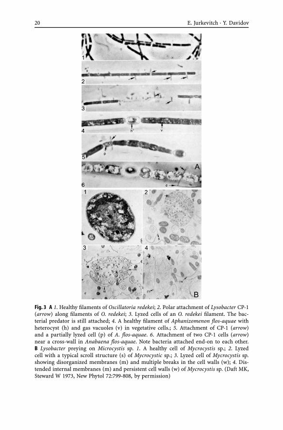
Lysobacter may lyze their prey using a “wolfpack” strategy, which requiresa high density of predators to ensure that the concentrations of the lyzingfactors remain high enough to act upon the prey cell wall (Dworkin 1999).However, cell to cell contact seems to also be the rule, and membrane-boundenzymes may be implicated in prey lysis (Shilo 1970; Daft and Stewart 1971,1973; Kobayashi 2005). Attachment occurs when one cell pole binds to theprey cell, often close to the cross-septa of the filaments, ending up perpen-dicular to the prey (Daft and Stewart 1973, Figure 3A). This would fit anepibiotic mode of predation (Martin 2002), reflected in the capacity of a sin-gle Lysobacter cell to lyze a Nostoc cell in 20 minutes (Shilo 1970, Fig. 3A).Prey search may involve an aerotactic response by which the sensing of anincreasing oxygen gradient leads towards photosynthetic prey cells (Reichen-bach 2001). Interestingly, the same parameter (oxygen concentration) butin reverse, may be used by Bdellovibrio bacteriovorus to locate its prey inenvironments slightly depleted in oxygen by the respiratory activity of thelatter (Rendulic et al. 2004). In opposition to Bdellovibrio and like organ-isms, no defined structure can be seen at the attachment site of Lysobacterto the prey. Lysis occurs rapidly after attachment (Gillespie and Cook 1965),with the peptidoglycan layer being the first structure to be attacked by thepredator (Reichenbach 2001). Lyzed cells contained the remains of internalmembranous complexes and the layers from the cell wall may form scroll-like structures (Fig. 3B). Similar scroll-like structures can also be observed inPseudomonas corrugata prey lyzed by Micavibrio (Fig. 2C).
Lysobacter spp. are a source of enzymes for the biotechnology industryas they produce a wide array of extracellular biopolymer-degrading enzymessuch as nucleases (von Tigerstrom 1980), chitinases (Christensen and Cook1978), proteases (Epstein and Wensink 1988; Wright et al. 1998), glucanases(Palumbo 2003), lipases (Folman et al. 2003), as well as antibiotic compounds(Christensen and Cook 1978; Kato et al. 1997), and a muramidase and twopeptidases endowed with bacteriolytic activities (Sitkin et al. 2003; Step-naya et al. 2004). These latter enzymes are positively charged and interactwith a negatively charged extracellular polysaccharide, forming lysoamidase,
20 E. Jurkevitch · Y. Davidov
Fig. 3 A 1. Healthy filaments of Oscillatoria redekei; 2. Polar attachment of Lysobacter CP-1(arrow) along filaments of O. redekei; 3. Lyzed cells of an O. redekei filament. The bac-terial predator is still attached; 4. A healthy filament of Aphanizomenon flos-aquae withheterocyst (h) and gas vacuoles (v) in vegetative cells.; 5. Attachment of CP-1 (arrow)and a partially lyzed cell (p) of A. flos-aquae. 6. Attachment of two CP-1 cells (arrow)near a cross-wall in Anabaena flos-aquae. Note bacteria attached end-on to each other.B Lysobacter preying on Microcystis sp. 1. A healthy cell of Mycrocystis sp.; 2. Lyzedcell with a typical scroll structure (s) of Mycrocystic sp.; 3. Lyzed cell of Mycrocystis sp.showing disorganized membranes (m) and multiple breaks in the cell walls (w); 4. Dis-tended internal membranes (m) and persistent cell walls (w) of Mycrocystis sp. (Daft MK,Steward W 1973, New Phytol 72:799-808, by permission)