Embed Size (px)
Citation preview
Vol. 195: 29-45.2000 MARINE ECOLOGY PROGRESS SERIES
Mar Ecol Prog Ser ' Published March 31
Separation of chlorophylls and carotenoids from marine phytoplankton: a new HPLC method using a reversed phase C8 column and pyridine-
containing mobile phases
Manuel Z a p a t a l . * , Francisco Rodriguezl, Jose L. ~ a r r i d o ~
'Centro de Investigacions Marinas. Conselleria de Pesca, Xunta de Galicia, Apdo.13.36620 Vilanova de Arousa. Spain 'Institute de Investigacions Maririas de Vigo (CSIC). Eduardo Cabello 6,36208 Vigo. Spain
ABSTRACT: A high-performance liquid chromatographic (HPLC) method based on a reversed-phase C8 column and pyridine-containing mobile phases was developed for the simultaneous separation of chlorophylls and carotenoids. The method is selective enough to resolve monovinyl (MV) and divinyl (DV) pairs of polar chlorophylls and DV chlorophyll a (chl a ) (the marker pigment for the prokaryote Prochlorococcus marinus) from chl a (the MV analogue). Only the pair DV chl alchl b was not resolved. This resolution capability for chlorophylls was only prev~ously achieved using polymeric C),, columns in combination with ammonium acetate or pyridine-containing mobile phases. The proposed method also allows the separation of taxon-specific carotenoids belonging to 8 algal classes, including some critical pigment pairs for previous HPLC methods using C,, columns. The method employs a binary gradient, so it can be used with both low-pressure and high-pressure mixing instruments. Method transferability was tested using 3 HPLC systems. Only a slight adjustment of gradient profile was required to obtain similar results with HPLC equipment having different dwell volumes. The selectiv- ity of the method towards some recently discovered chlorophyll and carotenoid pigments makes it especially suitable for studying not only fleld samples, but also for re-examining the pigment composi- tion of different algal classes.
KEY WORDS: HPLC pigment analysis . Phytoplankton pigments . C, column . Pyridine-containing mobile phases Chemotaxonomy
INTRODUCTION
The chemotaxonomic assessment of phytoplankton populations present in natural waters requires good biochemical markers and very efficient analytical tools. The analysis of photosynthetic pigments by high- performance liquid chromatography (HPLC) fulfils the above requirements as it allows the separation and quantification of taxon-specific chlorophylls and caro- tenoids, some of them present in seawater samples in trace amounts. The outstanding importance of HPLC- based phytoplankton pigment analysis in oceanographic studies has led to the publication of a comprehensive
'E-mail: rnzapata@c~macoron org
monograph in which both modern analytical methods and their application to biological oceanography were reviewed exhaustively (Jeffrey et al. 199713).
The photosynthetic pigments of phytoplankton in natural samples appear a s very complex mixtures whose separation has challenged analytical methods for decades. On the one hand, they cover a wide range of molecular structures, showing very different po- larities (from the acidic chlorophylls to the non-polar hydrocarbon carotenes). On the other hand, some chlorophylls and carotenoids are difficult to separate as they only differ in the presence or position of a dou- ble bond (e.g. monovinyl [MV] and divinyl [DV] chlorophyll pairs, P,P-carotene and p,€-carotene and their isomeric xanthophyll derivatives).
0 Inter-Research 2000 Resale o f full article not p e r n ~ ~ t t e d
30 Mar Ecol Prog Ser 195: 29-45, 2000
The classical HPLC methods were based on re- versed-phase octadecylsilica (ODS, C18) columns and gradient elution with aqueous methanol (Gieskes & Kraay 1983) or aqueous acetonitrile (Wright & Shearer 1984) as initial mobile phase. To retain the most polar acidic chlorophylls either the ion-pair reagent tetra- butylammonium acetate (Mantoura & Llewellyn 1983) or a simple ammonium acetate buffer solution (Zapata et al. 1987) was incorporated to the aqueous methanol mobile phase.
Combining advantages of earlier methods, Wright et al. (1991) developed a ternary gradient HPLC system which made use of the retention capacity of ammo- nlum acetate-containing mobile phase (Zapata et al. 1987) and the special selectivity of acetonitrile-based eluents for carotenoid separation (Wright & Shearer 1984). Wright et al.'s (1991) method was employed as a standard protocol in international oceanographic pro- 9ram.s (Joint Global Ocean Flux Study: JGOFS, UNESCO 1994), and recommended by SCOR Working Group 78 (Wright & Jeffrey 1997). Although these methods achieved good separation for most phyto- plankton carotenoids, none of them was able to separate acidic chlorophylls, with CO-elution of chl c,, chl cl and Mg-3,8-divinyl-pheoporphyrin a5 mono- methyl ester (MgDVP). After the discovery of the marine prokaryote Prochlorococcus marinus (Chis- holm et al. 1988, 1992) a new drawback was added, as these methods are not able to separate DV chls a and b (the marker pigments for P. marinus, Goericke &
Repeta 1992) from the MV (chls a and b) analogues. Once the simultaneous separation of pigments of
different polarities had been obtained, the next step was to improve the HPLC methods by increasing their capacity to resolve photosynthetic pigments with very similar structures. Thus, the resolution of polar and non-polar chls c was improved by using a high car- bon-loaded CI8 column (Kraay et al. 1992), polymeric CI8 columns (Garrido & Zapa.ta 1993, Van Heukelem et al. 1994. Van Lenning et al. 1995), and by increas- ing the mobile phase selectivity with changes in the solvents or the ion-pair reagent (Garrido & Zapata 1996, 1997).
The use of monomeric octylsilica (OS, C8) columns was first introduced by Goericke & Repeta (1993) to achieve the resolution of chl a and DV chl a. That method and further modifications, which employed the same stationary phase (Vidussi et al. 1996, Barlow et al. 1997), still failed in the separation of chl c-related pig- ments. In a recent paper (Rodriguez et al. 1998), we showed that using adequate gradient profiles and injection conditions, monomeric C8 columns can sepa- rate acidic chlorophylls simultaneously with other chlorophylls and carotenoids. However, this method, which obtained good results in the analysis of pigment
composition from many unialgal cultures, failed in the separation of certain pigment pairs from natural sam- ples, especially those composed by chls c and chl a acidic derivatives. To overcome such a problem, and knowing that the use of pyridine as the eluent modifier provides enhanced selectivity towards certain polar chlorophylls and carotenoids (Garrido & Zapata 1996, 1997, Zapata et al. 1998), we have evaluated the per- formance of pyridine-containing mobile phases on monomeric C8 stationary phase.
Here we present an HPLC method for the analysis of phytoplankton pigments which combines a CB column with an optimised mobile phase including an aqueous pyridine solution as an ion-pair reagent. The results obtained from analysis of unialgal culture extracts, complex pigment mixtures and natural samples show that the new method is able to separate, in a single run, most polar and non-polar chlorophylls and most taxon- specific carotenoids found in marine phytoplankton.
MATERIALS AND METHODS
HPLC. Method development was performed using Waters Alliance HPLC equipment (System l ) , includ- ing a 2690 separations module (low-pressure mixing system) and a Waters 996 diode-array detector (1.2 nm optical resolution) interfaced with a Waters 474 scan- ning fluorescence detector by means of a Sat/In analog interface. To verify the transferability of the new method to other HPLC systems, 2 additional instru- ments (Systems 2 and 3) were also employed. HPLC System 2 was a Beckman System Gold including a model 126 programmable solvent module (high-pres- sure mixing system), a model 168 diode-array detector (2 nm optical resolution) and a Rheodyne 77251 injec- tion valve fitted with a 200 p1 loop. HPLC System 3 was a Waters modular system (high dwell volume) including a Waters 600 S controller, a Waters 616 pump (low-pressure mixing system), a Waters 717 Plus autosampler (200 p1 loop) and a Waters 996 diode- array detector (1.2 nm optical resolution).
Stationary phase. Analytical separations were per- formed using a Waters Symmetry C8 column (150 X
4.6 mm, 3.5 pm particle size, 100 A pore size). The col- umn was thermostatted at 25OC by means of a refriger- ated circulator water bath (Neslab RTE-200) connected to an HPLC column water jacket (Alltech).
Mobile phases. Eluent A was a mixture of meth- anol:acetonitrile:aqueous pyridine solution (0.25 M pyridine, see below) (50:25:25 v:v:v) while eluent B was either B1, methanol:acetonitrile:acetone (20:60:20 v:v:v), or B2, acetonitri1e:acetone (80:20 v:v). Organic solvents employed to prepare mobile phases were HPLC-grade. The aqueous pyridine solution (0.25 M)
Zapata et al.: C8 HPLC phy toplankton pigment analysis
was prepared as follows: 10 m1 of acetic acid and 20 m1 of pyridine (Merck) were added to 900 m1 of milli-Q (Millipore) water in a 1 1 flask and mixed using a mag- netic stirrer. Acetic acid was then added dropwise until the pH was 5.0. The mixture was diluted to 1000 m1 with water (final pyridine concentration 0.248 M) and the pH rechecked. All procedures were performed in a fume hood. The pyridine solution was filtered (0.45 pm GHP Gelman filter) after mixing with methanol and acetonitrile (eluent A). Different gradient profiles were adjusted for minimising differences of equipment dwell volume (see Table 1). The flow rate was fixed at 1 m1 min-l.
Algal cultures. Two sets of algal cultures were employed during this study. The first one was used in our laboratories for HPLC method development and evaluation, and was selected to include the most diagnostic pigments and algal classes found in marine phytoplankton: Alexandrium minuturn ALlV- IEO (Dinophyceae) from the Instituto EspaIiol de Oceanografia, Vigo, Spain; Emiliania huxleyi NIOZ CH 24 (Prymnesiophyceae) from the Netherlands Insti- tute for Sea Research, Texel, The Netherlands; Pavlova gyrans CCMP 608 (Prymnesiophyceae), Prochlorococ- cus marinus CCMP 1375 (Cyanophyceae) and Pelago- coccus subviridis CCMP 1429 (Pelagophyceae) from the Provasoli-Guillard National Center for Culture of Marine Phytoplankton (CCMP), West Boothbay Harbor, ME, USA; Rhodomonas baltica ICMA (Cry- ptophyceae), Dunaliella tertiolecta ICMA (Chlorophy- ceae), and Tetraselmis suecica ICMA (Prasinophyceae) from the Instituto de Ciencias Marinas de Andalucia (CSIC), Cadiz, Spain; Micromonas pusilla CCAP 1965/4 (Prasinophyceae) from the Culture Collection of Algae and Protozoa, Oban, UK; and Skeletonema costatum Sk-l (Bacillariophyceae) from the Centro de Investigacions Marinas, Vilanova de Arousa, Spain.
All cultures except Prochlorococcus marinus were grown on f/2 enriched seawater medium (Guillard & Ryther 1962) under 12:12 h L:D cycle with an irradi- ance of 42 pm01 photons m-2 S-' during the light period. Temperature was maintained at 16 + 1°C. P. marinus CCMP 1375 was grown as described by Moore et al. (1995).
In an additional study performed at the CSIRO Marine Laboratories in Hobart, Australia, the method was transferred to other HPLC equipment (System 3) and the following SCOR reference microalgal cultures (Jeffrey & LeRoi 1997) were analysed: Amphidiniurn carterae CS-2 12 (Dinophyceae), Dunaliella tertio- lecta CS-175 (Chlorophyceae), Erniliania huxleyi CS-57 (Prymnesiophyceae), Pavlova lutheri CS-182 (Prymnesiophyceae), Pelagococcus subviridis CS-99 (Pelagophyceae), Phaeodactylum tricornutum CS-29 (Bacillariophyceae), Porphyridium cruentum CS-25
(Rhodophyceae), and Pycnococcus provasoli CS-185 (Prasinophyceae). Culture conditions were as de- scribed by Jeffrey & LeRoi (1997). All cultures were harvested during the exponential phase of growth by filtering under reduced vacuum onto 25 mm diameter Whatman GF/F filters.
The macroalga Codium tomentosum (Chlorophy- ceae) was employed as source of siphonaxanthin and siphonein. Isolated pigments were injected in HPLC System 1 to establish retention time and spectral infor- mation.
Field samples. Seawater samples were obtained from different regions. During the FRUELA 96 cruise (January 1996) on board the RV 'Hesperides', a sample was collected from Gerlache Strait (64" 20'S, 61°48'W, at 5 m depth) near the Antarctic Peninsula. A sample from oligotrophic waters was collected (May 1999) from eastern subtropical North Atlantic (33' 03' N, 21" 16'W, at the deep chlorophyll maximum depth: 80 m). Seawater samples were filtered through a 47 mm diameter Whatman GF/F filters. An estuarine sample was collected (November 1998) from the Ria of Arousa (Galician coast, NW Spain). Seawater was size-fractionated by sequential filtration through 47 mm diameter Whatman GF/D filter (nominal pore size 2.7 pm) and a Whatman GF/F filter (nominal pore size 0.7 pm).
Filters were kept frozen prior to HPLC analysis. The FRUELA sample was stored at -30°C for 2 yr, the North Atlantic sample at -80°C for 10 d and filters from the estuarine sample at -30°C for 1 d.
Pigment extraction. Frozen filters from algal cultures and natural samples were extracted in a Teflon-lined screw-capped tube with 5 m1 95% methanol (2 m1 for the oligotrophic sample) using a stainless steel spatula for filter grinding. The tube was then placed in a beaker with ice and water, and the whole set placed in an ultrasonic bath for 5 min. Extracts were then filtered through 25 mm diameter polypropylene syringe filters (MFS HP020, 0.2 pm pore size) to remove cell and filter debris. An aliquot (1 ml) of methanol extract was mixed with 0.2 m1 of water (0.4 m1 for SCOR culture extracts) to avoid the shape distortion of earlier eluting peaks (Zapata & Garrido 1991). Each sample was injected just after water addition, as a decrease in non-polar pigment concentrations was observed m~hen diluted extracts were held inside the refrigerated autosampler (4OC) prior to injection. The injection volume was 200 p1. All samples were prepared under subdued light.
Pigment detection and identification. Chlorophylls and carotenoids were detected by diode-array spectroscopy (350 to 750 nm). Chlorophylls were also detected by fluorescence (Ex [excitation]: 440 nm, Em [emission]: 650 nm). Absorbance chromatograms were
32 Mar Ecol Prog Ser 195: 29-45, 2000
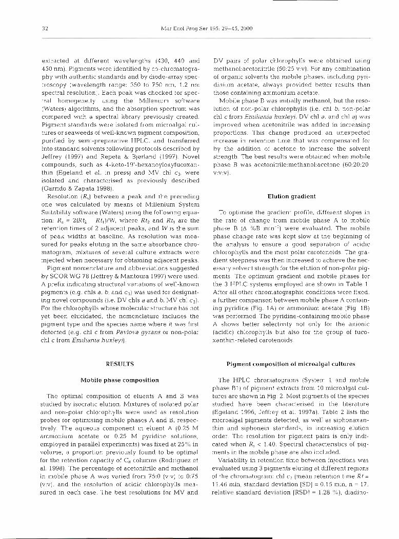
extracted at different wavelengths (430, 440 and 450 nm). Pigments were identified by co-chromatogra- phy with authentic standards and by diode-array spec- troscopy (wavelength range: 350 to 750 nm, 1.2 nm spectral resolution). Each peak was checked for spec- tral homogeneity using the Millenium software (Waters) algorithms, and the absorption spectrum was compared with a spectral library previously created. Pigment standards were isolated from microalgal cul- tures or seaweeds of well-known pigment composition, purified by semi-preparative HPLC, and transferred into standard solvents following protocols described by Jeffrey (1997) and Repeta & Bjarland (1997). Novel compounds, such as 4-keto-19'-hexanoyloxyfucoxan- thin (Egeland et al. in press) and MV chl cg, were isolated and characterised as previously described (Garrido & Zapata 1998).
Resolution (R,) between a peak and the preceding one was calculated by means of Millenium System Suitability software (Waters) using the following equa- tion: R, = 2(Rt, - Rt,)IW, where Rt2 and Rt, are the retention times of 2 adjacent peaks, and W is the sum of peak widths at baseline. As resolution was mea- sured for peaks eluting in the same absorbance chro- matogram, mixtures of several culture extracts were injected when necessary for obtaining adjacent peaks.
Pigment nomenclature and abbreviations suggested by SCOR WG 78 (Jeffrey & Mantoura 1997) were used. A prefix indicating structural variations of well-known pigments (e.g. chls a, b, and c3) was used for designat- ing novel compounds (i.e. DV chls a and b, MV chl c3). For the chlorophylls %.hose molecular structure has not yet been elucidated, the nomenclature includes the pigment type and the species name where it was first detected (e.g. chl c from Pavlova gyrans or non-polar chl c from Emiliania huxleyi].
RESULTS
Mobile phase composition
The optimal composition of eluents A and B was studied by isocratic elution. Mixtures of isolated polar and non-polar chlorophylls were used as resolution probes for optimising mobile phases A and B, respec- tively. The aqueous component in eluent A (0.25 M ammonium acetate or 0.25 M pyridine solutions, employed in parallel experiments) was fixed at 25 % in volume, a proportion previously found to be optimal for the retention capacity of C8 columns (Rodriguez et al. 1998). The percentage of acetonitrile and methanol in mobile phase A was varied from 75:O (v:v) to 03.5 (v:v), and the resolution of acidic chlorophylls mea- sured in each case. The best resolutions for MV and
DV pairs of polar chlorophylls were obtained using methano1:acetonitrile (50:25 v:v). For any combination of organic solvents the mobile phases, including pyri- dinium acetate, always provided better results than those containing ammonium acetate.
Mobile phase B was initially methanol, but the reso- lution of non-polar chlorophylls (i.e. chl b, non-polar chl c from Ernlliania huxleyi, DV chl a , and chl a) was improved when acetonitrile was added in increasing proportions. This change produced an unexpected increase in retention time that was compensated for by the addition of acetone to increase the solvent strength. The best results were obtained when mobile phase B was acetonitrile:methanol:acetone (60:20:20, v:v:v).
Elution gradient
To optimise the gradient profile, different slopes in the rate of change from mobile phase A to mobile phase B (A %B min-l) were evaluated. The mobile phase change rate was kept slow at the beginning of the analysis to ensure a good separation of acidic chlorophylls and the most polar carotenoids. The gra- dient steepness was then increased to achieve the nec- essary solvent strength for the elution of non-polar pig- ments. The optimum gradient and mobile phases for the 3 HPLC systems employed are shown in Table 1 After all other chromatographic conditions were fixed, a further comparison between mobile phase A contain- ing pyridine (Fig. 1A) or ammonium acetate (Fig. 1B) was performed. The pyridine-containing mobile phase A shows better selectivity not only for the anionic (acidic) chlorophylls but also for the group of fuco- xanthin-related carotenoids.
Pigment composition of microalgal cultures
The HPLC chromatograms (System 1 and mobile phase B1) of pigment extracts from 10 microalgal cul- tures are shown in Fig. 2. Most pigments of the species studied have been characterised in the literature (Egeland 1996, Jeffrey et al. 1997a). Table 2 lists the microalgal pigments detected, as well as siphonaxan- thin and siphonein standards, in increasing elution order. The resolution for pigment pairs is only indi- cated when R, < 1.40. Spectral characteristics of pig- ments in the mobile phase are also included.
Variability in retention time between injections was evaluated using 3 pigments eluting at different regions of the chromatogram: chl c2 (mean retention time Rt =
11.46 min, standard deviation [SDI = 0.15 min, n = 17, relative standard deviation [RSD] = 1.28 %), diadino-
Zapata e t al.: Cn HPLC phytoplankton pigment analysis 33
- -
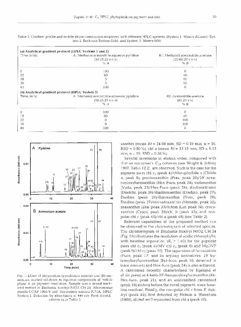
Table 1. Gradient profile and mobile phase composition employed with different HPLC systems. (System 1 : Waters Alliance; Sys- tem 2: 6eckman system old; and System 3: waters 600)
(a) Analytical gradient protocol (HPLC Systems 1 and 2) Time (min) A : Methano1:acetonitrile;aqueous pyridine Bl: Methano1:acetonitrile:acetone
(50:25:25 v:v:v) (20:60:20 v:v:v) % p, % B
(b) Analytical gradient protocol (HPLC System 3) Time (min) A : Methano1:acetonitnle:aqueous pyr~dine
(50:25:25 v:v:v) % A
Ammonium acetate I I
0 10 20 30 40 Time (rnin)
Fig. 1. Effect of (A) pyridine (pyridinium acetate) and (6) am- monium acetate solutions a s aqueous components of mobile phase A on pigment resolution, Sample was a mixed meth- anol extract of Emiliania huxleyi NIOZ C H 24, Micromonas pusilla C C A P 1965/4 and Tetraselmis suecica I C M A . HPLC System 1 . Detection by absorbance at 440 nm. Peak identifi-
cations as in Table 2
xanthin (mean Rt = 24.08 min, SD = 0.19 min, n = 16, RSD = 0.80%), chl a (mean Rt = 33.15 min, SD = 0.13 min, n = 19, RSD = 0.38%).
Several inversions in elution order, compared with that on monomeric Cis columns (see Wright & Jeffrey 1997, Table 12.2), are observed. Such is the case for the pigment pairs chl c3 (peak 4]/chlorophyllide a (Chlide a, peak 8); prasinoxanthin (Pras, peak 20)/19'-hexa- noyloxyfucoxanthin (Hex-Fuco, peak 24); violaxanthin (Viola, peak 23)/Hex-Fuco (peak 24); diadinochrome (Diadchr, peak 26)/diadinoxanthin (Diadino, peak 27); Diadino (peak 27)/dinoxanthin (Dino, peak 28); Diadino (peak 27)/monadoxanthin (Monado, peak 32); zeaxanthin (Zea, peak 33)/lutein (Lut, peak 34); croco- xanthin (Croco, peak 39)/chl b (peak 43); and non- polar chl c (peak 45)/chl a (peak 48) (see Table 2).
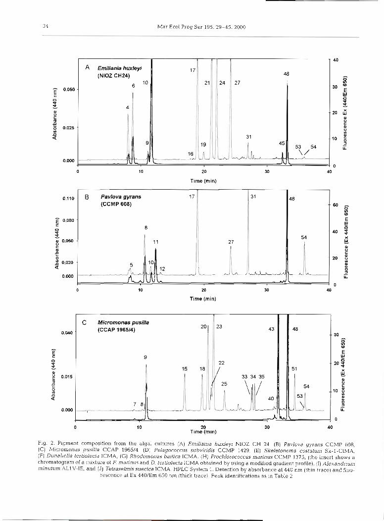
Relevant capabilities of the proposed method can be observed in the chromatogram of selected species. The chromatogram of Erniliania huxleyi NIOZ C H 24 (Fig. 2A) illustrates the resolution of acidic chlorophylls, with baseline separation (R , > 1.40) for the pigment pairs chl c3 (peak 4)/MV chl c3 (peak 6) and MgDVP (peak 9)/chl c2 (peak 10). The separation of fucoxanthin (Fuco, peak 17) and its acyloxy derivatives: 19'-bu- tanoyloxyfucoxanthin (But-fuco, peak 16, detected in trace amount) and Hex-fuco (peak 24) is also achieved. A carotenoid recently characterised by Egeland e t al. (in press) as 4-keto-19'-hexanoyloxyfucoxanthin (4k- Hex-fuco, peak 21), and an unidentified carotenoid (peak 19) eluting before the novel pigment, were base- line resolved. Finally, the non-polar chl c from E. hux- leyi (peak 45), first detected by Nelson & Wakeham (1989), eluted well separated from chl a (peak 48).
34 Mar Ecol Prog Ser 195: 29-45, 2000
A Emiliania huxleyi (NIOZ CH24)
20
Time (min)
20
Time (min)
C Micromonas pusilla (CCAP 196514) 43
---* -
20 Time (rnin)
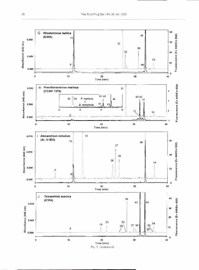
Fig. 2. Pigment composition from the algal cultures [A) Emiliania huxlep NIOZ CH 24, [B) Pavlova gyrans CCMP 608, (C) Micromonas pusifla CCAP 1965/4, ( D ) Pelagococcus subvindis CCMP 1429, ( E ) Skeletonema costatum Sk-1-CIMA, ( F ) Dunaliella tertiolecta ICMA, ( G ) Rhodomonas baltica ICMA, ( H ) Prochlorococcus marinus CCMP 1375, (the insert shows a chromatogram of a mixture of P. marinus and D. tertiolecta ICMA obtained by using a modified gradient profile), (I) Alexandnum minuturn AL1V-IE, and [J) Tetraselmis suedca ICMA. HPLC System 1. Detection by absorbance at 440 nm (thin trace) and fluo-
rescence at Ex 440/Em 650 nm (thick trace). Peak identifications as in Table 2
Zapata et al.. C8 HPLC phytoplankton pigment analysis 35
20
Time (rnin)
0.015 -
E Skeletonema costaturn (SK-1)
0.008 - id I 12
8 ' I 0.005 -
(CCMP 1429)
"I
20
Time (min)
F Dunaliella tertiolecta 0.075 - (ICMA)
20
Time (rnin)
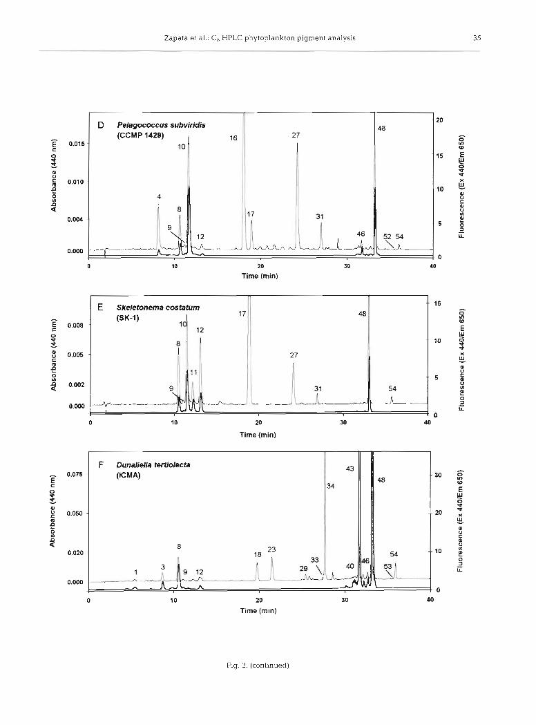
Fig. 2. (continued)
36 Mar Ecol Prog Ser 195: 29-45, 2000
H Prochlorococcus marinus 33
(CCMP 1375) - 2
0.050
c 0 ¥t ^ w ^ 0.020
a g
0.000
^
20 Time (min)
- Alexandrium minutum '. 1 I 3 (AL IV-IEO)
0 10 20 30 40 Time (min)
20 Time (min)
-. 40
30
- 20
10
- 0
G Rhodomonas baltica (ICMA) 10
J Tetraselmis suecica (ICMA) 34
0.010 - 43 - E 0 ¥ ¥ - 01 v . 0 4 0 - I m : (A n 8
0.000
-1
0 10 20 30 Time (min)
Fig. 2. (continued)
48
30
8
--.. J
SJ
39
53 -
- ,. ._.L - L - \.
Zapata et al.: C, HPLC phytoplankton pigment analysis 3 7
-p
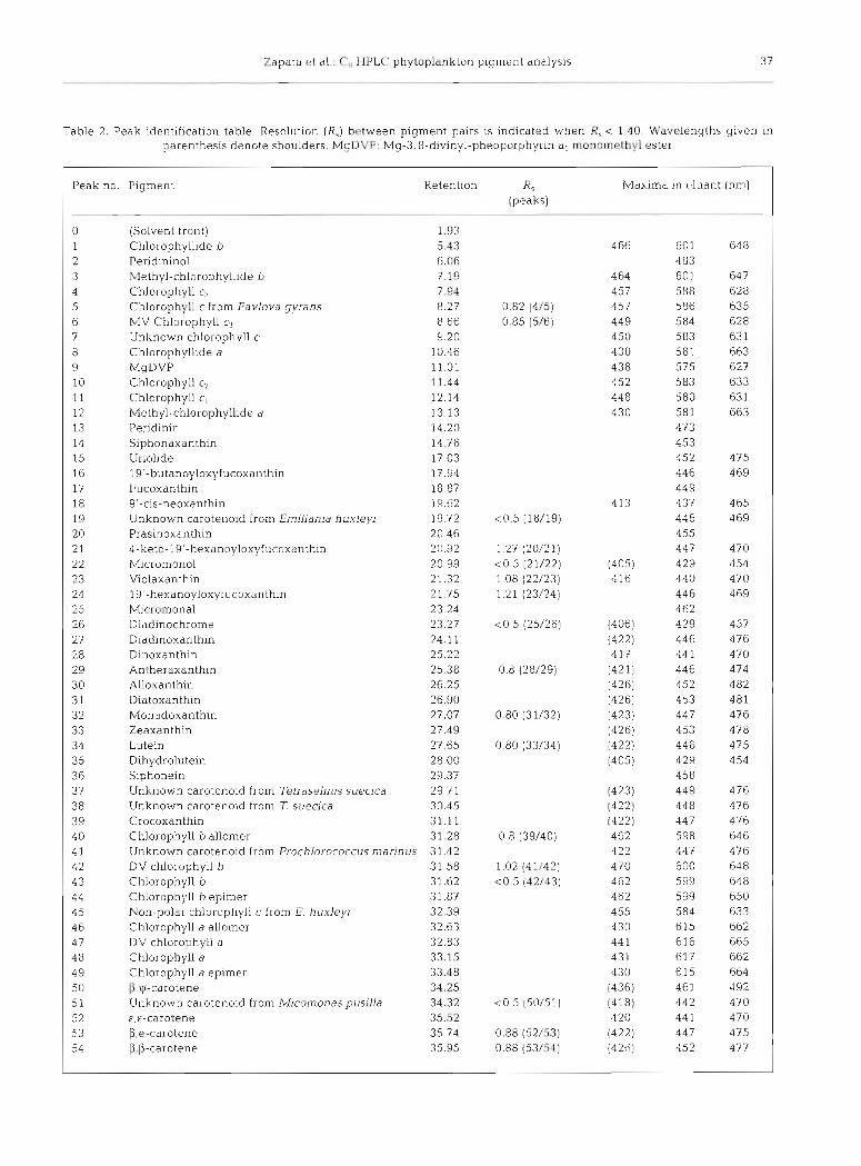
Table 2. Peak identification table. Resolution (R,) hctween pigment pairs is indicated when R, c 1.40. Wavelengths given in parenthesis denote shoulders. MgDVP: Mg-3,8-divinyl-pheoporphyrin a5 monomethyl ester
Peak no Pigment Retention RS Maxima in clluant (nm) (peaks)
. - -p
0 (Solvent front) 1.93 1 Chlorophyllide !J 5.43 4 66 60 1 64 8 2 Peridininol 6.06 483 3 Methyl-chlorophyllide b 7.19 4 64 601 647 4 Chlorophyll cn 7.94 457 588 628 5 Chlorophyll c from Pavlova gyrans 8.27 0.82 (4/5) 457 586 635 6 MV Chlorophyll c3 8.66 0.85 (5/6) 449 584 628 7 Unknown chlorophvll c 9.20 450 583 63 1 8 Chlorophyllide a 10.46 430 581 663 9 MgDVP 11.01 438 575 627 10 Chlorophyll c2 11.44 452 583 633 11 Chlorophyll c, 12.14 448 580 631 12 Methyl-chlorophyllide a 13.13 430 581 663 13 Pendinin 14.20 473 14 Siphonaxanthin 14.76 453 15 Uriolide 17.03 452 475 16 19'-butanoyloxyfucoxanthin 17.94 446 469 17 Fucoxanthin 18.87 449 18 9'-cis-neoxanthin 19.62 413 437 465 19 Unknown carotenoid from Emjljania huxleyi 19.72 <0.5 (18/19) 446 469 20 Prasinoxanthin 20.46 455 2 1 4-keto-19'-hexanoyloxyfucoxanthin 20.92 1.27 (20/21) 447 470 2 2 h4icromonol 20.99 <0.5 (21/22) (405) 429 454 2 3 Violaxanthin 21.32 1 08 (22/23) 416 440 470 24 19'-hexanoyloxyfucoxanth~n 21.75 1.21 (23/24) 446 469 25 Micromonal 23.24 462 26 Diadlnochrome 23.27 <O 5 (25/26) (406) 429 457 27 Diadinoxanthin 24.11 (422) 446 476 28 Dinoxanthin 25.22 4 17 44 1 470 29 Antheraxanthin 25.38 0.8 (28/29) (421) 446 474 30 Alloxanthin 26.25 (426) 452 482 31 Diatoxanthin 26.90 (426) 453 481 32 Monadoxanthin 27.07 0.80 (31/32) (423) 447 476 33 Zeaxanthin 27.49 (426) 453 478 34 Lutein 27.65 0.80 (33/34) (422) 446 475 3 5 D~hydrolutein 28.00 (405) 429 454 3 6 Siphonein 29.37 458 37 Unknown carotenoid from Tell-dselmls suecica 29.71 (423) 449 476 3 8 Unknown carotenoid from T. suecica 30.45 (422) 448 476 39 Crocoxanthin 31.11 (422) 447 476: 40 Chlorophyll b allomer 31.28 0.8 (39/40) 462 598 646 4 1 Unknown carotenoid from Prochlorococcus marinus 31.42 422 447 476 42 DV chlorophyll b 3 1.58 1.02 (41/42) 470 600 648 43 Chlorophyll b 31.62 <0.5 (-12/43) 462 599 648 4 4 Chlorophyll b epimer 31.87 462 599 650 4 5 Non-polar chlorophyll c from E. huxleyi 32.39 455 584 633 4 6 Chlorophyll a allomer 32.63 430 615 662 4 7 DV chlorophyll a 32.83 44 1 616 665 48 Chlorophyll a 33.15 43 1 617 662 49 Chlorophyll a epimer 33.48 430 615 664 5 0 P,ycarotene 34.25 (436) 461 492 5 1 Unknown carotenoid from Micomonaspusjlla 34.32 <0.5 (50151) (418) 442 470 52 €,E-carotene 35.52 420 44 1 470 53 p,e-carotene 35.74 0.88 (52/53) (422) 447 475 54 P,P-carotene 35.95 0.88 (53/54) (426) 452 477
38 Mar Ecol Prog Ser 195: 29-45, 2000
The resolution of other polar chlorophylls is shown in the chromatogram of Pavlova gyrans CCMP 608 (Fig. 2B) where the chl c-like pigment (peak 5), first detected in P, gyrans by Fawley (1989), Chlide a (peak 8), chl c2 (peak 10) and chl c l (peak 11) were baseline separated. Although peak 5 (chl c-like pigment) is not symmetric, it is spectrally homogeneous.
Some recently characterised carotenoids from Pra- sinophyceae (Egeland & Liaaen-Jensen 1995, Egeland et al. 1995) such as uriolide (Uri, peak 15), micromonol (Microl, peak 22), micromonal (Micral, peak 25) and dihydrolutein (Dihydrolut, peak 35) are detected in Micromonas pusilla CCAP 1965/4 (Fig. 2C). Although the separation of major peaks Pras (peak 20) and violaxanthin (Viola, peak 23) seems good, other carotenoids are only partially resolved: MicrolNiola (R, = 1.08), Zea/Lut (R, = 0.80). An unknown polar chl c-like pigment (peak 7), with spectral characteristics similar to chl c2 (see Table 2 ) , was detected in this strain of M. pusilla.
The chromatogram of Pelagococcus subviridis CCMP 1429 (Fig. 2D) shows a major peak of But-fuco (peak 16), usually employed as a marker pigment for the class Pelagophyceae (Andersen et al. 1993), eluting ahead of Fuco (peak 17). A minor peak, identified as &,E-carotene (&&-Car, peak 52), elutes before the other carotenes.
The chromatogram of the diatom Skeletonema costa- turn Sk-l (Fig. 2E), whose chlorophyllase activity pro- motes the conversion of chl a (peak 48) into Chlide a (peak 8) (Jeffrey & Hallegraeff 1987), shows the sepa- ration of this acidic derivative and its methyl ester (peak 12), probably generated during the extraction process using methanol as solvent.
Another species showing high chlorophyllase activ- ity is Dunaliella tertiolecta ICMA, whose chromato- gram (Fig. 2F) shows the presence of Chlide b (peak l), Chlide a (peak 8), and their methyl esters (peaks 3 and 12, respectively). Trace amounts of Zea (peak 33) eluted ahead of Lut (peak 34) achieving a partial reso- lution (R, = 0.80). Its characteristic monocyclic P,y- carotene ( 0 ~ - C a r , peak 50), was detected in trace amount eluting before p,&-carotene @&-Car, peak 53).
In the chromatogram of Rhodomonas baltica ICMA (Fig. 2G) the marker pigment alloxanthin (Allo, peak 30) was baseline resolved from monadoxanthin (Mon- ado, peak 32); and Croco (peak 39) and PE-Car (peak 53) were also detected.
The chromatogram of the cyanobacterium Pro- chlorococcus mannus CCMP 1375 (Fig. 2H) shows MgDVP (peak g), Zea (peak 33, the major carotenoid), an unknown carotenoid (peak 41, spectrally similar to PE-Car) eluting before a peak containing DV chl b (peak 42, the major component) plus chl b (peak 43, detected as a minor component eluting at the final part
of the DV chl b peak), and peaks corresponding to DV chl a (peak 47) and P&-Car (peak 53).
A mixture of methanol extracts from Prochlorococcus marinus and Dunaliella tertiolecta ICMA was used to study the effect of different gradient profiles to resolve the critical pair DV chl blchl b (see insert in Fig. 2H). Although the gradient steepness applied at minute 22 was changed from 40-95% B in 6 min (-A 10%B min-l, standard conditions) to 40-95 % B in 12 min (-A 5 % B min-l), the pigment pair remained unresolved. How- ever, an improvement in the resolution was observed for the pigment pairs Zea/Lut (from R, = 0.80 to R, =
1.08) and DV chl a/chl a (from R, = 1.42 to R, = 1.48). The chromatogram of the toxic dinoflagellate Alex-
andrium rninutum ALlV-IEO (Fig. 21) shows an inver- sion in elution order (with respect C18 columns) for the pigment pairs: Diadchr (peak 26)/Diadino (peak 27), and Diadino (peak 27)IDino (peak 28).
Two unknown carotenoids (peaks 37 and 38, Fig. 25) with similar visible absorbance spectra (see Table 2) were detected in Tetraselmis suecica ICMA. Consider- ing both spectral information and chromatographic behaviour the carotenoids were tentatively identified as loroxanthin esters having a different fatty acid com- position.
Mixed algal extracts
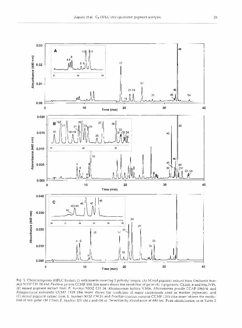
The performance of the method with mixed culture extracts simulating phytoplankton populations of field samples was also evaluated. Three regions of the resulting chromatograms -polar end, central region and non-polar end - were examined in detail (Fig. 3).
The behaviour of polar chlorophylls can be illus- trated by a mixture of Emiliania huxleyi NIOZ CH 24 and Pavlova gyrans CCMP 608 (Fig. 3A). The high resolution (R, = 2.31) for the pigment pair chl c3 (peak 4)/MV chl c3 (peak 6), allows the separation of chl c from P. gyrans (peak 5) between them, while Chlide a (peak 8) and MgDVP (peak 9) elute baseline separated after them, followed by the pair chl c;! (peak 10) and chl c, (peak ll), also well resolved (R, = 2.09).
Most of the taxon-specific carotenoids elute at the central part of the chromatogram. This is illustrated when a mixture of methanol extracts from Emiliania huxleyi NIOZ CH 24, Micromonas pusilla, Pelagococ- cus subvirids CCMP1429 and Rhodomonas baltica is analysed (Fig. 3B). Several carotenoids usually em- ployed as marker pigments for different algal classes are separated: Hex-fuco (peak 24) for Prymnesio- phyceae, But-fuco (peak 16) for Pelagophyceae, Allo (peak 30) for Crytophyceae, and Pras (peak 20) for Prasinophyceae.
Zapata et al. C , HPLC phytoplankton pigment analysis 39
10 Time (min) 20 30 40
Time (min)
0 10 20 30 Time (rnin) 40
Fig 3. Chrornatograrns (HPLC System 1) with inserts covering 3 polarity ranges. (A) Mixed pigment extract from Erniliania hux- lejrl NIOZ CH 24 and Pavlo~ra gyrans CCMP 608 (the insert shows the resolution of polar chl cpigments, Chlide a and MgDVP), (B) mixed pigment extract from E. huxlej/i NIOZ CH 24, Rhodomonas baltica ICMA. Micromonas pusllla CCAP 1965/4, and Pelagococcus subviridis CCMP 1429 (the insert shows the resolution of major carotenoids used as marker pigments); and (C) mixed pigment extract from E. huxleyi NIOZ CH 24 and Prochlorococcus marinus CCMP 1375 (the insert shows the resolu- tlon of non-polar chl cfrom E. huxleyi, DV chl a and chl a). Detection by absorbance at 440 nm. Peak identification as in Table 2
4 0 Mar Ecol Prog Ser 195: 29-45, 2000
Finally, the non-polar end of the chromatogram from a mixture of methanol extracts from Prochlorococcus marinus (Cyanophyceae) and Erniliania huxleyi NIOZ CH 24 (Prymnesiophyceae) (Fig. 3C) shows the coelu- tion of DV chl b (peak 42) and chl b (peak 43) present in P. marinus as trace amounts; the non-polar chl c from E. huxleyi NIOZ CH 24 (peak 45) is well resolved (Rs > 1.50) from DV chl a (peak 471, and DV chl a (peak 47) is separated (R, = 1 47) from chl a (peak 48).
Natural samples
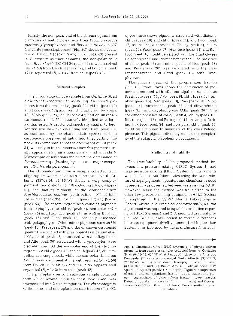
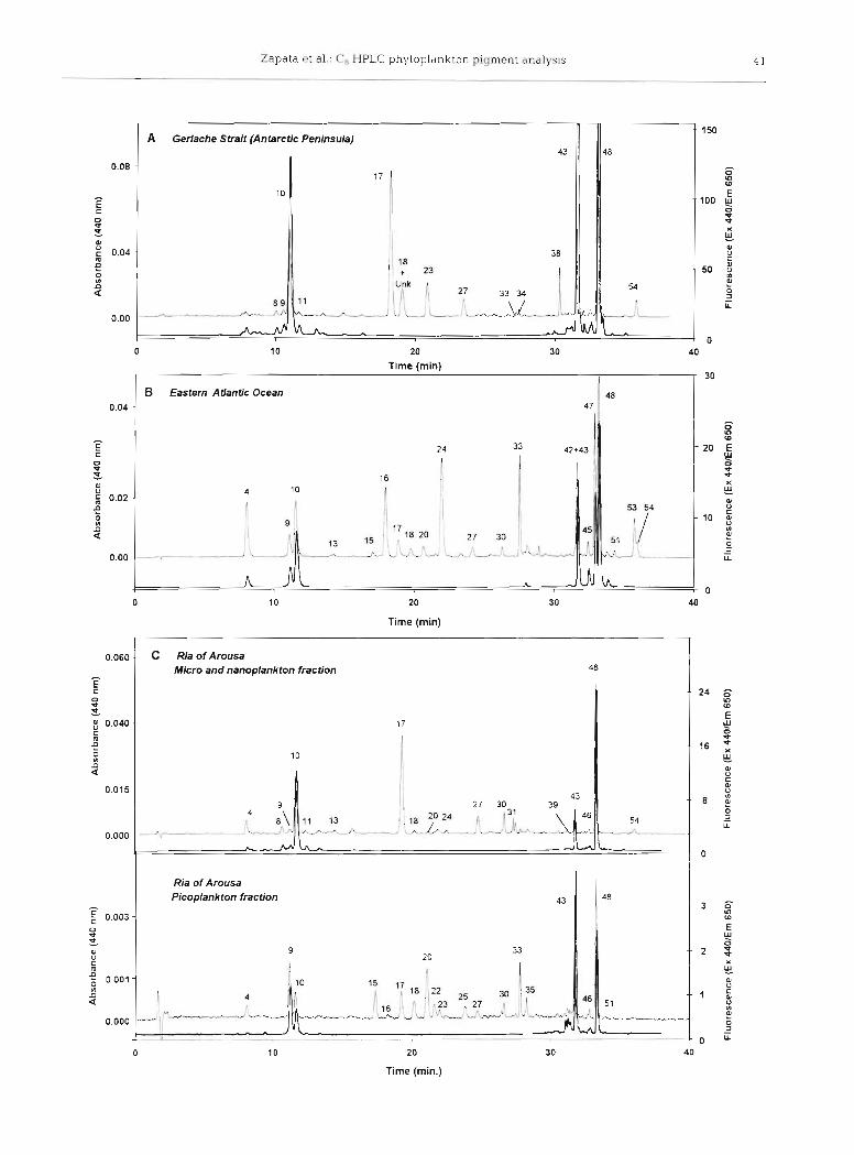
The chromatogram of a sample from Gerlache Strait close to the Antarctic Peninsula (Flg. 4A) shows pig- ments from diatoms: chl c2 (peak 10), chl c , (peak 11) and Fuco (peak 17); and from chlorophytes: Neo (peak 18), Viola (peak 23), chl b (peak 43) and an unknown carotenoid (peak 38) tentatively identified as a loro- xanthin ester. A carotenoid spectrally similar to loro- xanthin was detected CO-eluting with Neo (peak 18), as confirm.ed by the characteristic spectra, of both carotenoids observed at initial and final parts of the peak. It is remarkable that the occurrence of Lut (peak 34) was only in trace amounts, since this pigment usu- ally appears in higher amounts associated with chl b. Microscopic observations indicated the dominance of Pyrarnimonas sp. (Prasinophyceae) as a major compo- nent (M. Varela pers, comm.).
The chromatogram from a sample collected from oligotrophic waters of eastern subtrop~cal North At- lantic (33"03'N, 21" 16'W) shows a very complex pigment composition (Fig. 4B) including DV chl a (peak 4?), the marker pigment of the cyanobacterium Prochlorococcus marinus (contributing 40 % of total chl a ) , Zea (peak 33), DV chl b (peak 42) and P&-Car (peak 53). The chromatograrn also contains pigments from haptophytes as chl c3 (peak 4 ) , non-polar chl c (peak 45) and Hex-fuco (peak 241, as well as But-fuco (peak 16) and Fuco (peak l? ) , probably associated with pelagophytes. Other minor pigments such as Uri (peak 15), Pras (peak 20) and the unknown carotenoid (peak 51) associated with prasinophytes (Egeland et al. 1995), Perid (peak 13) associated w ~ t h dinoflagellates, and Allo (peak 30) associated with cryptophytes, were also identified. At the non-polar end of the chroma- togram, DV chl b (peak 42) and chl b (peak 43) elute to- gether as a single peak, while the non-polar chl c from Emiliania huxleyi (peak 45) is well resolved (R, > 1.50) from DV chl a (peak 47) and the latter appears well separated (R, = 1.42) from chl a (peak 48).
The phytoplankton of a seawater sample collected from Ria of Arousa (Galician coast, NW Spain) was fractionated into 2 size categories. The chromatogram of the nano- and microplankton size-fraction (Fig. 4C,
upper trace) shows pigments associated with diatoms: chl c, (peak 10) a.nd chl c, (peak I l ) , and Fuco (peak 17) as the major carotenoid. Chl c3 (peak 4), chl, cl (peak to), Fuco (peak l?), Hex-fuco (peak 24) and But- fuco (peak 16) could be related with the algal classes Pelagophyceae and Prymnesiophyceae. The presence of chl b (peak 43) and minor peaks of Neo (peak 18) and Pras (peak 20) was associated with the class Prasinophyceae and Perid (peak 13) with Dino- phyceae.
The chromatogram of the picoplankton fraction (Fig. 4C, lower trace) shows the dominance of pig- ments associated with different algal classes such as Prasinophyceae (MgDVP [peak 91, chl b [peak 431, uri- olide [peak IS], Neo [peak 181, Pras [peak 201, Viola [peak 231, micromonal [peak 221 and dlhydrolutein [peak 351) and Cryptophyceae (Allo [peak 301). The combined presence of chl c3 (peak 4), chl c2 (peak 10), But-fuco (peak 16) and Fuco (peak 17) in samples lack- ing Hex-fuco (peak 24) and non-polar chl c (peak 45) could be attributed to members of the class Pelago- phyceae. This pigment diversity reflects the complex- ity of the eukarotlc picoplankton community.
Method transferability
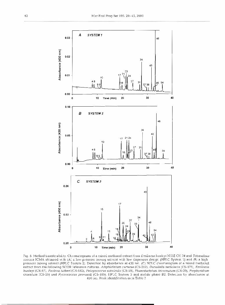
The transferability of the proposed method be- tween low-pressure mixing (HPLC System 1) and high-pressure mixing (HPLC System 2) instruments was checked In our laboratories using the same mix- ture of algal p~gments, operators and chemicals. A good agreement was observed between systems (Fig. 5A,B). However, when the method was transferred to the other low-pressure mixing equipment (HPLC System 3) employed at the CSIRO Marine Laboratories in Hobart, Australia, during a collaborative study, a slight adjustment was required to equal the resolution capac- ity of HPLC Systems 1 and 2. A modified gradient pro- file (see Table 1) was applied to correct differences between equipment dwell volumes (4 m1 higher than System 1, as informed by the manufacturer). In addi-
Fig. 4. Chromatograms (HPLC System 1) of phytoplankton pigments from seawater samples collected from (A) Gerlache Strait (64"20'S, 61°48' W, at 5 m depth) close to the Antarctic Peninsula, (B) eastern subtropical North Atlantic (33'03'N. 21" 16'W), sample from deep chlorophyll maximum layer (80 m depth); and (C) Ria of Arousa (Galician coast, NW Spain), integrated profile (15 m depth) Pigment composition of micro- and nanoplankton fraction (upper traces) and pig- ment cornposltlon of picoplankton fraction (lower traces). Detection by absorbance at 440 nm (thin trace) and fluores- cence Ex 440/Em 650 nm (thick trace). Peak identifications as
in Table 2
Zapata ~t dl . : CÃ H P L c phytoplankton pigment dndlysis 4 1
- ---
A Gerlache Strait (Antarctic Peninsula) 43
Time (min) 30
20
0 Q -3
x Ill a
10 u
B Eastern Atlantic Ocean
Time (min)
c Ria of Arousa Micro and nanoplankton fraction
Ria of Arousa Picoplankton fraction
20
Time (min.)
4 2 Mar Ecol Prog Ser 195: 29-45, 2000
0 10 Time (min) 20
10 Time (mln) 20
0 10 Time (mln) 20 30 40
Fig. 5 . Method transferability. Chromatograms of a mixed methanol extract from Emiliania huxleyi NIOZ CH 24 and Tetraselmis suecica ICMA obtained with (A) a low-pressure mixing solvent with low dispersion design (HPLC System 1) and (B) a high- pressure mixing solvent (HPLC System 2). Detection by absorbance at 430 nm. (C) HPLC chromatogram of a mixed methanol extract from the following SCOR reference cultures. Arnphidinium carterae (CS-212), Dunaliella tertiolecta (CS-175), Erniliania huxleyi (CS-57), Pavlova lutheri (CS-182), Pelagococcus subviridis (CS-99), Phaeodactylurn tricornuturn (CS-29), Porphyridium cruentum (CS-25) and Pycnococcus provasoli (CS-185). HPLC System 3 and mobile phase B2. Detection by absorbance at
450 nm. Peak identification as in Table 2
Zapata et al . . Cg HPLC phytoplankton pigment analysis 4 3
-
tion, the methanol of mobile phase B was substituted with acetonitrile (eluent B2). After that, a mixture of selected SCOR reference cultures was analysed and a similar retention time and resolution were obtained (see Fig. 5C).
DISCUSSION
Chromatographic aspects
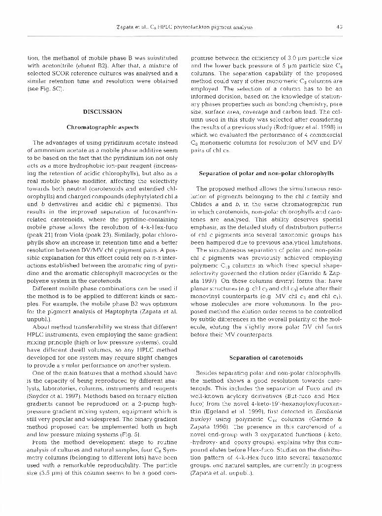
The advantages of using pyridinium acetate instead of ammonium acetate as a mobile phase additive seem to be based on the fact that the pyridinium ion not only acts as a more hydrophobic ion-pair reagent (increas- ing the retention of acidic chlorophylls), but also as a real mobile phase modifier, affecting the selectivity towards both neutral (carotenoids and esterified chl- orophyll~) and charged compounds (dephytylated chl a and b derivatives and acidic chl c pigments). This results in the improved separation of fucoxanthin- related carotenoids, where the pyridine-containing mobile phase allows the resolution of 4-k-Hex-fuco (peak 21) from Viola (peak 23). Similarly, polar chloro- phy l l~ show an increase in retention time and a better resolution between DV/MV chl cpigment pairs. A pos- sible explanation for this effect could rely on n-n inter- actions established between the aromatic ring of pyri- dine and the aromatic chlorophyll macrocycles or the polyene system in the carotenoids.
Different mobile phase combinations can be used if the method is to be applied to different kinds of sam- ples. For example, the mobile phase B2 was optimum for the pigment analysis of Haptophyta (Zapata et al. unpubl.).
About method transferability we stress that different HPLC instruments, even employing the same gradient mixing principle (high or low pressure systems), could have different dwell volumes, so any HPLC method developed for one system may require slight changes to provide a similar performance on another system.
One of the main features that a method should have is the capacity of being reproduced by different ana- lysts, laboratories, columns, instruments and reagents (Snyder et al. 1997). Methods based on ternary elution gradients cannot be reproduced on a 2-pump high- pressure gradient mixing system, equipment which is still very popular and widespread. The binary gradient method proposed can be implemented both in high and low pressure mixing systems (Fig. 5 ) ,
From the method development stage to routine analysis of cultures and natural samples, four Cg Sym- metry columns (belonging to different lots) have been used with a remarkable reproducibility. The particle size (3.5 pm) of this column seems to be a good com-
promise between the efficiency of 3.0 pm particle size and the lower back pressure of 5 pm particle size Ca columns. The separation capability of the proposed method could vary if other monomeric Cg columns are employed, The selection of a column has to be an informed decision, based on the knowledge of station- ary phases properties such as bonding chemistry, pore size, surface area, coverage and carbon load. The col- umn used in this study was selected after considering the results of a previous study (Rodriguez et al. 1998) in which we evaluated the performance of 4 commercial CÃ monomeric columns for resolution of MV and DV pairs of chl cs,
Separation of polar and non-polar chlorophylls
The proposed method allows the simultaneous reso- lution of pigments belonging to the chl c family and Chlides a and b, in the same chromatographic run in which carotenoids, non-polar chlorophylls and caro- tenes are analysed. This ability deserves special emphasis, as the detailed study of distribution patterns of chl c pigments into several taxonomic groups has been hampered due to previous analytical limitations.
The simultaneous separation of polar and non-polar chl c pigments was previously achieved employing polymeric Ci8 columns in which their special shape- selectivity governed the elution order (Garrido & Zap- ata 1997). On these columns divinyl forms that have planar structures (e.g. chl c2 and chl c3) elute after their monovinyl counterparts (e.g MV chl cy and chl c,), whose molecules are more voluminous. In the pro- posed method the elution order seems to be controlled by subtle differences in the overall polarity of the mol- ecule, eluting the slightly more polar DV chl forms before their MV counterparts.
Separation of carotenoids
Besides separating polar and non-polar chlorophylls, the method shows a good resolution towards caro- tenoids. This includes the separation of Fuco and its well-known acyloxy derivatives (But-fuco and Hex- fuco) from the novel 4-keto-19'-hexanoyloxyfucoxan- thin (Egeland et al. 1999), first detected in Emiliania huxleyj using polymeric Cis columns (Garrido & Zapata 1998). The presence in this carotenoid of a novel end-group with 3 oxygenated functions (-keto, -hydroxy- and -epoxy groups), explains why this com- pound elutes before Hex-fuco. Studies on the distribu- tion pattern of 4-k-Hex-fuco into several taxonomic groups, and natural samples, are currently in progress (Zapata et al. unpubl.).
Mar Ecol Prog Ser 195: 29-45,2000
Natural samples
The proposed method improves the resolution of a wide range of pigments present in field samples, achieving a baseline separation (R, > 1.40) of polar chlorophylls (including Chlide a and b and the diverse family of chl c pigments), non-polar chlorophylls (except the critical pair DV chl b/chl b), and excellent resolution of many taxonomically significant caro- tenoids in a reasonable run time of 36 min.
The coelution of DV chl b/chl b does not hamper the identification of Prochlorococcus marinus in seawater samples, since DV chl a is well separated from chl a. In addition, DV chl b is a less specific marker pigment for P. marinussince some cultured isolates are able to syn- thesise chl b as a response to high irradiance (Moore et al. 1995). A recent study has identified surface eco- types of P. marinus with low DV chl b/DV chl a ratio CO-existing, at intermediate water depths, with deep- water ecotypes characterised by high DV chl b/DV chl a ratio (Moore et al. 1998). The coelution of chl b and DV chl b in a single peak could be a drawback if the contribution of algal classes as Chlorophyceae, Euglenophyceae and Prasinophyceae has to be evalu- ated when DV chl a-containing cyanobacteria are pre- sent.
A matrix factorisation program (Chemical taxonomy. CHEMTAX) recently developed by Mackey et al. (1996) is able to resolve such limitations. It exploits the capability of the chemotaxonomic approach to infer the contribution of different algal groups to natural phyto- plankton assemblages. This approach has been suc- cessfully applied to HPLC pigment data obtained from field samples (Wright et al. 1996, Mackey et al. 1998, Pinckney et al. 1998), allowing the quantitative estima- tion of algal class abundance from marker pigments.
The combination of new HPLC methods, able to sep- arate additional marker pigments, and the new gener- ation of mathematical tools for interpreting the HPLC pigment data, will provide invaluable information about the variability of phytoplankton populations from different oceanic regions.
Acknowledgements. Thls work was supported by the project PGIDT-CIMA-99/9 of the Xunta de Gal.ici.a, Spain. We espe- cially than.k Dr Shuley Jeffrey for scientific help, and Lesley Clementson for methodological advice during HPLC pigment analyses of SCOR reference algal cultures performed at CSIRO Marine Laboratories, Hobart, Australia.
LITERATURE CITED
Andersen RA, Saunders GW. Paskind MP, Sexton JP (1993) Ultrastructure and 18s RNA gene sequence for Pel- agomonas calceolata gen. et sp. nov. and the description of a new algal class, the Pelagophyceae classis nov. J Phyc0129:701-715
Barlow RG, Cummings DG, Gibb SW (1.991) Improved resolu- tion of mono- and divinyl chlorophylls a and b and zea- xanthln and lutein in phytoplankton extracts using reverse phase C-8 HPLC. Mar Ecol Prog Ser 161:303-307
Chisholm SW, Olsen RJ, Zettler ER, Goericktl R, Waterbury JB, Welschmeyer NA (1988) A novel free-living prochloro- phyte abundant in the oceanic euphotic zone. Nature 334:340-343
Chisholm SW, Frankel SL, Goericke R, Olson RJ, Palenik B, Waterbury JB, West-Johnsrud L Zettler ER (1992) Pro- chlorococcus marinus nov. gen. nov. sp.: an oxyphoto- trophic marine prokaryote containing divinyl chlorophyll a and b. Arch Microbiol 151,297-300
Egeland. ES (1996) Algal carotenolds and chemosystematics. PhD thesis, Institute for Organlc Chemistry, Trondheim
Egeland ES, Liaaen-Jensen S (1995) Ten minor carotenoids from Prasinophyceae (Chlorophyta). Phytochemistry 40: 515-520
Egeland ES, Eikrem W, Throndsen J. Wilhem C, Zapata M, Liaaen-Jensen S (1995). Carotenoids from further prasino- phytes. Biochem Syst Ecol23:747-755
Egeland ES, Garrido JL, Zapata M, Maestro MA. Liaaen- Jensen S (in press) Algal carotenoids. Part 64. Structure and chemistry of 4-keto-19'-hexanoyloxyfucoxanthin with a novel end group. J Chem Soc, Perkin Trans 1
Fawley MW (1989) A new form of chlorophyll c involved in light-harvesting. Plant Physiol91:727-732
Garrido JL, Zapata M (1993) High performance liquid chro- matographic separation of polar and non-polar chloro- phyll pigments in algae uslng a wide pore polymeric octadecyl silica column J High Resolut Chromatogr 16: 229-233
Garrido JL, Zapata M (1996) Ion-pair reversed phase high- performance liquid chromatography of algal chlorophylls. J Chromatogr 738:285-289
Garrido JL, Zapata M (1997) Reversed-phase high-perfor- mance liquid chromatography of mono- and divinyl chlo- rophyll forms using pyridine-containing mobile phases and polymeric octadecylsilica column. Chromatographia 44:43-49
Garrido JL, Zapata M (1998) Detection of new pigments from Emiliania huxleyi (Prymnesiophyceae) by high perfor- mance liquid chromatography, liquid chromatography- mass spectrometry, vlsible spectroscopy and fast atom bombardment-mass spectrometry. J Phycol34:70-78
Gieskes WWC, Kraay GW (1983) Dominance of Crypto- phyceae during the phytoplankton spring bloom in the central North Sea in 1983. Mar Biol 75:179-185
Goericke R, Repeta DG (1992) The pigments of Prochlorococ- cus marinus: the presence of divinyl chlorophyll a and b in a marine procaryote. Limnol Oceanogr 37:425-433
Goericke R, Repeta DG (1993) Chlorophylls a and b and divinyl chlorophylls a and bin the open subtropical North Atlantic Ocean. Mar Ecol Prog Ser 101:307-313
GuiUard RRL, Ryther JH (1962) Studies of marine plankton diatoms. I. Cyclotella nand Hustedt and Detonula confer- vacea (Cleve) Gran Can J M~crobiol 8:229-239
Jeffrey SW (1997) Preparation of chlorophyll standards. In. Jeffrey SIV, Mantoura RFC, Wright SW (eds) Phytoplank- ton pigments in oceanography: guidelines to modern methods. UNESCO, Paris, p 207-238
Jeffrey SW, Hallegraeff GM (1987) Chlorophyllase distribu- tion in 10 algal classes of phytoplankton-a problem for chlorophyll analysis. Mar Ecol Prog Ser 35:293-304
Jeffrey SW, LeRoi JM (1997) Simple procedures for growing SCOR reference microalgal cultures In: Jeffrey SW, Man- toura RFC, Wright SW (eds) Phytoplankton pigments in
Zapata et al.: CH HPLC phytoplankton pigment analysis 45
oceanography: guidelines to modern methods. UNESCO, Paris, p 181-205
Jeffrey SW. Mantoura RFC (1997) Pigment abbrev~ations used by SCOR WG 78. In. Jeffrey SW, Mantoura RFC, Wright SW (eds) Phytoplankton pigments in oceanogra- phy: guidelines to modern methods. UNESCO. Paris, p 564-565
Jeffrey SW. Mantoura RFC, Bjornland T (1997a) Data for the identification of 47 key phytoplankton pigments. In: Jef- frey SW, Mantoura RFC, Wnght SW (eds) Phytoplankton pigments in oceanography. guidelines to modern meth- ods. UNESCO, Paris, p 449-559
Jeffrey SW. Mantoura RFC, \tVnght SW (eds) (1997b) Phyto- plankton pigments in oceanography: guidelines to mod- ern methods. UNESCO. Pans
Kraay GW. Zapata M, Veldhuis MJMJ (1992) Separation of chlorophylls c,, c2 and c, of marine phytoplankton by reversed-phase CI8 high-performance liquid chromatog- raphy. J Phyco128:708-712
Mackey DJ, Higgins HW, Mackey MD, Holdsworth D (1998) Algal class abundances in the western equatorial Pac~fic: estimation from HPLC measurements of chloroplast pig- ments using CHEMTAX. Deep-Sea Res 451441-1468
Mackey MD, blackey DJ, Higgins HW. Wright SW (1996) CHEMTAX - a program for estimating class abundances from chemical markers, application to HPLC measure- ments of phytoplankton Mar Ecol Prog Ser 144.265-283
Mantoura RFC, Llewellyn CA (1983) The rapid determinat~on of algal chlorophyll and carotenoid pigments and their breakdown products in natural waters by reverse-phase high-performance liquid chromatography. Anal Chim Acta 151x297-314
Moore LR, Goericke R, Chisholm SW (1995) Comparative physiology of Synechococcus and Prochlorococcus: influ- ence of light and temperature on growth, pigments, fluo- rescence and absorptive properties. Mar Ecol Prog Ser 116:259-275
Moore LR, Rocap G, Chisholm SW (1998) Physiology and mol- ecular phylogeny of coexisting Prochlorococcus ecotypes. Nature 393:464-467
Nelson JR. Wakeham SG (1989) A phytol-substituted chloro- phyll c from Emiliania huxleyi (Prymnesiophyceae). J Phy- c01 25:761-766
Pinckney JL, Paerl HW, Harnngton MB, Howe KE (1998) Annual cycles of phytoplankton community-structure and bloom dynamics in the Neuse River Estuary, North Car- olina. Mar Biol 131:371-381
Repeta DJ. Bsrnland T (1997) Preparation of carotenoid stan- dards. In: Jeffrey SW, Mantoura RFC, Wright SW (eds) Phytoplankton pigments in oceanography: guidelines to modern methods. UNESCO. Paris, p 239-260
Rodriguez F, Zapata M, Garrido JL (1998) High performance liquid chromatographic separation of chlorophyll c forms from marine phytoplankton using octylsilica bonded phases. Chromatographia 48.677-680
Snyder RL, Kirkland JJ , Glalch JL 11997) Completing the
Editorial responsibility: Otto Kinne (Editor), Oldendorf/Luhe, Germany
method: validation and transfer. In: Snyder RL, Kirkland JJ, Glajch JL (eds) Practical HPLC method development. J Wiley & Sons, New York, p 685-713
UNESCO (1994) Protocols for the Joint Global Ocean Flux Study (JGOFS) core measurements. IOC. Manuals and Guides, UNESCO, Paris
Van Heukelem L, Lewitus AJ, Kana TM, Craft NE (1994) Improved separation of phytoplankton pigments using temperature-controlled high performance liquid chro- matography. Mar Ecol Prog Ser 114:303-314
Van Lenning K , Garrido JL, Aristegui J , Zapata M (1995) Temperature-programmed high performance l~quid chro- matographic separation of mono and divinyl-chlorophyll forms from marine phytoplankton. Chromatographia 41. 539-543
Vidussi F, Claustre H, Bustillos-Guzman J , Cailliau C, Marty J C (1996) Determination of chlorophylls and carotenoids of marine phytoplankton: separation of chlorophyll a from divinyl chlorophyll a and zeaxanthin from lutein. J Plank- ton Res 18:2377-2382
Wright SW, Jeffrey SW (1997) High-resolution HPLC system for chlorophylls and carotenoids of marine phytoplankton. In: Jeffrey SW, Mantoura RFC, Wright SW (eds) Phyto- plankton pigments in oceanography: guidelines to mod- ern methods. UNESCO. Paris. p 327-360
Wright SW, Shearer JD (1984) Rapid extraction and high-per- formance liquid chromatography of chlorophylls and carotenoids from marine phytoplankton. J Chromatogr 294.281-295
Wright SW, Jeffrey SW, Mantoura RFC, Llewellyn CA, Bjsrn- land T, Repeta D, Welschmeyer N (1991) Improved HPLC method for the analysis of chlorophylls and carotenoids from marine phytoplankton. Mar Ecol Prog Ser 77: 183-196
Wright SW, Thomas, DP, Marchant HJ, Higgins, HW, Mackey MD, Mackey DJ (1996) Analysis of phytoplankton of the Australian sector of the Southern Ocean: comparisons of microscopy and size frequency data with interpretations of pigment HPLC data using the 'CHEMTAX' matnx fac- torisation program. Mar Ecol Prog Ser 144:285-298.
Zapata M. Garrido JL (1991) Influence of injection conditions in reversed-phase high-performance liquid chromatogra- phy of chlorophylls and carotenoids. Chromatographia 31:589-594
Zapata M, Ayala AM, Franco JM, Garrido JL (1987) Separ- ation of chlorophylls and their degradation products in marine phytoplankton by reversed-phase high-perfor- mance liquid chromatography. Chromatographia 23: 26-30
Zapata M, Freire J , Garrido JL (1998) Pigment composition of several harmful algae as determined by HPLC using pyri- dine-containmy mobile phases and a polymeric octadecyl- silica column. In: Reguera B, Blanco J , Fernandez ML, Wyatt T (eds) Harmful algae. Xunta de Galicia and Inter- governmental Oceanograph~c Commission of UNESCO, Paris, p 304-307
Submitted: January 21, 1999; Accepted: October 13, 1999 Proofs received from author(s): March 14, 2000