Embed Size (px)
Citation preview
DOI: 10.1007/s11099-018-0792-x PHOTOSYNTHETICA 56 (1): 11-43, 2018
11
REVIEW
Living off the Sun: chlorophylls, bacteriochlorophylls and rhodopsins A.W.D. LARKUM*, R.J. RITCHIE**, and J.A. RAVEN***,* Global Climate Cluster, Building 4, University of Technology Sydney, Broadway, NSW 2007, Australia* Tropical Environmental Plant Biology Unit, Faculty of Technology and Environment, Prince of Songkla University Phuket, Kathu 83120, Phuket, Thailand** University of Dundee at the James Hutton Institute, Invergowrie, Dundee DD2 5DA, UK***(permanent address) Abstract Pigments absorbing 350–1,050 nm radiation have had an important role on the Earth for at least 3.5 billion years. The ion pumping rhodopsins absorb blue and green photons using retinal and pump ions across cell membranes. Bacteriochlorophylls (BChl), absorbing in the violet/blue and near infra red (NIR), power anoxygenic photosynthesis, with one photoreaction centre; and chlorophylls (Chl), absorbing in the violet/blue and red (occasionally NIR) power oxygenic photosynthesis, with two photoreaction centres. The accessory (bacterio)chlorophylls add to the spectral range (bandwidth) of photon absorption, e.g., in algae living at depth in clear oceanic water and in algae and photosynthetic (PS) bacteria in microbial mats. Organism size, via the package effect, determines the photon absorption benefit of the costs of synthesis of the pigment–protein complexes. There are unresolved issues as to the evolution of Chls vs. BChls and the role of violet/blue and NIR radiation in PS bacteria. Introduction The Sun’s energy has been potentially available to power energy transduction on the Earth since its formation, although early physical conditions would have made this unlikely. The Earth orbits a G-type star (Figs. 3–7) with biologically significant characteristics of the spectrum <400 nm and various H2O and CO2 absorption bands >700 nm. Even allowing for the faint young sun (see “Implications of a Faint Sun”) the Sun’s spectrum would not have changed enough to influence the development of pigments involved in energy transduction. Likewise one can be fairly confident in predicting what systems would develop on extra-terrestrial planets (Wolstencroft and Raven 2002, Kiang et al. 2007a,b; Ritchie et al. 2017).
Today on the Earth energy-transducing pigment–protein complexes are widespread. Ion-pumping rhodo-psins (bacteriorhodopsin, halorhodopsin, proteorhodopsin, and xanthorhodopsin) occur in Archaea, Bacteria, and Eukarya. BChls and Chls occur in Bacteria, while Chls
occur in all photosynthetic Eukarya. Photochemistry by (bacterio)chlorophylls yields an oxidant and a reductant with a large redox potential difference between them; the energy-transducing rhodopsins do not generate reduced and oxidised products but do generate an electrochemical potential for protons or other ions.
Interestingly, and not fully resolved at the present time, is the divergence between BChl in the photosynthetic (PS) anoxygenic bacteria and Chl in the oxygenic cyano-bacteria, eukaryotic algae, and embryophytes. If one takes the view that the PS bacteria employ earlier forms of photosynthesis, albeit anoxygenic, i.e. not extracting electrons from water (and not forming oxygen as a by-product) and that cyanobacteria and eukaryotic PS organisms are derived from these, then an explanation is at hand (Mauzerall 1973, Bjorn 1976, 2009; Hohmann-Marriott et al. 2011). However, this leaves the puzzle
———
Received 8 September 2017, accepted 20 December 2017, published as online-first 8 February 2018. +Corresponding author; e-mail: [email protected] Abbreviations: AAPB – aerobic anoxygenic aerobic photosynthesis; BChl – bacteriochlorophyll; Chl – chlorophyll; ETR – electron transport rate; GOE – Great Oxidation Event; HPLC – high performance liquid chromatography; LED – light-emitting diode; LGT – lateral gene transfer; MgDVP – magnesium-2,4-divinyl phaeoporphyrin monomethyl ester A5; PBP – phycobiliprotein; RC – reaction center; TLC – thin layer chromatography; UV – ultraviolet. Acknowledgements: The University of Dundee is a registered Scottish charity, No. 015096. RJR wishes to thank Prince of Songkla University - Phuket campus for providing the facilities for the project. AWDL thanks the Global Climate Cluster, University of Technology Sydney for ongoing support.
A.W.D. LARKUM et al.
12
that BChl in contemporary organisms is formed on a biosynthetic pathway that first forms Chl and its molecular relatives (Fig. 1) (Granick 1965, Larkum 2006, 2008). An alternative view, much less popular, is that PS evolved in primitive procyanoabacterial organisms (Mulkidjanian et al. 2006, Cardona 2016, Cardona et al. in review) which subsequently gave rise on the one hand to the cyano-bacteria with photosystem I and II (Govindjee et al. 2017), and on the other to anoxygenic PS bacteria, with either PSI-like or PSII-like reaction centres. According to this view, the first anoxygenic PS organisms produced Chl rather than BChl. Absorptance curves, however, show that the Soret (QX – see Reimers et al. 2013) window, available for blue light-absorbing BChl and Chl pigments, absorb about as many photons per absorption band as the QY band windows for both Chl a and BChl a/b (Ritchie et al. 2017, and this paper, see Figs. 3–7). Perhaps this common use of similar blue bands subsequently drove more cryptic anoxygenic PS bacteria to adopt BChl which can harvest visible light but just as importantly can harvest far-red radiation, depending on the organism, up to 1050 nm (Larkum 1991, 2003, 2006, 2008; Kiang et al. 2007a,b; Stomp 2007, Ritchie et al. 2017). However, the use in photochemistry of these lower-energy far-red photons means that the energy stored in photochemistry is limited and less than half of the excitation energy of absorbed photons at 450 nm is usable in photochemistry at 950 nm, as compared to two-thirds for photochemistry at 680 nm (using Chl a). Unfortunately, we are dealing here with a time more than 3.5 billion years ago (BYA) when few identifiable fossils exist (but see Rosing et al. 2004, Nutman et al. 2016) and molecular phylogenetic analysis becomes unreliable because so many of the various photosynthetic lines seem to be about the same very ancient age (Harel et al. 2015).
Much has been written on the reasons why BChls are used in photoredox reactions of anoxygenic photosyn-thesis, and Chl a (very occasionally Chl d) in the photo-redox reactions of oxygenic photosynthesis (Mauzerall 1973, Nishio 2000, Wolstencroft and Raven 2002, Kiang et al. 2007a,b; Stomp et al. 2007, Björn et al. 2009, Milo 2009, Raven 2009, Marosvölgyi and van Gorkum 2010, van Hohman-Mariott and Blankenship 2011, Raven and Ralph 2015, Kume et al. 2016, Kume 2017). Provisional conclusions are that the main consideration is energy per photon relative to the energy stored in the products of photochemistry, with subsidiary effects of spectral attenuation by the medium.
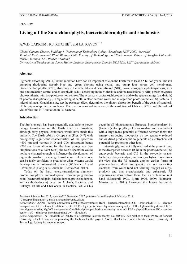
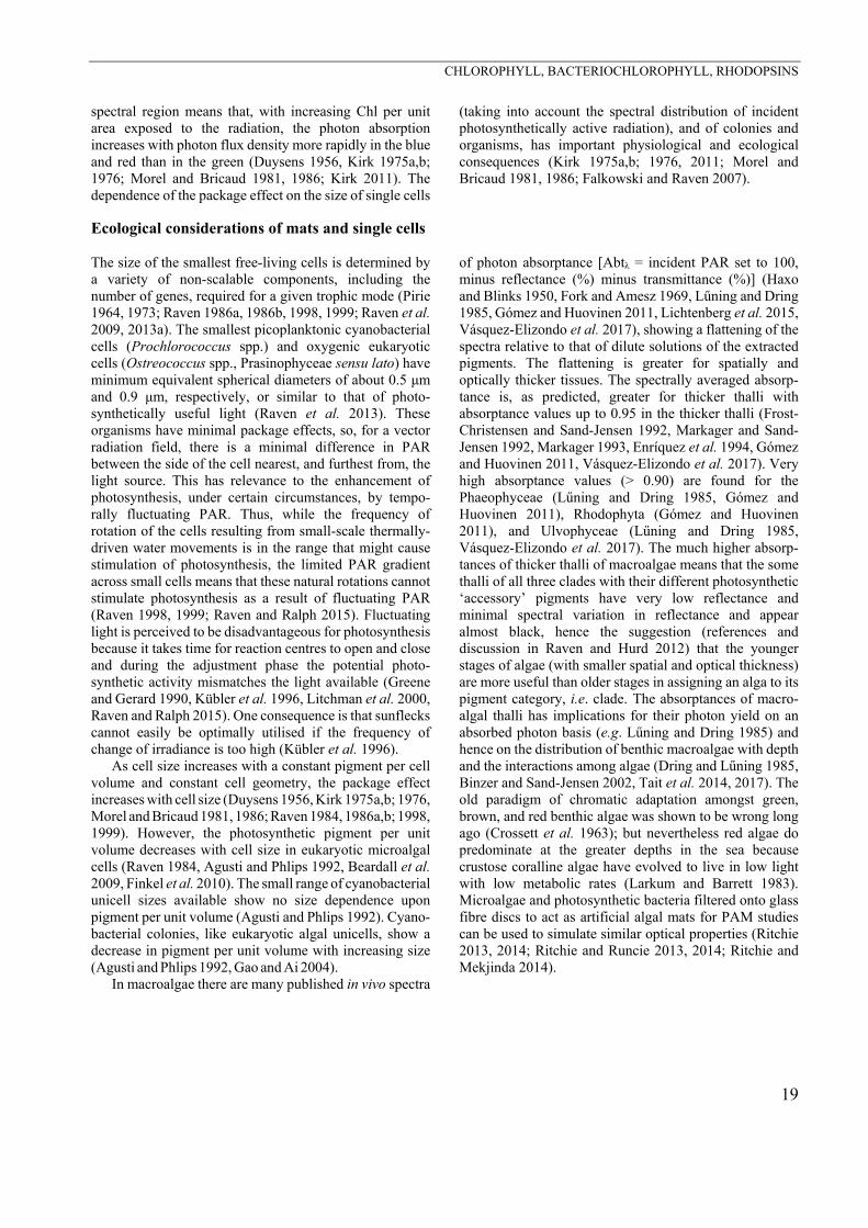
There are four other Chls besides Chl a (Fig. 1). These are Chls b, c (various subtypes), d, and f. The absence of a Chl e is because reports of its occurrence in Tribonema bombycium and Vaucheria hamata (Allen 1996, see Meeks 1974) proved unfounded; the original reports of Chl e did not provide a chemical structure and no further investigations have been published. Chl b and c [(c1, c2, and c3 and magnesium 2.4-divinyl phaeoporphyrin mono-methyl ester A5 (MgDVP)] have been labelled accessory Chls because they act purely in a light-harvesting role, are not present in reaction centres, and do not carry out (productive) photochemistry; and the same may be true for Chl f. Chl d and Chl f, absorb near-infra-red (NIR) radiation using their far-red QY bands. Both occur in cyanobacteria and not in any known eukaryotic algae (for Chl d, Larkum and Kühl 2005, for Chl f, Chen et al. 2010). These Chls allow those cyanobacteria which possess them, to live in environments enriched in NIR, e.g. Acaryo-chloris (Miyashita et al. 2006) and Halomicronema (Chen et al. 2012), and also have a contribution, with carotenoids, and have the additional, rarely noted, effect of widening the blue absorption band (Fig. 4–5). Whether other Chls,
Fig. 1. Structure of chlorophylls a, b, d, and f. The R groups (1 to 4) of various chlorophylls are shown in the Table as well as their QY peaks in acetone solvent. Chls a, b, d, and f all have 10 double bonds in the tetrapyrrole ring (20 π electrons), i.e. they are chlorins. Chlorophylls c1 and c2 do not have a phytol tail and ring D is oxidised to yield 22 π electrons in what is a porphyrin ring. Chlorophyll c3 is phytylated (Zapata and Garrido 1997) but also has 22 π electrons. BChl a, b, and g are bacteriochlorins with reduced B and D pyrrole rings (18 π e-) and BChls c, d, e, and f are chlorins with a reduced ring D (20 π e-) (refer to Scheer 2006). Phy – phytol tail.
CHLOROPHYLL, BACTERIOCHLOROPHYLL, RHODOPSINS
13
potentially with NIR absorption, will be found it has been discussed (Chen and Blankenship 2011, Schliep et al. 2013) but none so far have been discovered or bio-engineered. Interestingly, there are special Chl a–protein complexes that allow absorption of radiation up to 760 nm, such as the special form in Ostreobium spp. (see below); the reason is the conjugation of the Chl a to a special protein (Trissl 2003, see also Chen et al. 2005). In all these far-red and NIR-absorbing systems it is necessary for the absorption of long-wavelength photons to pass on their energy to pigments whose absorption maxima are at shorter wavelengths. In the case of Chls a it is migration of energy from ~760 nm to the absorption maximum of P680, the major reaction centre pigment of PSII. This is called “uphill” energy transfer. It is quite feasible to explain by quantum physics; for Ostreobium (Trissl 2003, Wilhelm and Jacob 2006), and for Acaryochloris see Nieuwenburg et al. (2003), but it comes with an energy cost. Also in the case of Acaryochloris Chl d is used in both reaction centres, and the PSII RC has a P705 so the uphill energy migration is not so steep. In the case of Chl f organisms, the nature of the Chl in reaction centres (RCs) is not known but provisionally it is assumed that it is Chl a in P680 and P700 (Chen et al. 2012, Chen 2014).
What the existence of Chl and BChl tells us in terms of the evolution of photosynthesis is, as mentioned above, a matter of debate (see for example Chen 2014). Just as interesting is what the existence of Chl d and Chl f tell us about the evolution of the Chls. These Chls are formed from Chl a or chlorophyllide a by modification of one of the hydroxyls on the porphyrin ring A to a formyl group (Fig. 1). At the most simple level of interpretation, this says that Chl a was the first pigment in the cyanobacteria. But in reality this does not necessarily mean that Chl a preceded Chl d or Chl f in the early cyanobacterial or procyanobacterial organisms. In the case of Chl f it is now known that the final enzymatic step is the addition of an oxygen to a methyl group on the first porphyrin ring (A)
and is brought about by a protein with affinities to a very early form of D1 protein, which does not have the Mn4Ca1O5 centre (Ho et al. 2016). This is not incompatible with the hypothesis that at this early stage Chl f was the major chlorophyll of procyanobacteria. However, a corollary would be that Chl f once functioned as the redox Chl in the PSII RC, as does Chl d in Acaryochloris.
It would be a hope that the phylogeny of cyanobacteria would tell us which group gave rise to the plastids of eukaryotic algae and whether or not there was a mono-phyletic plastid origin. A polyphyletic origin could easily give rise to obscure results. The results so far are not conclusive (e.g. Harel et al. 2015) but molecular phylo-genies together with fossil evidence are improving the time estimates and yield indications of the nearest living ancestor (Cardona 2016, Shih et al. 2017, Sánchez-Baracaldo et al. 2017, and references therein). What is clear is that there are three types of primary plastid in eukaryotic algae, i.e. plastids having a single set of outer (envelope) membranes; these occur in the three phyla: Chlorophyta and allies [with Chl (a+b)], Rhodophyta (with Chl a and PBP including rhodophytan phyco-erythrin), and Glaucocystophyta (with cyanelles posses-sing Chl a and either cyanobacterial PC or PE) (Larkum 2003, 2006, 2008; Larkum et al. 2007, Price et al. 2012; algal taxonomy here and subsequently follows Graham et al. 2016 and http://www.algaebase.org). It is usually assumed that these primary plastids arose by a single endosymbiosis. All other algae have plastids that show indications of two or more serial endosymbioses (Larkum 2006).
The discussion that follows addresses the relationship between the energy content of a photon at the wavelength of photochemistry, migration of the absorbed photonic energy to the site of photochemistry and the energy stored in the photoproduct, and the incident photon spectrum, both where the organisms occur today, and where and when they evolved.
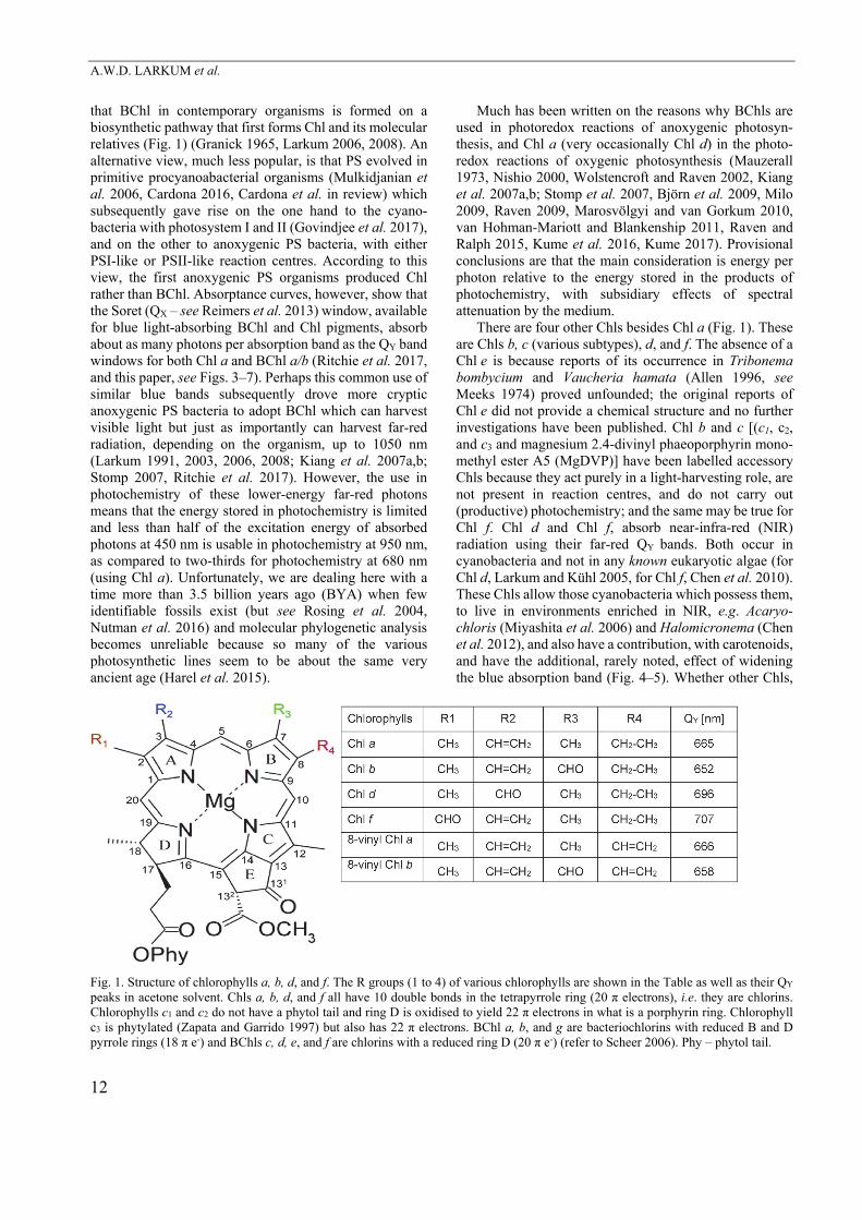
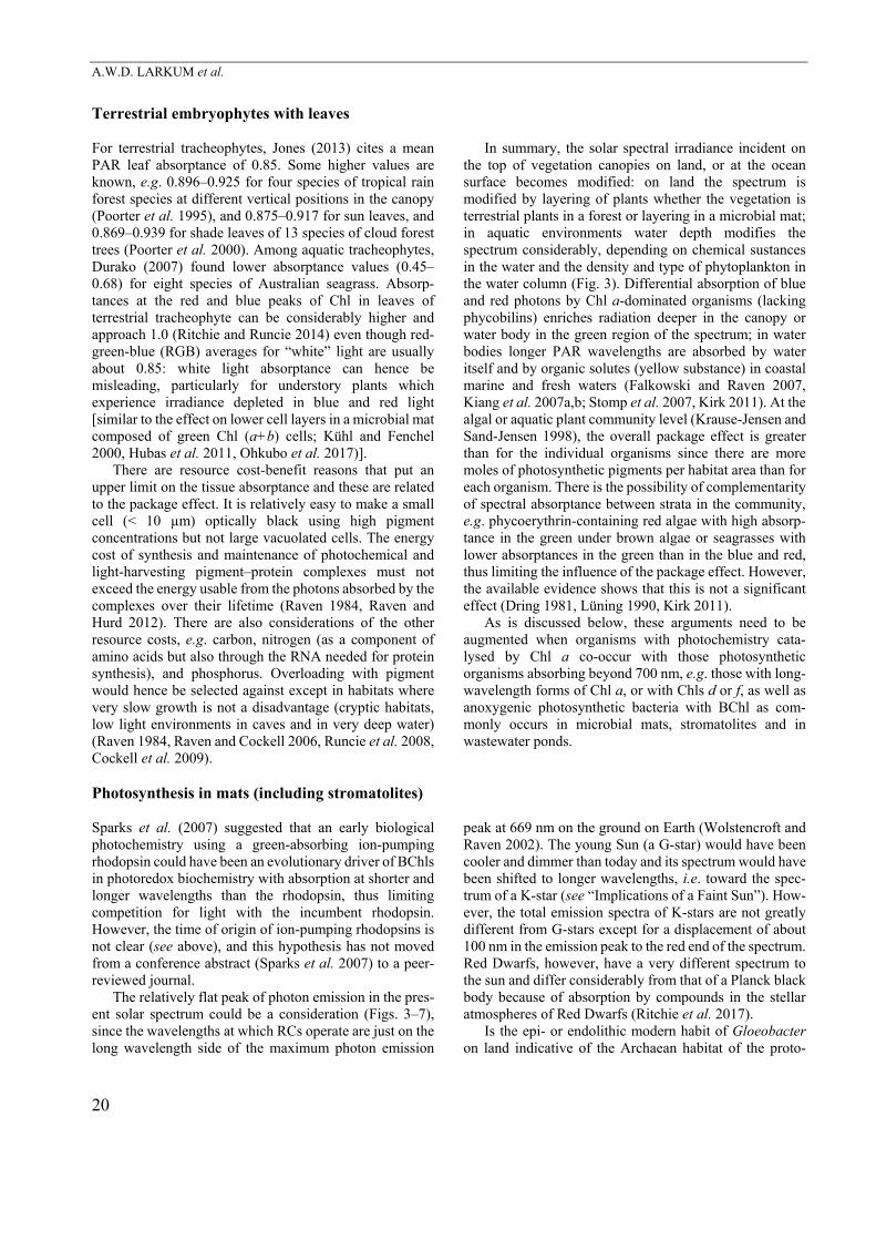
Relation of the energy per photon used in photochemistry to the energy stored in a stable form Ion-pumping rhodopsins The ion-pumping rhodopsins have a retinal chromophore (Fig. 2) and an apoprotein that uses the photon energy absorbed by the retinal pigment to pump the ions. H+ is the most commonly transported ion (although Na+ and Cl– can also be pumped, see below), and the H+-pumping bacteriorhodopsin from the Archaean Halobacterium halobium was the first ion-pumping rhodopsin to be identified (Osterhelt and Stoeckenius 1971, Kandori 2015). Most rhodopsins are barrel-shaped membrane spanning porins with the retinal head wrapped in its hydrocarbon tail in a pocket on the outer part of the protein (Baumann et al. 2014). The direction of H+ transport is almost always from the cytosol to the external medium in Archaea and Bacteria, including the oxygenic photo-synthetic cyanobacterium Gloeobacter violaceous that
lacks thylakoids (Rexroth et al. 2011). In Gloeobacter, H+
efflux across the plasma membrane can be driven by the respiratory electron transport chain in the dark, with the addition of both photosynthetic and rhodopsin-based H+ efflux in the light (Rexroth et al. 2011, Choi et al. 2014).
Fig. 2. The structure of retinal showing its head group and phytol-like tail. Most rhodopsins are barrel-shaped membrane spanning porins with the retinal head wrapped in its hydrocarbon tail in a pocket on the outer part of the protein (Baumann et al. 2014).
A.W.D. LARKUM et al.
14
The roles of the two mechanisms could be tested in cells by illumination in the presence of DCMU, when linear electron flow between PSII and PSI is prevented. The remaining light-dependent H+ efflux would be driven by the H+-pumping rhodopsin or by cyclic electron flow around PSI: theoretically distinguishable by their action spectra; however, thus far, experimentally measured action spectra have not fully distinguished between these two means of H+ energization (Choi et al. 2014).
The aerobic anoxygenic photosynthetic bacterium, Fulvimarina pelagi, contains bacteriorhodopsin- and xanthodopsin-based H+ pumps (Kang et al. 2010) (Table 1). The archaean Halobacterium halobium with halo-rhodopsin (Matsuno-Yagi and Mukohata 1977, Schobert and Lanyi 1982) normally pumps Cl– from the medium to the cytosol powered by green light, but halorhodopsin can also catalyse the downhill entry of H+ using blue light (Hegemann et al. 1985, Oesterhelt et al. 1985, Bamberg et al. 1993). The flavobacterium Nonlabens marinus has a proteorhodopsin Cl– pump, proteorhodopsin NM3, which evolved independently of the archaean Cl– pump (Yoshizawa et al. 2014, Tsukamoto et al. 2017). Gaudana et al. (2014) suggested that cyanobacterial proteo-rhodopsin could be genetically engineered to catalyse active transport of HCO3
- into cells, but this has not yet been achieved.
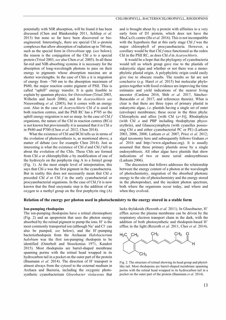
Table 1. Ion-pumping rhodopsins: phylogenetic distribution, nature of the ion pumped, wavelength of maximum absorption, and occurrence in organisms with chlorophyll-based photo-chemistry. Sources: Fuhrman et al. (2008), Raven (2009), Slamowitz et al. (2011), Inoue et al. (2013), Kirchman and Hanson (2013), Yoshikawa et al. (2014), Bogachev et al. (2016), Kato et al. (2016), Tsukamoto et al. (2017). 1H+-pumping proteorhodopsins occur in some oxygenic photosynthetic cyano-bacteria (Gaudana et al. 2014); the Gloeobacter H+-pumping rhodopsin is relatively distantly related to other proteorhodopsins and has been termed GR to distinguish it from PR (the core proteorhodopsins): Ganapathy et al. (2017). An H+-pumping xanthorhodopsin is also found in an aerobic anoxygenic photo-heterotrophic bacterium (Kang et al. 2010). 2H+-pumping proteo-rhodopsins occur in the oxygenic photosynthetic ulvophycean marine green alga Acetabularia (Raven 2009, Tsunuda et al. 2006), in some marine oxygenic photosynthetic diatoms
(Slamowitz et al. 2011, Marchetti et al. 2012), and in some marine
oxygenic photosynthetic dinoflagellates (Slamowitz et al. 2011, Marchetti et al. 2012) as well as some non-photosynthetic dinoflagellates (Slamowitz et al. 2011, Marchetti et al. 2012, Jahnke et al. 2013, Guo et al. 2014).
Category of ion-pumping rhodopsin
Wavelength of maximum absorption
Archaea Bacteria Eukarya
Bacteriorhodopsin 568 nm + (H+) - - Halorhodopsin 578 nm + (Cl-) - - Proteorhodopsin 490 nm or
525 nm + (H+) + (H+)1
+(Na+) + (Cl-)
+ (H+)2
Xanthorhodopsin 560 nm - + (H+)1 -
More recently, rhodopsins that pump Na+ out of bacterial cells have been found to pump H+ in the same direction in the absence of Na+ (Inoue et al. 2013, Bogachev et al. 2016, Kato et al. 2016, Suomovuori et al. 2017). Nonlabens marinus also has a proteorhodopsin H+ efflux pump (NM1) and a proteorhodopsin Na+ efflux pump (NM2), as well as the Cl– pump NM3 mentioned above (Yoshizawa et al. 2014), as well as the Cl–. There are two possible exceptions in eukaryotes to the paradigm that ion-pumping rhodopsins catalyse H+ or Na+ efflux, or Cl– influx, at the plasmalemma. One is the work of Tsunuda et al. (2006), who examined the proteorhodopsin of the marine ulvophycean Acetabularia acetabulum (formerly A. mediterreana). When the proteorhodopsin was heterologously expressed (in Xenopus oocytes) it pumped H+ from the cytosol to the medium, although this proteorhodopsin expressed in Acetabularia acetabulum catalyses an inwardly-directed, downhill, current carried by H+. The other example is proteorhodopsin of the phagotrophic, non-photosynthetic dinoflagellate Oxyrrhis marina (Jahnke et al. 2013), which is apparently expressed in the endomembrane system where it operates in the topologically identical manner to efflux across the plasmalemma, i.e. from a compartment of high protein diversity to a compartment of low protein diversity. This would acidify the endomembrane lumen and possibly aid digestion in food vacuoles if proteases with acid pH optima exist (Slamovitz et al. 2011). Such food vacuole acidification could help to explain the increased survival of starving Oxyrrhis marina in the light relative to that in the dark (Guo et al. 2014) (Table 1).
Longer light survival than dark survival of cells expressing ion-pumping rhodopsins has been more widely documented for Archaea and Bacteria (Fuhrman et al. 2008, Kirchman and Hanson 2013), as is the case for aerobic anoxygenic photosynthetic bacteria (AAPB) with BChl-based photochemistry (Kirchman and Hanson 2013). More direct estimates of the role of light than simple survival tests are needed: for example, nobody appears to have attempted to measure photosynthetic electron transport rates (pETR) in cultured AAPBs using PAM fluorometry, which is known to work in a variety of RC-2 type photosynthetic bacteria (Ritchie 2013, Ritchie and Runcie 2013, Ritchie and Mekjinda 2015). This might be technically difficult because AAPB bacteria have very low amounts of BChls per cell. Nevertheless, it seems feasible because it has been shown that there is a correlation of 800-nm fluorescence at the ocean surface with the BChl content (Kolber et al. 2000, 2001). In the cases of both the ion-pumping rhodopsin-based and BChl-based systems there are rather fewer cases of light-stimulation of growth (Kirchman and Hanson 2013, Pinhassi et al. 2016). The analysis of Kirchman and Hanson (2013) shows that the net energy gain (energy harvested, corrected for the cost of production of the light energy transduction apparatus) is greater for BChl-based than rhodopsin-based systems. This difference between
CHLOROPHYLL, BACTERIOCHLOROPHYLL, RHODOPSINS
15
the BChl-based and rhodopsin-based energy transduction systems is reflected in both contributions to maintenance costs, and to growth rates (Gómez-Consarnau et al. 2007, 2010; Fuhrman et al. 2008, Kirchman and Hanson 2013, Courties et al. 2015). Ferrera et al. (2017) showed, for the first time, that light enhances growth of aerobic anoxy-genic phototrophic (AAPB) in natural populations. An important point is that organisms with rhodopsin-based energy transduction lack the machinery specific to auto-trophic CO2 assimilation (Palovaara et al. 2014), unless the organism also has oxygenic photosynthesis. Aerobic an-oxygenic photosynthetic bacteria also lack the machinery specific to autotrophic CO2 assimilation (Fuchs et al. 2007, Li et al. 2013, Zeng et al. 2013, Mathusamy et al. 2014, Zheng et al. 2015) and so live photoheterotrophically.
The absorption maxima are 490 nm for the blue-absorbing proteorhodopsins, 525 nm for the green-absorbing proteorhodopsins, 560 nm for xanthorhodopsin, 568 nm for bacteriorhodopsin, and 578 nm for halo-rhodopsin (Fuhrman et al. 2008). The energy of 490-nm photons is 244 kJ mol–1(photon), and for 578-nm photons it is 207 kJ mol–1(photon). The largest H+ electrochemical potential difference generated across a membrane by a H+-pumping rhodopsin is 27 kJ mol–1 in the archaean Halobacterium halobium in the presence of N,N’-dicyclohexylcarbodiimide (DCCD), which limits the re-entry of the H+ pumped out of the cells (Michel and Oesterhelt 1980). In comparing the energy per photon with the maximum energy stored in the ion gradient it is important to consider the photon efficiency of the light- driven pumps. For BChl the quantum efficiency of H+
transport was determined by Govindjee et al. (1990) as 0.64 ± 0.04, with previously published values in the range of 0.3–0.79 (Hagemann et al. 1985, Oesterhelt et al. 1985).
As indicated above, organisms with ion-pumping rhodopsins lack photoautotrophic CO2-assimilating machinery unless the rhodopsins occur in organisms with Chl-based photosynthetic autotrophy (e.g. oxygenic photosynthesis as in Gloeobacter violaceous). However, it is worth exploring the “what if” question of the energy, as photons, cost of autotrophic CO2 fixation with ion-pumping rhodopsins as the light energy transducers. For comparison with a (bacterio)chlorophyll-based photosyn-thetic system, the purple proteobacterial anoxygenic photosynthetic system is used because here light energy transduction is also solely through H+-pumping in cyclic electron flow, around its PSII-like photosystem (Klamt et al. 2008, Feniouk and Junge 2009, Nicholls and Ferguson 2013). In both cases an electron donor capable of reducing ubiquinone is used, as is autotrophic CO2 fixation using the Benson-Calvin cycle in the absence of O2 with a ratio of 3 ATP:2 NADH:1 CO2. The absence of O2 is essential for the synthesis of BChl in anoxygenic photosynthetic bacteria and is consistent with the presence of S2– or other reductants of similar or lower redox potential as an electron donor able to reduce ubiquinone. Many photo-synthetic bacteria will grow quite well in an unstirred
conical flask open to the atmosphere because such a culture has redox gradients near the surface of the culture: thus lower levels of the culture are suitable for BChl synthesis and most photosynthetic bacteria are highly motile. If such cultures are aerated over several hours they turn white. Photosynthetic electron transport can be demonstrated in anaerobic anoxygenic photosynthetic bacteria filtered onto filter disks under atmospheric oxygen concentrations (Ritchie 2013, Ritchie and Runcie 2013, Ritchie and Mekjinda 2015) even though they require anoxic conditions to actually make BChl. Streaks on aerobically incubated agar plates typically appear white on the edges with a pink interior. This explains why anoxygenic photosynthetic bacterial blooms often occur on the surfaces of mudflats, on lake surfaces and parti-cularly in sewage and wastewater ponds (Ritchie et al. 2017). The metropolitan sewage pond in Phuket in Thailand has a floating photosynthetic bacterium: just about the least likely organism one would expect to encounter in today’s modern atmosphere (Ritchie, unpublished). Aerobic anoxygenic photosynthetic bacteria are obligately aerobic for growth (Yurkov and Beatty 1998). They only synthesise BChl in the dark (e.g. the dark phase of the light–dark cycle). While BChl is obviously only used in photochemistry in the light (e.g. the light phase of the light–dark cycle), BChl is also lost in the light as a result of dilution in growth, rather than breakdown in the absence of synthesis (Yurkov and Beatty 1998).
For the proteobacterial anoxygenic PS bacteria it is assumed that one photon absorbed by the photosynthetic pigments moves one electron from reduced cytochrome c to ubiquinone. Cyclic electron flow returns the electron from ubiquinol to oxidised cytochrome c and pumps 2 H+
from the cytosol to the thylakoid lumen or the external medium. Each electron moved from ubiquinol (produced by reduction of ubiquinone by S2–) to NAD+ by a NADH-ubiquinone oxidoreductase involves 2 H+ moving from the thylakoid lumen or the external medium to the cytosol (Klamt et al. 2008, Nichols and Ferguson 2013). The reduction of 2 NAD+ to 2 NADH therefore needs 4 photons. With an H+:ATP ratio of 3.3 for the FoF1 ATP synthase (Klamt et al. 2008, Feniouk and Junge 2009, Nicholls and Ferguson 2013) the production of 3 ATP needs 10 H+ moving from the thylakoid lumen or the external medium to the cytosol, involving 5 photons. The photon costs of the production of the 2 NADH and 3 ATP used to reduce 1 CO2 to the redox level of carbohydrate means that the autotrophic assimilation of 1 CO2 requires 9 absorbed photons. This is a lower limit of the photon cost, since the photon cost of moving an electron from reduced cytochrome c to ubiquinone in purple photo-synthetic proteobacteria using their PSII-like photosystem is 0.3–0.6 electrons moved per mol(photon absorbed) as estimated from fluorescence studies (Kolber et al. 2000). This is consistent with maximum photosynthetic effi-ciencies found in photosynthetic bacteria by PAM fluorometry (Ritchie 2013, Ritchie and Runcie 2013,
A.W.D. LARKUM et al.
16
Ritchie and Mekjinda 2015). However, a direct estimate of the photon cost of photochemistry of Rhodopseudomonas spheroides gives a value not statistically distinguishable from 1.0 (see above), contrasting with the directly comparable fluorescence-derived value of 0.7 (Wraight and Clayton 1974).
Turning to the hypothetical case of autotrophic CO2
assimilation with energization using a H+-pumping rhodo-psin, we assume one H+ pumped per photon absorbed (a low photon cost relative to the measured values discussed above) and make all the assumptions on the use of the H+
gradient in generating 2 NADH and 3 ATP as were made above for the anoxygenic BChl-based system in proteo-bacteria. The outcome is that the conversion of 1 CO2 to the redox level of carbohydrate means that the autotrophic assimilation of 1 CO2 would require 18 absorbed photons. In such a system NADH would have to be made by processes unrelated to the photochemical reactions. In the case of RC-2 photosynthetic organisms, the photosynthetic electron transport chain makes ATP and not NADH, so NADH has to be otherwise manufactured: estimates of the number of photons required to fix CO2 using RC-1 photosynthesis are about 11 but this rises to about 16 to 18 in the case of RC-2 organisms (Ritchie et al. 2017).
For comparison, Raven et al. (2014) calculated a minimum absorbed photon cost of 9.9–11.0 photons per CO2 conversion to fixed carbon (CH2O) for oxygenic photosynthetic organisms, depending on the inorganic acquisition pathway. Raven et al. (2014) made the following assumptions:
(1) One electron moved through PSI per photon absorbed by that photosystem, and 0.8 electron moved through PSII per photon absorbed by that photosystem.
(2) 3 H+ moved from the stroma (eukaryotes) or cytosol (cyanobacteria) to the thylakoid lumen per electron moving from H2O to a neutral (CO2 or O2) in linear electron flow.
(3) 4 H+ moved from the stroma (eukaryotes) or cytosol (cyanobacteria) to the thylakoid lumen per electron cyc-ling in cyclic electron flow passing from reduced ferre-doxin to oxidised cytochrome c6 or oxidised plastocyanin.
(4) 4 H+ moved from the thylakoid lumen to the stroma (eukaryote) or cytosol (cyanobacteria) per ATP produced from ADP plus phosphate.
(5) Where a CO2 concentrating mechanism is used, half of the CO2 accumulated round Rubisco leaks back to the medium.
The assumptions all minimize the photon cost of CO2
conversion to fixed carbon (CH2O), apart from assumption (5) where there are values of CO2 leakage below 0.5, decreasing energy costs, as well as values above 0.5.
To summarise, the predicted minimum absorbed photon cost of 1 CO2 conversion to 1 (CH2O) with H2S as
electron donor and proteorhodopsin photochemistry is ≈18, much greater than the minimum value of ≈11 for Type 1 (RC-1, PSI-like) BChl photochemistry and is about the same as estimates of about 16 to 18 for Type 2 (RC-2, PSII-like) photochemistry (see Ritchie et al. 2017 for a discussion), and the minimum values of 9.9–11.0 for the Chl a photochemistry of oxygenic photolithotrophy with H2O as the electron donor. Thus, the ability to make NADH as part of the photochemistry of PSII/PSI oxygenic photosynthesis and RC-1 photosynthesis cuts the photon requirement for carbon fixation by about half compared to RC-2 organisms.
(Bacterio)chlorophylls For all BChls energy loss occurs, since photons absorbed at the shorter wavelength (Soret absorption, QX band, second excited state) can only pass on energy via the first excited state (QY) in the (infra)red in photochemistry (Scheer 2006). There are also absorption losses (Ritchie and Runcie 2014) that need to be measured either with a reflectance-absorptance-transmission (RAT) apparatus or using integrating sphere spectroscopy (Ritchie 2013, Ritchie and Runcie 2013, Ritchie and Mekjinda 2015). Williams and Laurens (2010) calculated a 20% loss of absorbed energy in this conversion for 400700 nm. In lamentable ignorance of this paper, Raven and Donnelly (2013) did the calculation for 350700 nm and found 21% loss. Zhu et al. (2008) just used the difference in energy between the first and second excited states of Chl a, rather than integrating the absorption over the 400–700 nm, or 350–700 nm, to calculate a minimum loss of 6.6% of the incident solar radiation. The energy loss as a fraction of the total absorbed photons must be greater for BChl-based photochemistry with longer wavelengths for photochemistry. For organisms with RC-1 reaction centres the wavelengths are 798 nm (Heliobacteria) and 840 nm (Chlorobi), while for RC-2 reaction centres, the wavelengths are 865 nm (Chloroflexi) and 850 or 880 nm (Proteobacteria) (Blankenship et al. 1995, Ritchie et al. 2017). Figs. 6 and 7 show absorptance curves for four different anoxygenic photosynthetic bacteria. All are very good at harvesting blue light, using the Soret (QX) band, and perform at least as well as oxygenic organisms (Figs. 4, 5). QY band absorption, although useful as a selection mechanism in the microbiology laboratory (using NIR diodes or incandescent light bulbs) may give a misleading picture of what light these organisms are actually using in nature (Ritchie et al. 2017). Action spectra based on simple growth experiments under different irradiances can be highly informative (Hellingwerf et al. 1982) because action spectra using PAM methods would be very difficult for technical reasons.
CHLOROPHYLL, BACTERIOCHLOROPHYLL, RHODOPSINS
17
Relation of the wavelength at which photochemistry occurs to the incident photon spectrum, both where the organisms occur today, and where and when they evolved
Ion-pumping rhodopsins As indicated above, the range for absorption maxima for naturally occurring energy-transducing rhodopsins is from 490 to 578 nm (Fuhrman et al. 2008). The red-sensitive cone photoreceptor rhodopsins of vertebrate retinas have absorption maxima in the range of 552–620 nm (Havosi 1976, Bowmaker et al. 1980, Merbs and Nathans 1992). No known naturally occurring ion-pumping rhodopsins have absorption maxima red-shifted to 620 nm or greater wavelengths. However, constructs involving genetic modification of opsins from proteorhodopsins and retinal analogues, such as 3-methylamino-16-nor-1,2,3,4-di-hydro-retinal (MMAR) (compare to Fig. 2), have red shifts of the absorption maxima of up to 200 nm (Ganapathy et al. 2017). However, the present constructs have relatively low H+- pumping specific reaction rates at light saturation (Ganapathy et al. 2017), and there appear to be no published measurements of the photon yield of H+
pumping by the constructs. The organisms with energy-transducing rhodopsins are
found on the surfaces of leaves of terrestrial plants (Atamna-Ismaeel et al. 2012) and in/on soils (Finkel et al. 2013) as well as in freshwater and brackish habitats (Atamno-Ismaeel et al. 2008). They also occur in marine environments (Béjà et al. 2001, Man et al. 2003, Tsunuda et al. 2006, Gómez-Consarnau et al. 2007, Sabehi et al. 2007, Slamowitz et al. 2011, Inouye et al. 2013, Janke et al. 2013, Kirchman and Hanson 2013, Guo et al. 2014, Palovaara et al. 2014, Bogachev et al. 2016), hypersaline habitats (Michel and Oesterhelt 1980, Oesterhelt et al. 1980, Hegemann et al. 1985, Bamberg et al. 1993), and within marine stromatolites (Albarracin et al. 2016). Some of the data on the occurrence of energy-transducing rhodopsins depend solely on genomic data, but trans-criptomics data are available for some, and ion-pumping has been shown experimentally for only a few of the rhodopsins thought to be energy-transducing based on genomics data (references in previous sentence).
Bacteria with ion-pumping rhodopsins that occur on the adaxial surface of leaves at the top of the canopy, and on the soil surface, are exposed to the solar spectrum as modified by the atmosphere (Figs. 4, 5) (Jones 2013). Ion-pumping rhodopsins below terrestrial plant canopies, and those in water bodies, are subjected to a solar spectrum significantly modified by absorption by organisms, water, and chemical substances (Falkowski and Raven 2007, Kirk 2011). There are some correlations of spectral absorption and habitat, with green-absorbing ion-pumping rhodopsins nearer the surface of the ocean, and blue-absorbing forms deeper in the ocean (Man et al. 2003, Sabehi et al. 2007). For the data from the extremely oligotrophic Eastern
Mediterranean see Pinhassi et al. (2016). Furthermore, green-absorbing ion-pumping rhodopsins are more common in coastal marine waters and blue-absorbing forms predominate in the open ocean (Brindefalk et al. 2016, Pinhassi et al. 2016). However, in the Sargasso Sea, blue-absorbing forms occur at all depths (data of Sabehi et al. 2007, Pinassi et al. 2016), and both green- and blue-absorbing forms occur in the brine channels of sea ice at the surface of the Antarctic Ocean (Kohl et al. 2010); the green form predominates, while the blue form occurs in the mid-section of the ice. The green form of proteorhodopsin occurs in marine stromatolites (Albracin et al. 2016).
Light stimulation of growth of the proteorhopsin-expressing psychrophilic Psychroflexus torquis increases with increasing salinity (Feng et al. 2013), consistent with additional involvement of ion-pumping rhodopsins when the bacteria are incorporated into brine channels of sea ice. Proteorhodopsin transcripts in marine diatoms from Fe-limited habitats are increased when Fe is limiting the growth rate in cultures (Marchetti et al. 2012). This increased expression could relate to the much lower Fe cost of energy transduction than that of alternatives, such as photosynthetic and oxidative phosphorylation, both processes have heavy Fe requirements for synthesis of cytochromes (Raven 2009, Marchetti et al. 2012).
There is evidence of (positive) selection of proteo-rhodopsin in response to different irradiances (Bieliwski et al. 2004), and directed evolution has been shown for spectral properties of the proteorhodopsin of the oxygenic photosynthetic cyanobacterium Gloeobacter violaceous (Engquist et al. 2015). Redistribution of pre-existing genetic variants of proteorhodopsins by horizontal gene transfer has clearly taken place, e.g. from a marine planktonic bacterium to an Archaean (Friggard et al. 2006). One mechanism of horizontal transmission is via viruses, since proteorhodopsin sequences are found in giant viruses of eukaryotic microorganisms (Yutin and Koonin 2012, Brindefalk et al. 2016). The prediction by Yutin and Koonin (2012) of the function of the viral proteorhodopsins is that they are primarily for sensory signalling (as electrical potential) rather than bulk ion-pumping, but see Brindefalk et al. (2016).
Although it has been suggested that ion-pumping rhodopsins evolved before (bacterio)chlorophyll-based photosynthesis (e.g. Raven and Smith 1981, Sparks et al. 2007), there seem to be no estimates of when the ion-pumping rhodopsins evolved. The question might never be satisfactorily resolved because both rhodopsins and Chls evolved very early in evolutionary history where it is not possible to accurately calibrate molecular clocks.
A.W.D. LARKUM et al.
18
(Bacterio)chlorophylls Chlorophyll a General features Much has been written on the “greenness” of Chl a, and the “red edge” effect, i.e. a large decrease in absorption of vegetation above about 700 nm (Figs. 4, 5) and the associated wavelengths of photochemistry of PSI (700 nm) and PSII (680 nm) (Mauzerall 1973, Björn 1976, Nishio 2000, Raven and Wolstencroft 2002, Wolstencroft and Raven 2002, Kiang et al. 2007a,b; Stomp et al. 2007, Björn et al. 2009, Milo 2009, Raven 2009, Marosvölgyi and van Gorkum 2010, van Hohman-Mariott and Blankenship 2011, Kume et al. 2016, Kume 2017, Ritchie et al. 2017). The red drop is often suggested as a diagnostic test for the presence of land-based vegetation on an extrasolar planet but Ritchie et al. (2017) pointed out that its application is not as straightforward as it might appear. Björn et al. (2009) cite Sir Walter Rayleigh as asking ‘‘Man cannot give a true reason for the green under our feet. Why it should be green, rather than red or some other colour”. The ability to distinguish green has been demonstrated in pre-verbal infants with normal colour vision (Skelton et al. 2017), so distinguishing ‘green’ in the visible spectrum is not just by lexical convention.
The blue and red absorption maxima, of in vivo Chl a, with a green minimum (Figs. 6, 7), has been criticized in natural selection terms since the maximum solar radiant
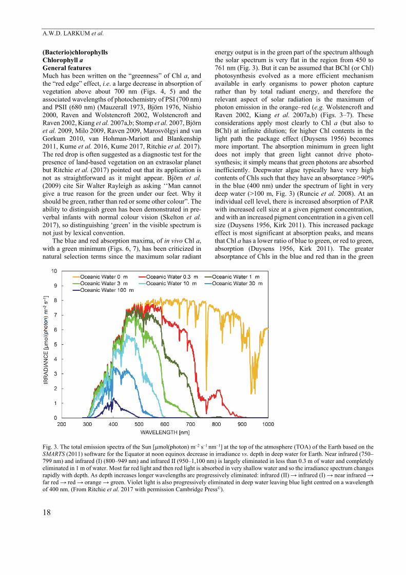
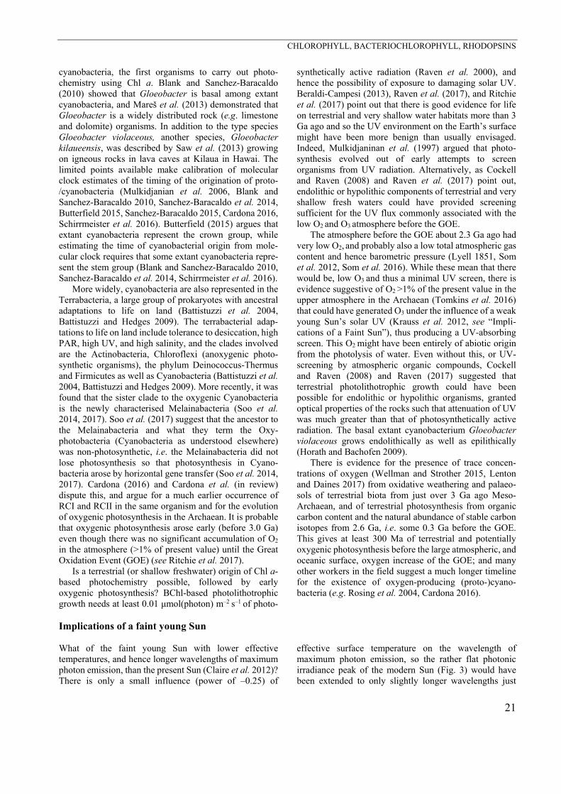
energy output is in the green part of the spectrum although the solar spectrum is very flat in the region from 450 to 761 nm (Fig. 3). But it can be assumed that BChl (or Chl) photosynthesis evolved as a more efficient mechanism available in early organisms to power photon capture rather than by total radiant energy, and therefore the relevant aspect of solar radiation is the maximum of photon emission in the orange–red (e.g. Wolstencroft and Raven 2002, Kiang et al. 2007a,b) (Figs. 3–7). These considerations apply most clearly to Chl a (but also to BChl) at infinite dilution; for higher Chl contents in the light path the package effect (Duysens 1956) becomes more important. The absorption minimum in green light does not imply that green light cannot drive photo-synthesis; it simply means that green photons are absorbed inefficiently. Deepwater algae typically have very high contents of Chls such that they have an absorptance >90% in the blue (400 nm) under the spectrum of light in very deep water (>100 m, Fig. 3) (Runcie et al. 2008). At an individual cell level, there is increased absorption of PAR with increased cell size at a given pigment concentration, and with an increased pigment concentration in a given cell size (Duysens 1956, Kirk 2011). This increased package effect is most significant at absorption peaks, and means that Chl a has a lower ratio of blue to green, or red to green, absorption (Duysens 1956, Kirk 2011). The greater absorptance of Chls in the blue and red than in the green
Fig. 3. The total emission spectra of the Sun [µmol(photon) m–2 s–1 nm–1] at the top of the atmosphere (TOA) of the Earth based on the SMARTS (2011) software for the Equator at noon equinox decrease in irradiance vs. depth in deep water for Earth. Near infrared (750–799 nm) and infrared (I) (800–949 nm) and infrared II (950–1,100 nm) is largely eliminated in less than 0.3 m of water and completely eliminated in 1 m of water. Most far red light and then red light is absorbed in very shallow water and so the irradiance spectrum changes rapidly with depth. As depth increases longer wavelengths are progressively eliminated: infrared (II) → infrared (I) → near infrared → far red → red → orange → green. Violet light is also progressively eliminated in deep water leaving blue light centred on a wavelength of 400 nm. (From Ritchie et al. 2017 with permission Cambridge Press©).
CHLOROPHYLL, BACTERIOCHLOROPHYLL, RHODOPSINS
19
spectral region means that, with increasing Chl per unit area exposed to the radiation, the photon absorption increases with photon flux density more rapidly in the blue and red than in the green (Duysens 1956, Kirk 1975a,b; 1976; Morel and Bricaud 1981, 1986; Kirk 2011). The dependence of the package effect on the size of single cells
(taking into account the spectral distribution of incident photosynthetically active radiation), and of colonies and organisms, has important physiological and ecological consequences (Kirk 1975a,b; 1976, 2011; Morel and Bricaud 1981, 1986; Falkowski and Raven 2007).
Ecological considerations of mats and single cells The size of the smallest free-living cells is determined by a variety of non-scalable components, including the number of genes, required for a given trophic mode (Pirie 1964, 1973; Raven 1986a, 1986b, 1998, 1999; Raven et al. 2009, 2013a). The smallest picoplanktonic cyanobacterial cells (Prochlorococcus spp.) and oxygenic eukaryotic cells (Ostreococcus spp., Prasinophyceae sensu lato) have minimum equivalent spherical diameters of about 0.5 μm and 0.9 μm, respectively, or similar to that of photo-synthetically useful light (Raven et al. 2013). These organisms have minimal package effects, so, for a vector radiation field, there is a minimal difference in PAR between the side of the cell nearest, and furthest from, the light source. This has relevance to the enhancement of photosynthesis, under certain circumstances, by tempo-rally fluctuating PAR. Thus, while the frequency of rotation of the cells resulting from small-scale thermally-driven water movements is in the range that might cause stimulation of photosynthesis, the limited PAR gradient across small cells means that these natural rotations cannot stimulate photosynthesis as a result of fluctuating PAR (Raven 1998, 1999; Raven and Ralph 2015). Fluctuating light is perceived to be disadvantageous for photosynthesis because it takes time for reaction centres to open and close and during the adjustment phase the potential photo-synthetic activity mismatches the light available (Greene and Gerard 1990, Kübler et al. 1996, Litchman et al. 2000, Raven and Ralph 2015). One consequence is that sunflecks cannot easily be optimally utilised if the frequency of change of irradiance is too high (Kübler et al. 1996).
As cell size increases with a constant pigment per cell
volume and constant cell geometry, the package effect
increases with cell size (Duysens 1956, Kirk 1975a,b; 1976, Morel and Bricaud 1981, 1986; Raven 1984, 1986a,b; 1998,
1999). However, the photosynthetic pigment per unit
volume decreases with cell size in eukaryotic microalgal
cells (Raven 1984, Agusti and Phlips 1992, Beardall et al. 2009, Finkel et al. 2010). The small range of cyanobacterial
unicell sizes available show no size dependence upon
pigment per unit volume (Agusti and Phlips 1992). Cyano-bacterial colonies, like eukaryotic algal unicells, show a
decrease in pigment per unit volume with increasing size
(Agusti and Phlips 1992, Gao and Ai 2004). In macroalgae there are many published in vivo spectra
of photon absorptance [Abtλ = incident PAR set to 100, minus reflectance (%) minus transmittance (%)] (Haxo and Blinks 1950, Fork and Amesz 1969, Lűning and Dring 1985, Gómez and Huovinen 2011, Lichtenberg et al. 2015, Vásquez-Elizondo et al. 2017), showing a flattening of the spectra relative to that of dilute solutions of the extracted pigments. The flattening is greater for spatially and optically thicker tissues. The spectrally averaged absorp-tance is, as predicted, greater for thicker thalli with absorptance values up to 0.95 in the thicker thalli (Frost-Christensen and Sand-Jensen 1992, Markager and Sand-Jensen 1992, Markager 1993, Enríquez et al. 1994, Gómez and Huovinen 2011, Vásquez-Elizondo et al. 2017). Very high absorptance values (> 0.90) are found for the Phaeophyceae (Lűning and Dring 1985, Gómez and Huovinen 2011), Rhodophyta (Gómez and Huovinen 2011), and Ulvophyceae (Lűning and Dring 1985, Vásquez-Elizondo et al. 2017). The much higher absorp-tances of thicker thalli of macroalgae means that the some thalli of all three clades with their different photosynthetic ‘accessory’ pigments have very low reflectance and minimal spectral variation in reflectance and appear almost black, hence the suggestion (references and discussion in Raven and Hurd 2012) that the younger stages of algae (with smaller spatial and optical thickness) are more useful than older stages in assigning an alga to its pigment category, i.e. clade. The absorptances of macro-algal thalli has implications for their photon yield on an absorbed photon basis (e.g. Lűning and Dring 1985) and hence on the distribution of benthic macroalgae with depth and the interactions among algae (Dring and Lűning 1985, Binzer and Sand-Jensen 2002, Tait et al. 2014, 2017). The old paradigm of chromatic adaptation amongst green, brown, and red benthic algae was shown to be wrong long ago (Crossett et al. 1963); but nevertheless red algae do predominate at the greater depths in the sea because crustose coralline algae have evolved to live in low light with low metabolic rates (Larkum and Barrett 1983). Microalgae and photosynthetic bacteria filtered onto glass fibre discs to act as artificial algal mats for PAM studies can be used to simulate similar optical properties (Ritchie 2013, 2014; Ritchie and Runcie 2013, 2014; Ritchie and Mekjinda 2014).
A.W.D. LARKUM et al.
20
Terrestrial embryophytes with leaves For terrestrial tracheophytes, Jones (2013) cites a mean PAR leaf absorptance of 0.85. Some higher values are known, e.g. 0.896–0.925 for four species of tropical rain forest species at different vertical positions in the canopy (Poorter et al. 1995), and 0.875–0.917 for sun leaves, and 0.869–0.939 for shade leaves of 13 species of cloud forest trees (Poorter et al. 2000). Among aquatic tracheophytes, Durako (2007) found lower absorptance values (0.45–0.68) for eight species of Australian seagrass. Absorp-tances at the red and blue peaks of Chl in leaves of terrestrial tracheophyte can be considerably higher and approach 1.0 (Ritchie and Runcie 2014) even though red-green-blue (RGB) averages for “white” light are usually about 0.85: white light absorptance can hence be misleading, particularly for understory plants which experience irradiance depleted in blue and red light [similar to the effect on lower cell layers in a microbial mat composed of green Chl (a+b) cells; Kühl and Fenchel 2000, Hubas et al. 2011, Ohkubo et al. 2017)].
There are resource cost-benefit reasons that put an upper limit on the tissue absorptance and these are related to the package effect. It is relatively easy to make a small cell (< 10 µm) optically black using high pigment concentrations but not large vacuolated cells. The energy cost of synthesis and maintenance of photochemical and light-harvesting pigment–protein complexes must not exceed the energy usable from the photons absorbed by the complexes over their lifetime (Raven 1984, Raven and Hurd 2012). There are also considerations of the other resource costs, e.g. carbon, nitrogen (as a component of amino acids but also through the RNA needed for protein synthesis), and phosphorus. Overloading with pigment would hence be selected against except in habitats where very slow growth is not a disadvantage (cryptic habitats, low light environments in caves and in very deep water) (Raven 1984, Raven and Cockell 2006, Runcie et al. 2008, Cockell et al. 2009).
In summary, the solar spectral irradiance incident on the top of vegetation canopies on land, or at the ocean surface becomes modified: on land the spectrum is modified by layering of plants whether the vegetation is terrestrial plants in a forest or layering in a microbial mat; in aquatic environments water depth modifies the spectrum considerably, depending on chemical sustances in the water and the density and type of phytoplankton in the water column (Fig. 3). Differential absorption of blue and red photons by Chl a-dominated organisms (lacking phycobilins) enriches radiation deeper in the canopy or water body in the green region of the spectrum; in water bodies longer PAR wavelengths are absorbed by water itself and by organic solutes (yellow substance) in coastal marine and fresh waters (Falkowski and Raven 2007, Kiang et al. 2007a,b; Stomp et al. 2007, Kirk 2011). At the algal or aquatic plant community level (Krause-Jensen and Sand-Jensen 1998), the overall package effect is greater than for the individual organisms since there are more moles of photosynthetic pigments per habitat area than for each organism. There is the possibility of complementarity of spectral absorptance between strata in the community, e.g. phycoerythrin-containing red algae with high absorp-tance in the green under brown algae or seagrasses with lower absorptances in the green than in the blue and red, thus limiting the influence of the package effect. However, the available evidence shows that this is not a significant effect (Dring 1981, Lüning 1990, Kirk 2011).
As is discussed below, these arguments need to be augmented when organisms with photochemistry cata-lysed by Chl a co-occur with those photosynthetic organisms absorbing beyond 700 nm, e.g. those with long-wavelength forms of Chl a, or with Chls d or f, as well as anoxygenic photosynthetic bacteria with BChl as com-monly occurs in microbial mats, stromatolites and in wastewater ponds.
Photosynthesis in mats (including stromatolites) Sparks et al. (2007) suggested that an early biological photochemistry using a green-absorbing ion-pumping rhodopsin could have been an evolutionary driver of BChls in photoredox biochemistry with absorption at shorter and longer wavelengths than the rhodopsin, thus limiting competition for light with the incumbent rhodopsin. However, the time of origin of ion-pumping rhodopsins is not clear (see above), and this hypothesis has not moved from a conference abstract (Sparks et al. 2007) to a peer-reviewed journal.
The relatively flat peak of photon emission in the pres-ent solar spectrum could be a consideration (Figs. 3–7), since the wavelengths at which RCs operate are just on the long wavelength side of the maximum photon emission
peak at 669 nm on the ground on Earth (Wolstencroft and Raven 2002). The young Sun (a G-star) would have been cooler and dimmer than today and its spectrum would have been shifted to longer wavelengths, i.e. toward the spec-trum of a K-star (see “Implications of a Faint Sun”). How-ever, the total emission spectra of K-stars are not greatly different from G-stars except for a displacement of about 100 nm in the emission peak to the red end of the spectrum. Red Dwarfs, however, have a very different spectrum to the sun and differ considerably from that of a Planck black body because of absorption by compounds in the stellar atmospheres of Red Dwarfs (Ritchie et al. 2017).
Is the epi- or endolithic modern habit of Gloeobacter on land indicative of the Archaean habitat of the proto-
CHLOROPHYLL, BACTERIOCHLOROPHYLL, RHODOPSINS
21
cyanobacteria, the first organisms to carry out photo-chemistry using Chl a. Blank and Sanchez-Baracaldo (2010) showed that Gloeobacter is basal among extant cyanobacteria, and Mareš et al. (2013) demonstrated that Gloeobacter is a widely distributed rock (e.g. limestone and dolomite) organisms. In addition to the type species Gloeobacter violaceous, another species, Gloeobacter kilaueensis, was described by Saw et al. (2013) growing on igneous rocks in lava caves at Kilaua in Hawai. The limited points available make calibration of molecular clock estimates of the timing of the origination of proto-/cyanobacteria (Mulkidjanian et al. 2006, Blank and Sanchez-Baracaldo 2010, Sanchez-Baracaldo et al. 2014, Butterfield 2015, Sanchez-Baracaldo 2015, Cardona 2016, Schirrmeister et al. 2016). Butterfield (2015) argues that extant cyanobacteria represent the crown group, while estimating the time of cyanobacterial origin from mole-cular clock requires that some extant cyanobacteria repre-sent the stem group (Blank and Sanchez-Baracaldo 2010, Sanchez-Baracaldo et al. 2014, Schirrmeister et al. 2016).
More widely, cyanobacteria are also represented in the Terrabacteria, a large group of prokaryotes with ancestral adaptations to life on land (Battistuzzi et al. 2004, Battistuzzi and Hedges 2009). The terrabacterial adap-tations to life on land include tolerance to desiccation, high PAR, high UV, and high salinity, and the clades involved are the Actinobacteria, Chloroflexi (anoxygenic photo-synthetic organisms), the phylum Deinococcus-Thermus and Firmicutes as well as Cyanobacteria (Battistuzzi et al. 2004, Battistuzzi and Hedges 2009). More recently, it was found that the sister clade to the oxygenic Cyanobacteria is the newly characterised Melainabacteria (Soo et al. 2014, 2017). Soo et al. (2017) suggest that the ancestor to the Melainabacteria and what they term the Oxy-photobacteria (Cyanobacteria as understood elsewhere) was non-photosynthetic, i.e. the Melainabacteria did not lose photosynthesis so that photosynthesis in Cyano-bacteria arose by horizontal gene transfer (Soo et al. 2014, 2017). Cardona (2016) and Cardona et al. (in review) dispute this, and argue for a much earlier occurrence of RCI and RCII in the same organism and for the evolution of oxygenic photosynthesis in the Archaean. It is probable that oxygenic photosynthesis arose early (before 3.0 Ga) even though there was no significant accumulation of O2 in the atmosphere (>1% of present value) until the Great Oxidation Event (GOE) (see Ritchie et al. 2017).
Is a terrestrial (or shallow freshwater) origin of Chl a-based photochemistry possible, followed by early oxygenic photosynthesis? BChl-based photolithotrophic growth needs at least 0.01 μmol(photon) m–2 s–1 of photo-
synthetically active radiation (Raven et al. 2000), and hence the possibility of exposure to damaging solar UV. Beraldi-Campesi (2013), Raven et al. (2017), and Ritchie et al. (2017) point out that there is good evidence for life on terrestrial and very shallow water habitats more than 3 Ga ago and so the UV environment on the Earth’s surface might have been more benign than usually envisaged. Indeed, Mulkidjaninan et al. (1997) argued that photo-synthesis evolved out of early attempts to screen organisms from UV radiation. Alternatively, as Cockell and Raven (2008) and Raven et al. (2017) point out, endolithic or hypolithic components of terrestrial and very shallow fresh waters could have provided screening sufficient for the UV flux commonly associated with the low O2 and O3 atmosphere before the GOE.
The atmosphere before the GOE about 2.3 Ga ago had very low O2, and probably also a low total atmospheric gas content and hence barometric pressure (Lyell 1851, Som et al. 2012, Som et al. 2016). While these mean that there would be, low O3 and thus a minimal UV screen, there is evidence suggestive of O2 >1% of the present value in the upper atmosphere in the Archaean (Tomkins et al. 2016) that could have generated O3 under the influence of a weak young Sun’s solar UV (Krauss et al. 2012, see “Impli-cations of a Faint Sun”), thus producing a UV-absorbing screen. This O2 might have been entirely of abiotic origin from the photolysis of water. Even without this, or UV-screening by atmospheric organic compounds, Cockell and Raven (2008) and Raven (2017) suggested that terrestrial photolithotrophic growth could have been possible for endolithic or hypolithic organisms, granted optical properties of the rocks such that attenuation of UV was much greater than that of photosynthetically active radiation. The basal extant cyanobacterium Gloeobacter violaceous grows endolithically as well as epilithically (Horath and Bachofen 2009).
There is evidence for the presence of trace concen-trations of oxygen (Wellman and Strother 2015, Lenton and Daines 2017) from oxidative weathering and palaeo-sols of terrestrial biota from just over 3 Ga ago Meso-Archaean, and of terrestrial photosynthesis from organic carbon content and the natural abundance of stable carbon isotopes from 2.6 Ga, i.e. some 0.3 Ga before the GOE. This gives at least 300 Ma of terrestrial and potentially oxygenic photosynthesis before the large atmospheric, and oceanic surface, oxygen increase of the GOE; and many other workers in the field suggest a much longer timeline for the existence of oxygen-producing (proto-)cyano-bacteria (e.g. Rosing et al. 2004, Cardona 2016).
Implications of a faint young Sun What of the faint young Sun with lower effective temperatures, and hence longer wavelengths of maximum photon emission, than the present Sun (Claire et al. 2012)? There is only a small influence (power of –0.25) of
effective surface temperature on the wavelength of maximum photon emission, so the rather flat photonic irradiance peak of the modern Sun (Fig. 3) would have been extended to only slightly longer wavelengths just
A.W.D. LARKUM et al.
22
before the GOE than is the case today. The young Sun (G spectral type) would have had a spectrum slightly closer to that of a cooler star of K spectral type. The faint young Sun wavelength of maximum photon emission would have been on the short wavelength side of the 680 nm and 700 nm absorption maxima of the pigments doing the photochemistry of PSII and PSI for the great majority of oxygenic photosynthetic organisms with Chl a in the reaction centres of both photosystems. As discussed above, one major constraint on the wavelength of photons used in photochemistry is the energy content of the photons relative to the energetics of the reactions catalysed and as will be seen below slightly longer wavelengths for photochemistry occur in Chl d- and f-containing cyano-bacteria, lower energy input to the reactions driven by the photochemistry has been shown to have little effect on the rate and efficiency of the reactions examined (Loughlin et
al. 2013, Allakhverdiev et al. 2016). The higher fraction of photons on the land and sea
surface >700 nm under the weak young Sun might have favoured organisms with Chl d and f (see below) because reaction centres and LHCs equipped with Chl d or Chl f would have with red absorption maxima at longer wavelengths (Ritchie et al. 2017) but would have made attenuation of longer wavelength irradiance by water more severe than found today (Fig. 3). While the lower colour temperature of the young Sun means a lower steady UV flux at the top of the atmosphere, it is possible that greater periodic UV flare activity of the young Sun would mean an overall higher UV flux over long (millions of years) periods (Krauss et al. 2012). Flare activity is a major limitation for the habitability of planets of Red Dwarfs (Ritchie et al. 2017).
Cyanobacteria vs. eukaryotic algae The capacity to make significant use of photons at >700 nm in photosynthetic O2 production is not limited to cyanobacteria. Öquist (1969) cultivated the freshwater trebouxiophycean green alga Chlorella pyrenoidosa auto-trophically under incandescent irradiation with a 0.1-m water filter. Growth was slower in the far-red cultures, and acclimation to the two radiation regimes was complete in six weeks (Öquist 1969). Exposure to infrared radiation increased the absorbance and the capacity for light-limited photosynthesis in the wavelength range 700–740 nm by about two-fold. However, the capacity for light-saturated photosynthesis (Pmax) was lower for the infrared-grown cells. An Emerson enhancement effect has been shown for Chlorella acclimated (less likely to be genetically acclimated) to growth in far-red radiation (Öquist 1969). The increased rate of light-limited photosynthesis, when radiation was below 700 nm and above 700 nm are supplied simultaneously relative to the sum of the photosynthetic rates, when the two radiation regimes are supplied independently, occurred in both the far-red acclimated (adapted?) Chlorella cells and the control grown cells (Öquist 1969).
In some other eukaryotic algae there are genetic adap-tations that enable increased photosynthesis in radiation regimes above 700 nm than is the case in the majority of eukaryotic algae. A long wavelength Chl a–apoprotein combination can energize PSII in the marine ulvophycean green alga Ostreobium sp. (Fork and Larkum 1989, Koehne et al. 1999, Wilhem and Jakob 2006), and the chromerophycean (Alveolata) marine Chromera velia (Bína et al. 2014, Katabova et al. 2014). Two species of the Eustigmatophyeae (Ochrista or Heterokontophyta) can also use photons >700 nm for photosynthesis; one is the freshwater planktonic Trachydiscus minutus (Prĭbyl et al. 2012, Pazderninik 2015). The one as yet un-named freshwater eustigmatophycean Forest Park Isolate 5 (FP5) has Chl a as the sole Chl but is able to grow when
illuminated solely by 740 nm LEDs (Wolf et al. 2017). This ability means that one or more of the Chl a–protein complexes with significant absorbance >700 nm isolated from FP5 is associated with PSII (Wolf et al. 2017). Low rates of photosynthetic O2 production occur out to 780 nm in the flowering plant Helianthus annuus and Phaseolus vulgaris (Pettai et al. 2005a,b) and PSII activity occurs out to 780 nm in the flowering plant Spinacia oleracea (Hughes et al. 2006, Thapper et al. 2009). Shlodder et al. (2014) showed that PSI activity can be excited by photons out to 808 nm; for wavelengths longer that 750 nm direct excitation of a charge transfer complex is proposed. There seem to have been no published attempts to demonstrate Emerson-type enhancement by far-red light in Acaryo-chloris even though it would yield important information about PSI/PSII in the organism.
The role of these long wavelengths in light harvesting involve uphill excitation energy transfer from wavelengths longer than that of the reaction centres, with a decreasing fraction of the absorbed energy from longer wavelength excitation reaching the reaction centre as the wavelength difference between incident radiation and the reaction centre increases (Nieuwenburg et al. 2003, Pettai et al. 2005b, Wilhelm and Jakob 2006, Mimuro et al. 2007, Toms et al. 2014, Behrend et al. 2015, Raven and Ralph 2015). As pointed out above, such mechanisms are thermodynamically possible but come at considerable thermodynamic cost.
Finally, it is necessary to relate the discussion above to that of other Chls, especially the redox active Chl d. The first excited states of Chl a, as moderated by apoproteins reaction centres of PII and PSII clearly energize NADP+
reduction, H2O oxidation, and associated H+ pumping using 2 photons per electron, moderated by the maximal efficiency of ≈0.8 for PSII electron transfer per photon. The kinetics and efficiency of redox reactions are not markedly different in organisms with Chl d as opposed to
CHLOROPHYLL, BACTERIOCHLOROPHYLL, RHODOPSINS
23
Chl a, in the sense of photochemical and maximal light-harvesting efficiency of the harvesting pigment (Allakhverdiev et al. 2016). This means that there is not an absolute energetic requirement to use Chl a in oxygenic
photochemistry, as demonstrated by Chl d (see below) and there is a real possibility that Chl f could prove to be a third redox active chlorophyll.
Chlorophyll b Chl b differs from Chl a in molecular structure in having a hydroxyl (OH) at position C7 on porphyrin ring B (Fig. 1) [instead of a methyl (CH3) group]. This brings about a shift to the red in the Soret (Qx) peak from 436 nm (in acetone solvent) to 465 nm and a similar shift in vivo, apparent in Fig. 4. In the QY peak the shift is to the blue (batho-chromic) from 665 nm to 645 nm with a similar shift in vivo (Fig. 4). The overall effect is to narrow the gap in absorption of light between the Soret and QY peaks of Chl a. Both shifts are significant and hence improve the light-harvesting ability of organisms that possess both pigments conjugated to a joint protein, since the peak wavelengths of Chl b augment those of Chl a and the energy is passed on efficiently to the RCs. Considering that MgDVP (and by inference the various types of Chl c), with only a small red-shift to ~452 nm (and only a small peak in the red region at about 630 nm), have been successful light-harvesting pigments (Raven 1996; and see below),
Chl b must have been even more successful and this is borne out in terms of its widespread occurrence in nature, especially in embryophytes: however, in the oceans MgDVP and Chls c (see below) predominate, not only in numbers of species using them. However, in terms of biomass abundance: Prochlorococcus, with ‘Chls (a+b)’ has been one of the most successful organisms there; it has divinyl forms of Chls a and Chl b, and it also possesses MgDVP (Goericke and Repeta 1992, 1993; Ritchie et al. 2017) and therefore has the best of both worlds. It is true that a number of carotenoids harvest light efficiently in the blue region but the widespread occurrence of Chl b in these same organisms must mean that this pigment makes a strong contribution to gross productivity. In fact most light-harvesting Chl (LHC) proteins bind both Chl a and Chl b, as well as carotenoids, but, depending on the LHC and the organism, in different amounts (Larkum 2006). The overall effect, on land and in shallow aquatic
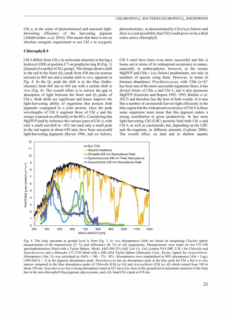
Fig. 4. The solar spectrum at ground level is from Fig. 3. In vivo absorptances (Abt) are based on integrating (Taylor) sphere measurements of the transmission (T, %) and reflectance (R, %) of cell suspensions. Measurements were made on two UV-VIS spectrophotometers fitted with a Taylor Sphere: Model A&E-S90-2D (A&E Lab Co. Ltd, London N14 5BP, U.K.) for Chlorella and Synechococcus and a Shimadzu UV-2550 fitted with a ISR-240A Taylor Sphere (Shimadzu Corp., Kyoto, Japan) for Acaryochloris. Absorptance (Abt, %) was calculated as Abt% = 100 - T% - R%. Absorptances were standardised to 90% absorptance [Abt = Log10 (100/Abt%) = 1] at the pigment absorptance peak. Synechococcus has an absorptance peak at the blue peak for Chl a but it is very narrow compared to the blue absorptance peaks of Chlorella [Chl (a+b)] and Acaryochloris [Chl (a+d)] which extend from 350 to about 550 nm. Synechococcus has a strong absorptance band at 627 nm (very close to the ground-level maximum emission of the Sun) due to the non-chlorophyll blue pigment, phycocyanin, and a QY band Chl a peak at 678 nm.
A.W.D. LARKUM et al.
24
environments, of the presence of Chl b in vivo, is to help close the gap in light harvesting between the blue and red peaks of Chl a (Fig. 4), but while this results in only a “knee” on the shorter wavelength (bathochromic) side of the Chl a red peak it has a substantial effect in widening blue light absorption (in conjunction with carotenoids) towards longer wavelengths (Fig. 4) compared to cyano-bacteria such as Synechococcus. Thus, the red-shift in the Soret band of Chl b is highly significant in increasing harvesting of light.
Interestingly, the cyanobacteria Prochlorococcus, Prochloron, and Prochlorothrix are the only cyanobacteria to have incorporated Chl b into their photosynthetic systems alongside Chl a. How Chl b evolved is not clear, but presumably it did so as a bioenergenically “cheaper” and perhaps space-saving light-harvesting adaptation that dispensed with phycobiliproteins (PBPs) in favour of Chl b in LHCs (Larkum 2003, 2006). Since PBPs are nitrogen expensive, because they are proteins, this strategy presumably evolved as a nitrogen conservation adaptation: and an important hint in support of this hypothesis is that none of the organisms mentioned have nitrogen fixation genes. Furthermore, Prochlorococcus and Prochloron live in environments where N is scarce (the first, most success-fully, in deep layers of the ocean and the second in nitrogen poor coral reef waters, albeit in the cloacal cavity of didemnid ascidians). The special Chl a/b-binding proteins of Prochlorococcus and Prochloron, which are similar, have been identified (La Roche et al. 1996); and they are distinct from the family of proteins that conjugate Chl a/b and Chl a/c in eukaryotic algae and Chl a/b in terrestrial plants (La Roche et al. 1996). A similar protein complex exists in Prochlorothrix (Herbstova et al. 2010), although there it has also been shown that Chl b binds to a ring around PSI (Bumba et al. 2005, Boichenko et al. 2007).
In this discussion it is also relevant to discuss the presence of divinyl forms of Chls a and b (Fig. 1) in Pro-chlorococcus (Chisholm et al. 1992, Goericke and Repeta 1992, 1993). This chemical change modifies the absorp-tion peaks. For the Soret peaks the shift is from 436 nm to 446 nm for divinyl Chl a and from 465 nm to 475 for divinyl Chl b. This means that for a deep-water phyto-plankton organism the absorption spectrum in the blue band is widened and more closely matches the blue light spectrum of the ocean at depth for these organisms (Figs. 3, 8) (Goericke and Repeta 1993, Ritchie et al. 2017).
Chl b was somehow inherited by a group of eukaryotic
algae collectively known as the green algae, i.e. Chlorophyta comprising the Palmophyllophyceae, Prasinophyceae, Chlorophyceae, Trebouxiophyceae, and Ulvophyceae, and the algal members (Charophyceae) of the Streptophyta (Kantz et al. 1990, Leliaert et al. 2016), with MgDVP in some Prasinophyceae. Thus, in eukaryotic algae Chl b is restricted to the Chlorophyta and Strepto-phyta, and secondary endosymbioses involving endosym-biosis of a chlorophyte alga: in the Chlorarachniophyta, in the Rhizaria, in Euglenophyta, and in the Excavata (Graham et al. 2016). The restriction of Chl b to these photosynthetic eukaryotes can be rationalised in terms of a single endosymbiotic primary plastid, i.e. a monophyletic Archaeplastida (Price et al. 2012, Sanchez-Báracaldo et al. 2017) with no lateral gene transfer (LGT). This requires the presence of both Chl b and phycobilins in the cyanobacterium ancestral to primary plastids (Tomitani et al. 1999), with loss of Chl b in the Glaucocystophyta and the Rhodophyta, and hence the algae that gained plastids by secondary endosymbiosis of red algae. Furthermore, it would require loss of MgDVP in the cyanobacterial ancestor of eukaryotic plastids; and its reappearance (by LGT?) in some prasinophytes (Larkum 2006) and the Palmophyllophyceae (Leliaert et al. 2016). The absence of
PBP in the Chlorophyta and Streptophyta would require loss
of PBPs at the origin of these clades. Another explanation is
a polyphyletic origin of plastids, with multiple endo-symbiosis, but also with much LGT (Larkum et al. 2007). Monophyly does not preclude the involvement of LGT.
It could be coincidental (Gould and Lewontin 1979) that the group of algae which gave rise to land plants (embryophytes) came from one group of Chlorophyta, the streptophytes with Chl (a+b) (Waters 2003, Raven 2017). Thus, the embryophytes inherited only the light-harvesting strategies of the streptophytes and more broadly of Chlorophyta. Clearly this light-harvesting strategy has been very successful; however, had the line that conquered the land also possessed PBP the light-harvesting strategies might have been just as successful (compare Figs. 4 and 5) (Ritchie et al. 2017). The spectral width gain in the blue part of the spectrum in cells with Chls (a+b) is substantial and appears to be advantageous compared to a phyco-cyanin-type Synechococcus cyanobacterium with no phycoerythrin (Fig. 4); but this needs further experimental examination at an ecological/physiological level. However, Chl (a+b) organisms are much better at using terrestrial irradiance than is sometimes thought (Fig. 4).
Chlorophyll c and MgDVP The various types of Chl c as mentioned above have a Soret (blue) band not far different from Chl a with peaks at 447 nm (solvent acetone) for Chl c1, 450 nm for Chl c2,
and 452 nm for Chl c3 (Zapata and Garrido 1997). These wavelengths are very close to the Soret peak for Chl a. So what is to gain from their synthesis? Furthermore,
considering the fact that the in vivo QY band peak (≈630 nm) is very small (Fig. 5), brings into question the role of these Chl c accessory pigments. However, Fig. 5 shows that the blue absorptance peak of Chl c-containing organisms (Isochrysis and Chaetoceros) is conspicuously wider than in Synechococcus which only has Chl a and
CHLOROPHYLL, BACTERIOCHLOROPHYLL, RHODOPSINS
25
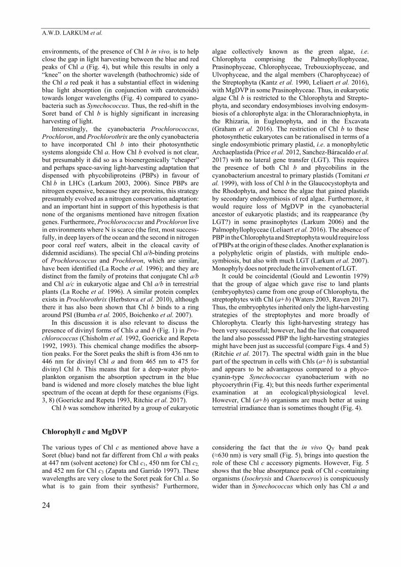
Fig. 5. The solar spectrum at ground level is from Fig. 3. In vivo absorptances (Abt) are based on integrating (Taylor) sphere measure-ments of the transmission (T, %) and reflectance (R, %) of Isochrysis (Cryptophyceae) and the diatom Chaetoceros (Bacillariophyceae) cell suspensions measured using the A&E spectrophotometer fitted with a Taylor Sphere. Absorptances were standardised to 90% absorptance [Abt = Log10 (100/Abt %) = 1] at the pigment absorptance peak. The absorptance spectra of Isochrysis [Chl (a+c2)] and Chaetoceros [Chl (a + c1 + c2)] are very similar. Both have a very wide blue absorptance peak (350 to about 550 nm) with peaks at 436 and 439 nm, respectively. Isochrysis and Chaetoceros have a QY band red peak at about 676 nm but note how narrow it is compared to the blue band. The red Chl c absorptance peak is at about 639 nm but absorption by Chl c at its Q-band peak is only a very minor component of the total absorptance of these chromophytes. phycocyanin (Fig. 4). Chlorella which is a classic chlorophyte with Chl (a+b), also has a much wider blue absorptance band than Synechococcus (Fig. 4). Is this enhanced harvesting of blue light the main reason for the use of Chl c as an accessory pigment or does it perform a role in some other way in light harvesting? The mere fact that there are three major Chls c, other less well Chl c variants and magnesium 2,4-divinyl-pheoporphyrin mono-methyl ester A5 (MgDVP) may suggest that they perform some other function or act in association with some special carotenoids (such as fucoxanthin in Phaeophyta and peridinin in dinoflagellates). The situation is further complicated by the existence of MgDVP as a physio-logically functional pigment of Chl–protein complexes (Larkum, 2003, 2006, 2008): it is very close to Chl c biosynthetically and in molecular structure, is on the biosynthetic pathway to all BChls and Chl c (Hunter and Coomber 1988, Burke et al. 1993), and has a Soret peak very close to that of the Chls c. It is found in a few chlorophytan planktonic algae in the Prasinophyceae (Rowan 1989, Kantz et al. 1990, Zingone et al. 2002, Latasa et al. 2004) and the Palmophyllophyceae (Leliaert et al. 2016). Raven (1984, 1996) surmised that MgDVP and by implication Chls c do contribute to a significant degree to light harvesting in the light-harvesting complexes (LHC) where MgDVP and Chls c occur. Other
authors, e.g. Larkum (2008), have speculated that these pigments play a role in energy migration from the pigment perhaps via specific carotenoids (see above) to the reaction centres. The latter possibility would go some way to explain why there are a variety of Chl c compounds.
The various types of Chl c are found in a wide range of eukaryote chromist algae (Larkum 2006), i.e. those algae that do not possess Chl b and, apart from the Cryptophyta, do not possess PBP: the chromists are the Cryptophyta, Haptophyta, and Ochrista (= Ochrophyta) (e.g. brown alga and diatoms), as well as the basal, peridinin-containing, Dinoflagellata (Pyrrophyta), also including dinoflagellates with diatom tertiary endosymbiotic plastids. Two excep-tions here are the cryptophytes which possess Chl c2 and PBP (Larkum 2006) and Prochloron (one of the Chl b-containing cyanobacteria) that also possesses MgDVP (Larkum et al. 1994) as well as a number of species of the Chl b-containing Prasinophyceae (formerly Micromona-dophyceae; Fawley2004, Rowan 1989, Zingone et al. 2002, Latasa et al. 2004) and Palmophyllophyceae (a new group of early chlorophytes allied to the Prasinococcales and separate from the Prasinophyceae; Leliaert et al. 2016). MgDVP also occurs in Chl d-containing organisms (Miyashita et al. 1997, Schliep et al. 2007), so it is now apparent that it is a rather widespread pigment in Chl b- and Chl c-containing algae. The PBP of Cryptophytes,
A.W.D. LARKUM et al.
26
however, is not in the form of a phycobilisome that is “plugged into” PSII as is found in Glaucocystophytes, Rhodophytes, and cyanobacteria (Kana et al. 2009, Graham et al. 2016).
As mentioned above, for Chl b, the possession of Chl c in only one of the three groups, which possess primary plastids poses a problem for the proposition of a mono-phyletic origin of plastids. One possible explanation is that Chl c arose, alongside the special carotenoids that such algae possess de novo to satisfy special absorption criteria; but beware of the Panglossian argument that every part of the phenology of an organism must necessarily have a purpose rather than being incidental (Gould and Lewontin 1979). However, no such special advantageous criterion has been satisfactorily put forward; but it has been carefully pointed out here that it would more likely involve blue light absorption (Soret band) rather than of 630 nm light (Figs. 4, 5). Nevertheless, Chl c could have arisen independently and the fact that two groups of algae, the
dinoflagellates, and cryptophytes have only Chl c2 whereas the other groups have Chl c1 and c2 (as well as, in some instances, Chl c3) suggests that Chl c2 evolved first: and this fits with the biosynthetic pathway in which Chl c2 is a precursor to Chl c1 (Larkum 2003, 2006). Most forms of Chl c do not have a phytol tail but the existence of phytylated forms in some eukaryotic algae, but as yet in no known prokaryote, suggests independent evolution (Zapata and Garrido 1997).
It is interesting, furthermore, that certain lines of chromophytic algae with Chl c-containing relatives have either Chl a alone or no Chl at all (but loss of all Chl is not restricted to chromophytes, e.g. Prototheca, Polytoma, and Polytomella). As is the case in many organisms of obvious Chl (a+b) affinities, there are examples of chromophytes lacking any variety of Chl c such as the Eustigmato-phyceae (e.g. Nannochloropsis) and chromerids such as Chromera and Vitrella (Pan et al. 2012).
Chlorophyll d The unique Chl, Chl d (Fig. 1) serves a similar function to Chl a in that it functions as both a redox active special pair substituting for Chl a in the equivalent position of P680 (possibly at 713 nm) and P700 (P740) of cyanobacteria that possess it and additionally as a light-harvesting pigment (Larkum 2006, Telfer et al. 2007, Ohashi et al. 2008, Mohr et al. 2010, Loughlin et al. 2013, 2014). The only exception here, for the substitution of Chl d for Chl a in the reaction centres, is the need for phaeophytin a as the primary (acceptor of the special pair, P697) of PSII, which is synthesised from Chl a, and therefore requires the formation of a small amount of Chl a. Apart from that and a small metabolic pool of Chl a, Acaryochloris marina seems to be able to function with mostly Chl d (up to
~97%); with Chl d also acting as a light-harvesting Chl with its own Chl–protein (Chen et al. 2005). The relative abundance of Chl d (the Chl d/a ratio) varies depending on the light regime under which it is grown (Miyashita et al. 1996, Chen et al. 2005, Gloag et al. 2007, Chen 2014), varying from a minority to majority Chl under favourable conditions. For the in vivo absorptance of Acaryochloris compared to Synechococcus (Chl a) and Chlorella [Chl (a+b)] see Fig. 4.
Chl d is synthesised from Chl a by substituting a methyl group on position C5 of ring A with a hydroxyl group with the oxygen derived from O2 (see Fig. 1) (Schliep et al. 2010). How this oxygen is added is not known, but see Loughlin et al. (2014).
Chlorophyll f Chl f (Fig. 1) was discovered in a filamentous cyano-bacterium from living stromatolite material collected from intertidal columnar stromatolites from Hamelin Pool, Shark Bay, Western Australia (Chen et al. 2010, Niedzwiedzki et al. 2014, Tomo et al. 2014). It has since been found in a number of other cyanobacteria, both filamentous and globular (Chen 2014, Itoh et al. 2015). It is found in hot spring cyanobacteria and a number of the latter organisms have been well researched by Bryant and his group (Ho et al. 2016, Ho et al. 2017), who have shown that Chl f is induced under far-red light (Ho et al. 2016a). As previously pointed out, the final synthetic step, the addition of oxygen to C3 methyl to form a formyl group (Fig. 1) is brought about by a primitive form of D1 protein (Ho et al. 2016). In all these organisms Chl f constitutes
only a small proportion of the total Chl (<10%), the rest being Chl a; and the formation of Chl f is NIR-dependent, i.e. the amount of Chl f in white light is very small (Ho et al. 2016a).
As far as is known, Chl f functions only in a light-harvesting role (Chen 2014). The energy transfer mechanisms have been studied by Li et al. (2012) and Akimoto et al. (2015) and the structure of Chl f has been presented by Willows et al. (2013). Details on the evolution of this pigment are not known. It has a red-shifted QY peak at 707 vs. 696 nm for Chl d (in organic solvents) (Chen et al. 2010, 2015), which means that it is able to function further into the NIR than Chl d, but this depends on the conjugation to specific proteins.
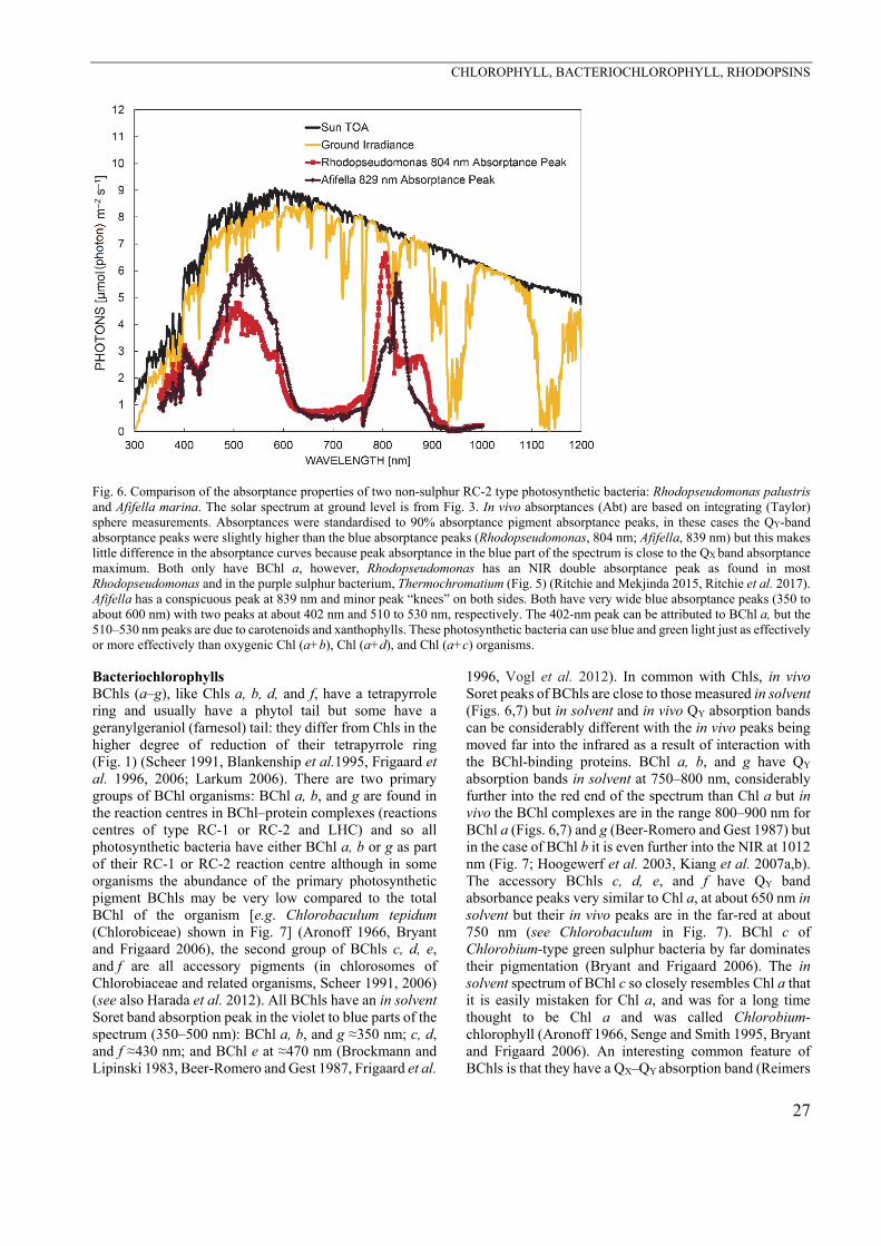
CHLOROPHYLL, BACTERIOCHLOROPHYLL, RHODOPSINS
27
Fig. 6. Comparison of the absorptance properties of two non-sulphur RC-2 type photosynthetic bacteria: Rhodopseudomonas palustris and Afifella marina. The solar spectrum at ground level is from Fig. 3. In vivo absorptances (Abt) are based on integrating (Taylor) sphere measurements. Absorptances were standardised to 90% absorptance pigment absorptance peaks, in these cases the QY-band absorptance peaks were slightly higher than the blue absorptance peaks (Rhodopseudomonas, 804 nm; Afifella, 839 nm) but this makes little difference in the absorptance curves because peak absorptance in the blue part of the spectrum is close to the QX band absorptance maximum. Both only have BChl a, however, Rhodopseudomonas has an NIR double absorptance peak as found in most Rhodopseudomonas and in the purple sulphur bacterium, Thermochromatium (Fig. 5) (Ritchie and Mekjinda 2015, Ritchie et al. 2017). Afifella has a conspicuous peak at 839 nm and minor peak “knees” on both sides. Both have very wide blue absorptance peaks (350 to about 600 nm) with two peaks at about 402 nm and 510 to 530 nm, respectively. The 402-nm peak can be attributed to BChl a, but the 510–530 nm peaks are due to carotenoids and xanthophylls. These photosynthetic bacteria can use blue and green light just as effectively or more effectively than oxygenic Chl (a+b), Chl (a+d), and Chl (a+c) organisms. Bacteriochlorophylls BChls (a–g), like Chls a, b, d, and f, have a tetrapyrrole ring and usually have a phytol tail but some have a geranylgeraniol (farnesol) tail: they differ from Chls in the higher degree of reduction of their tetrapyrrole ring (Fig. 1) (Scheer 1991, Blankenship et al.1995, Frigaard et al. 1996, 2006; Larkum 2006). There are two primary groups of BChl organisms: BChl a, b, and g are found in the reaction centres in BChl–protein complexes (reactions centres of type RC-1 or RC-2 and LHC) and so all photosynthetic bacteria have either BChl a, b or g as part of their RC-1 or RC-2 reaction centre although in some organisms the abundance of the primary photosynthetic pigment BChls may be very low compared to the total BChl of the organism [e.g. Chlorobaculum tepidum (Chlorobiceae) shown in Fig. 7] (Aronoff 1966, Bryant and Frigaard 2006), the second group of BChls c, d, e, and f are all accessory pigments (in chlorosomes of Chlorobiaceae and related organisms, Scheer 1991, 2006) (see also Harada et al. 2012). All BChls have an in solvent Soret band absorption peak in the violet to blue parts of the spectrum (350–500 nm): BChl a, b, and g ≈350 nm; c, d, and f ≈430 nm; and BChl e at ≈470 nm (Brockmann and Lipinski 1983, Beer-Romero and Gest 1987, Frigaard et al.
1996, Vogl et al. 2012). In common with Chls, in vivo Soret peaks of BChls are close to those measured in solvent (Figs. 6,7) but in solvent and in vivo QY absorption bands can be considerably different with the in vivo peaks being moved far into the infrared as a result of interaction with the BChl-binding proteins. BChl a, b, and g have QY absorption bands in solvent at 750–800 nm, considerably further into the red end of the spectrum than Chl a but in vivo the BChl complexes are in the range 800–900 nm for BChl a (Figs. 6,7) and g (Beer-Romero and Gest 1987) but in the case of BChl b it is even further into the NIR at 1012 nm (Fig. 7; Hoogewerf et al. 2003, Kiang et al. 2007a,b). The accessory BChls c, d, e, and f have QY band absorbance peaks very similar to Chl a, at about 650 nm in solvent but their in vivo peaks are in the far-red at about 750 nm (see Chlorobaculum in Fig. 7). BChl c of Chlorobium-type green sulphur bacteria by far dominates their pigmentation (Bryant and Frigaard 2006). The in solvent spectrum of BChl c so closely resembles Chl a that it is easily mistaken for Chl a, and was for a long time thought to be Chl a and was called Chlorobium-chlorophyll (Aronoff 1966, Senge and Smith 1995, Bryant and Frigaard 2006). An interesting common feature of BChls is that they have a QX–QY absorption band (Reimers
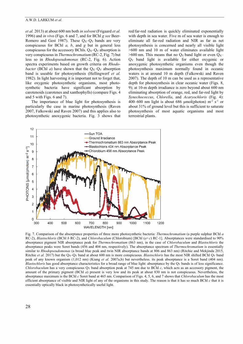
A.W.D. LARKUM et al.
28
et al. 2013) at about 600 nm both in solvent (Frigaard et al. 1996) and in vivo (Figs. 6 and 7, and for BChl g see Beer-Romero and Gest 1987). These QX–QY bands are very conspicuous for BChl a, b, and g but in general less conspicuous for the accessory BChls. QX–QY absorption is very conspicuous in Thermochromatium (RC-2, Fig. 7) but less so in Rhodopseudomonas (RC-2, Fig. 6). Action spectra experiments based on growth criteria on Rhodo-bacter (BChl a) have shown that the QX–QY absorption band is useable for photosynthesis (Hellingwerf et al. 1982). In light harvesting it is important not to forget that, like oxygenic photosynthetic organisms, most photo-synthetic bacteria have significant absorption by carotenoids (carotenes and xanthophylls) (compare Figs. 4 and 5 with Figs. 6 and 7).
The importance of blue light for photosynthesis is particularly the case in marine photosynthesis (Raven 2007, Falkowski and Raven 2007) and this applies also to photosynthetic anoxygenic bacteria. Fig. 3 shows that
red/far-red radiation is quickly eliminated exponentially with depth in sea water. Five m of sea water is enough to eliminate all far-red radiation and NIR as far as net photosynthesis is concerned and nearly all visible light >600 nm and 10 m of water eliminates available light >600 nm. This means that no QY band light or even QX–QY band light is available for either oxygenic or anoxygenic photosynthetic organisms even though the photosynthesis maximum normally found in oceanic waters is at around 10 m depth (Falkowski and Raven 2007). The depth of 10 m can be used as a representative depth for photosynthesis in clear oceanic water (Figs. 8, 9); at 10-m depth irradiance is zero beyond about 600 nm eliminating absorption of orange, red, and far-red light by Synechococcus, Chlorella, and Acaryochloris (Fig. 4): 400–600 nm light is about 686 μmol(photon) m-2 s-1 or about 31% of ground level but this is sufficient to saturate photosynthesis of most aquatic organisms and most terrestrial plants.
Fig. 7. Comparison of the absorptance properties of three more photosynthetic bacteria: Thermochromatium (a purple sulphur BChl a RC-2), Blastochloris (BChl b RC-2), and Chlorobaculum (Chlorobium) [BChl (a+c) RC-1]. Absorptances were standardised to 90% absorptance pigment NIR absorptance peak for Thermochromatium (863 nm), in the case of Chlorobaculum and Blastochloris the absorptance peaks were Soret bands (456 and 404 nm, respectively). The absorptance spectrum of Thermochromatium is essentially similar to Rhodopseudomonas (a broad blue peak and twin NIR absorptance bands at 806 and 863 nm) (Ritchie and Mekjinda 2015, Ritchie et al. 2017) but the QX–QY band at about 600 nm is more conspicuous. Blastochloris has the most NIR shifted BChl QY band peak of any known organism (1,012 nm) (Kiang et al. 2007a,b) but nevertheless. its peak absorptance is a Soret band (404 nm). Blastochloris has good absorptance characteristics for a broad range of blue light: absorptance by the QY bands is of less significance. Chlorobaculum has a very conspicuous QY band absorption peak at 745 nm due to BChl c, which acts as an accessory pigment, the amount of the primary pigment (BChl a) present is very low and its peak at about 830 nm is not conspicuous. Nevertheless, the absorptance maximum is the BChl c Soret band at 465 nm. Comparison of Figs. 4, 5, 6, and 7 shows that Chlorobaculum has the most efficient absorptance of visible and NIR light of any of the organisms in this study. The reason is that it has so much BChl c that it is essentially optically black in photosynthetically useful light.
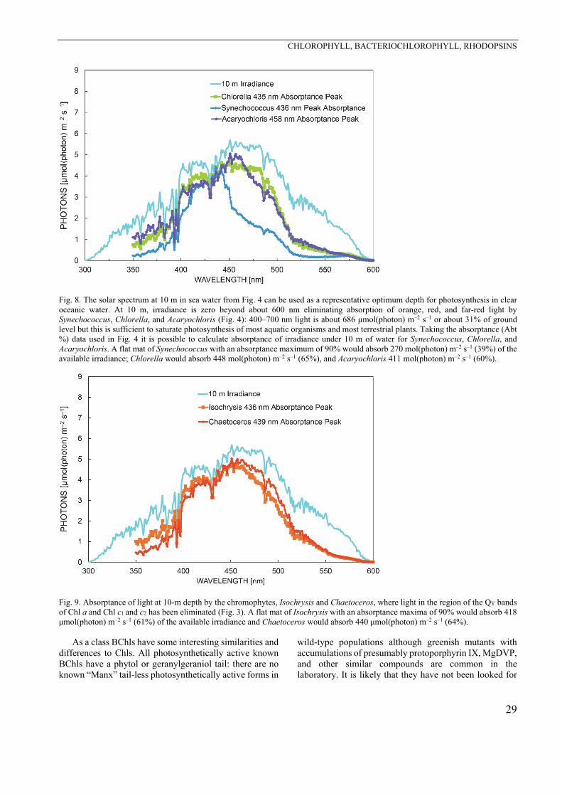
CHLOROPHYLL, BACTERIOCHLOROPHYLL, RHODOPSINS
29
Fig. 8. The solar spectrum at 10 m in sea water from Fig. 4 can be used as a representative optimum depth for photosynthesis in clear oceanic water. At 10 m, irradiance is zero beyond about 600 nm eliminating absorption of orange, red, and far-red light by Synechococcus, Chlorella, and Acaryochloris (Fig. 4): 400–700 nm light is about 686 μmol(photon) m–2 s–1 or about 31% of ground level but this is sufficient to saturate photosynthesis of most aquatic organisms and most terrestrial plants. Taking the absorptance (Abt %) data used in Fig. 4 it is possible to calculate absorptance of irradiance under 10 m of water for Synechococcus, Chlorella, and Acaryochloris. A flat mat of Synechococcus with an absorptance maximum of 90% would absorb 270 mol(photon) m–2 s–1 (39%) of the available irradiance; Chlorella would absorb 448 mol(photon) m–2 s–1 (65%), and Acaryochloris 411 mol(photon) m–2 s–1 (60%).
Fig. 9. Absorptance of light at 10-m depth by the chromophytes, Isochrysis and Chaetoceros, where light in the region of the QY bands of Chl a and Chl c1 and c2 has been eliminated (Fig. 3). A flat mat of Isochrysis with an absorptance maxima of 90% would absorb 418 μmol(photon) m–2 s–1 (61%) of the available irradiance and Chaetoceros would absorb 440 μmol(photon) m–2 s–1 (64%).
As a class BChls have some interesting similarities and differences to Chls. All photosynthetically active known BChls have a phytol or geranylgeraniol tail: there are no known “Manx” tail-less photosynthetically active forms in
wild-type populations although greenish mutants with accumulations of presumably protoporphyrin IX, MgDVP, and other similar compounds are common in the laboratory. It is likely that they have not been looked for
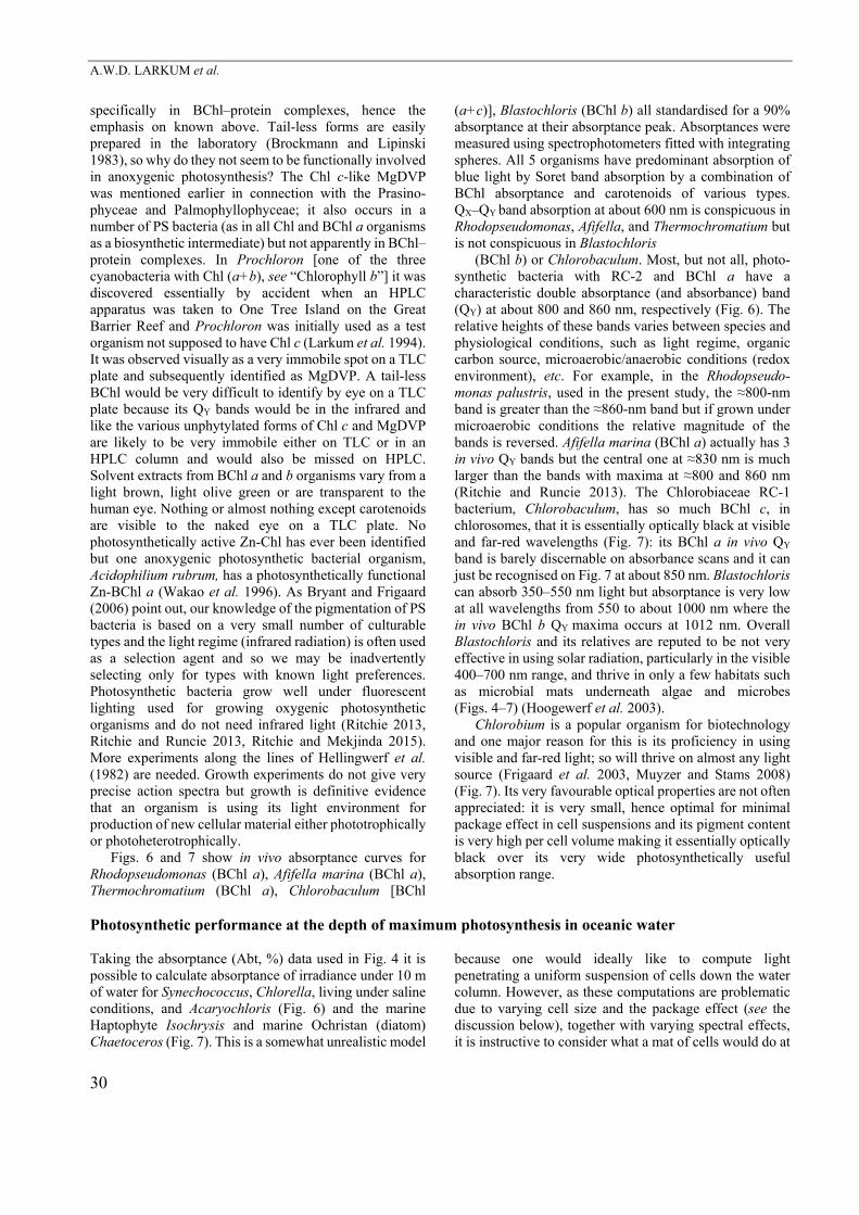
A.W.D. LARKUM et al.
30
specifically in BChl–protein complexes, hence the emphasis on known above. Tail-less forms are easily prepared in the laboratory (Brockmann and Lipinski 1983), so why do they not seem to be functionally involved in anoxygenic photosynthesis? The Chl c-like MgDVP was mentioned earlier in connection with the Prasino-phyceae and Palmophyllophyceae; it also occurs in a number of PS bacteria (as in all Chl and BChl a organisms as a biosynthetic intermediate) but not apparently in BChl–protein complexes. In Prochloron [one of the three cyanobacteria with Chl (a+b), see “Chlorophyll b”] it was discovered essentially by accident when an HPLC apparatus was taken to One Tree Island on the Great Barrier Reef and Prochloron was initially used as a test organism not supposed to have Chl c (Larkum et al. 1994). It was observed visually as a very immobile spot on a TLC plate and subsequently identified as MgDVP. A tail-less BChl would be very difficult to identify by eye on a TLC plate because its QY bands would be in the infrared and like the various unphytylated forms of Chl c and MgDVP are likely to be very immobile either on TLC or in an HPLC column and would also be missed on HPLC. Solvent extracts from BChl a and b organisms vary from a light brown, light olive green or are transparent to the human eye. Nothing or almost nothing except carotenoids are visible to the naked eye on a TLC plate. No photosynthetically active Zn-Chl has ever been identified but one anoxygenic photosynthetic bacterial organism, Acidophilium rubrum, has a photosynthetically functional Zn-BChl a (Wakao et al. 1996). As Bryant and Frigaard (2006) point out, our knowledge of the pigmentation of PS bacteria is based on a very small number of culturable types and the light regime (infrared radiation) is often used as a selection agent and so we may be inadvertently selecting only for types with known light preferences. Photosynthetic bacteria grow well under fluorescent lighting used for growing oxygenic photosynthetic organisms and do not need infrared light (Ritchie 2013, Ritchie and Runcie 2013, Ritchie and Mekjinda 2015). More experiments along the lines of Hellingwerf et al. (1982) are needed. Growth experiments do not give very precise action spectra but growth is definitive evidence that an organism is using its light environment for production of new cellular material either phototrophically or photoheterotrophically.
Figs. 6 and 7 show in vivo absorptance curves for Rhodopseudomonas (BChl a), Afifella marina (BChl a), Thermochromatium (BChl a), Chlorobaculum [BChl
(a+c)], Blastochloris (BChl b) all standardised for a 90% absorptance at their absorptance peak. Absorptances were measured using spectrophotometers fitted with integrating spheres. All 5 organisms have predominant absorption of blue light by Soret band absorption by a combination of BChl absorptance and carotenoids of various types. QX–QY band absorption at about 600 nm is conspicuous in Rhodopseudomonas, Afifella, and Thermochromatium but is not conspicuous in Blastochloris
(BChl b) or Chlorobaculum. Most, but not all, photo-synthetic bacteria with RC-2 and BChl a have a characteristic double absorptance (and absorbance) band (QY) at about 800 and 860 nm, respectively (Fig. 6). The relative heights of these bands varies between species and physiological conditions, such as light regime, organic carbon source, microaerobic/anaerobic conditions (redox environment), etc. For example, in the Rhodopseudo-monas palustris, used in the present study, the ≈800-nm band is greater than the ≈860-nm band but if grown under microaerobic conditions the relative magnitude of the bands is reversed. Afifella marina (BChl a) actually has 3 in vivo QY bands but the central one at ≈830 nm is much larger than the bands with maxima at ≈800 and 860 nm (Ritchie and Runcie 2013). The Chlorobiaceae RC-1 bacterium, Chlorobaculum, has so much BChl c, in chlorosomes, that it is essentially optically black at visible and far-red wavelengths (Fig. 7): its BChl a in vivo QY band is barely discernable on absorbance scans and it can just be recognised on Fig. 7 at about 850 nm. Blastochloris can absorb 350–550 nm light but absorptance is very low at all wavelengths from 550 to about 1000 nm where the in vivo BChl b QY maxima occurs at 1012 nm. Overall Blastochloris and its relatives are reputed to be not very effective in using solar radiation, particularly in the visible 400–700 nm range, and thrive in only a few habitats such as microbial mats underneath algae and microbes (Figs. 4–7) (Hoogewerf et al. 2003).
Chlorobium is a popular organism for biotechnology and one major reason for this is its proficiency in using visible and far-red light; so will thrive on almost any light source (Frigaard et al. 2003, Muyzer and Stams 2008) (Fig. 7). Its very favourable optical properties are not often appreciated: it is very small, hence optimal for minimal package effect in cell suspensions and its pigment content is very high per cell volume making it essentially optically black over its very wide photosynthetically useful absorption range.
Photosynthetic performance at the depth of maximum photosynthesis in oceanic water Taking the absorptance (Abt, %) data used in Fig. 4 it is possible to calculate absorptance of irradiance under 10 m of water for Synechococcus, Chlorella, living under saline conditions, and Acaryochloris (Fig. 6) and the marine Haptophyte Isochrysis and marine Ochristan (diatom) Chaetoceros (Fig. 7). This is a somewhat unrealistic model
because one would ideally like to compute light penetrating a uniform suspension of cells down the water column. However, as these computations are problematic due to varying cell size and the package effect (see the discussion below), together with varying spectral effects, it is instructive to consider what a mat of cells would do at

CHLOROPHYLL, BACTERIOCHLOROPHYLL, RHODOPSINS
31
10 m in oceanic water. A flat mat of Synechococcus with an absorptance maximum of 90% would absorb 270 mol(photon) m–2 s–1 (39%) of the available irradiance, Chlorella would absorb 448 mol(photon) m–2 s–1 (65%), and Acaryochloris 411 mol(photon) m–2 s–1 (60%), which shows, in the last example, the value of possessing PBPs. Long wavelength-absorbing green algae such as Ostreobium would also be able to perform well under 10 m of water using Soret-band absorption in a similar way to Acaryochloris even though far-red light would be absent (Koehne et al. 1999). A flat mat of Isochrysis with an absorptance maximum of 90% would absorb 418 μmol
(photon) m–2 s–1
(61%) of the available irradiance and Chae-toceros would absorb 440 μmol(photon) m–2
s–1 (64%).
Absorption efficiencies in the 60% range for all the algae with accessory Chls is impressive when compared to the relatively poor performance of Synechococcus R-2 used in the present study which only has phycocyanin (orange/red absorption peak ≈630 nm, which is outside the irradiance range available under 10 m of water). Synechococcus has an absorption efficiency of only 39% because phycocyanin has the wrong spectrum for absorption of light under 10 m of water. Cyanobacteria and Cryptophytes with phycoerythrin, e.g. Rhodomonas, would be expected to do as well or better under 10 m of water as do Chlorella, Isochrysis, and Chaetoceros.
Unfortunately, only absorbance data was available for Rhodomonas (a phycoerthryin-containing cryptophyte, Ritchie et al. 2017) but not absorptance data, which would be needed if it was to be included in the present study. Qualitatively Fig. 1 in Ritchie et al. (2017) shows that Rhodomonas would be expected to be able to use light at 10 m with very high efficiency. Considering that green algae and terrestrial plants [those with Chl (a+b)] are often perceived as being poor utilisers of light (Chen and Blankenship 2011, Chen and Scheer 2013, Ort et al. 2015) it is ironic that a Chlorella mat with a 90% absorptance maximum at 435 nm (Fig. 4) turns out to be the best absorber of light at the optimum photosynthetic depth in the open ocean of all four oxygenic photosynthetic organisms, which have accessory Chls and that are included in the present study. Just why are plants green indeed (Mauzerall 1973, Bjorn 1976, Nishio 2000, Larkum 2006, 2008; Björn et al. 2009, Raven 2009, Kume et al. 2016, Kume 2017)? It turns out that green plants [Chl (a+b)] have much better photosynthetic light absorption performance than they are often taken for at both ground level and even underwater (Figs. 4, 8). Perhaps why are plants green is not such an accident after all; perhaps emphasis on the blue-band absorption is the key to appreciating why land plants are green [Chl (a+b)].
Absorptance curves for photosynthetic bacteria using data shown in Figs. 6 and 7 can also be used to calculate absorptance curves in microbial mats under 10 m of water. The absorptance curves are essentially similar to those calculated for the oxygenic organisms (Figs. 8, 9) because at this depth the blue light absorption curves of the
photosynthetic bacteria closely resemble those of oxygenic organisms. The Rhodopseudomonas used here (Ritchie 2013) was a freshwater species but marine Rhodopseudo-monads are common. Afifella is a marine purple non-sulphur photosynthetic bacterium (Ritchie and Runcie 2013). BChl a in vivo is able to absorb 350-nm light and so the total irradiance from 350 to 600 nm was calculated [805 μmol(photon) m–2 s–1). At 10-m depth both the QX–QY and QY in vivo absorption bands of BChl a (Fig. 6) are ineffective because of the negligible irradiance at 600 nm and above, and hence the strong QX–QY band at ≈600 nm would be ineffective. The Soret band in vivo of BChl a is at about 380–400 nm (Figs. 6, 7). Flat mats of Rhodopseudomonas and Afifella with absorptance maxima of 90% would absorb 280 μmol(photon) m–2 s–1 (35%) of the available irradiance and 356 μmol(photon) m–2 s–1 (44%), respectively. Absorptance by Rhodopseudomonas and Afifella in the blue part of the spectrum is not as high as
found in the case of Chlorella, Acaryochloris, Isochrysis, and Chaetoceros, but nevertheless both are higher than
found in Synechococcus [270 mol(photon) m–2 s–1, 39%].
Rhodopseudomonas and Afifella are both RC-2 purple nonsulphur photosynthetic bacteria. The effective irradi-ance for these organisms was 350–600 nm because the blue peak of BChl a is below 400 nm. As in the case of Rhodopseudomonas and Afifella at 10-m depth, the QX–QY
in vivo absorption bands of BChl a, b, and c (at about 600 nm see Figs. 6 and 7) are ineffective because of the negligible irradiance at 600 nm and above. Flat mats of Thermochromatium with absorptance maxima of 90% absorbed ≈275 μmol(photon) m–2 s–1 (34% of the available irradiance) and the spectrum is little different to that found for Rhodopseudomonas although the QX–QY band at 600 nm is more conspicuous (Fig. 7). Blastochloris (BChl b) absorbed 233 μmol(photon) m–2 s–1 or only 29% of the available light and so Blastochloris has the poorest performance at 10 m of any of the photosynthetic organisms included in the present study. On the other hand, Chlorobaculum BChl (a+c) absorbed 465 μmol(photon) m–2 s–1 or 58% of 350–700 nm light at 10-m depth. This is the highest absorption capacity of any photosynthetic organism included in the present study and is a result of its extremely high absorptance in the 350–600 nm range (Fig. 7). Comparison of Figs. 4 and 5 with Figs. 6 and 7 suggest that photosynthetic organisms with only a single BChl are at a conspicuous disadvantage in using blue light and the role of accessory BChls is to broaden blue-band absorption by themselves and to enhance the efficiency of utilisation of light absorbed by carotenoids.
While the absorptance data give a good indication of the capacity for photon absorption, it is also important to have action spectrum data and to calculate absorptance for suspensions of small cells with large package effects over a wide range of dilutions. While several action spectra are available for oxygenic photosynthetic organisms, this is not the case for the anoxygenic photosynthetic bacteria. For the purple non-sulphur bacteria there are only two
A.W.D. LARKUM et al.
32
published action spectra. The early study with few wavebands (French 1937) suggests a lower quantum yield in the blue region than at longer wavelengths, but a later more detailed action spectrum study suggested an essentially constant quantum yield at all wavelengths and showed that Soret (QX), QX–QY, and QY bands were all used photosynthetically (Hellingwerf et al. 1982). For the oxygenic photosynthetic organisms the action spectrum also generally follows the absorption spectrum relatively closely (e.g. Lűning and Dring 1985). Researchers in the field have long been impressed by the fact that most oxygenic organisms are able to use blue and red light but are poor absorbers of green and orange light (550–625 nm) light and suggestions to improve the performance of pho-tosynthesis are frequently made (Chen and Blankenship 2011, Chen and Scheer 2013, Ort et al. 2015). This deficiency is often overestimated and is contrary to our conclusions drawn at several points in the present paper (Figs. 4, 5). Blankenship et al. (2011) has suggested that PSI in oxygenic organisms could be replaced by RC-1 from photosynthetic bacteria using recombinant DNA technology. Scrutiny of Figs. 4–7 for terrestrial and Figs. 8 and 9 for marine conditions strongly suggest that any gains from such a genetic recombination would be marginal because the number of photons absorbed by a Chl a based LHC/PSI/PSII would be of a similar magnitude as could be collected by an RC-1 organism with its associated LHCs. A recombinant organism with functional PSII/PSI and RC-1 (a Chl/BChl-hybrid version of a three photo-system model) (Wolstencroft and Raven 2002) for oxygenic photosynthesis (PSII/PSI/PSIII) might offer gains in total spectral light utilisation. Govindjee et al. (2017) give a historical overview of two and three photo-system models of oxygenic photosynthesis; Wolstencroft and Raven’s (2002) paper considers these two models in an astrobiological context.
The absorptance results shown in Figs. 4, 5, 8, and 9 tend to downplay the significance of the red, far-red absorption bands of Chls. However, the relatively larger blue absorption bands include contributions from carote-noids (some of which could be involved in photoprotection rather than light harvesting) as well as Chls. Ideally, action spectra of photosynthesis should also be used more extensively. It is notable that all the photosynthetic organisms considered in the present study in sunlight and even more so at 10-m depth in clear oceanic water are using predominantly blue light [Soret (QX) band] for photosynthesis. In the case of photosynthetic bacteria the routine use of far-red and NIR as a selection agent
(incandescent light bulbs and NIR diodes) and neglect of blue-light absorption has probably resulted in an excessive focus on photosynthesis under red light/far red irradiance to give the impression that they must be using far red and NIR as their primary light sources in nature. Certainly under oceanic conditions, all photosynthetic organisms are using only blue light for photosynthesis in water deeper than about 5 m (Fig. 3) and so the red/far red absorption properties of the photosynthetic pigments are not relevant to photosynthetic electron transport in anything but very shallow water or terrestrially. Even the trophically proble-matic aerobic anoxygenic photosynthetic bacteria (AAPB) (Jiao et al. 2007, Garcia-Chaves et al. 2016, Stoecker et al. 2017) would be using mainly blue light. For example, the very small QY absorptance maximum of Chl c1 and c2 in vivo (≈630 nm) make it very implausible that the signifi-cance of chromophytes having Chl c1 and c2 has much to do with absorptance at 630 nm: having Chl c as an accessory pigment is much more likely to have important consequences in the absorptance of blue light (Figs. 5, 9).
The conclusion for photosynthetic bacteria with BChls is that they do best using blue light; their famed ability to use far red irradiation only applies when they exist in light environments where only far red irradiation is available or in very low light environments where the use of any light available from 350 to 1,050 nm is an advantage. In this regard, the predominance of PS bacteria in modern stromatolites (Burns et al. 2004) may be informative in terms of early evolution (see “Microbial mats”, below). This again reinforces the conclusion that modern photosynthetic bacteria evolved after the evolution of Chl organisms in cryptic environments, where the ability to absorb far red radiation was a significant advantage in terrestrial environments but far red was only useable in very shallow water environments (Figs. 3, 6, 7) (Larkum 2006, 2008).
Further progress in comparing the spectral properties of organisms with different Chls and BChls requires basing the absorptance values on the quantity of the main BChls, with units of m2 mol–1(pigment) (Morel and Bricaud 1981), m2 g–1(cell organic C) (Falkowski et al. 1985) or spacial volume of the reaction centres (nm2 per reaction centre) (e.g. Key et al. 2009). Consideration is also
needed of the influence of the package effect on the values
obtained, with increasing influence of the package effect in
larger cells or colonies or clumps of cells and with higher
concentrations of pigment per unit cell volume or colony
volume (Kirk 2012, Klughammer and Schreiber 2015).
Microbial mats Microbial mats (including stromatolites) have aroused great interest in photosynthetic research and have been found to be the habitats of very interesting organisms (Wakao et al. 1996, Burns et al. 2004, Vopel and Hawes 2006, Mohr et al. 2010, Hohmann-Marriott and
Blankenship 2011, Ohkubo et al. 2017). Microbial mats are not necessarily aquatic, for example desert crusts (Lange et al. 1992). Much research has focused on the layering of organisms in microbial mats and the attenuation of light inside the mat matrix and redox
CHLOROPHYLL, BACTERIOCHLOROPHYLL, RHODOPSINS
33
gradients (Kühl and Fenchel 2000, Raven et al. 2000, Stomp et al. 2007, Marosvölgyi and van Gorkum 2010, Hubas et al. 2011, Chen et al. 2012, Ohkubo et al. 2017). They are the earliest known manifestations of photo-synthetic life on the Earth (Tice and Lowe 2004, Butterfield 2015, Wellman et al. 2015, Nutman et al. 2016). Chromatic gradients in microbial mats most likely date to the very beginnings of photosynthesis on the Earth.
Layered microbial mats are hence of great interest to those interested in photoadaptation of photosynthetic organisms. Serial absorption by different microbial layers is understood in qualitative terms but quantitative or semi-quantitative considerations are rare. Here we will consider a microbial mat with a green alga [Chl (a+b)] (Fig. 4) on the surface and cyanobacteria and Acaryochloris living underneath (consider the light environment under oxygenic organisms, Figs. 4, 5). Chlorella can be used as a model Chl (a+b) organism and has characteristic absorption and transmission properties although it is not usually found in microbial mats, but is used here as a model organism. Transmission [%] of cell suspensions was measured using an integrating sphere as part of the calculation of absorptance (Abt, %) of Chlorella (Fig. 4). Because Abt (%) is calculated from both transmission (T, %) and reflectance (R, %) data, T (%) is not simply proportional to Abt (%). To estimate the effect of a Chlorella overlayer in a microbial mat, the transmission was scaled to the absorptance maximum at 435 nm to set the minimum transmission to 10% at 435 nm. The layer of Chlorella cells shown in Fig. 4 would transmit about 1,020 μmol(quanta) m–2 s–1 (≈46%) of incident sunlight (350–750 nm). Under a layer of Chlorella, the cyanobacterium Synechococcus (Chl a) and Acaryochloris (Chl d) would be able to survive quite well on light that passed through
the layer of Chlorella cells [Abt350-750 nm Synechococcus ≈231 μmol(quanta) m–2 s–1; Abt350-750 nm Acaryochloris ≈309 μmol(quanta) m–2 s–1]. Ostreobium, the green alga with Chl (a+b) pigmentation but with Chl–protein complexes that allow it to use far-red light would also be able to grow well under a Chlorella shade mat (Fork and Larkum 1989, Koehne et al. 1999). Similar findings were made for the scenario of a Chaetoceros diatom mat with a 10% transmission minimum at 439 nm (Fig. 5) overlaying Synechococcus or Acaryochloris. The light regime under a layer of diatoms would easily support photosynthesis by other photosynthetic organisms with different light preferences.
Similar calculations can be made for the scenario where a layer of Chlorella cells overlays a layer of photosynthetic bacteria (Fig. 4, compared to Figs. 6 and 7). Here, Thermochromatium (BChl a), Chlorobaculum [BChl (a+c)], and Blastochloris (BChl b) will be used as examples. The layer of Chlorella cells would transmit about 2,553 μmol(quanta) m–2 s–1 (≈51%) of incident sun-light (350–1,100 nm). Under a layer of Chlorella acting as a green sunshade the RC-2 Thermochromatium would absorb ≈652 μmol(quanta) m–2 s–1 (Abt350-1100 nm), Chloro-baculum would absorb ≈1,175 μmol(quanta) m–2 s–1
(Abt350-1100 nm), and Blastochloris would absorb ≈331 μmol(quanta) m–2 s–1 (Abt350-1100 nm). Growth under a layer of Chlorella provides a very favourable light environment for all three photosynthetic bacteria, in parti-cular Chlorobaculum. As above for the case of an over-laying layer of the diatom Chaetoceros (Fig. 5), it can be shown that all three photosynthetic bacteria would have more than adequate light available under a layer of Chae-toceros with a 10% transmission minimum at 439 nm.
Cultures mentioned in the present study Chlorella sp. (freshwater and euryhaline marine) was grown in freshwater BG-11 medium although it grows equally well in enriched seawater (Seatae et al. 2014), Synechococcus PCC-7942 was also grown in BG-11 medium, Acaryochloris marina (Miyashita et al. 2003), Isochrysis sp., and Chaetoceros sp. were grown in f-2 enriched seawater as described previously (Ritchie 2008,
Ritchie et al. 2017). The photosynthetic bacteria were grown in PM media as previously described (Ritchie 2013, Ritchie and Runcie 2013, Ritchie and Mekjinda 2015). All were grown under fluorescent lights (cool-daylight) with no supplemental far-red or NIR light [≈150 μmol(photon) m–2 s–1, PAR 400–700 nm].
Global estimates of annual photosynthetic net primary production and organic carbon assimilation by organisms with photochemical energy transduction It is appropriate here to consider attempts to quantify global photolithotrophy by oxygenic and anoxygenic organisms, as well as the uses of photosynthetically produced organic carbon by organisms with photo-chemical energy transduction. Global oxygenic photo-lithotrophy has been much investigated, with significant variation in estimates. Some estimates for photosynthetic organisms suggest that anoxygenic photolithotrophy
contributes less than 0.1% of the total of anoxygenic plus oxygenic photolithotrophy. Johnston et al. (2009) and Raven (2009) have estimated that anoxygenic photolitho-trophy in marine habitats amounts to about 2.8–7.1 Tmol of fixed carbon per annum contrasting with estimates of 4,000–4,700 Tmol of fixed carbon per annum in all aquatic habitats (Field at al. 1998, Raven 2009, Buitenhuis et al. 2013), i.e. 0.07–0.15% of the total. Estimates of anoxy-
A.W.D. LARKUM et al.
34
genic photosynthesis is based on S2- oxidation in the global S cycle; this includes chemolithotrophy and excludes other inorganic electron donors such as Fe and also does not include use of organic carbon as electron donors, which can be regarded as a recycling process for already fixed organic carbon. In marine habitats almost all oxygenic primary productivity is by marine phytoplankton because the area occupied by marine macrophytes and marine angiosperms is such a small proportion of the surface area of the global ocean. Terrestrial oxygenic primary pro-duction (oxygenic photolithotrophy) is about 4,700–5,300 T mol(C) per year based on Field et al. (1998), Raven (2009), Edwards et al. (2015), and Rafique et al. (2016). Nearly all oxygenic primary productivity on land can be attributed to embryophytes; predominantly tracheophytes.
Although the estimates of global net primary production (NPP) show a range of values, there seem to be no estimates of how much respiratory CO2 production is spared by photoheterotrophy involving ion-pumping rhodopsins, BChl and Chl, on a global scale. Dissolved organic carbon (DOC) in the global ocean that is used as
an electron and carbon source by the abundant photo-heterotrophic bacteria (Kolber et al. 1991, 2001) is in effect recycled back into organic carbon in cells. This sparing of respiration of organic carbon from reaching the CO2 redox state by photoheterotrophic recycling of fixed carbon is discussed by Schmidt et al. (2015), Selosse et al. (2017), and Stoecker et al. (2017), for ion-pumping rhodopsins in bacteria and BChl in aerobic anoxygenic bacteria. Schmidt et al. (2015), Selosse et al. (2017), and Stoecker et al. (2017) also deal with eukaryotic oxygenic photosynthetic organisms that acquire organic carbon in parallel with phagotrophy in mixotrophic algae and in ciliates and Rhizaria that are photosynthetic through symbiosis with microalgae, or kleptoplast. In photosynthe-tically competent embryophytic land plants dissolved
organic carbon is acquired mainly via fungal symbionts, in
carnivorous plants through trapping animals then dissolving
their tissues by extracellular digestion, and sometimes by
ingestion of organic particles even in walled cells (Schmidt
et al. 2015, Selosse et al. 2017, Stoecker et al. 2017).
Conclusions A range of biologically produced photochemically active pigment–protein complexes absorbing radiation in the range 350–1050 nm are at the basis of almost all food webs on the Earth today, and may have been involved prior to 3.5 billion years ago (Ga). The ion-pumping rhodopsins use light absorbed by retinal to absorb energy from blue and green light to pump ions across the cell membranes. Chlorophylls have absorption bands in the blue and red (some just extending into the near infrared), and bacteriochlorophylls have absorption bands in the blue and red (QX and QX–QY) and far red (QY). Absorbed photons energize redox reactions and proton transport powering ADP phosphorylation, then oxidation of an external reductant and autotrophic assimilation of carbon dioxide. Anoxygenic photosynthesis involves external electron donors of a lower redox potential than water and a single photoreaction, while later-evolving oxygenic photosyn-thesis with two photoreaction centres in series, and water as the electron donor.
Two redox active Chls occur today, Chl a and Chl d; these two, usually with one or more of Chl b, Chl c, and Chl f, function as light-harvesting pigments. Chls a and b, Chls c, and MgDVP, absorb in the blue and, to lesser extent, the red, and Chls d and f in the blue plus near infrared (NIR). The additional light-harvesting chloro-phylls modify the absorption spectrum of the organism so that, especially with Chls c and MgDVP, there is an increase in blue absorption relative to that in the red. The ecological distribution of the various chlorophylls does not provide clear confirmation of, for example, particular occurrence of Chls c and MgDVP deep in clear oceanic waters with lowest radiation attenuation in the blue. The occurrence of divinyl Chl a and Chl b which yield
bathochromic shifts in their Soret (blue) bands, in the Chl a/b-containing Prochlorococcus, may be related to the generally deep open ocean habitat of these cyanobacteria. An additional factor is the size of the organisms, or dense populations of microorganisms; the package effect means that larger aggregations of pigments in larger organisms decreases the photon harvesting benefit and energy costs of increments in the pigment content.
The existence of bacteriochlorophylls raises important and unresolved issues as to the evolution of Chl vs. BChl. BChls occur in anoxygenic photosynthetic bacteria, with a small contribution to global primary productivity as anaerobic anoxygenic photosynthetic bacteria in anoxic habitats with often low photosynthetically usable photon fluxes (Table 2). However, anoxygenic bacteria are widespread and often abundant and so they should not be neglected (Kolber et al. 1990, 1991; Blankenship et al. 1995) despite their small contribution to global net primary productivity (Table 2). They often turn up in unexpected habitats: BChls occur in aerobic surface waters of marine and inland water habitats. The organisms found in these waters mostly do not carry out net primary production (only anaplerotic fixation of CO2), but the photon absorp-tion by BChl in these osmo-organotrophic organisms generate ion gradients and ATP that can decrease the loss of organic carbon in respiration during growth and maintenance through photo-osmo-organotrophic recycling of fixed carbon. A similar sparing of respiration can occur in the osmo-organotrophs with ion-pumping rhodopsins. Whether the PS bacteria, which possess either a PSI-like or a PSII-like system, evolved before the cyanobacteria has been discussed. BChl can harvest visible light but the possession of a variety of QY bands, enable PS bacteria to
CHLOROPHYLL, BACTERIOCHLOROPHYLL, RHODOPSINS
35
harvest far red radiation up to 1050 nm. MgDVP although present as a metabolite in photosynthetic bacteria does not appear to be incorporated into BChl–protein complexes.
Two things emerge from the present considerations. The first is that photosynthetic organisms often do not behave as expected from our reading of most textbooks (but see Kirk 2011) and our knowledge of the real diversity of photosynthesis is limited. The vast majority of microorganisms are not culturable. New organisms with novel pigmentation and photochemistry are being found all the time and usually accidentally and not always in exotic habitats. Even routine laboratory procedures can mask the presence of interesting organisms, for example the routine use of 750 nm as a blank for Chl assay effec-tively masks the presence of BChl. Very few researchers are familiar at the laboratory bench with both oxygenic and anoxygenic photosynthetic organisms, even excluding organisms that use rhodopsins. The second is that the characteristics of organisms are often incidental not Panglossian: the spandrels argument of Gould and Lewontin (1979). In the present considerations, just because far red radiation is a convenient selection agent for isolating anoxygenic photosynthetic bacteria does not mean that they are necessarily growing using primarily far red radiation in nature. Similarly, Chls and BChls are
measured using the QY band absorption maxima in solvent but that does not mean that oxygenic organisms are primarily using red light and photosynthetic bacteria are using NIR or far red in their natural habitats. Similarly, just because the QY absorption band of Chl c of the Cryptophyta, Haptophya, and most Ochrophyta (but not Eustigmatopyceae) is readily recognised in vivo and in solvent that does not mean that absorption of 630 nm light is their major function in vivo [see discussion above on the role of the Soret (QX) band of MgDVP and Chl c]. In many marine habitats the red/far-red absorption characteristics of photosynthetic organisms are not relevant in terms of natural selection and are incidental as far as absorption of light is concerned because in that particular environment red/far-red radiation is absent. A definite role of the QY bands is in defining the energetics of photochemistry.
A generalisation drawn from the present study is that both accessory chlorophylls and bacteriochlorophylls have an important role in widening the blue absorption band. This is of particular advantage in deeper water where blue light is still available but wavelengths longer than about 600 nm have been eliminated and so are not available for absorbtion by the QX–QY and QY bands of chlorophyll and bacteriochlorophylls.
References Agusti S., Phlips E.J.: Light absorption by cyanobacteria:
implications of the colonial growth form. – Limnol. Oceanogr. 37: 434-441, 1992.
Akimoto S., Shinoda T., Chen M. et al.: Energy transfer in the chlorophyll f-containing cyanobacterium, Halomicronema hongdechloris, analyzed by time-resolved fluorescence spectroscopies – Photosynth. Res. 125: 115-122, 2015.
Albarracín V.H., Kraiselburd I., Bamann C. et al.: Functional green-tuned proteorhodopsin from modern stromatolites. – PLoS ONE 11: e0154962, 2016.
Allakhverdiev S.I., Kreslavski V.D., Zharmukhamedov S.K.: Chlorophylls d and f and their role in primary photodynthetic processes of cyanobacteria. – Biochemistry-Moscow 81: 201-212, 2016.
Allen M.B.: Distribution of the chorophylls. – In: Vernon L.P., Seeley G.R. (ed.): The Chlorophylls: Physical, Chemical and Biological Properties. Pp. 511-519. Academic Press, New York and London 1996.
Aronoff S.: The chlorophylls – an introductory survey. – In: Vernon L.P., Seeley G.R. (ed.): The Chlorophylls: Physical, Chemical and Biological Properties. Pp. 3-20. Academic Press, New York and London 1966.
Atamna-Ismaeel N., Finkel O.M., Glaser F. et al.: Microbial rhodopsins on leaf surfaces of terrestrial plants. – Environ. Microbiol. 14: 140-146, 2012.
Atamna-Ismaeel N., Sabehi G., Sharon I. et al.: Widespread distribution of proteorhodopsins in freshwater and brackish ecosystems. – ISME J. 2: 656 -662, 2008.
Bamberg E., Tittor J., Oesterhelt D.: Light-driven proton or chloride pumping by halorhodopsin. – P. Natl. Acad. Sci. USA 90: 639-643, 1993.
Battistuzzi F.U., Feijao A., Hedges S.B.: A genomic timescale of
prokaryote evolution: insights into the origin of methano-genesis, phototrophy, and the colonization of land. – BMC Evol. Biol. 4: 44, 2004.
Battistuzzi F.U., Hedges S.B.: A major clade of prokaryotes with ancient adaptations to life on land. – Mol. Biol. Evol. 26: 335-343, 2009.
Baumann C., Bamberg E., Waxhveitl J.: Proteorhodopsin. – Biochim. Biophys. Acta 1837: 614-625, 2014.
Beardall J., Allen D., Bragg J.: Allometry and stoichometry of unicellular, colonial and multicellular phytoplankton. – New Phytol. 181: 295-309, 2009.
Beer-Romero P., Gest H.: Heliobacillus mobilis, a peritrichously flagellated anoxyphototroph containing bacteriochlorophyll g. – FEMS Microbiol. Lett. 41: 109-114, 1987.
Behrendt L., Brejnrod A., Schliep M. et al.: Chlorophyll f-driven photosynthesis in a cavernous cyanobacterium. – ISME J. 9: 2108-2111, 2015.
Béjà O., Spudich E.N., Spudich J.L. et al.: Proteorhodopsin phototrophy in the ocean. – Nature 411: 786-789, 2001.
Beraldi-Campesi H.: Early life on land and the first terrestrial ecosystems. – Ecol. Process. 2: 1, 2013.
Bieliwski J.P., Dunn K.A., Sabehi G. et al.: Darwinian adaptation of proteorhodopsin to different light intensities in the marine environment. – P. Natl. Acad. Sci. USA 101: 14824-14829, 2004.
Bína D., Gardian Z., Herbstová M. et al.: Novel type of red-shifted chlorophyll a antenna complex from Chromera velia II. Biochemistry and spectroscopy. – BBA-Bioenergetics 1837: 802-810, 2014.
Binzer T., Sand-Jensen K.: Importance of structure and density of macroalgae communities (Fucus serratus) for photo-synthetic production and light utilisation. – Mar. Ecol. Prog.
A.W.D. LARKUM et al.
36
Ser. 235: 53-62, 2002 Björn L.O., Papageorgiou G.C., Blankenship R.F. et al.: A view-
point: why chlorophyll a? – Photosynth. Res. 99: 85-98, 2009. Björn L.O.: Why are plants green – relationships between
pigment absorption and photosynthetic efficiency. – Photosynthetica 10: 121-129, 1976.
Blank C.E., Sánchez-Baracaldo P.: Timing of morphological and ecological innovations in the cyanobacteria – a key to understanding the rise in atmospheric oxygen. – Geobiology 8: 1-23, 2010.
Blankenship R.E., Madigan M.T., Bauer C.E. (ed.): Anoxygenic Photosynthetic Bacteria. Pp. 1331. Kluwer Academic Publications, Dordrecht – Boston – London 1995.
Blankenship R.E., Tiede D.M., Barber J. et al.: Comparing photosynthetic and photovoltaic efficiencies and recognising the potential for improvement. – Science 332: 805-809, 2011.
Bogachev A.V., Bertsova Y.V., Verkhovsakaya M.L. et al.: Real-time kinetics of electrogenic Na+ transport by rhodopsin from the marine flavobacterium Dokdonia sp. PRO95. – Sci. Rep. 6: 21397, 2016.
Boichenko V.A., Pinevich A.V., Stadnichuk I.N.: Association of chlorophyll a/b-binding Pcb proteins with photosystems I and II in Prochlorothrix hollandica. – BBA-Bioenergetics 1767: 801-806, 2007.
Borra C., Brunett C. et al.: Phylogenetic position of Crustomastix stigmata, sp.nov., and Dolichmastix tenuilepis in relation to the Mamiellaes (Prasiophyceae, Chlorophyta). – J. Phycol. 38: 1024-1039, 2002
Bowmaker J.K., Dartnall H.J.: Visual pigments of rods and cones in a human retina. – J. Physiol. 298: 501-511, 1980.
Brindefalk B., Ekman M., Ininbergs K. et al.: Distribution and expression of microbial rhodopsins in Baltic Sea and adjacent waters. – Environ. Microbiol. 18: 4442-4455, 2016.
Brockmann H. Jr., Lipinski A.: Bacteriochlorophyll g. A new bacteriochlorophyll from Heliobacterium chlorum. – Arch. Microbiol. 136: 17-19, 1983.
Bryant D.B., Frigaard N.-U.: Prokaryotic photosynthesis and phototrophy illuminated. – Trends Microbiol. 14: 488-496, 2006.
Buitenhuis E.T., Hasioka T., Le Quéré R.: Combined constraints on global ocean production using observations and models. – Glob. Change Biol. 27: 847-858, 2013.
Bumba L. Prášil O., Vácha F.: Antenna ring around trimeric photosystem I in chlorophyll b containing cyanobacterium Prochlorothrix hollandica. – BBA-Bioenergetics 1708: 1-5, 2005.
Burke D.H., Alberti M., Hearst J.E.: bchFNBH bacterio-chlorophyll synthesis genes of Rhodobacter capsulatus and identification of the third subunit of light-independent protochlorophyllide reductase in bacteria and plants. – J. Bacteriol. 175: 2414-2422, 1993.
Burns B.P, Goh F., Allen M., Neilan B.A.: Microbial diversity of extant stromatolites in thehypersaline marine environment of Shark Bay, Australia. – Environ. Microbiol. 6: 1096-1101, 2004.
Butterfield N.J.: Proterozoic photosynthesis – a critical review. – Paleontology 58: 953-972, 2015.
Cardona T.: Reconstructing the origin of oxygenic photo-synthesis: Do assembly and photoactivation recapitulate evolution? – Front. Plant Sci. 7: 257, 2016.
Chen M.: Chlorophyll modifications and their spectral extension in oxygenic photosynthesis. – Annu. Rev. Biochem. 83: 317-340, 2014.
Chen M., Blankenship R.E.: Expanding the solar spectrum used by photosynthesis. – Trends Plant Sci. 16: 427-431, 2011.
Chen M., Eggink L., Hoober J.K. et al.: Influence of structure on binding of chlorophylls to peptide ligands. – J. Am. Chem. Soc. 127: 2052-2053, 2005.
Chen M., Hiller R.H., Howe C.J. et al.: Unique origin and lateral transfer of prokaryotic chlorophyll b and chlorophyll d light-harvesting systems. – Mol. Biol. Evol. 22: 21-28, 2005.
Chen M., Schliep M., Willows R. et al.: A red-shifted chlorophyll. – Science 329: 1318-1319, 2010.
Chen M., Li Y., Birch D., Willows R.D.: A cyanobacterium that contains chlorophyll f – a red-absorbing photopigment. – FEBS Lett. 586: 3249-3254, 2012.
Chen M., Scheer H.: Extending the limits of natural photosynthesis and implications for technical light harvesting. – J. Porphyr. Phthalocyan. 17: 1-15, 2013.
Chisholm S.W., Frankel S.L., Goericke R. et al.: Prochloro-coccus-marinus nov. gen. nov. sp. – an oxyphototrophic marine prokaryote containing divinyl chlorophyll-a and chlorophyll-b. – Arch. Microbiol. 157: 297-300, 1992.
Choi A.R., Shi L., Brown L.S. et al.: Cyanobacterial light-driven
proton pump, Gloeobacter rhodopsin: complementarity
between rhodopsin-based energy production and photo-synthesis. – PLoS ONE 9: e110643, 2014.
Claire M.W., Sheets J., Cohen M. et al.: The evolution of solar flux from 0.1 nm to 160 μm: quantitative estimates for planetary studies. – Astrophys. J. 757: 95, 2012.
Cockell C.S., Kaltenegger L., Raven J.A.: Cryptic photosynthesis – extrasolar planetary oxygen without a surface biological signature. – Astrobiology 9: 623-636, 2009.
Cockell C.S., Raven J.A.: Ozone and life on the Archaean Earth. – Philos. T. Roy. Soc. A 365: 1889-1907, 2008.
Courties A., Riedel T., Rapaport A. et al.: Light-driven increase in carbon yield is linked to maintenance in the proteobacterium Photobacterium angustum S14. – Front. Microbiol. 6: 688, 2015.
Crossett R.N., Drew E.A., Larkum A.W.D.: Chromatic adapta-tion in benthic marine algae. – Nature 207: 547-548, 1965.
Dring M.J.: Chromatic adaptation of photosynthesis in benthic marine algae: an examination of its ecological significance using a theoretical model. – Limnol. Oceanogr. 26: 271-284, 1981.
Durako M.J.: Leaf optical properties and photosynthetic leaf absorptances in several Australian seagrasses. – Aquat. Bot. 87: 83-89, 2007.
Duysens L.N.M.: The flattening of absorption spectrum of suspensions, compared to that of solutes. – Biochim. Biophys. Acta 19: 1-12, 1956.
Edwards D., Cherns L., Raven J.A.: Could land-based early photosynthesis ing ecosystems have bioengineered the planet in mid-Palaeozoic times? – Palaeontology 58: 803-837, 2015.
Engqvist M.K.M., McIsaac R.S., Dollinger P. et al.: Directed evolution of Gloeobacter violaceous rhodopsin spectral properties. – J. Mol. Biol. 427: 205-220, 2015.
Enríquez S., Agustí S., Duarte C.M.: Light Absorption by marine macrophytes. – Oecologia 98: 121-129, 1994.
Falkowski P.G., Dubinsky Z., Wyman K.: Growth-irradiance relationships in phytoplankton. – Limnol. Oceanogr. 30: 311-321, 1985.
Falkowski P.G., Raven J.A.:Aquatic Photosynthesis, 2nd Ed. Pp. 481. Princeton University Press, Princeton 2007.
Fawley M.W.: Photosynthetic pigments of Pseudoscourfieldia
CHLOROPHYLL, BACTERIOCHLOROPHYLL, RHODOPSINS
37
marina and select green flagellates and coccoid ultraphyto-plankton: implications for the systematics of the Micro-monadophyceae (Chlorophyta). – J. Phycol. 28: 26-31, 1992.
Feng S., Powell S.M., Wilson R. et al.: Light-stimulated growth of proteorhodopsin-bearing psychrophilic Psychoflexus torquis is salinity dependent. – ISME J. 7: 2206-2213, 2013.
Feniouk B.A., Junge W.: Proton translocation and ATP synthesis by the FoF1 –ATPase of purple bacteria. – In: Hunter N., Daldal F., Thurnauer M.C., Beatty J.T. (ed.): The Purple Phototrophic Bacteria. Pp. 475-493. Springer, Dordrecht 2009.
Ferrera I., Sánchez O., Kolářová E. et al.: Light enhances the growth rates of natural populations of aerobic anoxygenic phototrophic bacteria. – ISME J. 11: 2391-2393, 2017.
Field C.B., Behrenfeld M.J., Randerson J.T. et al.: Primary production of the biosphere: integrating terrestrial and oceanic components. – Science 281: 237-240, 1998.
Finkel Z.V., Beardall J., Flynn K.J. et al.: Phytoplankton in a changing world: cell size and elemental stoichiometry. – J. Plankton Res. 32: 118-137, 2010.
Fork D.C., Amesz J.: Action spectra and energy transfer in photosynthesis. – Annu. Rev. Plant Phys. 20: 305-328, 1969
Fork D.C., Larkum A.W.D.: Light harvesting in the green alga Oestreobium sp., a coral symbiont adapted to extreme shade. – Mar. Biol. 103: 381-385, 1989.
French C.S.: The quantum yield of hydrogen and carbon dioxide accumulation in purple bacteria. – J. Gen. Physiol. 20: 711-735, 1937.
Frigaard N.-U., Chew A.G.M., Li H. et al.: Chlorobium tepidum: insights into the structure, physiology, and metabolism of a green sulphur bacterium derived from the complete genome sequence. – Photosynth. Res. 78: 93-117, 2003.
Frigaard N.-U., Larsen K.L., Cox R.P.: Spectrochromatography of photosynthetic pigments as a fingerprinting technique for microbial phototrophs. – FEMS Microbiol. Ecol. 20: 69-77, 1996.
Frigaard N.-U., Martinez A., Mincer T.J. et al.: Proteorhodopsin lateral gene transfer between marine planktonic Bacteria and Archaea. – Nature 439: 847-850, 2006.
Frost-Christensen H., Sand-Jensen K.: The quantum efficiency of photosynthesis in macroalgae and submerged angiosperms. – Oecologia 91: 377-384, 1992.
Fuhrman J.A, Schwalbach M.S., Stingl U.: Proteorhodopsins: an array of physiological roles? – Nat. Rev. Microbiol. 6: 488-494, 2008.
Fuchs B.M., Spring S., Teeling H. et al.: Characterisation of a marine gamma proteobacterium capable of aerobic anoxygenic photosynthesis. – P. Natl. Acad. Sci. USA 108: 2891-2896, 2007.
Ganapathy S., Venselaar H., Chen Q. et al.: Retinal-based proton pumping in the near-infrared. – J. Am. Chem. Soc. 139: 2338-2344, 2017.
Gao K., Ai H.: Relationship of growth and photosynthesis with colony size in an edible cyanobacterium, Ge-Xian-Mi Nostoc (Cyanophyceae). – J. Phycol. 40: 523-526, 2004.
Garcia-Chaves M.C., Cottrell M.T., Kirchman D.L. et al.: Single-cell activity of freshwater aerobic anoxygenic phototrophic bacteria and their contribution to biomass production. – ISME J. 10: 1579-1588, 2016.
Gaudana S.B., Zarzycki J., Moparthi V.K. et al.: Bioinformatic analysis of the distribution of inorganic carbon transports and prospective targets for bioengineering to increase Ci uptake by cyanobacteria. – Photosynth. Res. 126: 99-109, 2015.
Gloag R.S., Ritchie R.J., Chen M. et al.: Chromatic photo-acclimation, photosynthetic electron transport and oxygen evolution in the Chlorophyll d-containing oxyphotobacterium Acaryochloris marina Miyashita. – BBA-Bioenergetics 1767: 127-135, 2007
Goericke R., Repeta D.J.: Chlorophylls a and b and divinyl chlorophylls a and b in the open subtropical North Atlantic Ocean. – Mar. Ecol. Prog. Ser. 101: 307-313, 1993.
Goericke R., Repeta D.J.: The pigments of Prochlorococcus marinus: the presence of divinylchlorophyll a and b in a marine procaryote. – Limnol. Oceanogr. 37: 425-433, 1992.
Gómez I., Huovinen P.: Morpho-functional patterns and zonation of South Chilean seaweeds: the importance of photosynthetic and bio-optical traits. – Mar. Ecol. Prog. Ser. 422: 77-91, 2011.
Gómez-Consarnau L., Akram N., Lindell K. et al.: Proteorhodopsin phototrophy promotes survival of marine bacteria during starvation. – PLoS Biol. 8: e1000358, 2010.
Gómez-Consarnau L., González J.M., Coll-Lladó M. et al.: Light stimulates growth of proteorhodopsin-containing marine Flavobacteria. – Nature 445: 210-213, 2007.
Gould S.J., Lewontin R.C.: The spandrels of San Marco and the Panglossian paradigm: a critique of the adaptionist programme. – P. Roy. Soc. B-Biol Sci 205: 581-598, 1979.
Govindjee R., Balashov S.P. Ebrey T.G.: Quantum efficiency of the photochemical cycle of bacteriorhodopsin. – Biophys. J. 58: 597-606, 1990.
Govindjee, Shevela D., Björn L.O.: The evolution of the Z-scheme of photosynthesis: a perspective. – Photosynth. Res. 133: 5-15, 2017.
Graham J.E., Wilcox L.W., Graham L.E. et al.: Algae. 3rd Edition. eBook; www.lijmpress.com/algae.html, 2016.
Granick S.: Evolution of heme and chlorophyll. – In: Bryson C., Vogel H.J. (ed.): Evolving Genes and Proteins. Pp. 67-88. Academic Press, New York, 1965.
Greene R.M., Gerard V.A.: Effects of high frequency light fluctuations on growth and photoacclimation of the red alga Chondrus crispus. – Mar. Biol. 105: 337-344, 1990.
Guo Z., Zhang H., Lin S.: Light-promoted rhodopsin expression and starvation survival in the marine dinoflagllate Oxyrrhis marina. – PLoS ONE 9: e114941, 2014.
Harada J., Mizoguchi T., Tsukatani Y. et al.: A seventh bacterial chlorophyll driving a large light-harvesting antenna. – Sci. Rep-UK 2: 671, 2012.
Harel A., Karkar S., Cheng S. et al.: Deciphering primordial cyanobacterial genome functions from protein network analysis. – Curr. Biol. 25: 628-634, 2015.
Harvey W.H.: A Manual of the British Algae. Pp. 52. John van Voorst, London 1849.
Havosi F.I.: Spectral relation of core pigments in goldfish. – J. Gen. Physiol. 68: 65-80, 1976.
Haxo F.T., Blinks L.R.: Action spectra of marine algae. – J. Gen. Physiol. 33: 389-422, 1950.
Hegemann P., Oesterbelt D., Steiner M.: The photocycle of the chloride pump halorhodopsin. I: Azide-catalysed deproto-nation of the chromophore is a side reaction of photocycle intermediates inactivating the pump. – EMBO J. 4: 2347-2350, 1985.
Hellingwerf K.J. de Vrij W., Konings W.N.: Wavelength dependence of energy transduction in Rhodopseudomonas sphaeroides: action spectrum for growth. – J. Bacteriol. 151: 534-541, 1982.
Herbstová M., Litvín R., Gardian Z. et al.: Localization of Pcb
A.W.D. LARKUM et al.
38
antenna complexes in the photosynthetic prokaryote Prochlo-rothrix hollandica. – BBA-Bioenergetics 1797: 89-97, 2010.
Ho M., Gan F., Shen G. et al.: Far-red light photoacclimation (FaRLiP) in Synechococcus sp. PCC 7335: I. Regulation of FaRLiP gene expression. – Photosynth. Res. 131: 173-186, 2017.
Ho M.-Y., Shen G., Canniffe D.P. et al.: Light-dependent chlorophyll f synthase is a highly divergent paralog of PsbA of photosystem II. – Science 353: 886-887, 2016.
Hohmann-Marriott M.F., Blankenship R.E.: Evolution of photosynthesis. – Annu. Rev. Plant Biol. 62: 515-548, 2011.
Hoogewerf G.J, Jung D.O., Madigan M.T.: Evidence for limited species diversity of bacteriochlorophyll b-containing purple non-sulphur anoxygenic phototrophs in freshwater habitats. – FEMS Microbiol. Lett. 218: 359-364, 2003.
Horath T., Bachofen R.: Molecular characterisation of an endolithic microbial community in dolomite rock in the Central Alps (Switzerland). – Environ. Microbiol. 58: 290-306, 2009.
Hubas C., Jesus B., Passarelli C. et al.: Tools providing new insight into coastal anoxygenic purple bacterial mats. – Res. Microbiol. 162: 858-868, 2011.
Hughes J.L., Smith P., Pace R. et al.: Charge separation in photosystem II core complexes induced by 690-730nm excitation at 1.7 K. – BBA-Bioenergetics 1757: 841-851, 2006.
Hunter C.N., Coomber S.A.: Cloning and oxygen-regulated expression of the bacteriochlorophyll biosynthesis genes bch,E, B, A and C of Rhodobacter spheroids. – J. Gen. Microbiol. 134: 1491-1497, 1988.
Itoh S., Ohno T., Noji T. et al.: Harvesting far-red light by chlorophyll f in photosystems I and II of unicellular cyanobacterium strain KC1. – Plant Cell Physiol. 56: 2024-2034, 2015.
Janke C., Scholz F., Becker-Baldus J. et al.: Photocycle and vectorial proton transfer in a rhodopsin from the eukaryote proton transfer in a rhodopsin from the eukaryote Oxyrrhis marina. – Biochemistry 52: 2750-2763, 2013.
Jiao N., Zhang Y., Zeng Y. et al.: Distinct distribution pattern of abundance and diversity of aerobic anoxygenic phototrophic bacteria in the global ocean. – Environ. Microbiol. 9: 3091-3099, 2007.
Johnston D.T., Wolfe-Simon F., Pearson A. et al.: Anxygenic photosynthesis modulated Proterozoic oxygen and sustained Earth’s middle age. – P. Natl. Acad. Sci. USA 106: 16925-16929, 2009.
Jones H.G.: Plants and Microclimate. A Quantitative Approach to Environmental Plant Physiology. 3rd Edition. Pp. 527. Cambridge University Press, Cambridge 2013.
Kaňa R., Prášil O., Mullineaux C.C.: Immobility of phycobilins in the thylakoid lumen of as cryptophyte suggests that protein diffusion in the lumen is very restricted. – FEBS Lett. 583: 670-674, 2009.
Kandori H.: Ion-pumping microbial rhodopsins. – Front. Mol. Biosci. 2: 52, 2015.
Kang I., Oh H.M., Lim S.I. et al.: Genome sequence of Fulvimarina pelagi HTCC2506(T), a Mn(II)-oxidising alphaproteobacterium possessing an aerobic anoxygenic photosynthetic gene cluster and xanthorhodopsin. – J. Bacteriol. 192: 4798-4799, 2010.
Kantz T.S., Theriot E.C., Zimmer E.A. et al.: The Pleurastrophyceae and Micromonadophyceae: a cladistic analysis of nuclear ribosomal RNA sequence data. – J. Phycol. 26: 711-721, 1990.
Kato H.E., Inoue K., Kandori H. et al.: The light-driven sodium
ion pump: a new player in rhodopsin research. – Bioessays 38: 1274-1282, 2016.
Key T., McCarthy A., Campbell D.A. et al.: Cell size trade-offs govern light exploitation strategies in marine phytoplankton. – Environ. Microbiol. 12: 95-104, 2009.
Kiang N.Y., Segura A., Tinetti G. et al.: Spectral signatures of photosynthesis. II. Coevolution with other stars and the atmosphere on extrasolar worlds. – Astrobiology 7: 252-274, 2007b.
Kiang N.Y., Siefert J., Govindjee et al.: Spectral signatures of photosynthesis. I. Review of Earth organisms. – Astrobiology 7: 222-251, 2007a
Kirchman D.L., Hanson T.E.: Bioenergetics of photohetero-trophic bacteria in the oceans. – Env. Microbiol. Rep. 5: 188-199, 2013.
Kirk J.T.O. A theoretical analysis of the contribution of algal cells to the attenuation of light within natural waters. I. General treatment of suspensions of pigmented pigmented cells. – New Phytol. 75: 11-20. 1975a.
Kirk J.T.O. A theoretical analysis of the contribution of algal cells to the attenuation of light within natural waters. II. Spherical cells. – New Phytol. 75: 21-36.1975b.
Kirk J.T.O. A theoretical analysis of the contribution of algal cells to the attenuation of light within natural waters. III. Cylindrical and spheroidal cells. – New Phytol. 76: 341-258. 1976.
Kirk J.T.O.: – Light and Photosynthesis in Aquatic Ecosystems. 3rd Edition. Pp. 649. Cambridge University Press, Cambridge UK, 2011.
Klamt S., Grammel H., Straube R. et al.: Modelling the electron transport chain of purple non-sulfur bacteria. – Mol. Syst. Biol. 4: 156, 2008.
Klughammer C., Schreiber U.: Apparent PS II absorption cross-section and estimation of mean PAR in optically thin and thick suspensions of Chlorella. – Photosynth. Res. 123: 77-92, 2015.
Koehne B., Elli G., Jennings R.C. et al.: Spectroscopic and molecular characterization of a long wavelength absorbing antenna of Ostreobium sp. – BBA-Bioenergetics 1412: 94-107, 1999.
Koh E.Y., Atamna-Ismaeel N., Martin A. et al.: Proteorhodopsin-bearing bacteria in Antarctic sea ice. – Appl. Environ. Microb. 76: 5918-5925, 2010.
Kolber Z.S., van Dover C.L., Niederman R.A. et al.: Bacterial photosynthesis in surface waters of the open ocean. – Nature 407: 177-179, 1991.
Kolber Z.S., Plumley F.G., Lang A.S. et al.: Contribution of photoheterotrophic bacteria to the carbon cycle in the ocean. – Science 292: 2492-2495, 2001.
Kolber Z.S., van Dover C.L., Niederman R.A. et al.: Bacterial photosynthesis in surface waters of the open ocean. – Nature 407: 177-179, 2000.
Kotabová E., Jarešová J., Kaňa R. et al.: Novel type of red-shifted chlorophyll a antenna complex from Chromera velia. I. Physiological relevance and functional connection to photosystems. – Biochim. Biophys. Acta 1837: 734-743, 2014.
Krauss S., Fichtinger B., Lammer H. et al.: Solar flares as a proxy for the young Sun: Satellite observed thermosphere response to an X17.2 flare of Earth’s upper atmosphere. – Ann. Geophys. 30: 1129-1141, 2012.
Kübler J.E., Raven J.A.: Nonequilibrium rates of photosynthesis and respiration under dynamic light supply. – J. Phycol. 32: 963-969, 1996.
Kühl M., Fenchel T.: Bio-optical characteristics and the vertical
CHLOROPHYLL, BACTERIOCHLOROPHYLL, RHODOPSINS
39
distribution of photosynthetic pigments and photosynthesis in an artificial cyanobacterial mat. – Microb. Ecol. 4: 94-103, 2000.
Kume A., Akitsu T., Nasahara K.N.: Leaf color is fine-tuned on the solar spectra to avoid strand direct solar radiation. – J. Plant Res. 129: 615-624, 2016.
Kume A.: Importance of the green color, absorption gradient, and spectral absorption of chloroplasts for the radiation energy balance of leaves. – J. Plant Res. 130: 501-514, 2017.
La Roche J., van der Staay G.W.M., Partensky F. et al.: Independent evolution of the prochlorophyte and green plant chlorophyll a/b light harvesting proteins. – P. Natl. Acad. Sci. USA 93: 15244-15248, 1996.
Lange O.L., Kidron G.J., Budel B. et al.: Taxonomic composition and photosynthetic characteristics of the `Biological Soil Crusts' covering sand dunes in the Western Negev Desert. – Funct. Ecol. 6: 519-527, 1992.
Larkum A.W.D.: The Evolution of Chlorophylls. – In: Scheer H. (ed.): .Chlorophylls. Pp. 367-383. CRC Publ., Boca Raton 1991.
Larkum A.W.D.: Light-harvesting systems in algae. – In: Larkum A.W.D., Douglas S.E., Raven J.A. (ed.): Photosynthesis of Algae. Pp. 277-304. Kluwer Acad. Publ., Dordrecht 2003.
Larkum A.W.D.: The evolution of chlorophylls and photosynthesis. – In: Grimm B., Porra R.J., Rüdiger E., Scheer H. (ed.): Chlorophylls and Bacteriochlorophylls: Biochemistry, Biophysics, Functions and Applications. Advances in Photosynthesis and Respiration, Vol. 25. Pp. 261-282. Springer, New York 2006.
Larkum A.W.D.: Evolution of the reaction centers and photo-systems. – In: Renger G. (ed.): Primary Processes of Photosynthesis: Principles and Apparatus, Vol. 2. Pp. 489-521. Royal Society of Chemistry, Cambridge 2008
.Larkum A.W.D. and Barrett J.: Light Harvesting Processes in Algae. Advances in Botanical Research 10, 1-219. Academic Press, New York, 1983.
Larkum A.W.D., Scaramuzzi C., Cox G.C. et al.: Light-harvesting chlorophyll c-like pigment in Prochloron. – P. Natl. Acad. Sci. USA 91: 678-683, 1994.
Larkum A.W.D, Lockhart P.J., Howe C.J.: Shopping for plastids. – Trends Plant Sci. 12: 189-195, 2007.
Latasa M., Scharek R., Le Gall F. et al.: Pigment suites and taxonomic group in Prasinophyceae. – J. Phycol. 40: 1149-1155, 2004.
Leliaert F., Tronholm A., Lemieux C. et al.: Chloroplast phylogenomic analysis reveal the deepest-branching lineage of the Chlorophyta, Palmophyllophyceae class nov. – Sci. Rep. 6: 25367, 2016.
Lenton T.M., Daines S.J.: Matworld – the biogeochemical effects of early life on land. – New Phytol. 215: 531-537, 2017.
Li X., Koblížek M., Feng F. et al.: Whole-genome sequence ofa freshwater aerobic anoxygenic phototroph, Porphyrobacter sp. strain AAP82, isolated from the Huguangyan Maar Lake in Southern China. – Genome Announc. 1: e00034-13, 2013.
Li Y-Q., Scales N., Blankenship R.E. et al.: Extinction coefficient for red-shifted chlorophylls: Chlorophyll d and chlorophyll f. – BBA-Bioenergetics 1817: 1292-1298, 2012
Lichtenberg M., Kühl M.Y.: Pronounced gradients of light, photosynthesis and O2 consumption in the tissue of the brown alga Fucus serratus. – New Phytol. 207: 559-569, 2015.
Litchman E.: Growth rates of phytoplankton under fluctuating light. – Freshwater Biol. 44: 223-235, 2000.
Loughlin P., Lin Y., Chen M.: Chlorophyll d and Acayochloris
marina: current status. – Photosynth. Res. 116: 277-293, 2013. Loughlin P.C., Willows R.D., Chen M.: In vitro conversion of
vinyl to formyl groups in naturally occurring chlorophylls. – Sci. Rep. 4: 6069, 2014.
Lüning K., Dring M.R.: Action spectra and spectral quantum yield of photosynthesis in marine macroalgae with thin and thick thalli. – Mar. Biol. 87: 119-129, 1985.
Lüning K.: Seaweeds. Their Environment, Biogeography, and Ecophysiology. Pp. 527. John Wiley & Sons, New York 1990.
Lyell C.: On fossil rain-marks of the Recent, Triassic and Carboniferous periods. – J. Geol. Soc. 7: 338-347, 1851.
Man D., Wang W., Sabehei G. et al.:. Diversification and spectral tuning in marine proteorhodopsin. – EMBO J. 22: 1725-1731, 2003.
Mareš J., Hrouzek P., Kaňa R. et al.: The primitive thylakoid-less cyanobacterium Gloeobacter is a common rock-dwelling organism. – PLoS ONE 8: e66323, 2013.
Marchetti A., Schruth D.M., Durkin C.A. et al.: Comparative metatranscriptomics identifies molecular basis for the physiological response phytoplankton to varying iron availability. – P. Natl. Acad. Sci. USA 109: E317-E325, 2012.
Markager S.: Light absorption and quantum yield for growth in five species of marine macroalga. – J. Phycol. 29: 54-63, 1993.
Markager S., Sand-Jensen K.: Light requirement and depth zonation of marine macroalgae. – Mar. Biol. Prog. Ser. 88: 83-92, 1992
Marosvölgyi M.A., van Gorkum H.J.: Cost and color of photosynthesis. – Photosynth. Res. 103: 105-109, 2010.
Mathusamy S., Baltar F., González J.M. et al.: Dynamics of metabolic activities and gene expression in the roseobacter clade bacterium Phaeobacter sp. strain med153 during growth with thiosulfate. – Appl. Environ. Microbiol. 80: 6933-6942, 2014.
Matsunoto-Yagi A., Mukohata Y.: Two possible roles of bacteriorhodopsin; a comparative study of strains of Halo-bacterium halobium differing in pigmentation. – Biochem. Bioph. Res. Co. 78: 237-243, 1977.
Mauzerall D.: Why chlorophyll? – Ann. NY Acad. Sci. 206: 483-494, 1973.
Meeks J.C.: Chlorophylls. – In: Stewart W.D.P. (ed.): Algal Physiology and Biochemistry. Pp. 161-175. Blackwell, Oxford 1974.
Merbs S.L., Nathans J.: Absorption spectra of human cone pigments. – Nature 356: 433-435, 1992.
Michel H., Oesterhelt D.: Electrochemical proton gradient across the cell membrane of Halobacterium halobium: effect of N,N’-dicyclohexylcarbodiimide, relation to adenosine triphosphate, adenosine diphosphate and phosphate concentration, and influence of the potassium gradient. – Biochemistry 19: 4607-4614, 1980.
Milo R.: What governs the reaction centre excitation wavelengths of photosystems I and II. – Photosynth. Res. 101: 59 -67, 2009.
Mimuro M., Hirayani K., Uezono K. et al.: Uphill energy transfer in a chlorophyll d-dominated oxygenic photosynthetic pro-karyote, Acaryochloris marina. – Biochim. Biophys. Acta 456: 27-34, 2007.
Miyashita H., Adachi K., Kurazo N. et al.: Pigment composition of a novel oxygenic photosynthetic prokaryote containing chlorophyll d as a major chlorophyll. – Plant Cell Physiol. 38: 274-281, 1997.
Miyashita H., Ikemoto H., Kurano N. et al.: Acaryochloris marina Gen. et Sp. Nov. (Cyanobacteria), an oxygenic photosynthetic prokaryote containing Chl d as a major pigment.
A.W.D. LARKUM et al.
40
– J. Phycol. 39: 1247-1253, 2003. Miyashita H., Ikemoto H., Kurano N. et al.: Chlorophyll d as a
major pigment. – Nature 383: 402-403, 1996. Mohr R., Voβ B., Schliep M. et al.: A new chlorophyll d-
containing cyanobacterium: evidence for niche adaptation in the genus Acaryochloris. – ISME J. 4: 1456-1469, 2010.
Morel A., Bricaud A.: Theoretical results concerning light absorption in a discrete medium, and application to specific absorption of phytoplankton. – Deep Sea Res. 28: 1375-1393, 1981.
Morel A., Bricaud A.: Inherent optical properties of algal cells including phytoplankton: theoretical and experimental results. – Can. Bull. Fish. Aquat. Sci. 214: 521-559, 1986.
Mulkidjanian A.Y., Koonin E.V., Makarova K.S. et al.: The cyanobacterial genome core and the origin of photosynthesis. – P. Natl. Acad. Sci. USA 103: 13126-13131, 2006.
Muyzer G., Stams A.J.M.: The ecology and biotechnology of sulphate-reducing bacteria. – Nat. Rev. Microbiol. 6: 441-454, 2008.
Niedzwiedzki D.M., Liu H., Chen M. et al.: Excited state properties of chlorophyll f in organic solvents at ambient and cryogenic temperatures. – Photosynth. Res. 121: 25-34, 2014.
Nieuwenburg P., Clark R.J. Chen M. et al.: Explanation of proposed “uphill energy transfer” in chlorophyll d, the major pigment of Acaryochloris marina. – Photochem. Photobiol. 77: 628-637, 2003.
Nicholls D.G., Ferguson S.J.: Bioenergetics 4. Pp. 419. Academic Press, Amsterdam 2013.
Nishio J.N.: Why are higher plants green? Evolution of the higher plant photosynthetic pigment complement. – Plant Cell Environ. 23: 539-548, 2000.
Nutman A.P., Bennett V.C., Friend C.R.L. et al.: Rapid emergence of life shown by discovery of 3,700-million-year-old microbial structures. – Nature 537: 535-538, 2016.
Oesterhelt D., Hegemann P., Tittor J.: The photocycle of the chloride pump halorhodopsin. II: Quantum yields and a kinetic model. – EMBO J. 4: 2351-2356, 1985.
Oesterhelt D., Stoeckenius W.: Rhodopsin-like protein from the purple membrane of Halobacterium halobium. – Nature New Biol. 233: 149-152, 1970.
Ohashi S., Miyashita H., Okada N. et al.: Unique photosystems in Acaryochloris marina. – Photosynth. Res. 98: 141-149, 2008.
Ohkubo S., Miyashita H.: A niche for cyanobacteria producing chlorophyll f within a microbial mat. – ISME J. 11: 2368-2378, 2017.
Öquist G.: Adaptations in pigment composition and photo-synthesis by far red radiation in Chlorella pyrenoidosa. – Physiol. Plantarum 22: 516-528, 1969.
Ort D.R., Merchant S.S., Alric J. et al.: Redesigning photo-synthesis to sustainability meet global food and bioenergy demand. – P. Natl. Acad. Sci. USA 112: 8529-8536, 2015.
Palovaara J., Akram N., Baltar F. et al.: Stimulation of growth of proteorhodopsin phototrophy involves regulation of central metabolic pathways in marine planktonic bacteria. – P. Natl. Acad. Sci. USA 111: E3650-E3658, 2014.
Pan H., Slapeta J., Carter D. et al.: Phylogenetic analysis of the light-harvesting system in Chromera velia. – Photosynth. Res. 111: 19-28, 2012.
Pazderník M.: Light harvesting complexes and chromatic adaptation of Eustigmatophyte alga Trachydiscus minutus. MSc Thesis, University of Southern Bohemia, České Budějovice 2015.
Pettai H., Oja V., Freiberg A. et al.: Photosynthetic activity of far-red light in green plants. – BBA-Bioenergetics 1708: 311-321, 2005b.
Pettai H., Oja V., Freiberg A. et al.: The long-wavelength limit of plant photosynthesis. – FEBS Lett. 579: 4017-4019, 2005a.
Pinhassi J., deLong E.F., Béjà,O. et al.: Marine bacterial and archael ion-pumping rhodopsins: genetic diversity, physiology, and ecology. – Microbiol. Mol. Biol. R. 80: 929-954, 2016.
Pirie N.W.: The size of small organisms. – P. Roy. Soc. B-Biol. Sci. 160: 149-166, 1964.
Pirie N.W.: On being the right size. – Annu. Rev. Microbiol. 27: 119-166, 1973.
Poorter L., Oberbauer S.T., Clark D.B.: Leaf optical properties along a vertical gradient in a tropical rain forest canopy in Costa Rica. – Am. J. Bot. 82: 1257-1263, 1995.
Poorter L., Kwant R., Hernández R., Medina E., Werger M.J.A.: Leaf optical properties in Venezuelan cloud forest trees. –Tree Physiol. 20: 519-526, 2000.
Price D.C., Chan C-X., Yoon H-S. et al.: Cyanophora paradoxa genome elucidates origin of photosynthesis in algae and plants. – Science 335: 843-847, 2012.
Přibyl P., Eliáš M., Cepák V. et al.: Zoosporogenesis, morpho-logy, ultrastructure, pigment composition and phylogenetic position of Trachydiscus minutus (Eustigmatophyceae, Heterokontophyta). – J. Phycol. 48: 231-242, 2012.
Rafique R., Zhao F., de Jong R. et al.: Global and regional variability and change in terrestrial ecosystems in net primary productivity and NDVI: a model-data comparison. – Remote Sens.-Basel 8: 177, 2016.
Raven J.A.: A cost-benefit analysis of photon absorption by photosynthetic unicells. – New Phytol. 98: 583-625, 1984.
Raven, J.A. Evolution of plant life forms. – In: Givnish T (ed.): On the Economy of Plant Form and Function. Pp. 421-492. Cambridge University Press, Cambridge 1986a. Raven J.A. Physiological consequences of extremely small size
for autotrophic organisms in the sea. – In: Platt T, Li W.K.W. (ed.): Photosynthetic Picoplankton. Pp. 1-70. Department of Fisheries and Oceans, Ottawa 1986b.
Raven J.A.: The bigger the fewer: size, taxonomic diversity and the range of chlorophyll(ide) pigments in oxygen evolving marine photolithotrophs. – J. Mar. Biol. Assoc. UK 76: 211-217, 1996
Raven J.A.: Small is beautiful. The picophytoplankton. – Funct. Ecol. 12: 503-513. 1998.
Raven J.A.: Picophytoplankton. – Prog. Phycol. Res. 13: 33-106, 1999.
Raven J.A.: Astrobiology. Photosynthesis in watercolours. – Nature 448: 418, 2007.
Raven J.A.: Functional evolution of photochemical energy transformation in oxygen-producing organisms. – Funct. Plant Biol. 36: 505-515, 2009a.
Raven J.A.: Contribution of anoxygenic and oxygenic phototrophy and chemolithotrophy to carbon and oxygen fluxes in aquatic environments. – Aquat. Microb. Ecol. 56: 177-192, 2009b.
Raven J.A.: Evolution and palaeophysiology of the vascular system and other means of long distance transport. – Philos. T. R. Soc. B 373: 20160497, 2018.
Raven J.A., Smith F.A.: H+ transport in the evolution of photosynthesis. – Biosystems 14: 95-111, 1981.
Raven J.A., Cockell C.S.: Influence on photosynthesis of starlight, moonlight, planetlight and light pollution (Reflections
CHLOROPHYLL, BACTERIOCHLOROPHYLL, RHODOPSINS
41
on photosynthetically active radiation in the universe). – Astrobiology 6: 668-675, 2006.
Raven J.A., Hurd C.J.: Ecophysiology of photosynthesis in macroalgae. – Photosynth. Res. 113: 105-125, 2012.
Raven J.A., Ralph P.J.: Enhanced biofuel production using optimality, pathway modification and waste minimization. – J. Appl. Phycol. 27: 1-31, 2015.
Raven J.A., Beardall J.: The ins and outs of CO2. – J. Exp. Bot. 67: 1-13, 2016.
Raven J.A., Kübler J.E., Beardall J.: Put out the light, then put out the light. – J. Mar. Biol. Assoc. UK 80: 1-25, 2000.
Raven J.A., Wolstencroft R.D.: Constraints on photosynthesis on Earth and Earth-like planets. – In: Norris, R.P., Stooman, F.H. (ed.): Bioastronomy: Life among the Stars. Proceedings of the 213th Symposium of the International Astronomical Union. Hamilton Island, Great Barrier Reef, Australia. Pp. 305-308. Astronomical Society of the Pacific, San Francisco, USA, 2002.
Raven J.A., Beardall J., Flynn K.J., Maberly S.C.: Phagotrophy in the origins of photosynthesis in eukaryotes and as a complementary mode of nutrition in phototrophs: relation to Darwin’s Insectivorous Plants. – J. Exp. Bot. 60: 3975-3987, 2009.
Raven J.A., Beardall J., Larkum A.W.D., Sanchez-Baracaldo P.: Interaction of photosynthesis with genome size and function. – Philos. T. Roy. Soc. B 368: 20120264, 2013a.
Raven J.A., Donnelly S.: Brown Dwarfs and Black Smokers: the potential for photosynthesis using radiation from low-temperature black bodies. – In: de Vera J.-P., Seckbach J. (ed.): Habitats of other Planets and Satellites, Cellular Origin, Life in Extreme Habitats and Astrobiology. Pp. 267-284. Springer, Dordrecht 2013b.
Raven J.A., Beardall, J., Giordano M.: Energy cost of carbon dioxide concentrating mechanisms in aquatic organisms. – Photosynth. Res. 121: 111-124, 2014.
Raven J.A., Beardall J., Sánchez-Baracaldo P.: The possible evolution and future of CO2 concentrating mechanisms. – J. Exp. Bot. 68: 3701-3716, 2017.
Reimers J.R., Cai Z.-L., Kobayashi R. et al.: Assignment of the Q-bands of the chlorophylls: coherence loss via Qx - Qy Mixing. – Sci. Rep-UK 3: 2761, 2013.
Rexroth S., Mullineaux C.W., Ellinger E. et al.: The plasma membrane of the cyanobacterium Gloeobacter violaceous contains segregated bioenergetics domains. – Plant Cell 23: 2379-2390, 2011.
Ritchie R.J.: Fitting light saturation curves measured using PAM fluorometry. – Photosynth. Res. 96: 201-215, 2008.
Ritchie R.J.: The use of solar radiation by a photosynthetic bacterium, Rhodopseudomonas palustris: Model simulation of conditions found in a shallow pond or flatbed reactor. – Photochem. Photobiol. 89: 1143-1162, 2013
Ritchie R.J.: Photosynthesis in an encrusting Lĺichen (Dirinaria picta (Sw.) Schaer.ex Clem., Physiaceae) and its symbiont, Trebouxia sp, using PAM fluorometry. – Int. J. Plant Sci. 175: 450-466, 2014.
Ritchie R.J., Runcie J.W.: Photosynthetic electron transport in an oxygenic photosynthetic bacterium Afifella (Rhodopseudo-monas) marina measured using PAM fluorometry. – Photochem. Photobiol. 89: 370-383, 2013.
Ritchie R.J., Runcie J.W.: A portable reflectance-absorptance-transmittance meter for photosynthetic work on vascular plant leaves. – Photosynthetica 52: 614-626, 2014.
Ritchie R.J., Mekjinda N.: Measurement of photosynthesis using
PAM technology in a purple sulphur bacterium Thermo-chromatium tepidum (Chromatiaceae). – Photochem. Photobiol. 91: 350-358, 2015.
Ritchie R.J., Larkum A.W.D., Ribas I.: Could photosynthesis function on Proxima Centauri b? – Int. J. Astrobiol. 2017: 1-30, 2017
Rosing M.T., Frei R.: U-rich Archaean sea-floor sediments from Greenland – indications of > 3700 Ma oxygenic photosynthesis. – Earth Planet. Sc. Lett. 217: 237-244, 2004.
Rowan K.S.: Photosynthetic Pigments of Algae. Pp. 334. Cambridge University Press, Cambridge 1989.
Runcie J.W., Gurgel,C.F., Mcdermid K.J.: In situ photosynthetic rates of tropical marine macroalgae at their lower depth limit. – Eur. J. Phycol. 43: 377-388, 2008.
Sabehi G., Kirkup B.C., Rozenberg M.: Adaptation and spectral tuning in divergent marine proteorhodopsins from the eastern Mediterranean and the Sargasso Sea. – ISME J. 1: 49-55, 2007.
Sánchez-Baracaldo P., Pisani D., Raven J.A. et al.: Early photosynthetic eukaryotes inhabited low-salinity habitats. – P. Natl. Acad. Sci. USA 114: E7727-E7746, 2017.
Sánchez-Baracaldo P., Ridgwell P., Raven J.A.: A neo-proterozoic transition in the marine nitrogen cycle. – Curr. Biol. 24: 652-657, 2014.
Sánchez-Baracaldo P.: Origin of marine planktonic cyanobacteria. – Sci. Rep.-UK 5: 17418, 2015.
Saw J.H.W., Schatz M., Brown M.V. et al.: Cultivation and complete genome sequencing of Gloeobacter kilaueensis sp. nov., from a lava cave in Kīlauea Caldera, Hawai’i. – PLoS ONE 8: e76376, 2013.
Seatae P., Bunthawi, S., Ritchie R.J.: Environmental persistence of chlorine from prawn farm discharge monitored by measuring the light reactions of photosynthesis of phyto-plankton. – Aquacult. Int. 22: 321-338, 2014.
Selosse M.-A., Charpin M., Not F.: Mixotrophy everywhere on land and in the water: the grand écourt hypothesis. – Ecol. Lett. 20: 246-263, 2017.
Senge M.O., Smith K.M.: Biosynthesis and Structures of the Bacteriochlorophylls. – In: Blankenship R.E., Madigan M.T., Bauer C.E. (ed): Anoxygenic Photosynthetic Bacteria. Pp. 137-151. Kluwer Academic Publishers, Dordrecht 1995.
Shih P. M., Hemp J., Ward L.M. et al.: Crown group Oxyphotobacteria postdate the rise of oxygen. – Geobiology 15: 19-29, 2017.
Scheer H.: An overview of chlorophylls and bacterio-chlorophylls: biochemistry, biophysics, functions and appli-cations – In: Scheer H. (ed.): Chlorophylls and Bacterio-chlorophylls. Pp. 1-26. Springer, Dordrecht 2006.
Scheer H.: Structure and occurrence of chlorophylls. – In: Scheer H. (ed.): Chlorophylls and Bacteriochlorophylls. Pp. 3-30. CRC Press, Boca Raton 1991.
Schirrmeister B.E., Sánchez-Baracaldo P., Wacey D.: Cyanobacterial evolution during the Precambrian. – Int. J. Astrobiol. 15: 187-204, 2016.
Schliep M., Cavigliasso G., Quinnell R.G. et al.: Formyl group modification of chlorophyll a: a major evolutionary mechanism in oxygenic photosynthesis. – Plant Cell Environ. 36: 521-527, 2013.
Schliep M., Crossett B., Willows R.D. et al.: O-18 labeling of chlorophyll d in Acaryochloris marina reveals that chlorophyll a and molecular oxygen are precursors. – J. Biol. Chem. 285: 28450-28456, 2010.
Schliep M., Chen M., Larkum T. et al.: The function of MgDVP in a chlorophyll d-containing organism. – Photosynth. Res. 91:
A.W.D. LARKUM et al.
42
263-263, 2007. Schlodder E., Lendzian F., Meyer J. et al.: Long-wavelength
limit of photochemical energy conversion in photosystem I. – J. Am. Chem. Soc. 136: 3904-3918, 2014.
Schmidt S., Raven J.A., Paungfoo-Lonhienne C.: The mixotrophic nature of photosynthetic plants. – Funct. Plant Biol. 40: 425-438, 2013.
Schobert B., Lanyi J.K.: Halorhodopsin is a light-driven chloride pump. – J. Biol. Chem. 257: 10306-10313, 1982.
Skelton A.E., Catchpole G., Abbott J.T. et al.: Biological origins of color categorization. – P. Natl. Acad. Sci. USA 114: 5545-5550, 2017.
Slamowitz C.H., Okamoto N., Burri L. et al.: A bacterial proteorhodopsin proton pump in marine eukaryotes. – Nat. Commun. 2: 183, 2011.
SMARTS: Simple Model of Atmospheric Radiative Transfer of Sunshine (SMARTS): http://www.nrel.gov/rredc/smarts/ [Accessed 12/Aug/2011].
Som S.M., Buick R., Hagadorn J.W. et al.: Earth’s air pressure 2.7 billion years ago constrained to less than half of present levels. – Nat. Geosci. 9: 448-451, 2016.
Som S.M., Catling D.C., Hornmeijer J.P. et al.: Air density billion years ago limited to less than twice modern levels. – Nature 484: 359 -362, 2012.
Soo R.M., Hemp J., Parks D.H. et al.: On the origins of oxygenic photosynthesis and aerobic respiration in cyanobacteria. – Science 355: 1436-1440, 2017.
Soo R.M., Skennerton C.T., Sekiguchi Y. et al.: An expanded genomic representation of the phylum cyanobacteria. – Genome Biol. Evol. 6: 1031-1045, 2014.
Sparks W.B., DasSarma S., Reid I.N.: Evolutionary competition between primitive systems: existence of an early purple Earth? – In: Bulletin of the American Astronomical Society, Vol. 38. Pp. 901. University of Maryland Biotechnology Institute, Maryland 2007.
Stoecker D.K., Hansen P.J., Caron D.A. et al.: Mixotrophy in the marine plankton. – Annu. Rev. Mar. Sci. 9: 311-335, 2017.
Stomp M., Huisman J., Stal L.J. et al.: Colorfull niches of phototrophic microorganisms shaped by vibrations of the water molecules. – ISME J. 1: 271-282, 2007.
Suomivuori C-M., Gamiz-Hernandez A.P., Sundholm D. et al.: Energetics and dynamics of a light-driven sodium-pumping rhodopsin. – P. Natl. Acad. Sci. USA 114: 7043-7048, 2017.
Telfer A., Pascal A., Barber J. et al.: Electron transfer reactions in photosystem I and II of the chlorophyll d containing cyanobacterium, Acaryochloris marina. – Photosynth. Res. 91: 143-143, 2007.
Thapper A., Madedor F., Mokvist F. et al.: Defining the far-red limit of photosystem II in spinach. – Plant Cell 21: 2381-2401, 2009.
Tice M.M., Lowe D.R.: Photosynthetic microbial mats in the 3,416-Myr-old ocean. – Nature 431: 549-552, 2004.
Tomitani A., Okada K., Miyashita H. et al.: Chlorophyll b and phycobilins in the common ancestor of cyanobacteria and chloroplasts. – Nature 400: 159-162, 1999.
Tomkins A.G., Bowlt L., Genge M. et al.: Ancient micrometeorites suggestive of oxygen-rich Archaean upper atmosphere. – Nature 533: 235-238, 2016.
Toms T., Shinoda T., Chen M. et al.: Energy transfer processes in chlorophyll f-containing cyanobacteria using time-resolved fluorescence spectroscopy on intact cells. – Biochim. Biophys. Acta 1837: 1484-1489, 2014.
Trissl H.W.: Modelling the excitiation energy capture in
thylakoid membranes. – In: Larkum A.W.D., Douglas S.E. Raven J.A. (ed.): Photosynthesis in Algae. Pp. 245-276. Springer Verlag, Berlin 2003.
Tsukamoto T., Yoshizawa S., Kikukawa T. et al.: Implications for light driven chloride ion transport mechanism of Nonlabens marinus Rhodopsin 3 by its photochemical characteristics. – J. Phys. Chem. B 121: 2027-2038, 2017.
Tsunuda S.P., Ewers D.O., Gazzarrini S. et al.: H+-pumping rhodopsin from the marine alga Acetabularia. – Biophys. J. 91: 1471-1479, 2006.
Vásquez-Elizondo R.M., Legaria-Moreno L., Pérez-Castro M.A. et al.: Absorptance determinations on multicellular tissues. – Photosynth. Res. 132: 311-324, 2017.
Vogl K., Tank M., Orf G.S. et al.: Bacteriochlorophyll f: properties of chlorosomes containing the “forbidden chlorophyll”. – Front. Microbiol. 3: 298, 2012.
Vopel K., Hawes I.: Photosynthetic performance of benthic microbial mats in Lake Hoare, Antarctica. – Limnol. Oceanogr. 51: 1801-1812, 2006.
Wakao N., Yokoi N., Isoyama N. et al.: Discovery of natural photosynthesis using Zn-containing bacteriochlorophyll in an aerobic bacterium Acidophilium rubrum. – Plant Cell Physiol. 37: 889-893, 1996.
Waters E.R.: Molecular adaptation and the origin of land plants. – Mol. Phylogenet. Evol. 29: 456-463, 2003.
Wellman C.H., Strother P.K.: The terrestrial biota prior to the origin of land plants (embryophytes): a review of the evidence. – Palaeontology 58: 601-627, 2015.
Wilhelm C., Jakob T.: Uphill energy transfer from long-wavelength absorbing chlorophylls to PSII in Ostreobium sp. is functional in carbon assimilation. – Photosynth. Res. 87: 323-329, 2006.
Williams P.J.leB. , Laurens L.M.L.: Microalgae as biodiesel and biomass feedstocks: review and analysis of the biochemistry, energetics and economics. – Energ. Environ. Sci. 3: 554-590, 2010.
Willows R.D., Li Y., Scheer H. et al.: Structure of chlorophyll f. – Org. Lett. 15: 1588-1590, 2013.
Wolf B.J., Niedzwiedzki D.M., Magdaong N.C.M. et al.: Characterizarion of a newly isolated Eustigmatophyte alga capable of ultilizing far-red light as its sole light source. – Photosynth. Res. doi: 10.1007.s11120-017-0401-z, in press, 2017.
Wolstencroft R.D., Raven J.A.: Photosynthesis: likelihood of occurrence and possibility of detection on Earth-like planets. – Icarus 157: 535-548, 2002.
Wraight C.A., Clayton R.K.: The absolute quantum efficiency of bacteriochlorophyll photo-oxidation in reaction centres of Rhodopseudomonas spheroides. – Biochim. Biophys. Acta 333: 246-260, 1974.
Yoshizawa S., Kumagai Y., Kim H. et al.: Functional characterisation of flavobacteria rhodopsins reveal a unique class of light-driven chloride pump in bacteria. – P. Natl. Acad. Sci. USA 111: 6732-6737, 2014.
Yurkov V., Beatty J.T.: Aerobic anoxygenic photosynthetic bacteria. – Microbiol. Mol. Biol. Rev. 62: 695-724, 1998.
Yutin N., Koonin E.V.: Proteorhodopsin genes in giant viruses. – Biol. Direct 7: 34, 2012.
Zak E., Norling B., Maitra R. et al.: The initial steps of biogenesis of cyanobacterial photosystems occur in plasma membranes. – P. Natl. Acad. Sci. USA 98: 13443-13448, 2001.
Zapata M., Garrido J.L.: Occurrence of phytylated chlorophyll c in Isochrysis galbana and Isochrysis sp. (Clone T-Iso)

CHLOROPHYLL, BACTERIOCHLOROPHYLL, RHODOPSINS
43
(Prymnesiophyceae). – J. Phycol. 33: 209-214, 1997. Zeng Y., Feng F., Liu Y. et al.: Genome sequences and
photosynthetic gene cluster composition of a freshwater aerobic anoxygenic phototroph, Sandarakinorhabdus sp. strain AAP62, isolated from the Shahu Lake of Ningxia, China. – Genome Announc. 1: e00034-13, 2013a.
Zeng Y., Koblížek M., Feng F. et al.: Whole-genome sequence of an aerobic anoxygenic phototroph, Blastomonas sp. strain AAP 53, isolated from a freshwater desert lake in inner
Mongolia, China. – Genome Announcements 1: e00071-13, 2013b.
Zheng Q., Liu Y., Sun J. et al.: Whole-genome sequence of aerobic anoxygenic phototrophic bacterium Erythrobacter sp. JL457, isolated from the South China Sea. – Mar. Genom. 21: 15-16, 2015.
Zhu X.G., Long S.P., Ort D.R.: What is the maximum efficiency with which photosynthesis can convert solar energy into biomass. – Curr. Opin. Biotech. 19: 153-159, 2008.