Embed Size (px)
Citation preview
Huang et al. Molecular Brain 2013, 6:53http://www.molecularbrain.com/content/6/1/53
RESEARCH Open Access
Sensory input is required for callosal axontargeting in the somatosensory cortexYing Huang1,2†, Ning-Ning Song1,2†, Wei Lan1,2, Qiong Zhang1,2, Ling Zhang1,2, Lei Zhang1,2, Ling Hu1,2,Jia-Yin Chen1,2, Chun-Jie Zhao3, Lingjiang Li4, Lin Xu5 and Yu-Qiang Ding1,2*
Abstract
Background: Sensory input is generally thought to be necessary for refining and consolidating neuronalconnections during brain development. We here report that cortical callosal axons in somatosensory cortex requiresensory input for their target selection in contralateral cortex.
Results: Eliminating sensory input to either hemisphere by unilateral transection of infraorbital nerve (ION) preventstarget selection of callosal axons in contralateral cortex. Strikingly, blocking sensory input bilaterally, bysimultaneously transecting both IONs, results in rescued callosal projection. In contrast, non-simultaneous bilateralION transection has the same effect as unilateral transection. Similar results are obtained by lesion of whisker hairfollicles. c-Fos-positive neurons in brain slices treated with KCl is decreased more in contralateral cortex with unilateralremoval of sensory input, but decreased similarly in both cortices in mice with simultaneous bilateral removal ofsensory input. Frequency of sEPSC of cortical neurons is also reduced in contralateral cortex with the unilateral removalof sensory input, but equally reduced on both sides with the bilateral removal of sensory input, suggesting thatunbalanced bilateral sensory input might lead to mismatched neuronal activity between the two cortices andcontribute to the formation of callosal projection.
Conclusion: Our data demonstrate a critical role of balanced bilateral somatosensory input in the formation ofcallosal connections, and thus reveal a new role of sensory input in wiring brain circuits.
Keywords: Callosal projection, Sensory input, Axon pathfinding, Somatosensory cortex
BackgroundThe corpus callosum is the largest commissural systemin the mammalian brain and responsible for communi-cation between the two cerebral hemispheres, which ishighlighted by the findings from “split brain” patientswith corpus callosotomy [1,2]. Callosal neurons aremainly located in layers II-III and V. Callosal axons crossthe midline region and then find correct cortical areas inopposite hemisphere to establish callosal connection [3].In mouse, the early-born layer V callosal neurons crossthe midline before birth, while the late-born layer II-III
* Correspondence: [email protected]†Equal contributors1Key Laboratory of Arrhythmias, Ministry of Education, East Hospitial, TongjiUniversity School of Medicine, 1239 Siping Road, Shanghai 200092, China2Department of Anatomy and Neurobiology, Tongji University School ofMedicine, 1239 Siping Road, Shanghai 200092, ChinaFull list of author information is available at the end of the article
© 2013 Huang et al.; licensee BioMed CentralCommons Attribution License (http://creativecreproduction in any medium, provided the orwaiver (http://creativecommons.org/publicdomstated.
callosal neurons cross the midline several days afterbirth [3-5].Agenesis of the corpus callosum is a birth defect that
occurs in over 50 different human congenital syndromes[3] and malformation of the corpus callosum is associatedwith mental retardation, cerebral palsy and schizophrenia[6-8]. Therefore, numerous studies have explored the mo-lecular mechanism underlying the development of thecorpus callosum. Evidence from mutant mice has shownthat guidance molecules, such as Netrin [9] and Slit[10], transcription factors, such as Emx1 [11] and Pax6 [12],and intracellular molecules, such as P35 and Cdk5 [13],are involved in the midline crossing of callosal axons.However, how callosal axons select the correct target inthe contralateral cortex is unclear.Recent evidence shows that callosal neuron activity
also plays an important role in the formation of theircallosal connection. Expression of the inward rectifyingpotassium channel Kir2.1, which lowers the neuronal
Ltd. This is an Open Access article distributed under the terms of the Creativeommons.org/licenses/by/2.0), which permits unrestricted use, distribution, andiginal work is properly cited. The Creative Commons Public Domain Dedicationain/zero/1.0/) applies to the data made available in this article, unless otherwise
Huang et al. Molecular Brain 2013, 6:53 Page 2 of 12http://www.molecularbrain.com/content/6/1/53
excitability, in callosal neurons in the somatosensory andvisual cortices dramatically reduces callosal projectionsto the corresponding contralateral cortex [4,5]. Afterbirth these two cortical areas undergo rapid develop-ment, such as formation of whisker-related barrel andtopographic arrangement of ocular dominance, andthese events are driven by continuous somatosensoryand visual inputs, respectively [14-17]. These findingspromote us to examine whether sensory input is re-quired for the formation of callosal connection in thesomatosensory cortex.In this study, we found that unilateral transection of
ION during early postnatal period arrested callosal pro-jection to the contralateral cortex. Surprisingly, simul-taneous bilateral transecting IONs did not obviouslyaffect the callosal projection, whereas non-simultaneousbilateral transection also arrested the callosal projection.Similar results were obtained by lesion of whisker hairfollicles. Our results not only show the requirement ofsensory input in wiring of callosal connection, but alsoreveal the critical role of balanced bilateral sensory inputin this developmental process.
ResultsUnilateral transection of ION on either side abolishescallosal projectionTo follow the outgrowth of callosal axons of somatosen-sory neurons, we delivered an EGFP expression con-struct into the pyramidal neurons of the somatosensorycortex by in utero electroporation at 15.5 dpc. Consist-ent with our previous finding [5], callosal axons of layerII-III cortical pyramidal neurons enter the white matterbeneath the contralateral somatosensory cortex at post-natal day (P) 5, and initiate dense projections to theborder region between the primary somatosensory cor-tex (S1) and secondary somatosensory cortex (S2) duringP6-P9. After P9, this callosal projection did not changemarkedly but an increase in axon aborization in layersII-III and V was observed [4,5].Thalamocortical circuits that relay somatosensory in-
put from the orofacial region to the somatosensory cor-tices mature around P5 [16,17], and lowering neuronalexcitability of the callosal neurons by expression ofKir2.1 dramatically reduces the callosal projections tothe S1/S2 border region [5]. We wondered whether sen-sory input influences the timing of target zone invasion.The ION on one side carries the major orofacial somato-sensory input to the contralateral somatosensory cortex[17], and we thus transected the ION at P2 on the sidecontralateral to the side of EGFP delivery in order toeliminate afferent sensory input to the callosal neurons.As shown in Figure 1A and B, callosal axons projectedvery densely to the S1/S2 border region in controlbrains, whereas after the ION transection few callosal
axons were detected in the border region although asubstantial number of callosal axons were situated in thewhite matter beneath the contralateral cortex (small tri-angles in Figure 1B). Transection of the ION at P2 leadsto a failure of barrel formation in the S1 [16,17], and thismay account for the targeting defect at the opposite S1/S2border. Transection at P5, however, does not disrupt bar-rel formation [16,17], but would be expected to eliminatethe later sensory input. Following transection of thecontralateral ION at P5, callosal axons had also failed toinvade the S1/S2 border region (Figure 1C, G), revealingthat sensory input, but not barrel structures per se, is re-quired for the callosal targeting in the contralateral cortex.It is unlikely that the drastic reduction of callosal
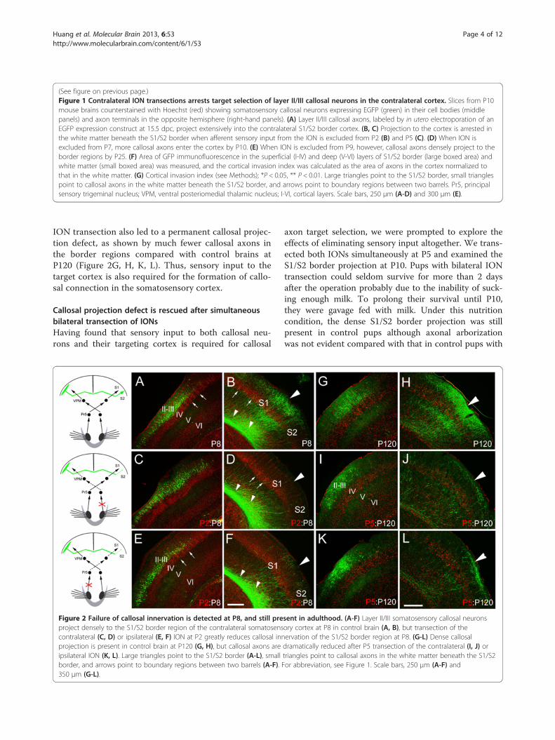
axons in the S1/S2 border cortex at P10 was due to theretraction of axons that have invaded the cortex at anearlier stage, because few axons were observed in thecortex at P8 after P2 (Figure 2C, D) and P5 ION transec-tion. In addition, the reduction of callosal projectioncaused by P2 and P5 ION transection (Figure 2I, J) wasalso observed at P120, reflecting a permanent targetingdefects. It should be noted that no ectopic callosal pro-jections were found in the ipsilateral or contralateralcortex, showing that the failure of targeting S1/S2 borderregion is not caused by a switch of targeting site follow-ing the removal of sensory input. As development pro-gressed, ION transection had progressively less effect.When ION transection was performed at P7, moreaxons were observed to have invaded the target cortexby P10 (Figure 1D, G). Sectioning the ION at P9 had nonotable effect, when callosal targeting was examined atP25 (Figure 1E). Taken together, these data suggest thatsensory input to callosal neurons is required continuallyfor S1/S2 border cortex invasion until targeting iscompleted.We next asked whether sensory input to the targeted
cortex is also required for the formation of this callosalprojection. To this end, we transected the ION ipsilat-eral to the side of electroporation (Figure 3). Transectionof the ipsilateral ION at P2 was also able to arrest callo-sal invasion of the contralateral S1/S2 border cortex(Figure 3B, E). This phenotype was likewise not due tobarrel structure malformations in the targeting somato-sensory cortex, because transection at P5 also precludedtarget innervation (Figure 3C, E). Ipsilateral ION tran-section at P7 or P9, however, had no notable effect ex-amined at P10 (Figure 3D, E) or P21.Similar to what were observed in the experiment of
contralateral transection of ION, a drastic reduction ofcallosal axons in the S1/S2 border region at P10 was notdue to the retraction of axons that have invaded the cor-tex at an earlier stage, because few axons were observedin the cortex at P8 after P2 (Figure 2E, F) and P5 ipsilat-eral ION transection. In addition, P2 and P5 ipsilateral
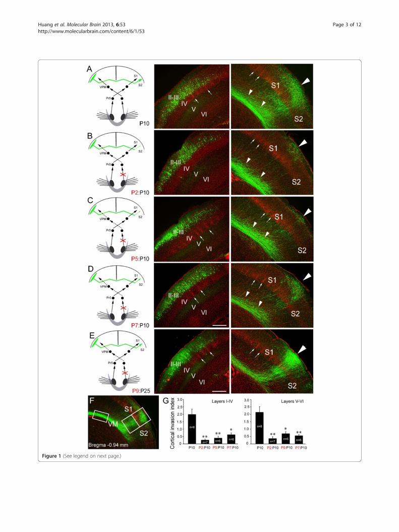
Figure 1 (See legend on next page.)
Huang et al. Molecular Brain 2013, 6:53 Page 3 of 12http://www.molecularbrain.com/content/6/1/53
(See figure on previous page.)Figure 1 Contralateral ION transections arrests target selection of layer II/III callosal neurons in the contralateral cortex. Slices from P10mouse brains counterstained with Hoechst (red) showing somatosensory callosal neurons expressing EGFP (green) in their cell bodies (middlepanels) and axon terminals in the opposite hemisphere (right-hand panels). (A) Layer II/III callosal axons, labeled by in utero electroporation of anEGFP expression construct at 15.5 dpc, project extensively into the contralateral S1/S2 border cortex. (B, C) Projection to the cortex is arrested inthe white matter beneath the S1/S2 border when afferent sensory input from the ION is excluded from P2 (B) and P5 (C). (D) When ION isexcluded from P7, more callosal axons enter the cortex by P10. (E) When ION is excluded from P9, however, callosal axons densely project to theborder regions by P25. (F) Area of GFP immunofluorescence in the superficial (I-IV) and deep (V-VI) layers of S1/S2 border (large boxed area) andwhite matter (small boxed area) was measured, and the cortical invasion index was calculated as the area of axons in the cortex normalized tothat in the white matter. (G) Cortical invasion index (see Methods); *P < 0.05, ** P < 0.01. Large triangles point to the S1/S2 border, small trianglespoint to callosal axons in the white matter beneath the S1/S2 border, and arrows point to boundary regions between two barrels. Pr5, principalsensory trigeminal nucleus; VPM, ventral posteriomedial thalamic nucleus; I-VI, cortical layers. Scale bars, 250 μm (A-D) and 300 μm (E).
Huang et al. Molecular Brain 2013, 6:53 Page 4 of 12http://www.molecularbrain.com/content/6/1/53
ION transection also led to a permanent callosal projec-tion defect, as shown by much fewer callosal axons inthe border regions compared with control brains atP120 (Figure 2G, H, K, L). Thus, sensory input to thetarget cortex is also required for the formation of callo-sal connection in the somatosensory cortex.
Callosal projection defect is rescued after simultaneousbilateral transection of IONsHaving found that sensory input to both callosal neu-rons and their targeting cortex is required for callosal
Figure 2 Failure of callosal innervation is detected at P8, and still preproject densely to the S1/S2 border region of the contralateral somatosenscontralateral (C, D) or ipsilateral (E, F) ION at P2 greatly reduces callosal innprojection is present in control brain at P120 (G, H), but callosal axons areipsilateral ION (K, L). Large triangles point to the S1/S2 border (A-L), smallborder, and arrows point to boundary regions between two barrels (A-F). F350 μm (G-L).
axon target selection, we were prompted to explore theeffects of eliminating sensory input altogether. We trans-ected both IONs simultaneously at P5 and examined theS1/S2 border projection at P10. Pups with bilateral IONtransection could seldom survive for more than 2 daysafter the operation probably due to the inability of suck-ing enough milk. To prolong their survival until P10,they were gavage fed with milk. Under this nutritioncondition, the dense S1/S2 border projection was stillpresent in control pups although axonal arborizationwas not evident compared with that in control pups with
sent in adulthood. (A-F) Layer II/III somatosensory callosal neuronsory cortex at P8 in control brain (A, B), but transection of theervation of the S1/S2 border region at P8. (G-L) Dense callosaldramatically reduced after P5 transection of the contralateral (I, J) ortriangles point to callosal axons in the white matter beneath the S1/S2or abbreviation, see Figure 1. Scale bars, 250 μm (A-F) and
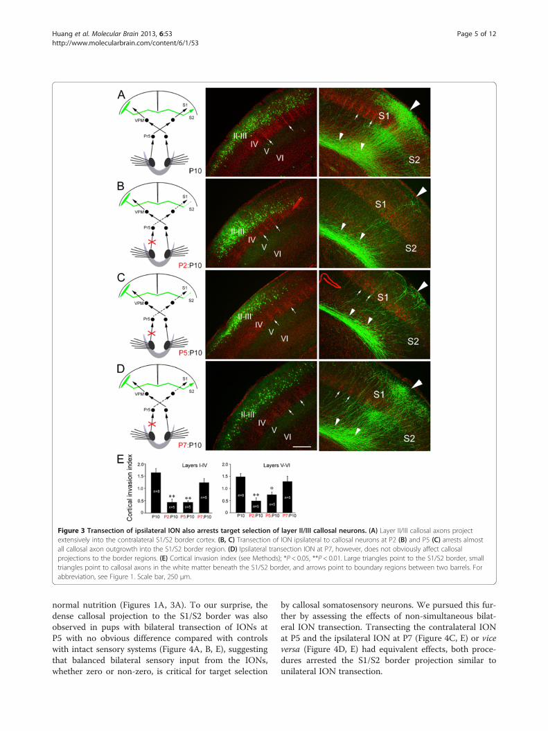
Figure 3 Transection of ipsilateral ION also arrests target selection of layer II/III callosal neurons. (A) Layer II/III callosal axons projectextensively into the contralateral S1/S2 border cortex. (B, C) Transection of ION ipsilateral to callosal neurons at P2 (B) and P5 (C) arrests almostall callosal axon outgrowth into the S1/S2 border region. (D) Ipsilateral transection ION at P7, however, does not obviously affect callosalprojections to the border regions. (E) Cortical invasion index (see Methods); *P < 0.05, **P < 0.01. Large triangles point to the S1/S2 border, smalltriangles point to callosal axons in the white matter beneath the S1/S2 border, and arrows point to boundary regions between two barrels. Forabbreviation, see Figure 1. Scale bar, 250 μm.
Huang et al. Molecular Brain 2013, 6:53 Page 5 of 12http://www.molecularbrain.com/content/6/1/53
normal nutrition (Figures 1A, 3A). To our surprise, thedense callosal projection to the S1/S2 border was alsoobserved in pups with bilateral transection of IONs atP5 with no obvious difference compared with controlswith intact sensory systems (Figure 4A, B, E), suggestingthat balanced bilateral sensory input from the IONs,whether zero or non-zero, is critical for target selection
by callosal somatosensory neurons. We pursued this fur-ther by assessing the effects of non-simultaneous bilat-eral ION transection. Transecting the contralateral IONat P5 and the ipsilateral ION at P7 (Figure 4C, E) or viceversa (Figure 4D, E) had equivalent effects, both proce-dures arrested the S1/S2 border projection similar tounilateral ION transection.
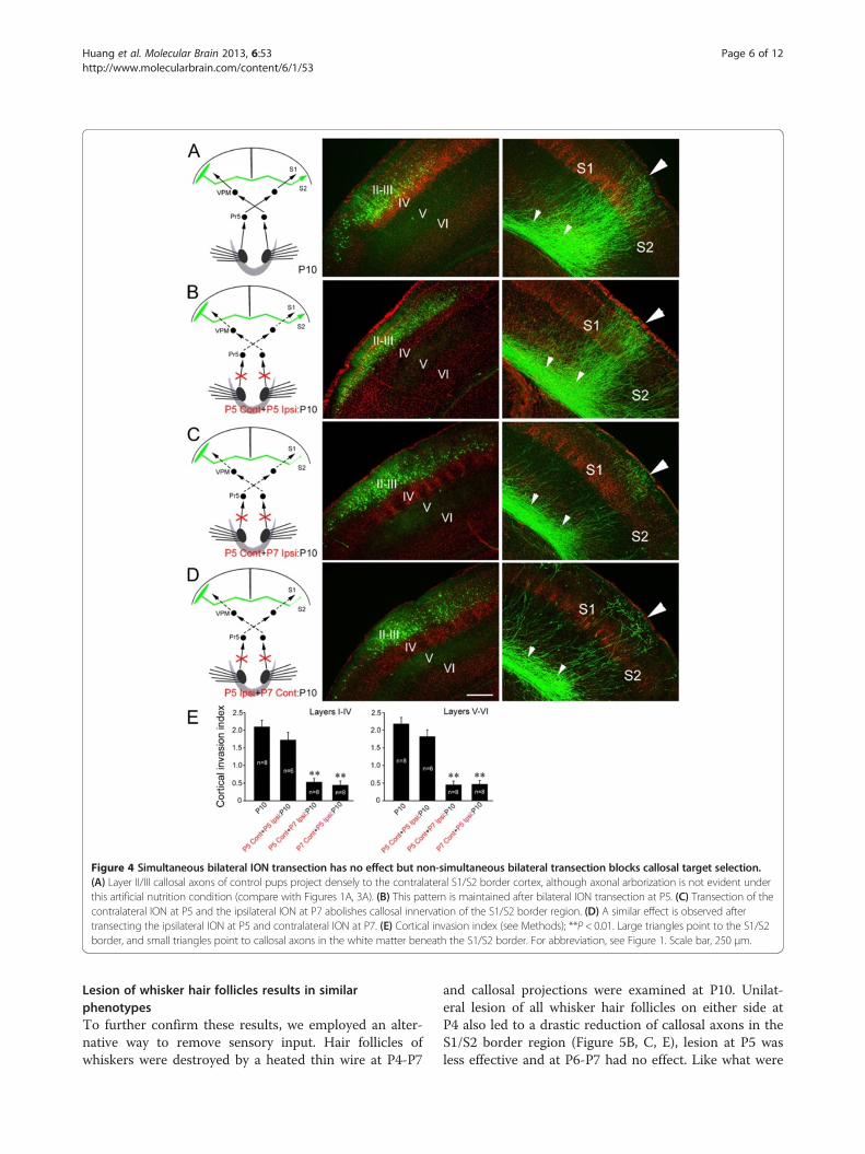
Figure 4 Simultaneous bilateral ION transection has no effect but non-simultaneous bilateral transection blocks callosal target selection.(A) Layer II/III callosal axons of control pups project densely to the contralateral S1/S2 border cortex, although axonal arborization is not evident underthis artificial nutrition condition (compare with Figures 1A, 3A). (B) This pattern is maintained after bilateral ION transection at P5. (C) Transection of thecontralateral ION at P5 and the ipsilateral ION at P7 abolishes callosal innervation of the S1/S2 border region. (D) A similar effect is observed aftertransecting the ipsilateral ION at P5 and contralateral ION at P7. (E) Cortical invasion index (see Methods); **P < 0.01. Large triangles point to the S1/S2border, and small triangles point to callosal axons in the white matter beneath the S1/S2 border. For abbreviation, see Figure 1. Scale bar, 250 μm.
Huang et al. Molecular Brain 2013, 6:53 Page 6 of 12http://www.molecularbrain.com/content/6/1/53
Lesion of whisker hair follicles results in similarphenotypesTo further confirm these results, we employed an alter-native way to remove sensory input. Hair follicles ofwhiskers were destroyed by a heated thin wire at P4-P7
and callosal projections were examined at P10. Unilat-eral lesion of all whisker hair follicles on either side atP4 also led to a drastic reduction of callosal axons in theS1/S2 border region (Figure 5B, C, E), lesion at P5 wasless effective and at P6-P7 had no effect. Like what were
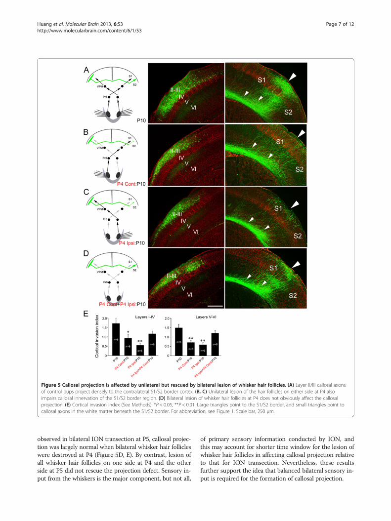
Figure 5 Callosal projection is affected by unilateral but rescued by bilateral lesion of whisker hair follicles. (A) Layer II/III callosal axonsof control pups project densely to the contralateral S1/S2 border cortex. (B, C) Unilateral lesion of the hair follicles on either side at P4 alsoimpairs callosal innervation of the S1/S2 border region. (D) Bilateral lesion of whisker hair follicles at P4 does not obviously affect the callosalprojection. (E) Cortical invasion index (See Methods); *P < 0.05, **P < 0.01. Large triangles point to the S1/S2 border, and small triangles point tocallosal axons in the white matter beneath the S1/S2 border. For abbreviation, see Figure 1. Scale bar, 250 μm.
Huang et al. Molecular Brain 2013, 6:53 Page 7 of 12http://www.molecularbrain.com/content/6/1/53
observed in bilateral ION transection at P5, callosal projec-tion was largely normal when bilateral whisker hair follicleswere destroyed at P4 (Figure 5D, E). By contrast, lesion ofall whisker hair follicles on one side at P4 and the otherside at P5 did not rescue the projection defect. Sensory in-put from the whiskers is the major component, but not all,
of primary sensory information conducted by ION, andthis may account for shorter time window for the lesion ofwhisker hair follicles in affecting callosal projection relativeto that for ION transection. Nevertheless, these resultsfurther support the idea that balanced bilateral sensory in-put is required for the formation of callosal projection.
Huang et al. Molecular Brain 2013, 6:53 Page 8 of 12http://www.molecularbrain.com/content/6/1/53
Neuronal activity is altered in the somatosensory cortexafter removal of sensory inputUnilateral or non-simultaneous bilateral removal of sen-sory input may disrupt the balance of bilateral sensoryinput to the two somatosensory cortices, which may inturn lead to mismatched activity between the two corti-ces. In contrast, neuron activity of the two cortices maymatch well after the simultaneous bilateral ION transec-tion, although it may be reduced. To test this possibility,c-Fos was used to examine cortical neuronal activity inP9 brain slices prepared from mice with P5 unilateral orsimultaneous bilateral removal of sensory input. It iswell known that neurons can be excited by high concen-tration of potassium in vitro. Brain slices were incubatedwith 50 mM KCl for 10 min and c-Fos staining wereperformed 40 min later. In brain slices prepared frommice with unilateral removal of sensory input (transec-tion of ION or lesion of whisker hair follicles on oneside), c-Fos-positive neurons were decreased more innumber in the contralateral brain slices compared withthe ipsilateral slices (Figure 6A-D, G). In brain sliceswith simultaneous bilateral removal of sensory input, thenumber of c-Fos-positive neurons on the two sides wassimilarly decreased (Figure 6E-G). These results suggestthat unilateral removal sensory input might lead to im-balanced neuronal activity between the two-side cortices,while simultaneous bilateral removal might not do so.We next used a whole cell patch clamp technique to
record the resting membrane potential, threshold forevoked action potential, and spontaneous excitatory post-synaptic current (sEPSC) of layer II/III cortical neuron inP7 mice with P5 unilateral or P5 bilateral ION transection.Compared to control mice, resting membrane potentialand threshold for evoked action potential showed nodifference in the two cortices of the mice with unilateralor bilateral ION transection, suggesting that electricalproperty of cortical neurons is not changed after P5ION transection. However, frequency of sEPSC decreaseddifferentially in the two side cortices with unilateral IONtransection, as shown by more reduction on the sidecontralateral to ION transection compared to that of theipsilateral side (Figure 6H). In contrast, in the mice withP5 bilateral ION transection, frequency of sEPSC wasreduced on both sides with no statistical difference(Figure 6H). Similar results were obtained in mice withunilateral or bilateral lesion of whisker hair follicles(Figure 6H). Amplitude of sEPSC was not changed inunilateral or bilateral ION-transected mice relative tocontrols. Considering the critical role of sensory inputin the maturation of somatosensory cortex, it might bepossible that the reduced frequency of sEPSC may re-flect less presynaptic glutamate release driven by per-ipheral sensory input, and imbalanced bilateral sensoryinput might lead to mismatched activity between the
two-side cortices whereby contributing to defective cal-losal targeting in the contralateral cortex.
DiscussionIn this study, we removed somatosensory input by trans-ecting the ION or lesion of whisker hair follicles toexamine the effects of sensory input on the developmentof the corpus callosum. Our results demonstrate thatsynchronous bilateral sensory input is required for targetselection of callosal neurons at system level. Unilateraltransection of ION on either side at P2-P5 arrested thecallosal projection, but it was less effective at P7 andshowed no effect after P7 (Figures 1, 3). These resultssuggest that the establishment of callosal projection re-quires peripheral sensory input, and, like the role of sen-sory input in barrel formation in the S1 [16,17], there isalso a “critical period” during which sensory input is re-quired but no longer required once the projection isformed.A previous study has shown that unilateral transection
of ION at birth in rat disrupts the callosal projection inthe S1/S2 border region when examined at postnatalone month [18]. Our data confirmed this finding inmouse, and further showed that the drastic reduction ofcallosal projection to the S1/S2 border region is dueto the failure of callosal invasion of the S1/S2 cortex(Figures 1, 2 and 3). The most interesting finding is thatbilateral transection of IONs at P5 did not obviouslyaffect the callosal projection, whereas bilateral transec-tion of IONs in the non-simultaneous way also arrestedthe projection (Figure 4). In addition to removal of per-ipheral sensory input, transection of IONs may also leadto other unknown effects that may contribute to the cal-losal projection phenotypes. To exclude this possibility,we employed an alternative method (lesion of whiskerhair follicles) to remove sensory input, and obtainedsimilar results (Figure 5). Thus, when bilateral sensoryinputs are removed in the same spatial and temporalfashion, callosal projection is maintained. On the otherhand, in the cases of unilateral or bilateral removal ofsensory input in a non-simultaneous way, the balance ofbilateral sensory input could be disrupted, and callosalinvasion of target cortex could not progress thus leadingto the drastic reduction of callosal projections. In sup-port of this, neuronal activity of two cortices shown byc-Fos immunostaining is matched in the mice with sim-ultaneous bilateral removal of sensory input although itis reduced on both sides, but not in those with unilateralremoval of sensory input (Figure 6). Considering the im-portant role of sensory input in the maturation of som-atosensory cortex [16,17] and the different reduction ofsEPSC frequency of cortical neurons (Figure 6), wespeculate that peripheral sensory input may be a majorfactor for this mismatched neuronal activity between the
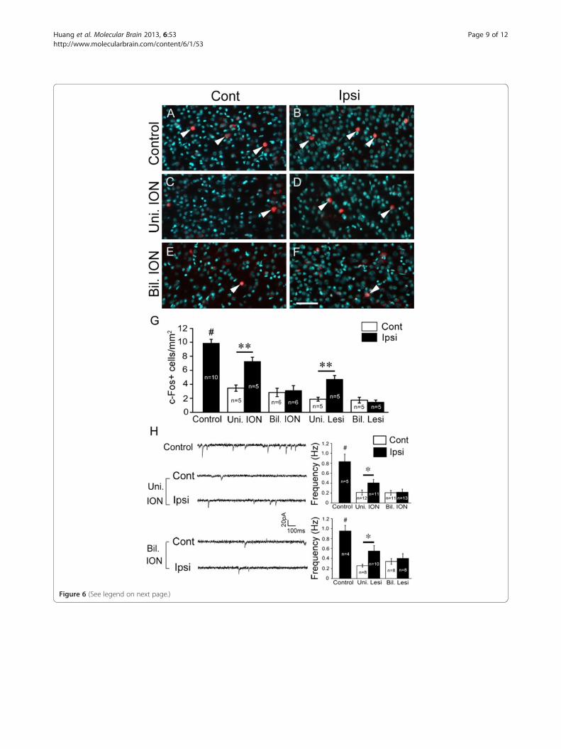
Figure 6 (See legend on next page.)
Huang et al. Molecular Brain 2013, 6:53 Page 9 of 12http://www.molecularbrain.com/content/6/1/53
(See figure on previous page.)Figure 6 Neuronal activity is altered in the somatosensory cortex after removal of sensory input. (A-G) c-Fos expression was examined inP9 brain slices. Compared with control brain (A, B), c-Fos-positive neurons are decreased more in number in P9 brain slices from the contralateralside compared with ipsilateral side (C, D) after P5 unilateral ION transection. While in the brain with P5 bilateral ION transection, c-Fos-positiveneurons are similarly reduced in number on both sides (E, F). Nuclear Hoechst counterstain is shown with pseudocolor (cyan), and c-Fos-positiveneurons are indicated by triangles. Comparison of numbers of c-Fos-positive neurons is shown in (G); animal numbers examined are indicated.**P < 0.01; #P < 0.01 (control vs the others). (H) Patch-clamp recording was performed in layer II/III neurons of the S1 region of brain slicesprepared from P7 mice. Frequency of sEPSC is reduced on both sides in mice with bilateral unilateral removal of sensory input compared withcontrol mice, but it shows more reduction in the contralateral side compared with that in the ipsilateral side. In mice with bilateral removal ofsensory input, frequency of sEPSC of the cortical neurons in the S1 is equally reduced on both sides. Representative recordings in brain sliceprepared from mice with ION transection are shown in left panel, and quantification of the sEPSC frequency is shown in right two panels. At leastthree mice were examined in each group, and neuron numbers for each recoding are indicated. *P < 0.05, #P < 0.01 (control vs the others). Bil.ION, bilateral transection of IONs; Bil. Lesi, bilateral lesion of whisker hair follicles; Cont, contralateral side; Ipsi, ipsilateral side; Uni. ION, unilateraltransection of ION; Uni. Lesi, unilateral lesion of whisker hair follicles. Scale bar, 60 μm.
Huang et al. Molecular Brain 2013, 6:53 Page 10 of 12http://www.molecularbrain.com/content/6/1/53
two cortices in mice with unilateral removal of sensory in-put. However, it should be noted that our neurophysio-logical results are not sufficient to establish a causal linkbetween the mismatched neuronal activity and defectivecallosal projection, and further studies are needed to ex-plore this question.Interestingly, unilateral and bilateral disruption of vis-
ual sensory input with monocular or binocular vision byenucleation, or dark rearing at birth does not preventtargeting of cortical callosal projections from innervatingthe contralateral cortex [19]. The essential role of bal-anced bilateral sensory input might explain this differ-ence. Unlike the somatosensory system, visual inputfrom one eye is delivered to both visual cortices, andthus either monocular or binocular enucleation or darkrearing would not disrupt the balanced sensory inputs inthe two hemispheres. In contrast, unilateral lesion of theoptic tract, which eliminates all visual input to onehemisphere, results in a nearly complete absence of cal-losal connections in the border region between the pri-mary and secondary visual cortices [19]. Thus, it mightbe possible that callosal neurons in both somatosensoryand visual cortices require matched bilateral sensory in-put to form their callosal connections.It is well accepted that prior to initial contact formation,
intrinsic developmental programs and external guidancecues are responsible for guiding growing axons into theirtarget field [20-23], and sensory input is required for refin-ing and consolidating neuronal connections in developingneuronal networks [24,25]. On the other hand, accumu-lated evidence indicates that neuronal activity is critical inaxon pathfinding. For example, spontaneous rhythmic ac-tivity in early chick spinal cord influences distinct motoraxon pathfinding decisions [26], and intraventricular injec-tion of tetrodotoxin blocks cortical target selection of tha-lamocortical axons [27]. On the basis of these findings, itis likely that during early postnatal development sensoryinput-driven neuronal activity plays an important role inguiding growing axons to select correct target to establishproper neuronal connections.
ConclusionsIn this study, we found that the callosal projection to thecontralateral cortex was arrested by unilateral transectionof ION or lesion of all whisker follicles before P7. Whilesimultaneous bilateral ION transection or lesion of whis-ker hair follicles did not obviously affect the callosal pro-jection, whereas non-simultaneous bilateral transection ofIONs or lesions whisker hair follicles arrested the callosalprojection. Besides, the neuronal activity is altered in thesomatosensory cortex after removal of sensory input. Ourresults not only show the requirement of sensory input inwiring of callosal connection, but also reveal the criticalrole of balanced bilateral sensory input in this develop-mental process.
Materials and methodsIn utero electroporationIn utero electroporation was performed on timed preg-nant C57BL/6 mice at 15.5 days post-coitum (dpc) aspreviously described [5]. After anesthesia with sodiumpentobarbital, pregnant mice were subjected to abdom-inal incision to expose the uterus. The CAG-EGFP plas-mid (1.5 μg/μl) was injected into the lateral ventriclewith a glass capillary through the uterine wall. Electricpulses were then delivered to the embryos by gentlyclasping their heads with forceps-shaped electrodes con-nected to a square-pulse generator, ECM-830 (BTX;Holliston, MA). The pups of either sex were allowed tosurvive to different postnatal stages, and perfused with4% of paraformaldehyde in 0.01 M phosphate bufferedsaline (pH 7.4) under deep anesthesia. Animal care andexperimental protocols were approved by the AnimalCenter of Tongji University School of Medicine, China.
ION transectionThe IONs of P2-P9 pups were severed as described pre-viously [16,17]. Since pups with bilateral IONs transec-tion were unable to suck breast milk, they were gavagefed with milk 3-5 times daily, and control pups with
Huang et al. Molecular Brain 2013, 6:53 Page 11 of 12http://www.molecularbrain.com/content/6/1/53
sham operation in this set of experiment were treated inthe same way.
Electrophysiological recordingTo examine electrophysiological changes of corticalneuron after unilateral or bilateral removal of sensory in-put, patch-clump recording was performed in brain slice.Brains from P7 mice of either sex were removed andtransverse slices (350 μm) were cut on a vibrating micro-slicer (Leica VT1200, Germany). Whole-cell patch-clamprecordings were performed after the brain slices at aproximate level of Bregma -0.22 – -1.94 mm were incu-bated for 1 h in external artificial CSF (in mM: NaCl 117,KCl 3.6, CaCl2 2.5, MgCl2 1.2, NaH2PO4 1.2, NaHCO3 25and glucose 11) which was bubbled continuously with car-bogen (95%O2/5%CO2). Recording pipettes with resis-tances of 3–5 MΩ were pulled from borosilicate glass(P-97; Sutter Instruments, Novato, CA) and filled with asolution of (in mM) potassium gluconate 135, KCl 5,CaCl2 0.5, MgCl2 2, EGTA 5, HEPES 5 and ATP-Mg 5.Resting membrane potential and the threshold for evok-ing action potential were measured in layer II-III corticalneurons in current clamp mode, and sEPSC (spontaneousexcitatory post synaptic current) was also recorded in volt-age clamp mode held at -70 mV. The signals were ampli-fied with an Axopatch 700B amplifier (Molecular Devices,Sunnyvale, CA), filtered at 2 kHz, and digitized at 5 kHz.Four-thirteen neurons were recorded in each group, anddata were stored with a personal computer using softwareof pCLAMP 10 and analyzed with Mini Analysis (Synap-tosoft Inc., Fort Lee, NJ). Comparisons were performedusing the One-Way ANOVA with post-hoc Fisher’s LSDtest, and P values less than 0.05 were considered statisti-cally significant.
ImmunohistochemistryFixed brains were cut into 40 μm-thick transverse sectionsand processed for GFP (1:2000; Invitrogen, Grand Island,NY) immunostaining as described in our previous study[5]. Sections were counterstained with Hoechst 33258(Sigma, St. Louis, MO) to clearly identify the morpho-logical features defining somatosensory cortex.To examine the changes of neuronal activity in the som-
atosensory cortex after unilateral or bilateral removal ofsensory input, c-Fos immunostaining was performed in P9brain slice (400 μm) prepared as mentioned above. Afterbeing incubated in the oxygenated artificial CSF for 1 h,brain slices were incubated in 50 mM KCl (Sigma) in CSFfor 10 min and then with CSF without KCl for 40 min.After fixation, brain slices were cut into 20 μm-thick sec-tions using a cryostat and processed for c-Fos immuno-staining (1:1000; Santa Cruz, Dallas, TX). Immunostainedsections were counterstained with Hoechst (Sigma), andc-Fos-positive neurons in the S1/S2 border region were
counted in all sections obtained. Comparisons wereperformed using the One-Way ANOVA with post-hocFisher’s LSD test and P values less than 0.05 wereconsidered statistically significant.
Image analysisMedio-lateral extents of GFP-labeled neurons in electro-porated cortices varied, and brains containing GFP+ neu-rons located within the S1-S2 region were included. Everysixth sections were collected as one set section, in whichabout three sections were included at the approximatelevel of Bregma -0.22 – -1.94 mm in most brain samples[28]. These three sections were used for image analysis be-cause callosal projection shows high consistency at thislevel along the rostro-caudal axis in control brains. Callo-sal axons projects primarily to the S1/S2 border region[5], and we thus quantified callosal axons in this area onthe contralateral side. As shown in Figure 1F, ImageJsoftware (NIH) was used to measure the area of GFPimmunofluorescence in the superficial (I-IV) and deep(V-VI) layers of the S1/S2 border region; this area cov-ered the entire S1/S2 border region target territory. Thecortical invasion index was calculated as the area ofaxons in the cortex normalized to that in the white mat-ter (a small boxed area in Figure 1F). Individual datawere pooled into groups and compared by One-WayANOVA with post-hoc Fisher’s LSD test for multiplecomparisons versus the control.
AbbreviationsBil. ION: Bilateral transection of IONs; Bil. Lesi: Bilateral lesion of whisker hairfollicles; Cont: Contralateral side; dpc: Days post-coitum; ION: Infraorbitalnerve; Ipsi: Ipsilateral side; P5: Postnatal day 5; Pr5: Principal sensorytrigeminal nucleus; S1: Primary somatosensory cortex; S2: Secondarysomatosensory cortex; sEPSC: Spontaneous excitatory postsynaptic current;VPM: Ventral posteriomedial thalamic nucleus; Uni. ION: Unilateral transectionof ION; Uni. Lesi: Unilateral lesion of whisker hair follicles.
Competing interestsThe authors declare that they have no competing interests.
Authors’ contributionYQD conceived the study. YH, NNS, CJZ, LJL, LX and YQD designed theresearch. YH, NNS, WL, QZ, LeiZ, LH and JYC performed immunostaining andanalyzed the data. LingZ performed electrophysiology and analyzed its data.LeiZ performed in utero electroporation. YH, NNS and YQD wrote the paper.All authors read and approved the final manuscript.
AcknowledgementsThis work was supported by the National Natural Science Foundation ofChina (81221001, 91232724, 81101026, 31100788, 31030034), Key StateResearch Program from Ministry of Science and Technology of China(2012CB966900, 2011CB510005), Science and Technology Commission ofShanghai Municipality (11140902200, 12XD1404800) and FundamentalResearch Funds for the Central Universities.
Author details1Key Laboratory of Arrhythmias, Ministry of Education, East Hospitial, TongjiUniversity School of Medicine, 1239 Siping Road, Shanghai 200092, China.2Department of Anatomy and Neurobiology, Tongji University School ofMedicine, 1239 Siping Road, Shanghai 200092, China. 3Key Laboratory ofDevelopmental Genes and Human Diseases, Ministry of Education, School of
Huang et al. Molecular Brain 2013, 6:53 Page 12 of 12http://www.molecularbrain.com/content/6/1/53
Medicine, Southeast University, 87 Dingjiaqiao Road, Nanjing, Jiangsu210009, China. 4Institute of Mental Health, Second Xiangya Hospital ofCentral South University, Changsha 410011, China. 5Key Laboratory of AnimalModels and Human Disease Mechanisms, Chinese Academy of Sciences andYunnan Province, Kunming Institute of Zoology, Kunming 650223, China.
Received: 24 November 2013 Accepted: 27 November 2013Published: 5 December 2013
References1. Polleux F, Ince-Dunn G, Ghosh A: Transcriptional regulation of vertebrate
axon guidance and synapse formation. Nat Rev Neurosci 2007, 8:331–340.2. Wolman D: A tale of two halves. Nature 2012, 483:260–263.3. Richards LJ, Plachez C, Ren T: Mechanisms regulating the development of
the corpus callosum and its agenesis in mouse and human. Clin Genet2004, 66:276–289.
4. Mizuno H, Hirano T, Tagawa Y: Evidence for activity-dependent corticalwiring: formation of interhemispheric connections in neonatal mousevisual cortex requires projection neuron activity. J Neurosci 2007,27:6760–6770.
5. Wang C-L, Zhang L, Zhou Y, Zhou J, Yang X-J, Duan S-m, Xiong Z-Q,Ding Y-Q: Activity-dependent development of callosal projections in thesomatosensory cortex. J Neurosci 2007, 27:11334–11342.
6. Chinnasamy D, Rudd R, Velakoulis D: A case of schizophrenia withcomplete agenesis of the corpus callosum. Aust Psychiatry 2006,14:327–330.
7. Motomura N, Satani S, Inaba M: Monozygotic twin cases of the agenesisof the corpus callosum with schizophrenic disorder. Psychiatry ClinNeurosci 2002, 56:199–202.
8. Serur D, Jeret JS, Wisniewski K: Agenesis of the corpus callosum: clinical,neuroradiological and cytogenetic studies. Neuropediatrics 1988, 19:87–91.
9. Serafini T, Colamarino SA, Leonardo ED, Wang H, Beddington R, Skarnes WC,Tessier-Lavigne M: Netrin-1 Is required for commissural axon guidance inthe developing vertebrate nervous system. Cell 1996, 87:1001–1014.
10. Bagri A, Mar1 n O, Plump AS, Mak J, Pleasure SJ, Rubenstein JLR, Tessier-LavigneM: Slit proteins prevent midline crossing and determine the dorsoventralposition of major axonal pathways in the mammalian forebrain. Neuron 2002,33:233–248.
11. Qiu M, Anderson S, Chen S, Meneses JJ, Hevner R, Kuwana E, Pedersen RA,Rubenstein JLR: Mutation of the Emx-1homeobox gene disrupts thecorpus callosum. Dev Biol 1996, 178:174–178.
12. Stoykova A, Fritsch R, Walther C, Gruss P: Forebrain patterning defects inSmall eye mutant mice. Development 1996, 122:3453–3465.
13. Chae T, Kwon YT, Bronson R, Dikkes P, Li E, Tsai L-H: Mice lacking p35, aneuronal specific activator of Cdk5, display cortical lamination defects,seizures, and adult lethality. Neuron 1997, 18:29–42.
14. Chapman B, Jacobson MD, Reiter HO, Stryker MP: Ocular dominance shiftin kitten visual cortex caused by imbalance in retinal electrical activity.Nature 1986, 324:154–156.
15. Frenkel MY, Bear MF: How monocular deprivation shifts oculardominance in visual cortex of young mice. Neuron 2004, 44:917–923.
16. Rice FL, Van der Loos H: Development of the barrels and barrel field inthe somatosensory cortex of the mouse. J Comp Neurol 1977,171:545–560.
17. Van der Loos H, Woolsey TA: Somatosensory cortex: structural alterationsfollowing early injury to sense organs. Science 1973, 179:395–398.
18. Koralek KA, Killackey HP: Callosal projections in rat somatosensory cortexare altered by early removal of afferent input. Proc Natl Acad Sci USA1990, 87:1396–1400.
19. Cusick CG, Lund RD: Modification of visual callosal projections in rats.J Comp Neurol 1982, 212:385–398.
20. Brose K, Tessier-Lavigne M: Slit proteins: key regulators of axon guidance,axonal branching, and cell migration. Curr Opin Neurobiol 2000, 10:95–102.
21. Friedman GC, O’Leary DDM: Eph receptor tyrosine kinases and theirligands in neural development. Curr Opin Neurobiol 1996, 6:127–133.
22. Imai T, Yamazaki T, Kobayakawa R, Kobayakawa K, Abe T, Suzuki M, Sakano H:Pre-target axon sorting establishes the neural map topography.Science 2009, 325:585–590.
23. Zou Y, Lyuksyutova AI: Morphogens as conserved axon guidance cues.Curr Opin Neurobiol 2007, 17:22–28.
24. Grubb MS, Thompson ID: The influence of early experience on thedevelopment of sensory systems. Cur Opin Neurobiol 2004, 14:503–512.
25. Innocenti GM, Price DJ: Exuberance in the development of corticalnetworks. Nat Rev Neurosci 2005, 6:955–965.
26. Hanson MG, Landmesser LT: Normal patterns of spontaneous activity arerequired for correct motor axon guidance and the expression of specificguidance molecules. Neuron 2004, 43:687–701.
27. Catalano SM, Shatz CJ: Activity-dependent cortical target selection bythalamic axons. Science 1998, 281:559–562.
28. Paxinos G, Franklin KB: The Mouse Brain in Stereotaxic Coordinates. 2ndedition. San Diego: Academic; 2001.
doi:10.1186/1756-6606-6-53Cite this article as: Huang et al.: Sensory input is required for callosalaxon targeting in the somatosensory cortex. Molecular Brain 2013 6:53.
Submit your next manuscript to BioMed Centraland take full advantage of:
• Convenient online submission
• Thorough peer review
• No space constraints or color figure charges
• Immediate publication on acceptance
• Inclusion in PubMed, CAS, Scopus and Google Scholar
• Research which is freely available for redistribution
Submit your manuscript at www.biomedcentral.com/submit