Embed Size (px)
DESCRIPTION
"Melanoma cells can switch between an elongated mesenchymal-type and a rounded amoeboid-type migration mode. The rounded‘amoeboid’ form of cell movement is driven by actomyosin contractility resulting in membrane blebbing. Unlike elongated A375melanoma cells, rounded A375 cells do not display any obvious morphological front–back polarisation, although polarisation isthought to be a prerequisite for cell movement. We show that blebbing A375 cells are polarised, with ezrin (a linker between the plasmamembrane and actin cytoskeleton), F-actin, myosin light chain, plasma membrane, phosphatidylinositol (4,5)-bisphosphate and b1-integrin accumulating at the cell rear in a uropod-like structure. This structure does not have the typical protruding shape of classicalleukocyte uropods, but, as for those structures, it is regulated by protein kinase C. We show that the ezrin-rich uropod-like structure(ERULS) is an inherent feature of polarised A375 cells and not a consequence of cell migration, and is necessary for cell invasion.Furthermore, we demonstrate that membrane blebbing is reduced at this site, leading to a model in which the rigid ezrin-containingstructure determines the direction of a moving cell through localised inhibition of membrane blebbing."(Lorentzen et al, 2010)
Citation preview

An ezrin-rich, rigid uropod-like structure directs movement of amoeboid blebbing cells
Anna Lorentzen, Jeffrey Bamber, Amine Sadok, Ilan Elson-Schwab and Christopher J. Marshall.
Tamara Heredia
27/11/2014
Blebbing
Fig. 1 Schematic of a bleb formation. (Fackler et al, 2008)

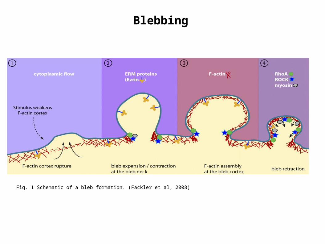
Mesenchymal-type elongated mode Rounded mode of movement or Ameboid mode
Requires degradation of the ECM by proteases
Is less dependent on proteases. Cells move by membrane blebbing or by squeezing through pores in the ECM.
Requires low actomyosin contractility High degree of actomyosin contractility
Elongated morphology Rounded morphology
Single migrating cells
(Amit Pathak et al, 2010)

Note: Uropod also contains protein kinase A (PKA) and phosphatidylinositol (4,5)-bisphosphate [PtdIns(4,5)P2]
Fig 2. Illustration of the establishment of two poles in migrating leukocytes, adhesion molecules and cytoskeletal elements. (Francisco Sanchez-Madrid1 et al, 1999)
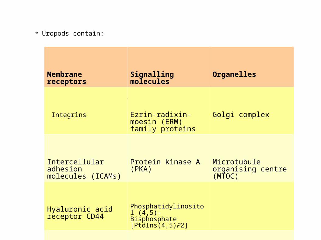
Uropods contain:
The uropod contains:
Membrane receptors Signalling molecules Organelles
Integrins Ezrin-radixin-moesin (ERM) family proteins
Golgi complex
Intercellular adhesion molecules (ICAMs)
Protein kinase A (PKA) Microtubule organising centre (MTOC)
Hyaluronic acid receptor CD44
Phosphatidylinositol (4,5)-Bisphosphate [PtdIns(4,5)P2]
P-selectin glycoprotein ligand-1 (PSGL-1)


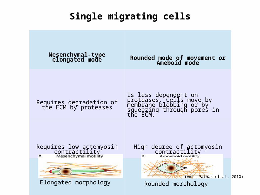
Nonadhesive amoeboid movement
Characterised by the formation of membrane blebs
It has been observed in Dictyostelium and cancer cells.
Cells with blebbing amoeboid movement lack apparent polarisation and have high actomyosin contractility .
.
Fig. 3 (A) Amoeboid cell crawling on a two-dimensional surface. Lamellipodium formation at the front determines the direction of movement. The front of the cell adheres to the substrate in focal adhesions through integrins. Retraction of the uropod at the rear creates the force that moves the cell forward. (B) Amoeboid cell squeezing through pores in a three-dimensional matrix. A membrane bleb grows into a gap in the ECM. (C )Movement by cells forming multiple small blebs. High contractility of the cell creates pressure that is released in membrane blebs.

In this research:
1. It is investigated the polarisation in blebbing “amoeboid” A375 melanoma cells in order to
understand cell movement driven by multiple blebs.
A375 cells: human melanoma cell with invasive and metastatic properties. A375 melanoma cells form multiple blebs over the membrane and would therefore not be able to move in one determined direction.
A375P means that the cell line contain low concentration of A375, A375M-medium and A375SM-high concentration.
2. It is shown that A375 cells form an ezrin-rich uropod-like structure (to which the autors call the
ERULS) at their cell rear, and that this is required for invasion into a three-dimensional matrix.
3. It is presented a model explaining how cells can move without a defined front and only a defined
rear.
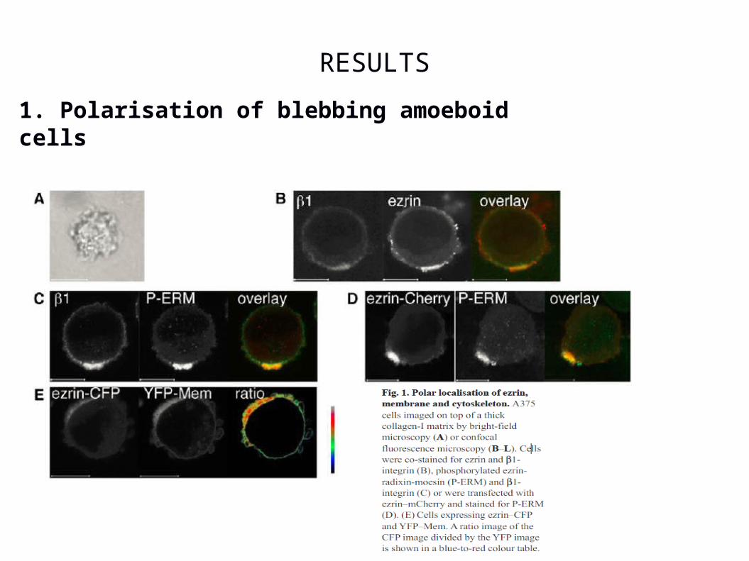
RESULTS
1. Polarisation of blebbing amoeboid cells
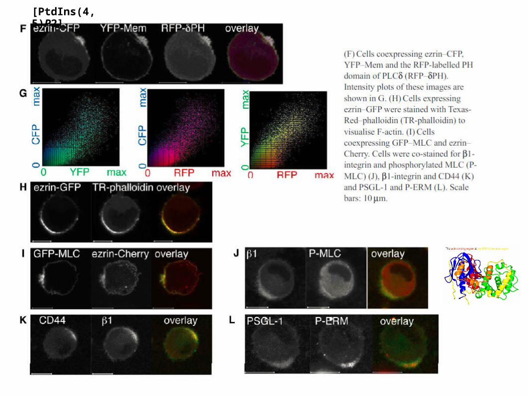
[PtdIns(4,5)P2]
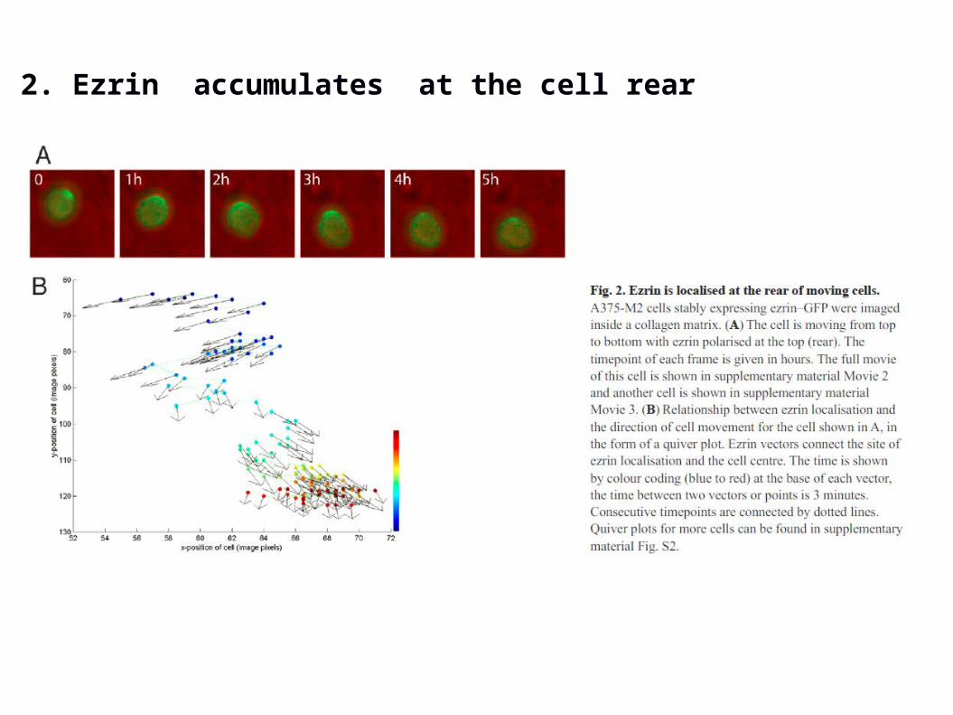
2. Ezrin accumulates at the cell rear
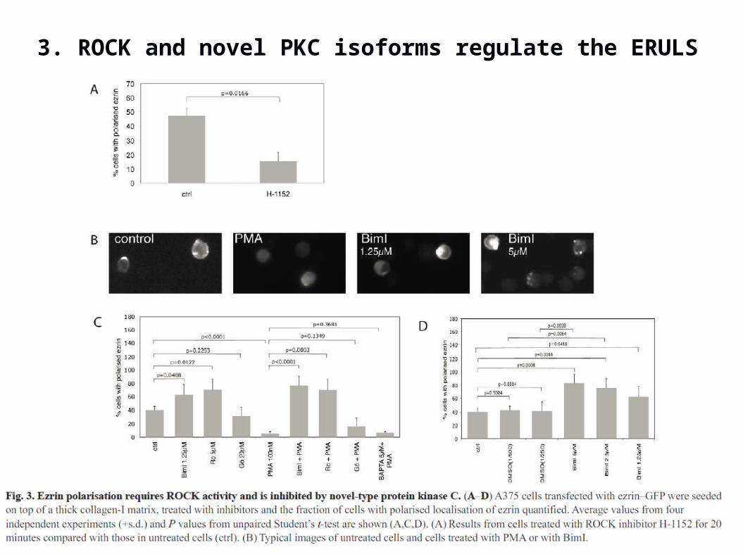
3. ROCK and novel PKC isoforms regulate the ERULS
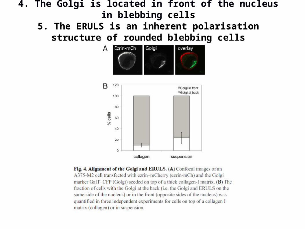
4. The Golgi is located in front of the nucleus in blebbing cells
5. The ERULS is an inherent polarisation structure of rounded blebbing cells
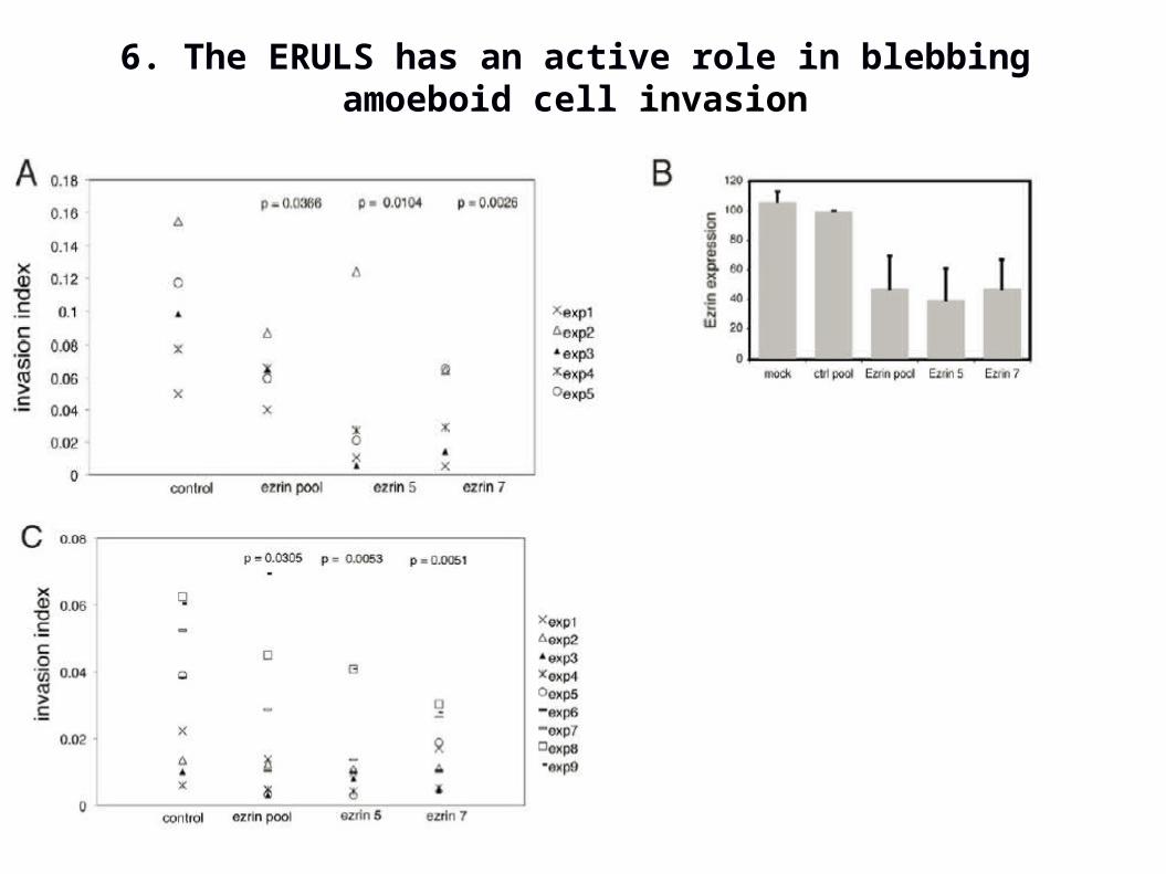
6. The ERULS has an active role in blebbing amoeboid cell invasion

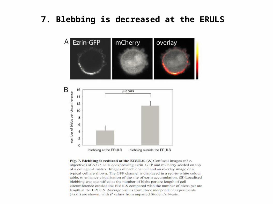
7. Blebbing is decreased at the ERULS
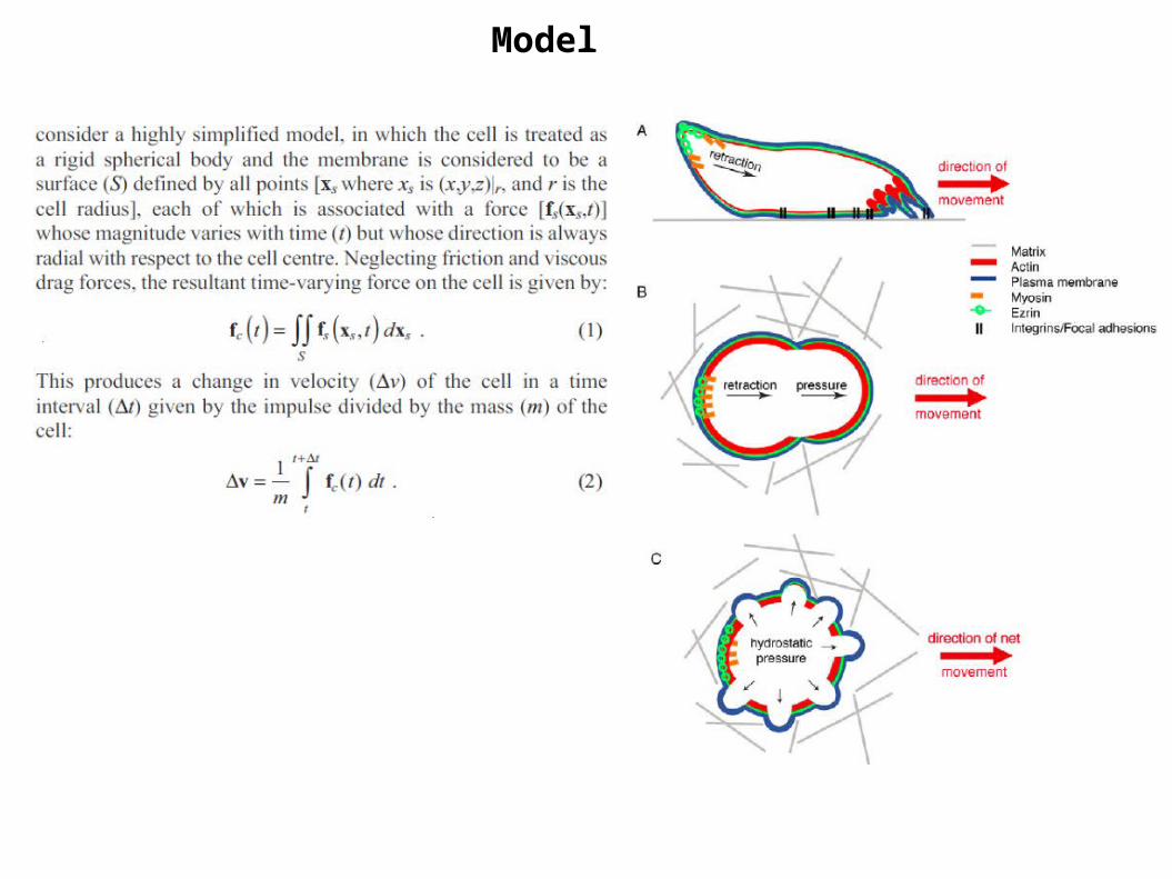
Model
.
.

Summary
It was investigated how melanoma cells with a round morphology and many small blebs over their surface can move without displaying morphological features of a front or a back.
It was shown that blebbing melanoma cells form a uropod-like structure (ERULS) at the rear that is enriched in ezrin, b1-integrin, actin, MLC and PtdIns(4,5)P2.
It was shown that this structure is a feature of polarised round blebbing cells and is not a consequence of cell movement.
It was shown that the ERULS is enriched in the phosphorylated active form of ezrin.
It was identified the Golgi alignment in front of the nucleus in blebbing melanoma cells, which is the opposite of the alignment in migrating leukocytes.
Also it was proposed a model explaining how cells can move without a defined front and only a defined rear.
References
A. Lorentzen, J. Bamber, A. Sadok, I. Elson-Schwab, C.J. Marshall, An ezrin-rich, rigid uropod-like structure directs movement of amoeboid blebbing cells, J Cell Sci, 124. 2010
Charras G., Paluch E. Blebs lead the way: how to migrate without lamellipodia. Nat Rev Mol Cell Biol. 2008
Fackler O.T., Grosse R. Cell motility through plasma membrane blebbing. J Cell Biol. 2008
Fehon R.G., McClatchey A.I., Bretscher A. Organizing the cell cortex: the role of ERM proteins. Nat Rev Mol Cell Biol. 2010
Yoshida K., Soldati T. Dissection of amoeboid movement into two mechanically distinct modes. J Cell Sci. 2006
Merkel R., Simson R., Simson D.A., Hohenadl M., Boulbitch A., Wallraff E., Sackmann E. A micromechanic study of cell polarity and plasma membrane cell body coupling in Dictyostelium. Biophys J. 2000
Vicente-Manzanares, M., Sanchez-Madrid, F, Cell polarization: a comparative cell biology and immunological. 2000