Embed Size (px)
Citation preview
1
CSS/HRT 451 CSS/HRT 451 CSS/HRT 451
Guo-qing Song & David DouchesJanuary 2009
Selectable Markers & Markers for Screening
2
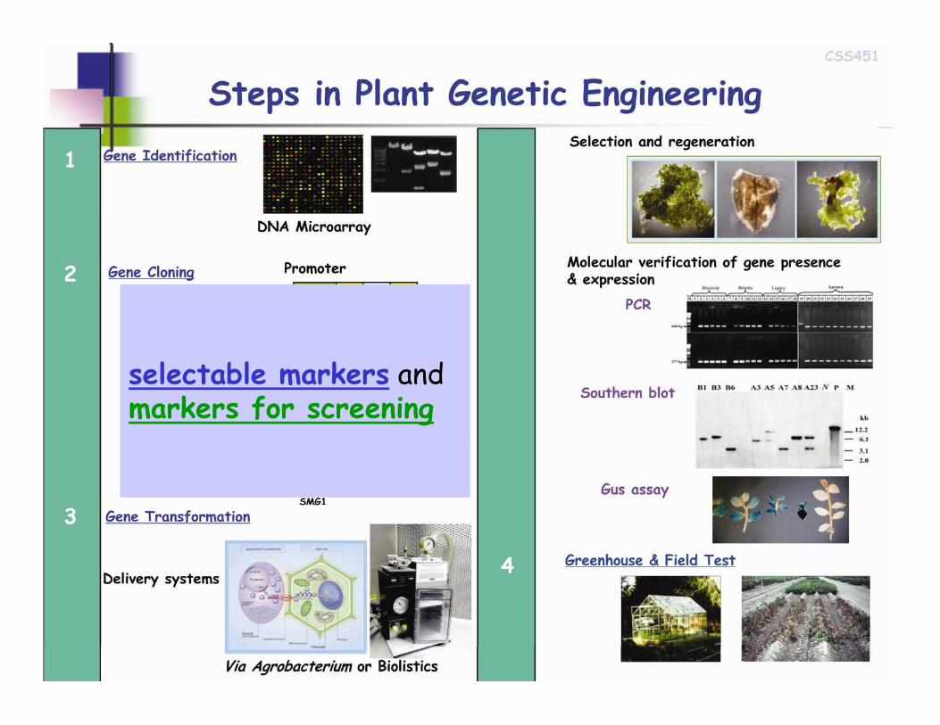
A marker gene is used in molecular biology to determine if a piece of DNA has been successfully inserted into the host organism. There are two types of marker genes: selectable markers and markers for screening.
Marker gene
3
Gene Clone and DNA Analysis in Agriculture
Chapter 15
Chapter 15.3 p341-344
4
selectable markers and markers for screening
5
Selectable markers &Markers for screening
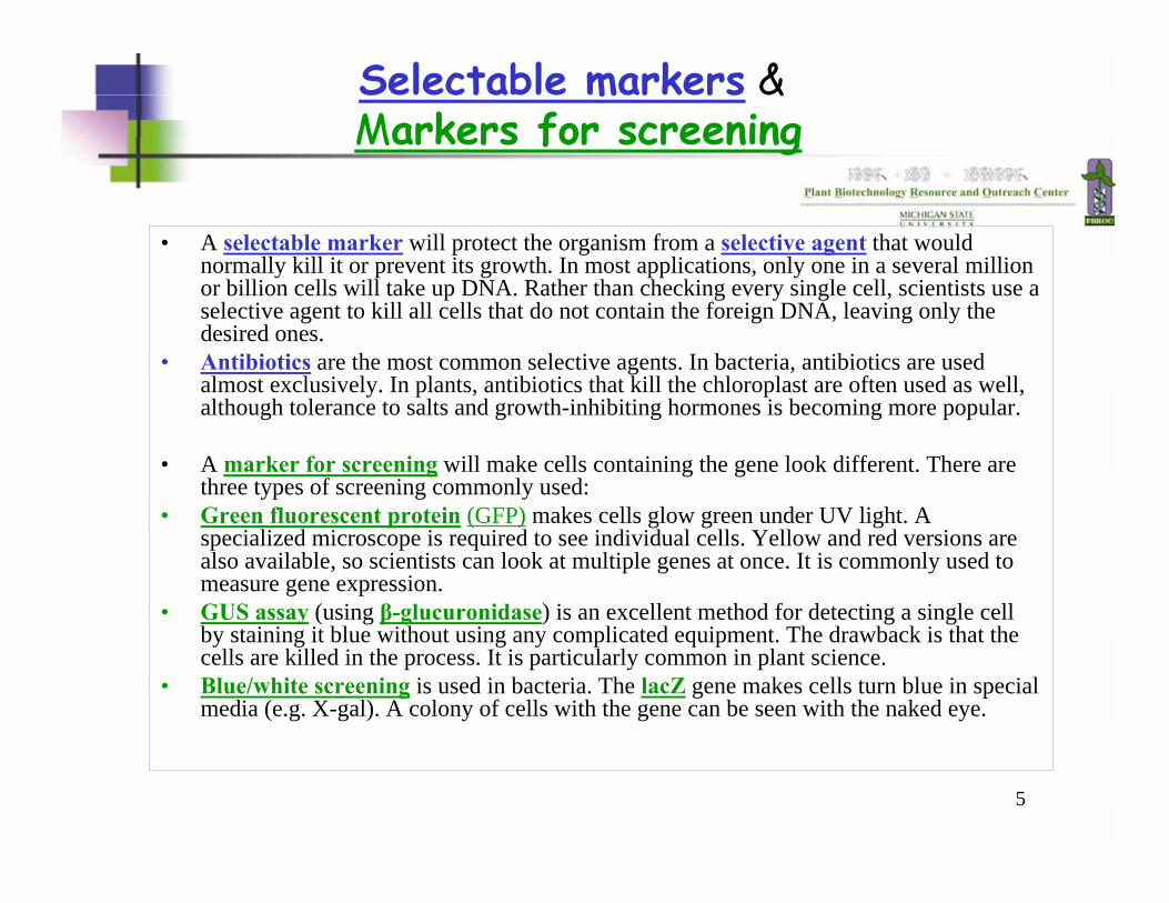
• A selectable marker will protect the organism from a selective agent that would normally kill it or prevent its growth. In most applications, only one in a several million or billion cells will take up DNA. Rather than checking every single cell, scientists use a selective agent to kill all cells that do not contain the foreign DNA, leaving only the desired ones.
• Antibiotics are the most common selective agents. In bacteria, antibiotics are used almost exclusively. In plants, antibiotics that kill the chloroplast are often used as well, although tolerance to salts and growth-inhibiting hormones is becoming more popular.
• A marker for screening will make cells containing the gene look different. There are three types of screening commonly used:
• Green fluorescent protein (GFP) makes cells glow green under UV light. A specialized microscope is required to see individual cells. Yellow and red versions are also available, so scientists can look at multiple genes at once. It is commonly used to measure gene expression.
• GUS assay (using β-glucuronidase) is an excellent method for detecting a single cell by staining it blue without using any complicated equipment. The drawback is that the cells are killed in the process. It is particularly common in plant science.
• Blue/white screening is used in bacteria. The lacZ gene makes cells turn blue in special media (e.g. X-gal). A colony of cells with the gene can be seen with the naked eye.
6
Literature
Miki, B., McHugh, S. Selectable Marker Genes in Transgenic Plants - Applications, Alternatives and Biosafety. Journal of Biotechnology. 2004. 107(3): 193-232.
Hare, P., Chua, N. Excision of Selectable Marker Genes from Transgenic Plants. Nature Biotechnology. 2002. 20(6): 575-580.
Goldstein et al. A Review - Human Safety and Genetically Modified Plants - A Review of Antibiotic Resistance Markers and Future Transformation Selection Technologies. Journal of Applied Microbiology. 2005. 99: 7-23.
Ramessar, K., Peremarti, A., Gomez-Galera, S., Naqvi, S., Moralejo, M., Munoz, P., Capell, T., Christou, P. Biosafety and Risk Assessment Framework for Selectable Marker Genes in Transgenic Crop Plants: A Case of the Science Not Supporting the Politics. Transgenic Research. 2007. 16(3): 261-280.

7
Selectable Markers

8
Selectable Markers
•• About About 50 50 selectable marker genesselectable marker genes
•• Six negative SMG: Six negative SMG: codAcodA, , aux2aux2, , tms2tms2, , dhlAdhlA, , CYP105ACYP105A, and , and cuecue
•• nptIInptII, , hpthpt, and , and barbar contribute to production of contribute to production of over 95% transgenic plantsover 95% transgenic plants
•• pmipmi (the (the E. coliE. coli manmanAA): mannose): mannose--dependent SMGdependent SMG
Miki & McHugh,J. Biotech. 2004, 107: 193-232
9
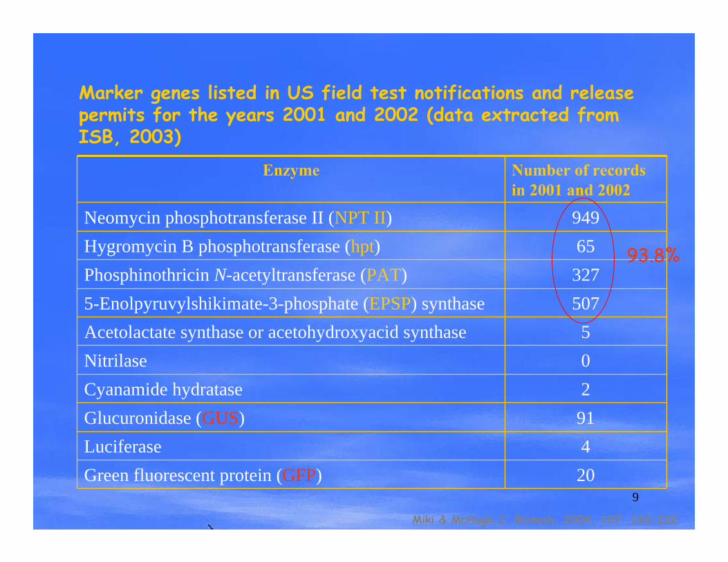
5075-Enolpyruvylshikimate-3-phosphate (EPSP) synthase327Phosphinothricin N-acetyltransferase (PAT)65Hygromycin B phosphotransferase (hpt)949Neomycin phosphotransferase II (NPT II)
Number of records in 2001 and 2002
Enzyme
0Nitrilase5Acetolactate synthase or acetohydroxyacid synthase
20Green fluorescent protein (GFP)4Luciferase91Glucuronidase (GUS)2Cyanamide hydratase
Marker genes listed in US field test notifications and release permits for the years 2001 and 2002 (data extracted from ISB, 2003)
Miki & McHugh,J. Biotech. 2004, 107: 193-232
93.8%
10
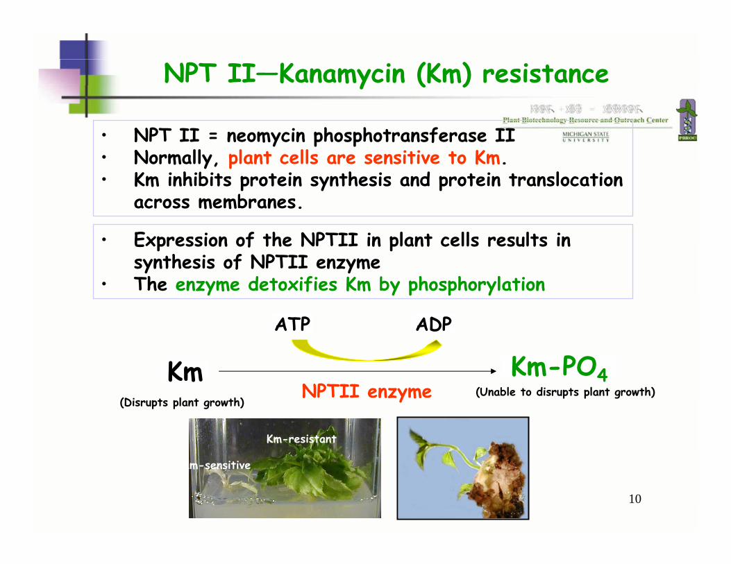
NPT II—Kanamycin (Km) resistance
• NPT II = neomycin phosphotransferase II • Normally, plant cells are sensitive to Km. • Km inhibits protein synthesis and protein translocation
across membranes.
• Expression of the NPTII in plant cells results in synthesis of NPTII enzyme
• The enzyme detoxifies Km by phosphorylation
Km
ATP ADP
NPTII enzymeKm-PO4
(Disrupts plant growth)(Unable to disrupts plant growth)
Km-sensitive
Km-resistant

11
Dose experiment on Km-resistance
Example 1: Km-selection
Km=0 (mg/L)
Km=10 Km=20
Km=30 Km=50 Km=100
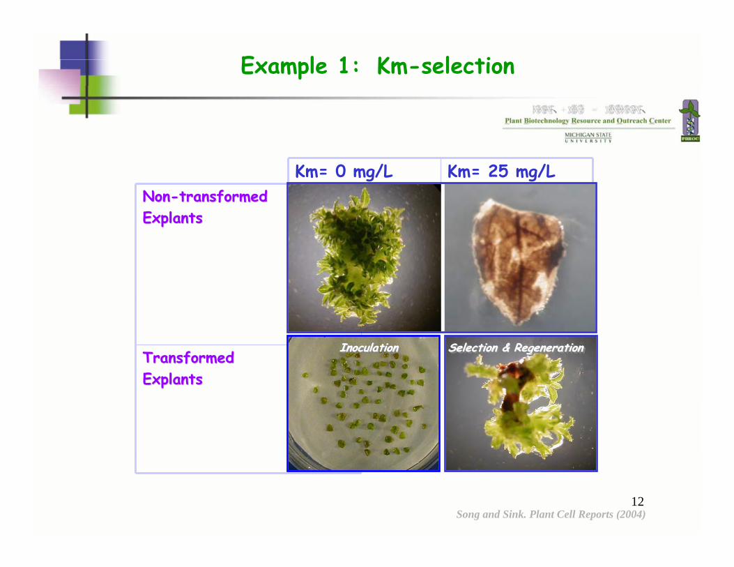
12
Transformed Transformed ExplantsExplants
NonNon--transformed transformed ExplantsExplants
Km= 25 mg/LKm= 0 mg/L
InoculationInoculationInoculation Selection & RegenerationSelection & RegenerationSelection & Regeneration
Example 1: Km-selection
Song and Sink. Plant Cell Reports (2004)
13
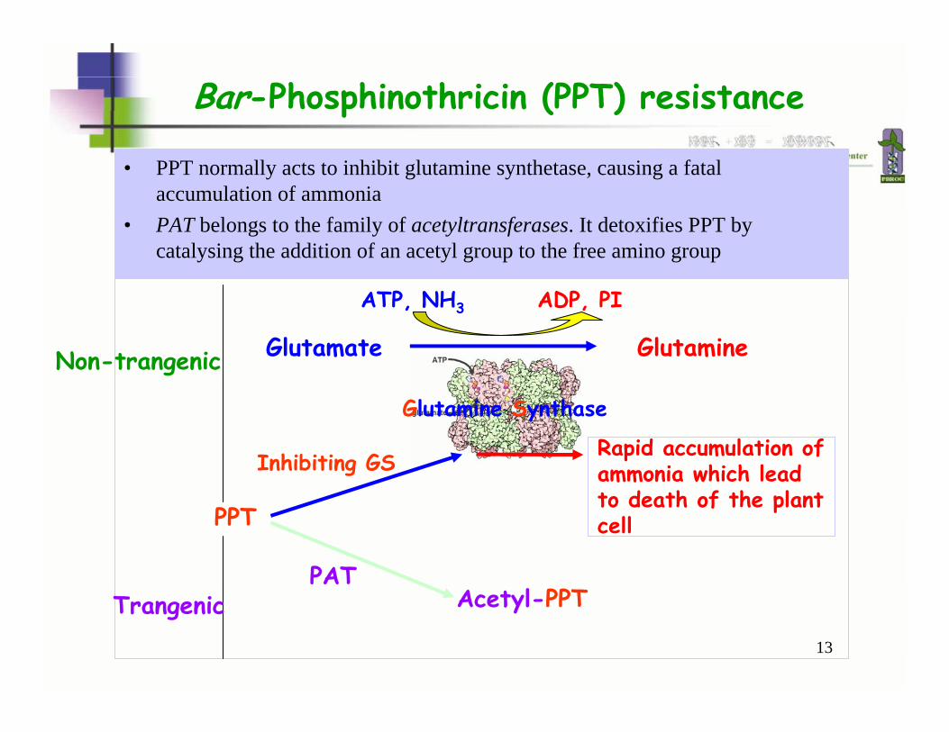
Rapid accumulation of ammonia which lead to death of the plant cell
• PPT normally acts to inhibit glutamine synthetase, causing a fatal accumulation of ammonia
• PAT belongs to the family of acetyltransferases. It detoxifies PPT by catalysing the addition of an acetyl group to the free amino group
Bar-Phosphinothricin (PPT) resistance
Glutamate Glutamine
ATP, NH3 ADP, PI
Glutamine Synthase
PPT
Inhibiting GS
Non-trangenic
TrangenicPAT
Acetyl-PPT
14
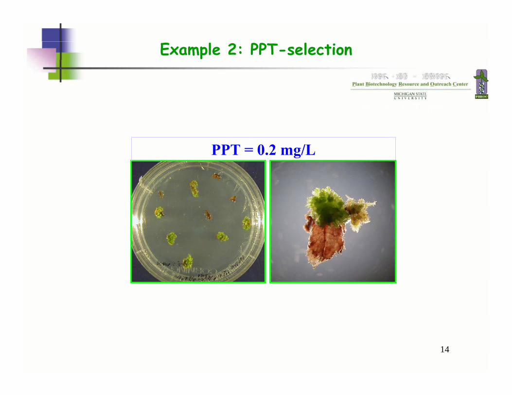
PPT = 0.2 mg/L
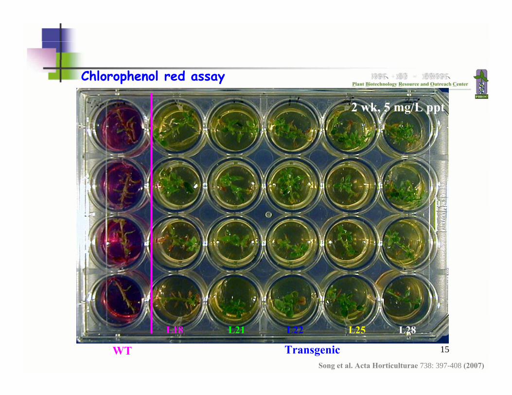
Example 2: PPT-selection
2 wk, 5 mg/L ppt
15
Chlorophenol red assay
WT Transgenic
2 wk, 5 mg/L ppt
L18 L25L21 L22 L28
Song et al. Acta Horticulturae 738: 397-408 (2007)
16
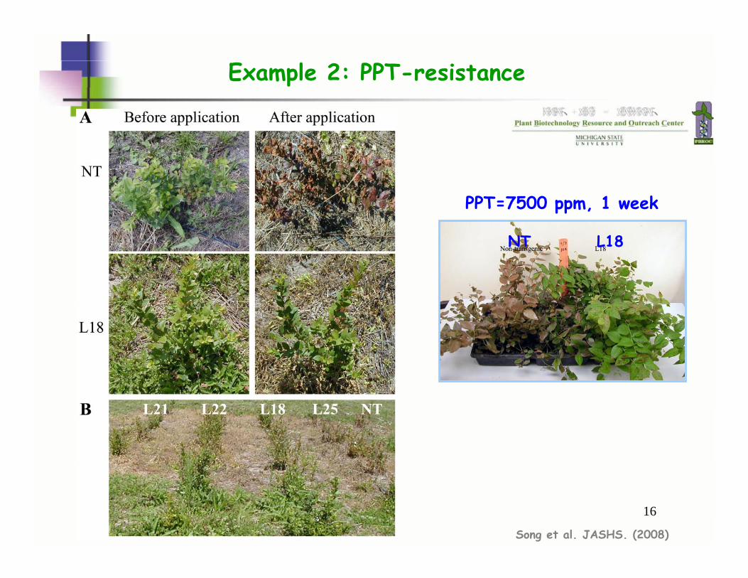
PPT=7500 ppm, 1 week
NT L18
Song et al. JASHS. (2008)
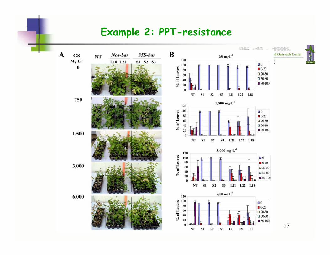
Example 2: PPT-resistance
17
Example 2: PPT-resistance
18
Hpt, hph or aphIV-Hygromycin (Hyg) B resistance
The hygromycin phosphotransferase (denoted hpt, hph or aphIV) gene was originally derived from Escherichia coli. The gene codes for hygromycin phosphotransferase (HPT), which detoxifies the aminocyclitol antibiotic hygromycin B. A large number of plants have been transformed with the hpt gene and hygromycin B has proved very effective in the selection of a wide range of plants, including monocotyledonous.
Most plants exhibit higher sensitivity to hygromycin B than to kanamycin, for instance cereals. Likewise, the hpt gene is used widely in selection of transformed mammalian cells.
Like kanamycin and other aminoglycoside antibiotics, hygromycin B inhibits protein synthesis by interfering with mRNA translation and causing mistranslocation of mRNA.

19
Example 3: Hyg-selection
HygHyg--Selection & Selection & RegenerationRegeneration(50 mg/L)(50 mg/L)
HygHyg--Selection (50 mg/L)Selection (50 mg/L)
20
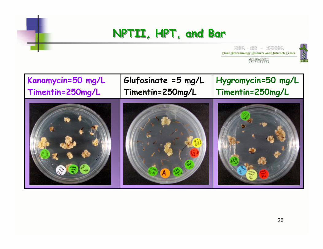
NPTII, HPT, and BarNPTII, HPT, and BarNPTII, HPT, and Bar
Hygromycin=50 mg/LTimentin=250mg/L
Glufosinate =5 mg/LTimentin=250mg/L
Kanamycin=50 mg/LTimentin=250mg/L
21
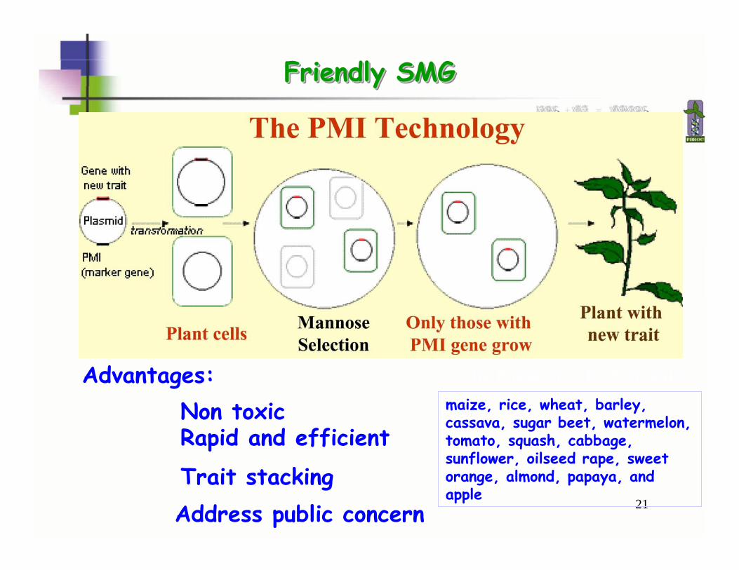
Non toxic
Friendly SMGFriendly SMGFriendly SMG
Advantages:
Rapid and efficient
Trait stackingAddress public concern
The PositechTM by Syngentamaize, rice, wheat, barley, cassava, sugar beet, watermelon, tomato, squash, cabbage, sunflower, oilseed rape, sweet orange, almond, papaya, and apple
22
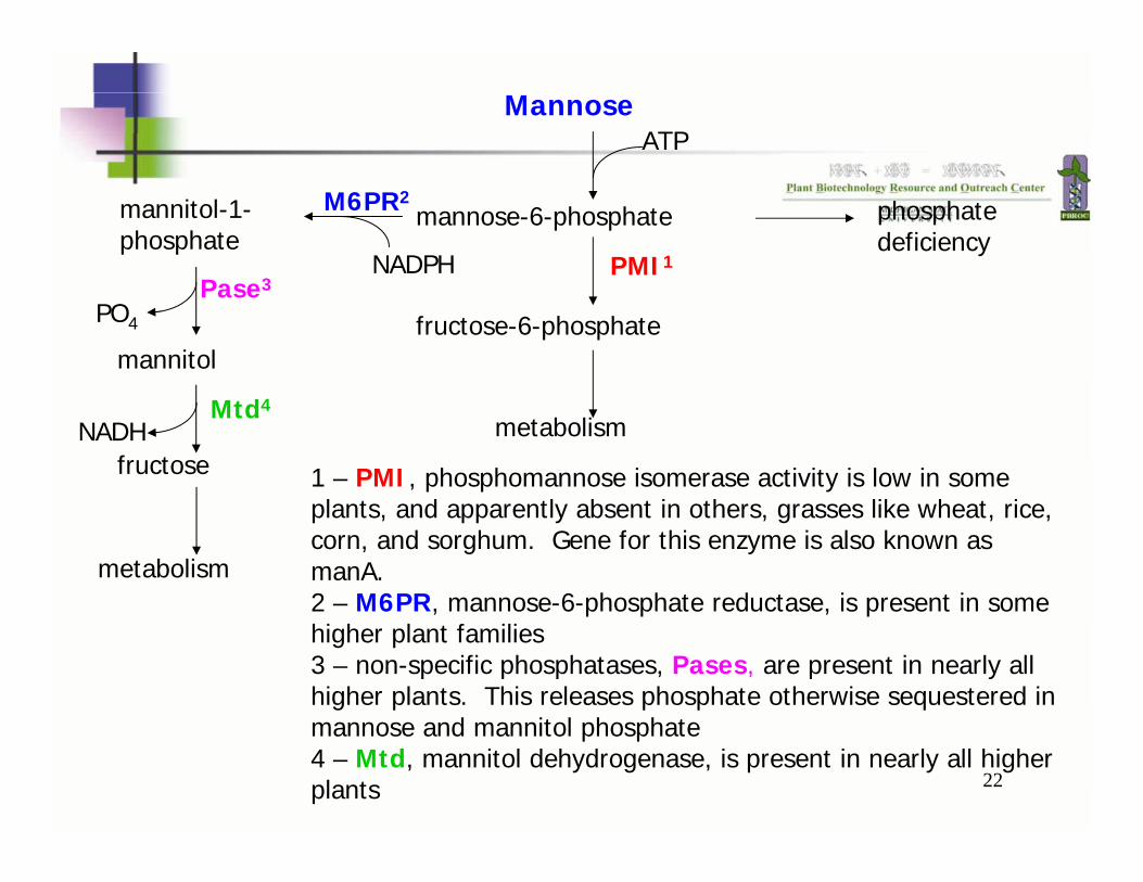
Mannose
mannose-6-phosphate
ATP
fructose-6-phosphate
metabolism
phosphate deficiency
PMI1
1 – PMI, phosphomannose isomerase activity is low in some plants, and apparently absent in others, grasses like wheat, rice, corn, and sorghum. Gene for this enzyme is also known as manA.2 – M6PR, mannose-6-phosphate reductase, is present in some higher plant families3 – non-specific phosphatases, Pases, are present in nearly all higher plants. This releases phosphate otherwise sequestered inmannose and mannitol phosphate4 – Mtd, mannitol dehydrogenase, is present in nearly all higher plants
Mtd4
Pase3
mannitol-1-phosphate
mannitol
fructose
metabolism
PO4
NADH
M6PR2
NADPH
23
Other SMG
Isopentyl transferasesHistidine kinase homologueHairy root-inducing genes

24
Markers for Screening
25
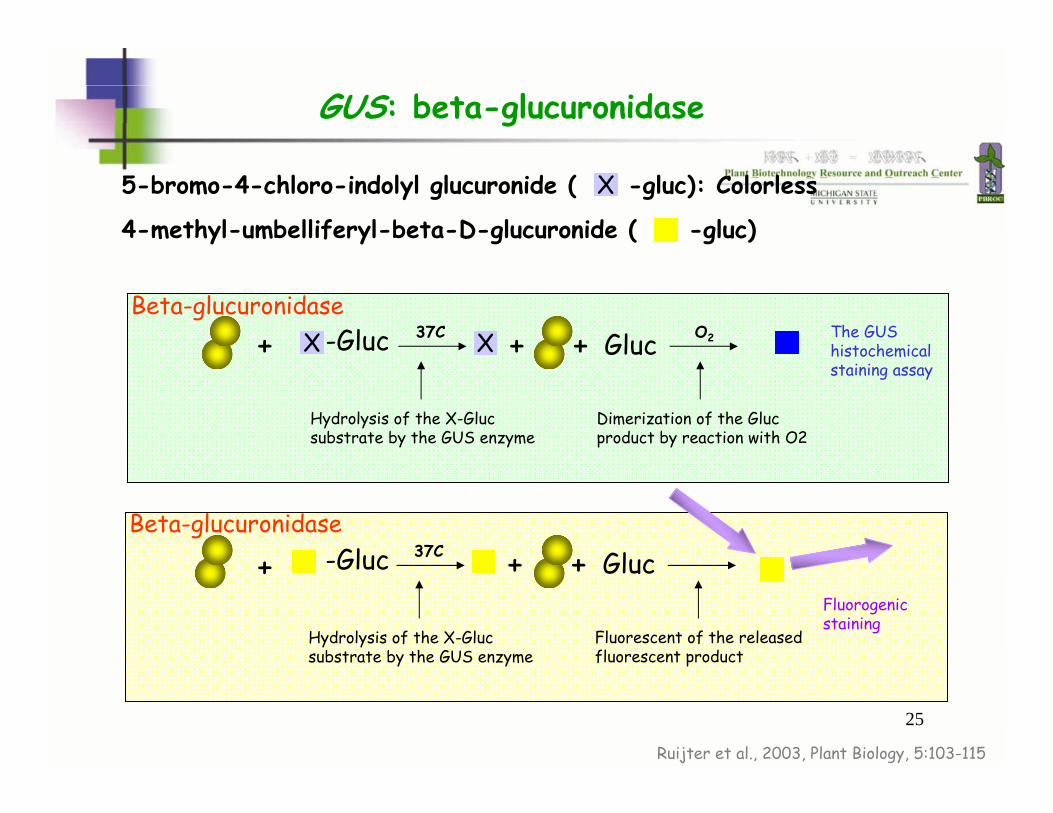
GUS: beta-glucuronidase
5-bromo-4-chloro-indolyl glucuronide ( -gluc): Colorless
Ruijter et al., 2003, Plant Biology, 5:103-115
+ X -Gluc 37C + Gluc+ O2
Hydrolysis of the X-Glucsubstrate by the GUS enzyme
Dimerization of the Glucproduct by reaction with O2
Beta-glucuronidase
+ -Gluc 37C + Gluc+
Hydrolysis of the X-Glucsubstrate by the GUS enzyme
Fluorescent of the released fluorescent product
Beta-glucuronidase
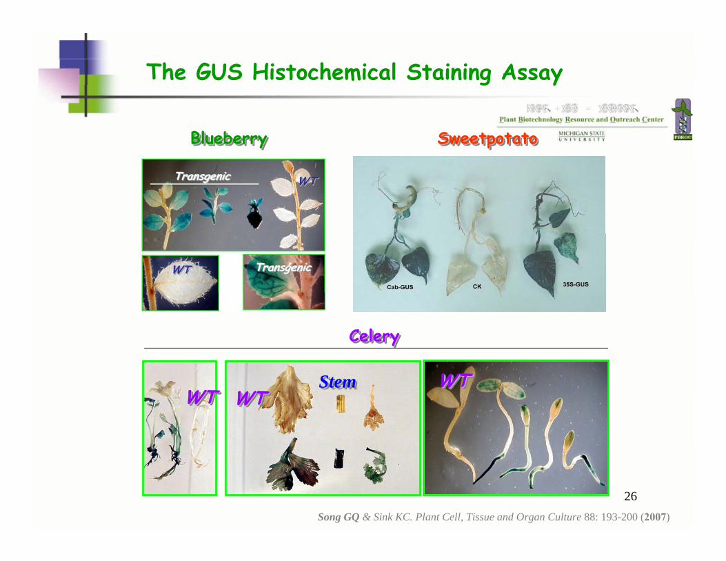
The GUS histochemicalstaining assay
X
4-methyl-umbelliferyl-beta-D-glucuronide ( -gluc)
X
Fluorogenicstaining
26
WTWTWT
WTWTWTStemStemStemWTWTWTWTWTWT
Blueberry Blueberry Blueberry SweetpotatoSweetpotatoSweetpotato
Celery Celery Celery
The GUS Histochemical Staining Assay
Song GQ & Sink KC. Plant Cell, Tissue and Organ Culture 88: 193-200 (2007)
27
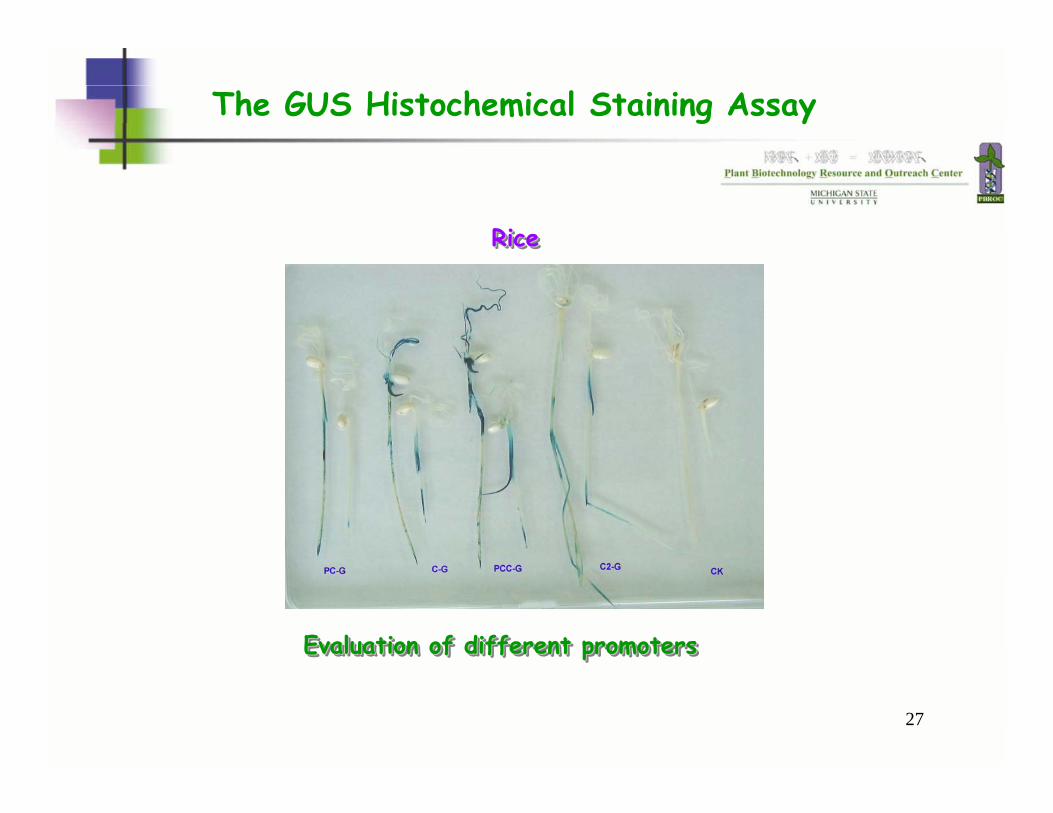
The GUS Histochemical Staining Assay
Rice Rice Rice
Evaluation of different promotersEvaluation of different promotersEvaluation of different promoters
28
Luciferase (LUC)
A conditional non-selectable marker gene
29
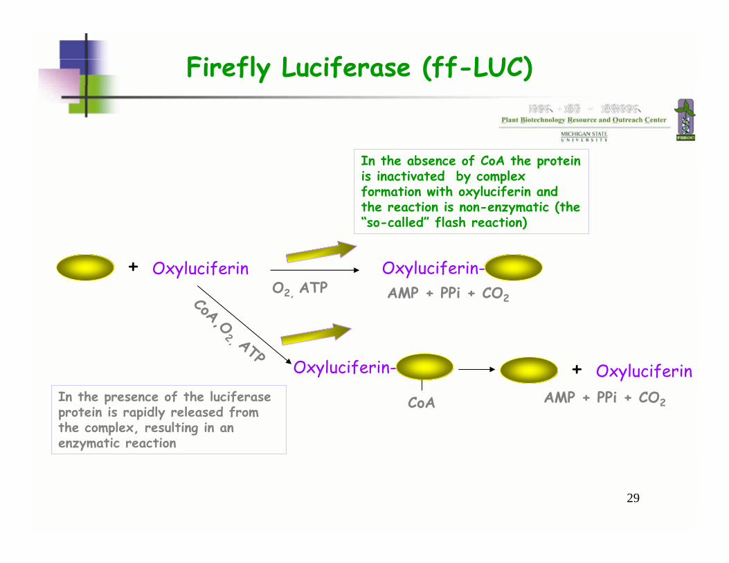
Firefly Luciferase (ff-LUC)
Oxyluciferin+O2, ATP
Oxyluciferin-AMP + PPi + CO2
In the absence of CoA the protein is inactivated by complex formation with oxyluciferin and the reaction is non-enzymatic (the “so-called” flash reaction)
CoA,O2, ATP
Oxyluciferin-
CoA
Oxyluciferin+AMP + PPi + CO2In the presence of the luciferase
protein is rapidly released from the complex, resulting in an enzymatic reaction
30
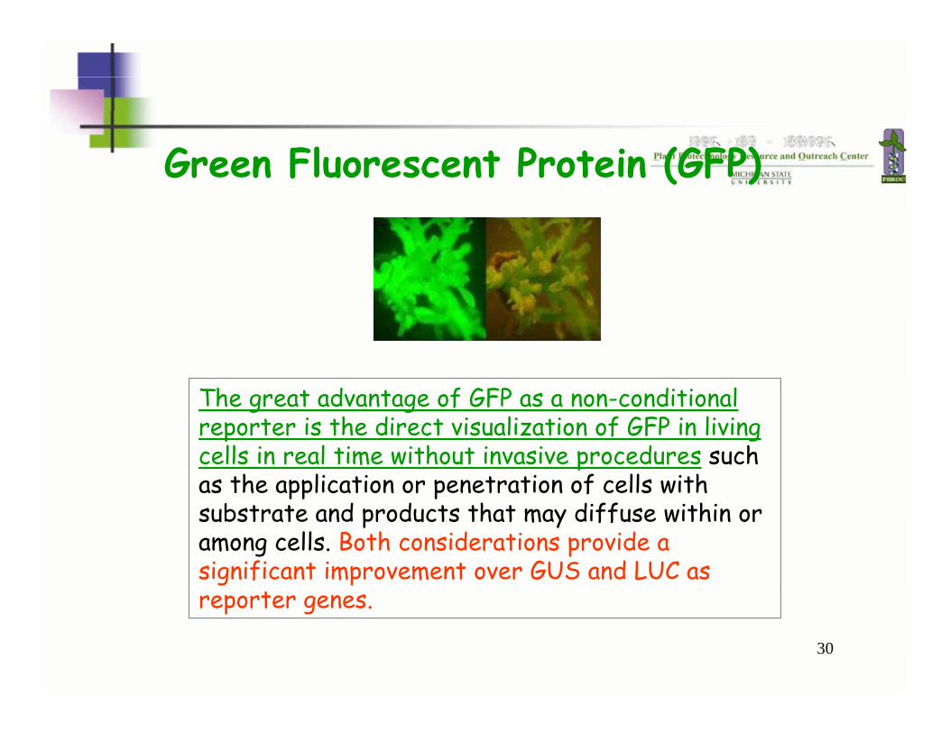
Green Fluorescent Protein (GFP)
The great advantage of GFP as a non-conditional reporter is the direct visualization of GFP in living cells in real time without invasive procedures such as the application or penetration of cells with substrate and products that may diffuse within or among cells. Both considerations provide a significant improvement over GUS and LUC as reporter genes.
31
In 1994 GFP was cloned. Now GFP is found in laboratories all over the world where it is used in every conceivable plant and animal. The GFP gene can be introduced into organisms and maintained in their genome through breeding, or local injection with a viral vector which can be used to introduce the gene.
GFP
32
Green Fluorescent Protein (GFP)
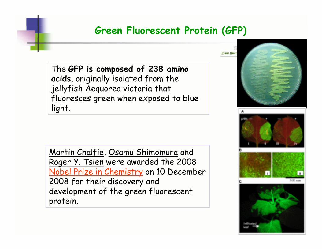
Martin Chalfie, Osamu Shimomura and Roger Y. Tsien were awarded the 2008 Nobel Prize in Chemistry on 10 December 2008 for their discovery and development of the green fluorescent protein.
The GFP is composed of 238 amino acids, originally isolated from the jellyfish Aequorea victoria that fluoresces green when exposed to blue light.
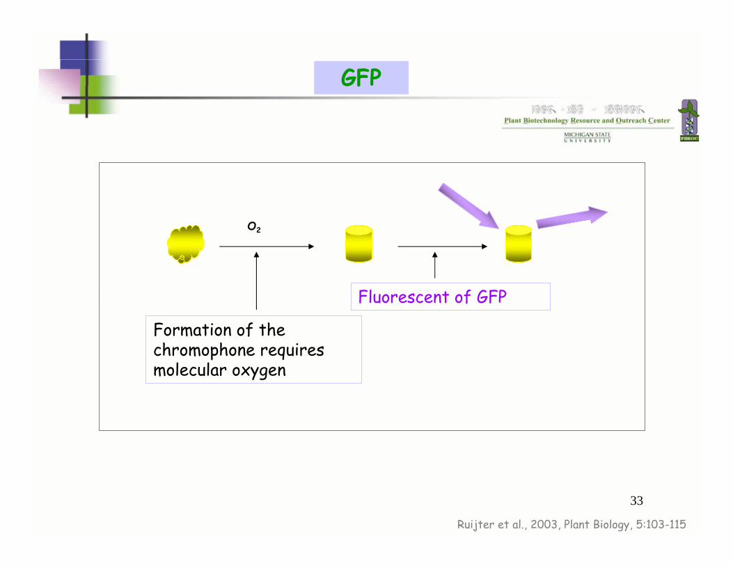
33
Formation of the chromophone requires molecular oxygen
Fluorescent of GFP
O2
Ruijter et al., 2003, Plant Biology, 5:103-115
GFP
34
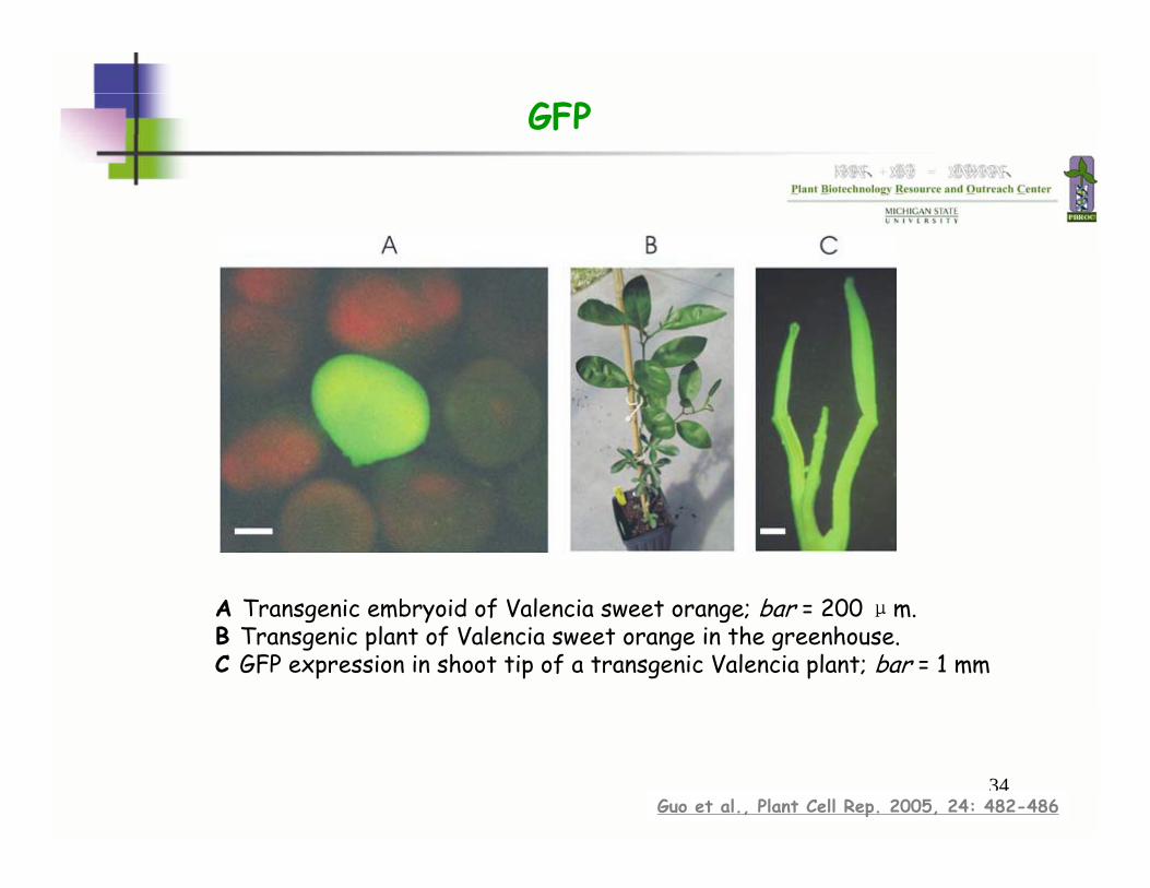
A Transgenic embryoid of Valencia sweet orange; bar = 200 μm. B Transgenic plant of Valencia sweet orange in the greenhouse. C GFP expression in shoot tip of a transgenic Valencia plant; bar = 1 mm
Guo et al., Plant Cell Rep. 2005, 24: 482-486
GFP
35
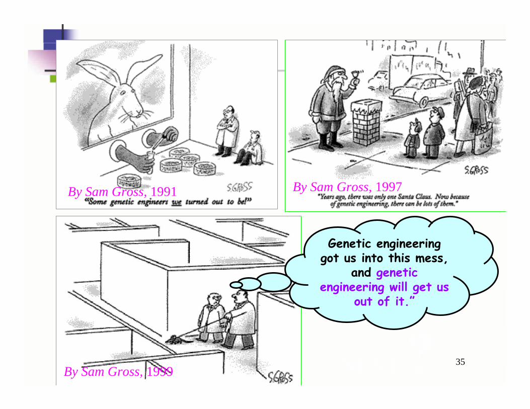
Genetic engineering got us into this mess,
and genetic engineering will get us
out of it.”
By Sam Gross, 1999
By Sam Gross, 1991 By Sam Gross, 1997
NEXT ?

36
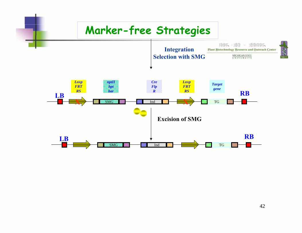
Marker-free Strategies
37
Although no adverse biosafety effects have been reported for the marker genes that have been adopted for widespread use, biosafety concerns should help direct which markers will be chosen for future crop development. Common sense dictates that marker genes conferring resistance to significant therapeutic antibiotics could not be used.
Concerns about the SMG
38
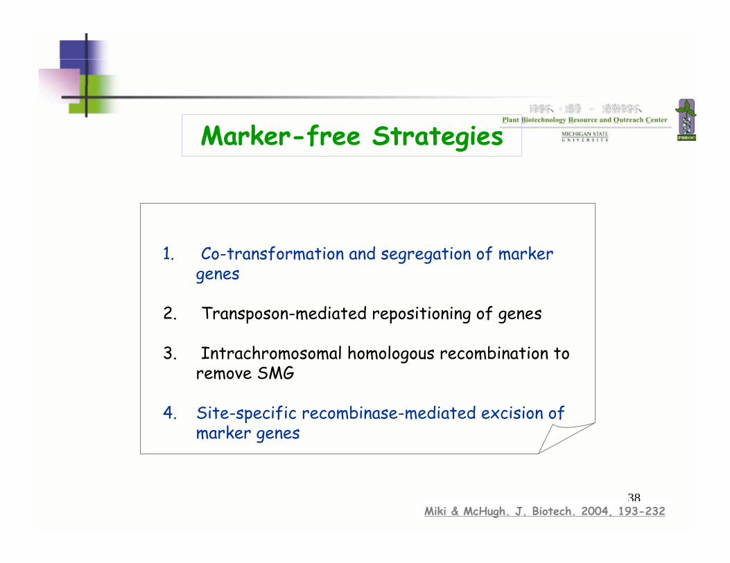
Marker-free Strategies
1. Co-transformation and segregation of marker genes
2. Transposon-mediated repositioning of genes
3. Intrachromosomal homologous recombination to remove SMG
4. Site-specific recombinase-mediated excision of marker genes
Miki & McHugh. J. Biotech. 2004, 193-232
39
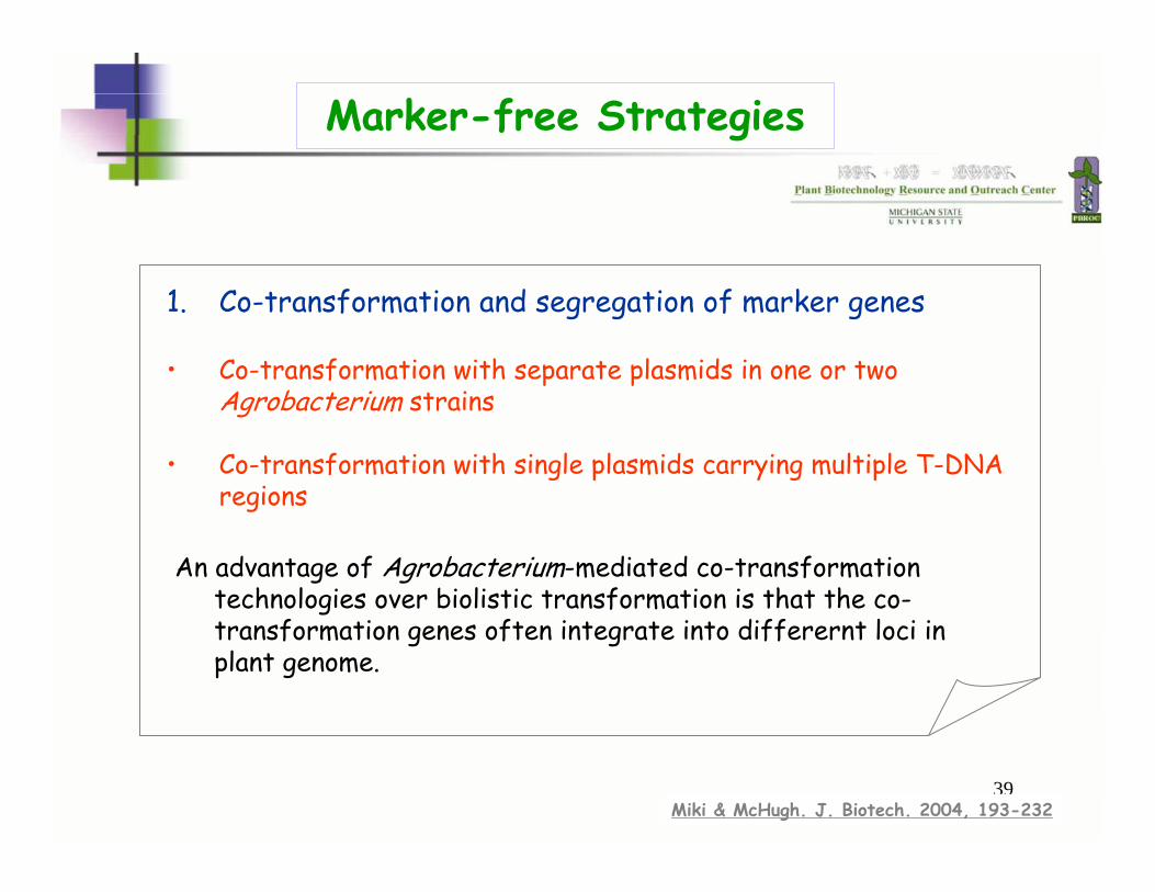
An advantage of Agrobacterium-mediated co-transformation technologies over biolistic transformation is that the co-transformation genes often integrate into differernt loci in plant genome.
1. Co-transformation and segregation of marker genes
• Co-transformation with separate plasmids in one or two Agrobacterium strains
• Co-transformation with single plasmids carrying multiple T-DNA regions
Marker-free Strategies
Miki & McHugh. J. Biotech. 2004, 193-232
40
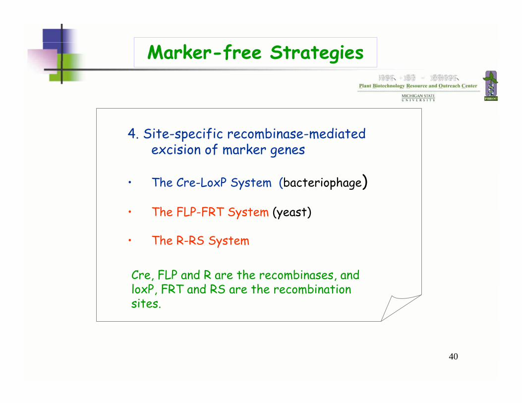
4. Site-specific recombinase-mediated excision of marker genes
• The Cre-LoxP System (bacteriophage)
• The FLP-FRT System (yeast)
• The R-RS System
Marker-free Strategies
Cre, FLP and R are the recombinases, and loxP, FRT and RS are the recombination sites.
41
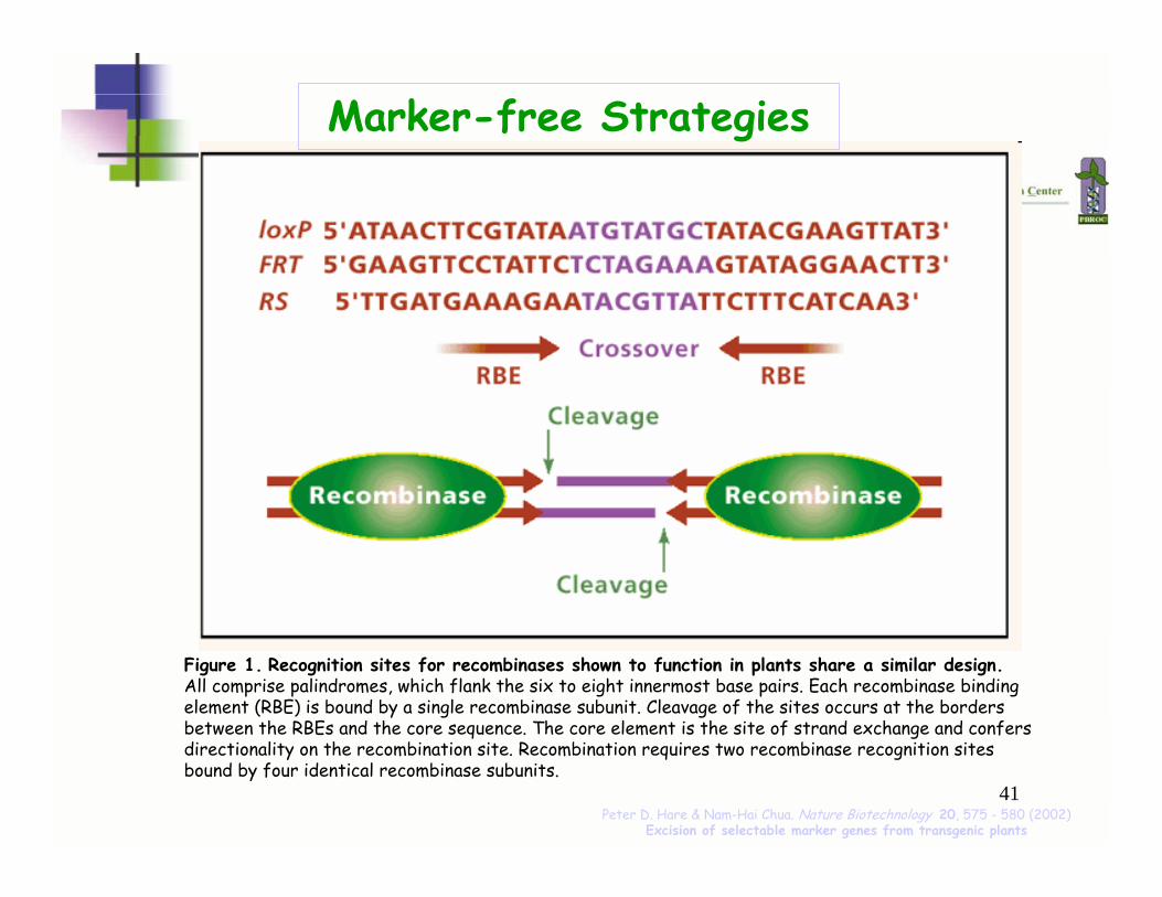
Figure 1. Recognition sites for recombinases shown to function in plants share a similar design.All comprise palindromes, which flank the six to eight innermost base pairs. Each recombinase binding element (RBE) is bound by a single recombinase subunit. Cleavage of the sites occurs at the borders between the RBEs and the core sequence. The core element is the site of strand exchange and confers directionality on the recombination site. Recombination requires two recombinase recognition sites bound by four identical recombinase subunits.
Peter D. Hare & Nam-Hai Chua. Nature Biotechnology 20, 575 - 580 (2002) Excision of selectable marker genes from transgenic plants
Marker-free Strategies
42
LoxpFRTRS
LoxpFRTRS
nptIIhptbar
CreFlpR
SMGLB RB
Ind TG
SMGLB RB
Ind TG
IntegrationSelection with SMG
Excision of SMG
XX
Targetgene
Marker-free Strategies
43
Experiment 6: Histochemical GUS assay
Objective:
To get familiar with using the gusA as a screening marker