Embed Size (px)
Citation preview

A CASE STUDY OF PERIARTICULAR FRACTURES
OF THE TIBIA MANAGED WITH HYBRID
EXTERNAL FIXATOR APPLICATION
DISSERTATION SUBMITTED FOR
M.S. DEGREE
(BRANCH II - ORTHOPAEDIC SURGERY)
MARCH 2009
THE TAMILNADU DR. M.G.R. MEDICAL UNIVERSITY
CHENNAI, TAMILNADU
brought to you by COREView metadata, citation and similar papers at core.ac.uk
provided by ePrints@TNMGRM (Tamil Nadu Dr. M.G.R. Medical University)

DEPARTMENT OF ORTHOPAEDICS MADURAI MEDICAL COLLEGE AND GOVERNMENT RAJAJI HOSPITAL MADURAI.
CERTIFICATE
This is to certify that the dissertation entitled “A CASE
STUDY OF PERIARTICULAR FRACTURES OF THE TIBIA
MANAGED WITH HYBRID EXTERNAL FIXATOR
APPLICATION” is a bonafide record of work done by Dr. P. SAI
PRASAD in the Department of Orthopaedics and Traumatology,
Government Rajaji Hospital, Madurai Medical College, Madurai,
under the direct guidance of me.
Prof. Dr. V. RAVIRAMAN, M.S.ORTHO., D.ORTHO.,
Professor and Head of the Department Department of Orthopaedics Madurai Medical College and Government Rajaji Hospital, Madurai.

DECLARATION
I Dr. P. SAI PRASAD, solemnly declare that the dissertation entitled
“A CASE STUDY OF PERIARTICULAR FRACTURES OF THE
TIBIA MANAGED WITH HYBRID EXTERNAL FIXATOR
APPLICATION’ has been prepared by me under the able guidance and
supervision of my guide Prof.Dr. V. Raviraman, M.S.ORTHO., D.ORTHO.,
Prof & HOD, Department of Orthopaedics and Traumatology, Madurai
Medical College, Madurai, in partial fulfillment of the regulation for the award
of M.S. (ORTHOPAEDIC SURGERY) degree examination of The
Tamilnadu Dr. M.G.R. Medical University, Chennai to be held in March 2009.
This work has not formed the basis for the award of any other degree or
diploma to me previously from any other university.
Place : Madurai
Date : DR. P. SAI PRASAD

ACKNOWLEDGEMENT
I am deeply indebted to my beloved chief and my teacher,
Prof. Dr. V. Raviraman, M.S.Ortho, D.Ortho., Professor and Head of
the Department of Orthopaedics, Madurai Medical College, Madurai
for the able guidance, inspiration and encouragement he has rendered
at every stage of this study.
I am grateful to my beloved teachers Prof. Dr. Malairaju, Prof. P.V.
Pughalenthi, Prof. Dr. A. Rajamani, Dr. T. Chandraprakasm for their
invaluable help and guidance rendered to me in preparing this dissertation.
I express my heartfelt gratitude to my beloved teacher
Dr.S.Shanmuganathan., for his able guidance, inspiration and
encouragement he has rendered at every stage of this study.
I express my heartfelt gratitude to Dr.V.Ramar,
Dr.R.Sivakumar, Dr.K.Ravichandran, Dr.V.R.Ganeshan, Dr.R.
Arivasan, Dr. M.N. Karthi, Dr.N.Thanappan, Assistant Professors of
Orthopaedics, Madurai Medical College, Madurai for their valuable
advice and help in carrying out this study.
I sincerely acknowledge with gratitude the guidance and
persistent encouragement given to me by Dr. B. Sivakumar,
Dr. S.Ramanathan, Dr. P.V.Thirumalai Murugan, Dr. K.P. Saravana

Kumar, Dr. J. Manikandan, Dr.T.C. Premkumar, Dr. Durai Murugan
and all our assistant professors.
My sincere thanks to Prof. Dr. S.M. Sivakumar, M.S., Dean,
Madurai Medical College, Madurai for permitting me to utilize the
clinical materials of the hospital.
I would like to thank my patients, friends and colleagues &
family who have stood by me throughout this work and above all the
GOD FOR HIS KINDNESS THROUGHOUT THIS STUDY.

CONTENTS
S. NO CONTENTS PAGE NO
PART - I
1. INTRODUCTION 1
2. AIM 4
3. REVIEW OF LITRATURE 5
4. BIOMECHANICS 8
5. ANATOMY OF PROXIMAL AND DISTAL TIBIA 13
6. CLASSIFICATION OF PROXIMAL TIBIA FRACTURES 15
7. CLASSIFICATION OF DISTAL TIBIA FRACTURES 18
8. MECHANISM OF INJURY 19
9. INVESTIGATIONS 21
10. PRINCIPLES OF MANAGEMENT 22
11. METHODS OF TREATMENT 23
12. COMPONENTS AND INSTRUMENTS OF HYBRID FIXATOR32
13. PREOPERATIVE PLANNING 34
14. SURGICAL TECHNIQUE 36
15. COMPLICATIONS 39
16. EVALUATION OF OUTCOME 44
PART - II
17. MATERIALS AND METHODS 46
18. PROCEDURE 51
19. ANALYSIS OF FUNCTIONAL OUTCOME 53
20. DISCUSSION 54
21. CONCLUSION 57
22. BIBLIOGRAPHY
23. MASTER CHART

INTRODUCTION
Increased incidence of Road Traffic Accidents, natural disasters,
industrial accidents claim most of human mortality and morbidity. Hence,
it forms the major epidemic of Modern Era. Of these, fractures of
Proximal and distal tibia have historically been difficult to treat. In this
modern era of increasing life expectancy, there is a rise of old age
population which, increases the incidence of these fractures in
osteoporotic bones, adding to the morbidity.
Because of the proximity of these fractures to the knee and ankle
joint, regaining full knee and ankle motion and function may be difficult.
Soft tissue damage, comminution, fracture extension into the knee or
ankle joint lead to unsatisfactory results in many cases regardless of the
treatment modality.
Better understanding of the injury patterns, availability of better
implants, the concept of early surgical fixation and early postoperative
mobilization of joint all have convincingly improved the functional
outcome of the patient to a large extent.

Main challenges encountered in the treatment of periarticular fractures
of tibia are
- these high energy fractures are associated with extremely damaged
soft tissue envelop leading to increased incidence of compound
injuries which results in increased complications following open
reduction and internal fixation. Also communition of metaphysis
and articular surface makes anatomical reduction difficult. The
resulting incongruency of articular surface leads to early secondary
OA.
- In metaphysis fixation is less satisfactory resulting in early
loosening of implant.
- communited fractures create difficulty in achieving rigid fixation
due to poor purchase and are less optimal to permit weight bearing
or even early joint mobilization.
Initially conservative treatment with POP was advocated as a
treatment option. But it lead to high incidence of malunion and stiffness
of adjacent joints. Also prolonged recumbency resulted in high incidence
of thromboembolic diseases and pneumonia. Open reduction and internal
fixation with plate osteosynthesis lead to skin necrosis and infection in
> 40% of patients eventually leading to malunion and implant failure.
Intramedullary devices gives inadequate stability due to wide medullary

cavity leading to implant failure and screw breakage. For compound
injury patients, initial treatment with external fixator for wound care and
then followed by definitive mode of fixation was advocated. This
involves multiple procedures which increased economical and mental
stress for the patients. But hybrid fixation is a single definitive
procedure, helping both in wound care and aiding in sound union of
fracture.
They also offer advantages of applying multiple pins according to
varying fracture pattern without disturbing the soft tissue cover; less
chances of infection, early mobilization of patient, reducing the tendency
for varus collapse and at the same time afford better stability.
The successful management of these injuries, demands thorough
knowledge of fracture personality, technical aspects of fracture fixation
and the tailored post operative management.
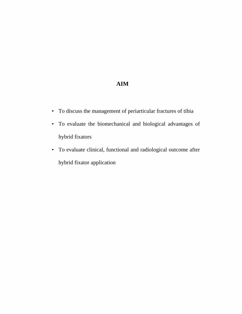
AIM
• To discuss the management of periarticular fractures of tibia
• To evaluate the biomechanical and biological advantages of
hybrid fixators
• To evaluate clinical, functional and radiological outcome after
hybrid fixator application

REVIEW OF LITRATURE
Intra-articular and juxtra articular fractures are the most commonly
occurring injuries. There has been a long debate on merits and demerits
of operative and non operative mode of management. Numerous
investigators have reported satisfactory reports with either closed or open
methods of treatment. The out come of fracture depends upon the
integrity of soft tissues.
Tracy Watson et al studied the biomechanics of hybrid fixator frame
and suggested that biomechanical data support the use of tensioned wire
and hybrid fixator for stabilizing complex fractures of proximal tibia.
In 1994 he studied 31 patients of tibial plateau fractures for whom
hybrid fixators were applied. Bone healed in an average of 15 weeks and
the knee range of movement was 106 degrees. He reported excellent
report in 27 of 31 patients.
Stamer et al36 reported 70% good to excellent results in tibial plateau
fracture patients treated with hybrid external fixator.
Micheal S sirkin et al25 from their study concluded that hybrid
external fixator is most appropriate for patients with tibial plateau
fractures.
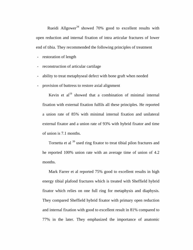
Rueidi Allgower34 showed 70% good to excellent results with
open reduction and internal fixation of intra articular fractures of lower
end of tibia. They recommended the following principles of treatment
- restoration of length
- reconstruction of articular cartilage
- ability to treat metaphyseal defect with bone graft when needed
- provision of buttress to restore axial alignment
Kevin et al19 showed that a combination of minimal internal
fixation with external fixation fulfils all these principles. He reported
a union rate of 85% with minimal internal fixation and unilateral
external fixator and a union rate of 93% with hybrid fixator and time
of union is 7.1 months.
Tornetta et al 39 used ring fixator to treat tibial pilon fractures and
he reported 100% union rate with an average time of union of 4.2
months.
Mark Farrer et al reported 75% good to excellent results in high
energy tibial plafond fractures which is treated with Sheffield hybrid
fixator which relies on one full ring for metaphysis and diaphysis.
They compared Sheffield hybrid fixator with primary open reduction
and internal fixation with good to excellent result in 81% compared to
77% in the later. They emphasized the importance of anatomic
reduction of articular surface which significantly improves the
out come.
Rocco Barbieri et al33 reported 37 distal tibia fractures fixed with
hybrid fixators and reported 12 excellent, 9 good, 7 fair and 6 poor
results. The results were acceptable in 82% and poor in 18%.
Leener et al20 studied the use of hybrid external fixators in
stabilizing peri articular, supracondylar and pilon fractures.
Enders et al 14 studied a total of 62 tibial pilon fractures and
reported good results in 87% of people treated with hybrid and only
38% of people treated with other conventional methods.
Robert et al32 studied strategies to improve frame stability of hybrid
external fixator and concluded that dramatic improvement of stability
can be achieved from addition of an anterior proximal half pin.

BIOMECHANICS
Hybrid external fixator presents mechanical characteristics that set
apart significantly from other systems of external fixator. It
characteristics can be called as solid-elastic. Solid enough for
stabilization and still providing micro motion which enhances good
callus formation.
Three theoretical and biomechanical foundation of hybrid external
fixator are
- minimal damage to vascularity and soft tissue
- solid yet elastic stabilization
- Immediate resumption of function.
Two separate components exits for hybrid external fixator
1. Ring fixator
2. AO external fixator
RING FIXATOR
Important factors governing rigidity is bone contact, which gives axial
stiffness. The number of wires gives torsional stiffness. 1.5 Or 1.8 mm K
wires are used. The amount of tension depends on frame stiffness and
weight of the patient. Generally a tension of 100 – 130 kg is given.
Tensioning reduces the deflection of the wire due to bending and
increases the bending stiffness. Bending stiffness is directly proportional
to fourth power of its diameter. A cluster of 4 or more k wires provides
increased stiffness. Increasing the number of wires distributes the load
from the injured region to intact trabecular bone surrounding the injury.
AO EXTERNAL FIXATOR
Factors affecting rigidity are
o number of pins
o diameter of pins distance of side bar from bone
o separation of pins around fracture site
o geometry of fixator
HALF PIN
It forms the back bone of the fixator. It ranges from 3-6 diameters
and it is self tapping. It is effective in stabilizing diaphysis and has less
chance of neurovascular injury. Stiffness of pin depends on core
diameter, long or short thread and diameter of shaft of pin.
When a long bone fracture is loaded in bending, the pins are loaded as
cantilever beam, with load at bone / pin interface. Highest stress is at
cortical surface where exits pin. Bone stress decreases by decreasing side
bar bone distance and increasing pin diameter.

DESIGN RATIONALE
The main aim is to hold the fragment in proper alignment while
allowing minimal axial dynamization at fracture site. Laryon and Rubin
showed that cyclic axial loading of bone is important for maintaining
bone mass and remodeling. Woodship, kenwight and Wolf et al
demonstrated that axial micromotion at fracture site increased fracture
healing. Fixation of metaphysis with K wire is safer since pin pull out is
greatly reduced. Fixation of diaphysis using half pin is safer by
decreasing neurovascular damage. Also early range of motion can be
started .
STIFFNESS
The slope of load deflection curve of fixation system is known as
fixator stiffness. In case of spiral fracture with cortical contact , direct lag
screw fixation produces 50- 70% of bending stiffness while DCP and
external fixator provides 82- 160% of stiffness. In axial compression ,
plate and external fixator provides 90- 115% of stiffness and ilizarow
provides 69- 117% of stiffness. Lag screw provides 9% of torsional
stiffness and plate and external fixator 40- 64% and intramedullary rod
6.5% of stiffness.
This shows external fixator is very effective for bending and axial
loading and ring fixator is very effective in metaphysis for torsional

stiffness. uniplanar fixator is stiffer than ilizarov in lateral stress whereas
ilizarow is stiffer in AP bending and torsion. So combination of these
both gives stiffness comparative to both of these fixators.
SHEAR STIFFNESS
Ability of fixator to resist translation shear at fracture site is shear
stiffness. Hybrid fixator resists shear forces as other fixators
AXIAL STIFFNESS
Ability of fixator to resist gap closure is known as axial stiffness.
Bone contact is most important in giving axial stiffness. Hybrid fixator
with good bone contact has axial stiffness less than external fixator and
equivalent to ilizarow fixator. This allows axial micromotion.
TORSIONAL STIFFNESS
Ring fixator with K wire increases torsional stiffness than external
fixator. Torsional stiffness increases upto 280 with pin angle of 90 deg.
MATERIAL AND PROPERTIES
The material used should with stand corrosion and should be
sufficient to with stand stresses. It depends on the composition of material

involved, grain size and porosity. The composition of stainless steel is
iron, 17- 20% chromium, 10-17% of nickel, 3% manganese, phosphorus,
sulfur, silicon, 2-4% molyblendum and < .03% carbon. The composition
of titanium alloy is titanium, 5.5- 6.5% alum, 3.5- 4.5% vanadium,
0.46%iron, carbon and oxygen. Titanium modulus of elasticity closes to
humerus bone and is very much corrosive resistant due to oxide film. It
has an excellent fatigue resistance. Other alloys are carbon fibre (side
rods), cobolt chromium alloy (bone screws).

ANATOMY OF PROXIMAL AND DISTAL TIBIA
Bony articular surface of proximal tibia slopes inferiorly 10 degree
from anterior to posterior. Between the medial and lateral plateau there
lies the intercondylar eminence which has medial and lateral tibial spines
which are areas of attachments for menisci and cruciate. Tibial tubercle
which is present on the anterior surface of tibial crest 2.5 to 3 cm below
joint line gives attachment for patellar tendon. Gerdy’s tubercle which
lies in the antero lateral surface of lateral tibial flare receives insertion of
ilio tibial band.
Proximal tibiofibular joint is located posterolaterally on tibial
condyl. The head of fibula gives attachment to fibular collateral ligament
and biceps tendon.
Tibial plateau is covered by hyaline cartilage 3mm thick on
medial plateau and 4mm thick on lateral side. Medial plateau is larger and
is concave from front to back as well as from side to side. Lateral plateau
is smaller in size and higher than medial. It is convex from front to back
and from side to side. Outer portion of each plateau is covered by
fibrocartilage.
Medial articular surface and subcondylar medial plateau is
stronger than lateral side. So in low violence injuries lateral condyl
fractures are more common.
The bony anatomy of ankle joint gives stability in dorsiflexion
and relative mobility in plantar flexion. In dorsiflexion stability is
provided by articular contact and in plantar flexion by the ligamentous
structures.
The talar dome is wider anteriorly than posteriorly. As the ankle
dorsiflexes, the fibula rotates externally to accommodate the widened
anterior surface of talus.

CLASSIFICATION OF PROXIMAL TIBIA FRACTURES
Various classification system were proposed since past to aid in
treatment of proximal tibial fractures. In 1956 Hohl and Luck were the
first to classify proximal tibial fractures into non displaced, local
depression, split depression and split fractures. Later Hohl expanded the
classification as local compression, split compression, total, split and
communited.
Then Moores classification came to role.
Type I split fractures (which also includes coronally split unstable
fractures)
Type II fracture involving entire condyl
Type III Rim avulsion fracture
Type IV Rim compression fractures
Type V communited bicondylar fracture with intercondylar eminence
as Separate fragment resulting in four part fracture
Again Hohl revised his classification including Moores classification.
He classified fractures into minimally displaced which are less than 4 mm
displaced and displaced. Displaced fractures are further classified into 6
types
1. Local compression
2. Split compression

3. Total depression
4. split fracture
5. Rim fracture
6. Bicondylar fracture
AO classification for proximal tibia fractures
A1 - Tibial spine avulsion fracture
A2 - extraarticular fracture
A3 - extraarticular communited fracture
B1 - Lateral condyl split fracture
B2 - lateral condyl compression fracture
B3 - lateral condyl communited fracture
C1 - Bicondylar fracture
C2 - Bicondylar fracture with metaphyseal communition
C3 - Bicondylar fracture with intra articular communition
Most commonly followed classifications for proximal tibia fractures
are that proposed by Schatzker.
Low energy fractures:
Type I
Split facture of Lateral plateau without articular depression. It
occurs in young adults with strong cancellous bone. If it is displaced
lateral meniscus is often torn.

Type II
Split depression fracture of lateral tibial condyl. It occurs more
commonly during 4th decade. It occurs due to lateral bending force with
axial loading.
Type III
Isolated depression fracture of lateral plateau. This type is usually
associated with joint instability
High energy fracture patterns
Type IV
Fracture of medial plateau. It is caused by varus force with axial
loading. This type is less common and is associated with cruciate, lateral
collateral, peroneal nerve and popliteal artery injuries.
Type V
Bicondylar plateau fracture
Type VI
Bicondylar tibial plateau fracture with diaphyseal metaphyseal
dissociation. This type is commonly associated with
compartmental syndrome, neurovascular compromise and
compromise to soft tissue.

CLASSIFICATION OF DISTAL TIBIA FRACTURES
Reudi and Allgower’s was the first classification came to use.
Type I - non displaced cleavage fractures of joint
Type II – displaced fractures with minimal communition
Type III – displaced fractures with severe communition.
This classification has a prognostic significance prognosis being poor
as the type increases from type I to type III.
AO/ OTA classification is now universally used for distal tibial
fractures.
Type A – non articular fractures
Type B – partially articular fractures
Type C – total articular fractures (tibial plafound fractures).
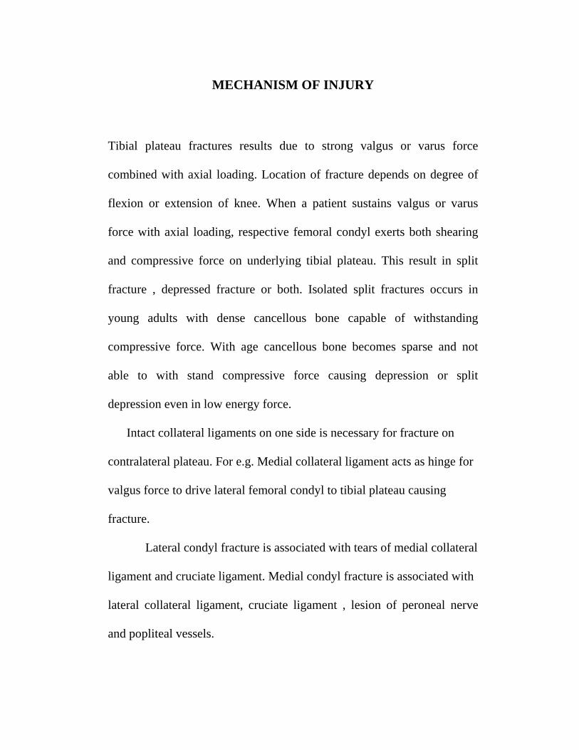
MECHANISM OF INJURY
Tibial plateau fractures results due to strong valgus or varus force
combined with axial loading. Location of fracture depends on degree of
flexion or extension of knee. When a patient sustains valgus or varus
force with axial loading, respective femoral condyl exerts both shearing
and compressive force on underlying tibial plateau. This result in split
fracture , depressed fracture or both. Isolated split fractures occurs in
young adults with dense cancellous bone capable of withstanding
compressive force. With age cancellous bone becomes sparse and not
able to with stand compressive force causing depression or split
depression even in low energy force.
Intact collateral ligaments on one side is necessary for fracture on
contralateral plateau. For e.g. Medial collateral ligament acts as hinge for
valgus force to drive lateral femoral condyl to tibial plateau causing
fracture.
Lateral condyl fracture is associated with tears of medial collateral
ligament and cruciate ligament. Medial condyl fracture is associated with
lateral collateral ligament, cruciate ligament , lesion of peroneal nerve
and popliteal vessels.
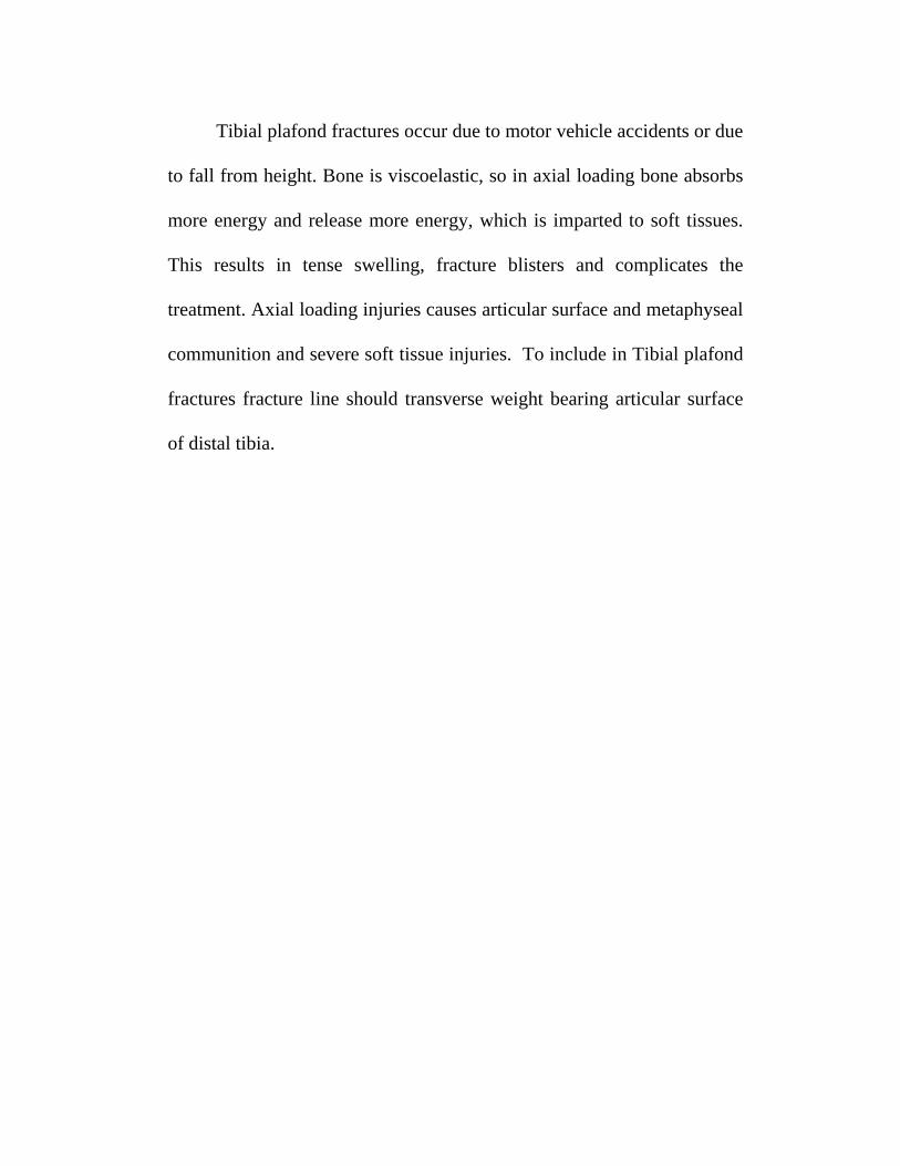
Tibial plafond fractures occur due to motor vehicle accidents or due
to fall from height. Bone is viscoelastic, so in axial loading bone absorbs
more energy and release more energy, which is imparted to soft tissues.
This results in tense swelling, fracture blisters and complicates the
treatment. Axial loading injuries causes articular surface and metaphyseal
communition and severe soft tissue injuries. To include in Tibial plafond
fractures fracture line should transverse weight bearing articular surface
of distal tibia.

INVESTIGATIONS
Clinically the patients may present with symptoms and signs either
of fractures (or) other major problems like hypovolemic shock.
All patients with fracture upper end of tibia should be looked for
peripheral pluses.
A good quality X ray in two perpendicular views is a must to look
for the joint involvement.
Computer tomography portrays the proximal and distal tibia
in cross- section, which helps to identify fracture lines in the
frontal plane. Two and three dimensional reconstructions
may also improve understanding of the fracture pattern in
preparation for surgery. But in our study, we haven’t taken
CT for any patients due to economical constraints.

PRINCIPLES OF MANAGEMENT
There are a lot of factors which play a dynamic role in
management. They include.
1. Amount of fracture displacement
2. Degree of comminution
3. Extent of soft tissue injury
4. Associated Neurovascular injuries
5. Magnitude of joint involvement
6. Degree of Osteoporosis
7. Associated injuries
8. Complex ipsilateral fractures (eg patella/plateau fracture)
The main goal of fracture treatment is obtaining a stable, aligned,
mobile and painless joint to minimize post traumatic osteo arthritis
So the objective of treatment of Periarticular fractures of Tibia is
1. To obtain and maintain satisfactory reduction and stable fixation.
2. To regain a functional range of motion of knee and ankle joint
3. To avoid varus collapse of knee joint.
4. To treat the associated injuries.
The prognosis of fracture depends on
- degree of articular depression
- extend of condylar separation or widening
- degree of diaphyseal metaphyseal communition and
dissociation
- integrity of soft tissue envelope

METHODS OF TREATMENT
The principles of treatment are
- An articular fracture with joint instability needs open
reduction and internal fixation
- Joint congruency should be restored
- Anatomical reduction and stable fixation of articular
fragment are necessary for articular cartilage regeneration
- If open reduction and internal fixation is inadvisable due to
Patient conditions skeletal traction and early motion should
be advocated
In the decade of 1960s, conservative treatment methods such as
traction and cast bracing, produced better results than operative treatment,
because of the lack of adequate internal fixation devices. It had a high
incidence of malunion and Stiffness of adjacent joints. Also prolonged
recumbency caused thromboembolic disease and pneumonia.
With the development of improved internal fixation devices,
treatment recommendations begin to change in 1980s. Open reduction
and internal fixation with buttress plates was done. Due to the poor soft
tissue cover it caused skin necrosis in both proximal and distal tibia
which leads to high chances of infection. Also low profile plate leads to
chances of implant failure and eventually malunion.

Intramedullary rods were used which had Inadequate stability due
to wide medullary cavity both in proximal and distal tibia which lead to
Implant failure and screw breakage. Also entry point was difficult in case
of proximal tibia due to extension of fracture line.
External fixation was used as either temporary (or) definitive fixation
in severe open proximal and distal tibia fractures especially those
associated with vascular injury. Since it is a form of Rigid fixation, union
was delayed. Also it Spans the joint and hence chances of joint stiffness
are more.
A recent advance in technology for the treatment of proximal and
distal tibial fractures includes minimally invasive reduction of fracture
and application of hybrid external fixator. They offer advantages of
applying multiple pins according to varying fracture pattern without
disturbing the soft tissue cover, less chances of infection, early
mobilization of patient, reducing the tendency for varus collapse and at
the same time afford better stability.
So management of distal femur fracture can be divided into two
broad categories.
1. conservative treatment
2. operative treatment

In operative treatment, various modalities include
1. Open Reduction Internal Fixation with buttress plate
2. Open Reduction Internal Fixation with locking compression
plate
3. Open Reduction Internal Fixation with Cancellous screws
4. Closed reduction & internal fixation with expert tibial locking
nails.
5. Ilizarov ring fixation
6. External fixation.
7. Hybrid external fixator application
CONSERVATIVE MANAGEMENT
Considerable controversy existed as to whether conservative (or)
surgical treatment leads to better results for management of periarticular
fractures of tibia. Early attempts at internal fixation of these complex
injuries were associated with high incidence of malunion, nonunion and
infection. Because of the increased risk of complications, numerous
authors concluded that closed methods were preferable to operative
treatment.
With the improvement in surgical techniques, availability of better
implants, prevalence of better antibiotics, the operative management has
got many recommendations in treatment of periarticular fractures of tibia.
The indications for conservative therapy include.

1. Undisplaced (or) Incomplete fractures with intact collateral
ligaments.
2. Fractures displaced less than 5mm
3. Elderly sedentary patients with depression less than 8 mm
4. Impacted stable fracture in elderly osteoporotic patients.
5. Lack of modern internal fixation devices.
6. Unfamiliarity or inexperience with surgical techniques.
7. Significant underlying medical disease
The goals of conservative treatment are not anatomical reduction of
fracture fragment but restoration of overall length and axial alignment.
The criteria’s for acceptable fracture management include
1. < 7 0 malalignment in frontal plane.
2. < 100 malalignment in sagittal plane
3. Limb shortening < 1.5 cm.
4. Articular incongruity < 2 mm
Various methods of conservative management include
1. Supramalleolar pin traction in a BB splint
2. Hinged knee brace locked in full extension. For the first two weeks
progressive range of movements are started aiming to achieve 90
degrees with in 4 weeks. Brace is worn for 8 -12 weeks

SURGICAL MANAGEMENT
INTRODUCTION:
In the past 25 years, various forms of treatment for fixation of
fractures of upper and lower end of tibia have evolved. The combination
of a better understanding of fracture pattern, importance of soft tissue
handling, judicious use of antibiotics, and improved imaging techniques
have brought the evaluation of different modes of minimally invasive
fixation practical. Since 1980, various studies have comparing the results
of various modes of fixation for periarticular fractures of tibia giving
variable results.
The goals of operative treatment of periarticular fractures of tibia
are
a) Anatomical Alignment
b) Stable fixation
c) Early Mobilization
d) Early functional rehabilitation of knee and ankle.
Indications for operative management include
1) Displaced intraarticular fracture
2) Patients with Multiple injuries
3) Open fractures
4) Associated vascular injuries requiring repair.

5) Severe ipsilateral limb injuries (patellar fracture, tibial
plateau fractures)
6) Major associated knee ligamentous injuries.
7) Irreducible fracture.
8) Pathological fracture
Contraindications to internal fixation include
1) Active infection
2) Inadequate facilities
3) Inexperienced surgeons
Timing of surgery
• for open injury, compartmental syndrome and vascular injury
immediate treatment is needed
• for displaced unstable fractures surgery is done as early as
condition of patient permits
• After stabilization of neurosurgical, abdominal and thoracic
injuries
• For critically ill patients, percutaneous fixation with temporary
joint spanning external fixator
• Open reduction and internal fixation is done after soft tissue
swelling subsides and local skin conditions permits

PROCEDURE
Sequences in the surgical management of tibial plateau and pilon
fracture includes
1) Restoration of articular surface
2) Metaphyseal alignment.
3) Defect filled with bone graft
4) Early mobilization of joint.
1. CANCELLOUS SCREW FIXATION:
Cancellous screw fixation alone can be done in case of low
violence fractures of tibial plateau (type I, II, III). Usually 6.5 mm screws
should be used. Cancellous screws can be applied percutaneously after
indirect reduction of fragments under fluoroscopic control or after open
reduction of fragments. If joint depression is present it is elevated, filled
with bone graft and then fixed with cancellous screws. It is also used in
conjugation with other modes of fixation e.g. Hybrid external fixator.
2. LATERAL BUTRESS PLATE:
Previously lateral buttress plates are used extensively for internal
fixation of tibial plateau fractures. Once it was placed after extensile
approach and open reduction which has lead to the problems of wound
dehiscence, infection and plate exposure. Nowadays lateral buttress plate
is used after minimal open reduction in which reduction is achieved using
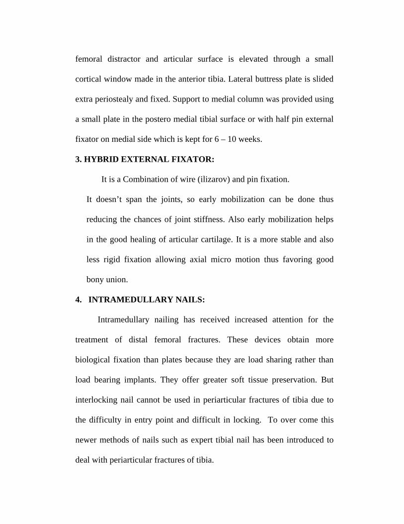
femoral distractor and articular surface is elevated through a small
cortical window made in the anterior tibia. Lateral buttress plate is slided
extra periostealy and fixed. Support to medial column was provided using
a small plate in the postero medial tibial surface or with half pin external
fixator on medial side which is kept for 6 – 10 weeks.
3. HYBRID EXTERNAL FIXATOR:
It is a Combination of wire (ilizarov) and pin fixation.
It doesn’t span the joints, so early mobilization can be done thus
reducing the chances of joint stiffness. Also early mobilization helps
in the good healing of articular cartilage. It is a more stable and also
less rigid fixation allowing axial micro motion thus favoring good
bony union.
4. INTRAMEDULLARY NAILS:
Intramedullary nailing has received increased attention for the
treatment of distal femoral fractures. These devices obtain more
biological fixation than plates because they are load sharing rather than
load bearing implants. They offer greater soft tissue preservation. But
interlocking nail cannot be used in periarticular fractures of tibia due to
the difficulty in entry point and difficult in locking. To over come this
newer methods of nails such as expert tibial nail has been introduced to
deal with periarticular fractures of tibia.
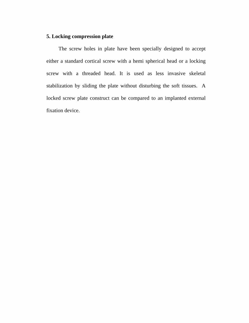
5. Locking compression plate
The screw holes in plate have been specially designed to accept
either a standard cortical screw with a hemi spherical head or a locking
screw with a threaded head. It is used as less invasive skeletal
stabilization by sliding the plate without disturbing the soft tissues. A
locked screw plate construct can be compared to an implanted external
fixation device.

COMPONENTS AND INSTRUMENTS OF HYBRID FIXATOR
Hybrid fixator consists of 3 components.
RING SYSTEM
It consists of k wires, olive wires, wire fixation bolts, 5/8th ring, bolts,
nuts and posts.
PIN SYSTEM
It consists of schanz screw, AO tube and universal clamp.
INTERCONNECTING SYSTEM
Male post connected with AO clamp
K WIRE
K wire was named after Martin Kirschner. Depending on the type of
tip it is differentiated into two types – trocar point has 3 cutting edges. It
is used in fixation of cancellous bones; diamond tip has beveled edge,
which is used in cortical bones. Stiffness of k wire is directly proportional
to 4th power of radius. For tibia and femur 1.8 mm k wire of 300mm
length is used.
HALF PINS
It does not pass beyond the far cortex. They are round at one end and
threaded at other end. They are self tapping and available in 3-6mm of
diameter. 4.5×175 mm is used for tibia and 4.5×200mm for femur
SIDE TUBE
It is the long stress bearing segment. It is 11mm in diameter and
available in lengths of 100-450in 50mm increments and 550&650. SS rod
is tubular with 11mm outer diameter and 9mm inner diameter. Carbon
fiber rod is solid with11mm diameter and is radiolucent.
UNIVERSAL CLAMP
It is used for connecting 4.5 or 5mm schanz screw with side tube.
This allows pin insertions for angle adjustments over 15 deg in frontal
plane.
RING
In hybrid 5/8th is used, availability ranging from 120-220.
INTERCONNECTION
Swivel from universal clamp is removed and connected to male post,
which is attached to 5/8th ring.
STABILITY OF FIXATOR:
Stability of fixator can be increased by the following techniques
• Increasing the number of wires in proximal fragment
• Adding a schanz screw in proximal fragment
• A minimum of three schantz screws are needed for each fragment
in case of diaphyseal extension.
• Adding a third rod to the apparatus
• Triangulating the fragment.

PREOPERATIVE PLANNING
X ray of the knee joint / ankle joint AP and Lateral view taken.
The need for reduction of articular surface is assessed.
The articular surface is reduced and fixed with cancellous
screws
Metaphysial fragment is fixed with two ilizarow wires and
connected to a 5/8th ring. For additional stability an extra
ilizarow wire can be added. For fixation of additional fragments
olive wires are used.

Tensioned ilizarow wires in metaphyseal fragments gives good
hold and stability than screws. It also gives multiplanar fixation
and aids in early joint mobilization.
In case of periarticular fractures of tibia extending to diaphysis
is 3 schantz screws are inserted in proximal fragment and three
schantz screws in distal fragment, to give adequate stability to
the fracture site.
Two AO rods are connected to the 5/8th ring and are
triangulated. Triangulation gives 360 degree stability which are
comparable to a full ring. For additional stability an additional
rod can be used.

SURGICAL TECHNIQUE
Preoperative planning
• Evaluate the need for distraction to aid reduction.
• Assess articular surface involvement.
• Plan and determine proper wire and Schanz screw placement
TECHNIQUE
PROXIMAL TIBIA
1. Reduce the articular surface
• Ligamentotaxis is achieved using femoral distractor
• If needed open reduction and bone graft.
• Screw fixation using 6.5mm cancellous screws provides
interfragmentary compression for the articular fragments
2. Determine wire position
• Minimum of two wires is needed.
• Position wires within the zones for safe pin placement ( location of
the peroneal nerve should be noted).
• Position the wires distal to cannulated screws and distal to the
capsular attachment of the knee joint.
• The location of wires to be used in proximal tibia is
- A. Anterolateral to posteromedial tibia
- B. Lateral to medial tibia

• Wires inserted should be below 14mm to joint line to avoid entry
into synovial recess posteriorly to avoid septic arthritis secondary
to pin tract infection
• Recently communication between tibiofibular joint and knee joint
was found. So fibular head should not be included in the wire
insertion.
• If a fragment is to be reduced an olive wire can be inserted
3. Insert wires
• Make a stab incision and insert a Protection Sleeve
• Manually push the wire through the sleeve until it contacts the
bone.
• Alternatively, push the point of the wire through the skin without
making an incision. Do not change the wire direction after
insertion.
• When the wire has pierced the opposite cortex proceed with gentle
blows of the hammer,
4. Place clamps on wires (central and eccentric)
5. Attach clamps to ring- 5/8th ring
6. Tension wires
- First wrench-tighten the wire-locking nut on one side and finger
tighten the wire-locking nut on the other (tensioning) side
- apply the tensioner to the wire and tension it
- wires are generally tensioned to 100-130 kg.
7. Attachment to AO rod to ring using male clamp or hybrid clamp
8. Additional rods attachment
Distal Tibial Fractures:
• Apply distraction using distractor
• Fix associated fibular fracture to restore correct length, rotation and
alignment
• Restore the articular surface
• Two typical wire positions :
A. Anterolateral to posteromedial tibia
B. Lateral to medial tibia

COMPLICATIONS
The surgical treatment for periarticular fractures of tibia now has a
better outcome than in the past because of improved implants. However
the new methods are not without problems.
Complication of fractures:
1. Infection
2. Vascular injuries
3. Nerve injuries
4. Nonunion
5. Malunion
6. Pulmonary complications
7. Missed ligamentous injuries
8. Knee stiffness
Complication of operative treatment:
1. Incomplete reduction
2. Incongruous reduction
3. Loss of knee motion
4. Infection
INFECTION:
The major draw back of operative fixation of periarticular fractures
of tibia is the risk of infection. However it should not exceed 5%. If

wound drainage develops postoperatively, aggressive irrigation and
debridement are indicated. Appropriate antibiotics should be given
intravenously for 3 to 6 weeks. In the presence of infection, the implants
should be retained because stable infected fractures are easy to manage
than unstable infected fractures. However if the implant is loose, it
should be removed and the fracture should be protected with external
fixation.
NONUNION:
It is much more common in conservatively treated cases than in
surgically treated cases, owing in part to the rich blood supply to the
proximal tibia and the predominance of cancellous bone. Nonunion
generally is due to presence of infection, unstable fixation, mechanical
failure of the implant or any combination of these factors. Treatment may
be difficult owing to preexisting osteopenia, proximity to knee joint and
prior surgical procedures. Aseptic nonunion should be treated by repeat
osteosynthesis. Septic nonunion should be treated with external
stabilization.
POST TRAUMATIC ARTHRITIS:
The incidence of post traumatic arthritis is unknown. However
incongruity of the joint surface is the leading cause of the early arthritis.
Unfortunately lot of patients developing post traumatic arthritis is young

patient becoming unsuitable for TKR. This complication can be reduced
by anatomical reduction and early mobilization. In patients where it is
limited to one compartment, a corrective osteotomy may be indicated. In
patients with severe disabling bicompartmental or Tricompartmental
arthritis, knee arthrodesis (or) TKR may be necessary. Factors such as
age, range of motion, presence or absence of flexion contractures and
infections play a major role in surgical decision making.
KNEE STIFFNESS:
Perhaps the most common complication that occurs after tibial
plateau fracture is loss of knee motion. This untoward complication
invariably results from damage to joint surface as a consequence of initial
trauma (or) surgical exposure for fixation (or) both. Arthrofibrosis of the
knee joint is thought to restrict knee movement. These effects are greatly
magnified by immobilization after fracture, or internal fixation.
Immobilization of the knee for a period of more than 3 weeks usually
results in some degree of permanent stiffness.
Early stable fixation of the fracture with meticulous soft tissue
handling and immediate mobilization of the knee maximize the chance
for an optimal outcome after periarticular fractures of tibia. Patients
should have 900 of knee flexion 4 weeks postoperatively. Patients who
failed to regain at least 900 of knee flexion between 8-10 weeks

postoperatively usually warrant additional treatment. Arthroscopic lysis
with manipulation can be done. Forcible manipulation should be avoided.
VASCULAR INJURIES:
The exact incidence of vascular injury accompanying proximal
tibial fracture is unknown but is estimated to be only 2-3 %. Vascular
injuries can be caused by direct laceration (or) contusion of the artery or
vein by fracture fragments or indirectly by stretching leading to initial
damage, clinical examination for signs of ischemia with evaluation of
pulses and motor and sensory function is essential.
MALUNION:
Malunion of one or both tibial condyles, distorts the articular
surface of the knee, but it produces much more severe disability. It should
be corrected and internally fixed with maintaining the articular surface.
PULMONARY COMPLICATIONS
When stabilization of the fractures was delayed in patients who had
multiple injuries, the incidence of pulmonary complications was higher,
patients who were treated conservatively or with late stabilization of
fractures in polytrauma had high incidence of fat embolism (22%).
ASSOCIATED LIGAMENTOUS INJURIES
Concomitant ligamentous injuries to the knee are common but are
rarely diagnosed preoperatively. Initially non operative treatment is
advocated as repair (or) reconstruction may produce further
comminution, prolonged operation time and increases the risk of loss of
knee motion and infection. Protected motion in conjunction with a knee
orthosis and vigorous rehabilitation may obviate the need for late
reconstructive surgery. If necessary late reconstruction should be done
after the fracture has healed.

EVALUATION OF OUTCOME
There is a lot of scoring system for evaluation of outcome. We
followed the rating system of Neer for evaluation of knee. Other systems
like the hospital total knee care system and Schatzker system are more
complicated to follow:
NEER’S RATING SYSTEM:
CHARACTER SCORE DEFINITION
Pain 4
3
2
1
No pain in all ranges of motion
Pain with normal daily activity
Minimal activity gives pain
Pain at rest
Movements
(In degrees)
4
3
2
1
Flexion > 120 ; No FFD
Full Extension, flexion 90 to 120
Loss of Extension less than 10 ;
Flexion 700 to 900
Flexion < 60
Function 4
3
2
1
Full weight bearing, Normal gait,
No functional impairment
Limp, No restriction of activity
Requires walking aid
Cannot walk
Shortening (cm) 4
3
2
1
0 – 0.5 cm
0.5 to 2.5 cm
2.5 to 5 cm
> 5 cm
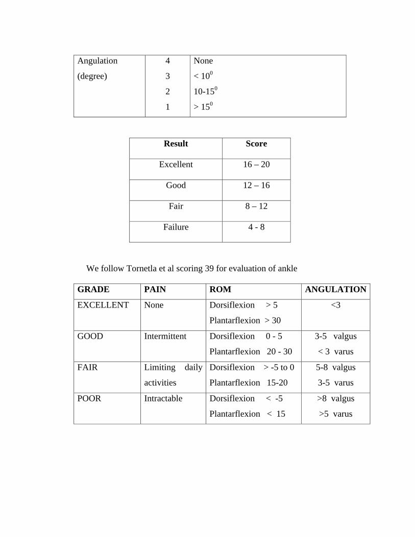
Angulation
(degree)
4
3
2
1
None
< 100
10-150
> 150
Result Score
Excellent 16 – 20
Good 12 – 16
Fair 8 – 12
Failure 4 - 8
We follow Tornetla et al scoring 39 for evaluation of ankle
GRADE PAIN ROM ANGULATION
EXCELLENT None Dorsiflexion > 5
Plantarflexion > 30
<3
GOOD Intermittent Dorsiflexion 0 - 5
Plantarflexion 20 - 30
3-5 valgus
< 3 varus
FAIR Limiting daily
activities
Dorsiflexion > -5 to 0
Plantarflexion 15-20
5-8 valgus
3-5 varus
POOR Intractable Dorsiflexion < -5
Plantarflexion < 15
>8 valgus
>5 varus
MATERIALS AND METHODS
The period of surgery and follow up extends from November 2006
to November 2008
It includes all grades of proximal and distal tibia fractures.
Pathological fractures and fractures in children were excluded.
The cases were analyzed as per the following criteria.
1. Age distribution
2. Sex distribution
3. Side of injury
4. Mode of injury
5. Anatomy of injury
6. Associated injuries
7. Open fractures
8. Duration for surgery
9. Time of union

1. AGE DISTRIBUTION
The age groups varied from 21 years to 51 years with the mean age of
36.5 years. Incidence of fracture was observed maximum between 30- 40
years of age.
Age Group Number of cases Percentage
20 – 30 years 3 16.7%
30 – 40 years 11 61%
40 – 50 years 3 16.7%
50 – 60 years 1 5%
2. SEX
Among the 18 cases, males were predominant with female to male
ratio being 1:19
Sex Number of
cases
Percentage
Male
Female
17
1
95 %
5 %

3. SIDE OF INJURY:
Right side was common in our series
Sex Right Left Percentage
Male
Female
10
0
7
1
56 %
44%
Total 10 8 100%
4. MODE OF INJURY:
Commonest mode of injury has been road traffic accident
Mode of Injury Number of cases Percentage
RTA
Fall
18
0
100 %
0%
5. ANATOMY
SITE Number of cases Percentage
Proximal tibia
Distal tibia
9
9
50%
50%

6. ASSOCIATED INJURIES
Head injury – 2
Distal radius fractures - 1
Patella fracture - 4
Supracondylar fracture femur - 1
7. OPEN FRACTURES
8. DURATION FOR SURGERY
TIME INTERVAL Number of cases Percentage
1 day
< 1 week
<2 weeks
6
8
4
34
45
21
Type Number of cases Percentage
Simple
Compound Gr II
Compound Gr III A
Compound Gr III B
5
2
4
7
28 %
11 %
22 %
39 %

9. TIME OF UNION
Time of union Number of cases Percentage
< 4 months
4-6 months
>6 months
11
4
2
66
23
11
OBSERVATION
• 80% of the patients were between 30- 50 yrs.
• Both male and female were included , majority being males.
• Right side was common and was no bilateral cases studied.
• 72% of the fractures were compound injuries.
• 45% of patients had associated injuries.
• Mean duration between injury and surgery was 1 week.
• 5 patients required additional surgeries like flap cover and SSG
(two immediately and others within 3 weeks).
• Average time for bone union was 4 1/2 months.
• Average knee joint flexion was 100 degrees and ankle dorsi
flexion was 20 degrees.
• The results were excellent in 54%, good in 29% and fair in 17%
of patients.
PROCEDURE
General Measures
All the patients were received in the casuality department and were
resuscitated. After the general condition improved X rays AP and lateral
views were taken. A detailed preoperative work up was done. All the
cases were taken for surgical procedure as soon as possible. Those cases
which were compound were initially treated with external fixator.
POST OPERATIVE PROTOCOL:
Second post op day:
ROM exercises started along with quadriceps Exercises. Partial
weight bearing started if bone grafting not done and delayed till 6 weeks
if grafted for settling of graft (None of our patients were grafted, so all
the patients were started partial weight bearing immediately).
After 6 weeks:
• Full weight bearing is started after radiological signs of union
appear (full weight bearing was started at an average of 6 weeks. In
3 of our patients it was delayed for 2 months due to poor
radiological signs of union).
• Fixator is removed after fracture is united radio logically

• Regular check up is done for half pin pull out, loss of wire tension,
deformity in half pins, side rod or 5/8th ring. Pin site care is given
according to oxford protocol 24.
Care in operating room – pin site dressing with non adhesive dressing
For 48 hrs – pin site not disturbed
After 48 hrs –
1. clean frame with normal saline prior to removing pin site dressing
2. clean each pin site with separate gauze and normal saline
3. cover half pin and wire site with nonadhesive dressing
4. continue daily pin dressing
After one week
- continue daily wash with cotton gauze and saline
- take daily shower
- leave all pin sites uncovered
Pin site infection is treated with twice daily dressing, oral antibiotics.
FOLLOW UP:
All the patients were followed up carefully looking for any
complication every fortnightly till fracture healing. And there after every
monthly upto 6 months. And every 6 monthly up to two years.

ANALYSIS OF FUNCTIONAL OUTCOME
Total 18 patients are included in the study. One case went for
septicemia and went for amputation. Other patients are evaluated and
studied for functional outcome.
Normal bone union – 15
Delayed union – 2
Shortening – 2
Joint stiffness – 2
Varus angulation – 2
Pin tract infection – 4
Deep infection - 2
OVERALL RESULTS
GRADING NO OF CASES PERCENTAGE
EXCELLENT 9 54
GOOD 5 29
FAIR 3 17
DISCUSSION
All the methods available for fixation of periarticular fractures of
tibia have good fracture union results but they do not address all of its
problems. The main problem of these fractures is severe communition
and more chance for compound injuries which becomes difficult to be
managed by open reduction and internal fixation. Even in closed injuries
internal fixation results in skin problems. Soft tissue handing becomes the
important criteria in the treatment of these fractures since it preserves the
hematoma aiding in quick fracture healing. Also it helps in early joint
mobilization thus preventing joint stiffness.
In case of compound fractures wound care becomes the prime
importance. In needs the preliminary application of a external fixator
followed by definitive mode of fixation after the wound has healed. But
the application of hybrid external fixator not only helps in wound care but
also is a definitive mode of fixation. This avoids two procedures for these
patients thus decreasing the stress for the patients and economically
friendly. The compliance of hybrid fixator is better than ilizarov fixator.
Also the simplicity of structure of this apparatus helps in easy application
of flap covers which is mostly needed in the due course of treatment of
compound injuries, which is not possible in case of ilizarov apparatus.

In our study of 18 cases of which 13 are compound cases 5 are simple
cases, hybrid fixator was applied in a mean time interval of 8 days. Time
interval ranged from day one to 2 weeks. The delay in surgery was
mainly due to medical problems of the patient.
All the cases were followed for a mean period of 14.2 months
averaging from 28 months to 4 months. Out of the 18 cases bony union
was obtained in 17 cases ( one of which was converted to ilizarov since
there was a bony defect after which bone transport and bony union was
obtained) and one patient went for below knee amputation due to
impending septicemia. 2 cases had delayed union one of which
maintained by prolonged PTB cast and one with fibulectomy. Main
reason for delayed union was intact fibula which made the fracture site to
distract. Even though hybrid provides axial micro movement,
compression cannot be applied to fracture after the application of fixator
which will be possible in ilizarov fixator. So at the time of application
fracture site should not be over distracted and particular attention should
paid in proper reduction of fracture. Comparing the other studies Barberie
et al 31 reported 3 non union in 34 patients managed by hybrid fixcator,
one case of non union in 31 patients in Tracy et al 41 study and no non
union out of 13 cases in a series by Kevin J Pugh.

There was one case of failure in a case of compound grade III b
fracture with severe contamination. This was due to severe infection and
extensive bone loss. Since the patient went to septicemia removal of
fixator was done and below knee amputation was done. This would have
been avoided by proper selection of case.
The average time of bony union was 4.5 months compared to 4
months by Barberi et al 33 and 4.2 months by Tornetla et al.
There were 4 cases of pin tract infections all which got settled
without surgical intervention. This was attributed to poor pin site care by
the patients after their discharge. There were two cases of deep infection
one persisted even after fixator removal which was treated by antibiotics.
Other patient presented lately after union in the site of Tension Band
Wiring applied for patella which settled after removal of implant.
There were 2 cases that were complicated by knee stiffness. Both the
patients had poor compliance in the post operative period which was the
result of knee stiffness.
One patient had knee instability which persisted after union of
fracture which was treated conservatively knee brace. Shortening of
<2 cm was seen in two patients both of which had highly communited
tibial plateau fractures with diaphyseal extension. They were managed
with heal rise.

CONCLUSION
To summarize, the advantages of hybrid external fixator are
- minimally invasive procedure
- good preservation of soft tissues
- better anchorage of thin tensioned wires than half pins in
cancellous bone and they give better stability.
- easy application of half pins in diaphysis without
neurovascular injury
- early mobilization of adjacent joint
- good skin care and easy application of flap cover
- less heavier than ilizarov gaining good acceptance of the
patient.
The disadvantages of hybrid fixator are
- risk of articular infection if pins are applied very close to
joint
- tough to obtain articular reduction
- the radio opaque ring obstructs x ray visualization of fracture

Hybrid External Fixator is very effective and useful treatment
modality for periarticular fractures of tibia. It aids in providing good skin
Care and it is easy for application of flap covers which is difficult in case
of ilizarov circular fixators. It also acts as a definitive fixation aiding in
good fracture union thereby avoiding multiple surgeries in compound
fractures.
Hybrid External fixator is a modular, safe and useful treatment option
for complex periarticular fractures of tibia. It is minimally invasive with
least complications compared to other methods which are used for peri
articular fractures of tibia.

BIBLIOGRAPHY
1. Ali AM, Saleh M, Bolongaro S, Yang L. The strength of different
techniques for bicondylar tibial plateau fractures – a biomechanical
study. Clinical bio mech( Bristol, Avon; Nov 2003, Vol 1819), No
864-870.
2. Allan.F. Tencer, Kenneth.D.Johnson, Biomechanics in orthopaedic
trauma. 1st edition, 1994, Lippincott.
3. Behrens F. Manual of internal fixation – Technic recommended by
AO – ASIF Group 1990, 3rd edition, chapter 5 , NO 367-411.
4. Benedetti G.B, Argneni.F – A.S.A.M.I group, operative principles
of ilizarov, Williams and Wilkins.
5. Beris A E, Glisson RR, Seaber Av, Urbaniak JR. “ Load tolerance
of tibial plateau depression with cluster of K wires”. Trans 34th
Orthopaedic research society meeting 1988; vol 13, No 30.
6. Bonel, Stegemann P, Mc Namara K, Seibel R. External fixation of
severely communited and open tibial pilon fractures. Clinical
orthop; 1993, vol 297, No 101-107.
7. Bronson DG , Samchukor MC, Birch JG. Journal peadiatric orthop
B; 2002 April, vol11, No 143-149.

8. Calhoum JH, LiF, Ledbetter BR , Gill CA. Biomechancs of
ilizarov for fracture fixation. Trans 37th orthopaedic Reserch
Society Meeting 1991; vol 16, No 438.
9. Browner and Jupiter, Text book of skeletal Trauma; 2nd edition
Chapter 56, No 2316 – 2324.
10. Catagni M- A.S.A.M.I group, operative principles of ilizarov,
Williams & Wilkins.
11. Decoster TA , hertderns DB, Downey DJ , Ferries JS, Jones W.
Optimizing bone screw pull out force. Journal of orthopaedic
Trauma; 1990, vol 4, No 169-174.
12. Dror paley. Problems, obstracles and complications of limb
lengthening by illizarow technique. Clinical orthop;1990, vol 250,
No81-104.
13. Egan JM, Shearer JR. Behavior of an external fixator frame
incorporating an angular separation of fixator pins; a finate element
approach. Clinical orthop; 1987, vol223, No 265-274.
14. Endres T, Grans R, Biewener A, Barthal S, Zwippu. Advantages of
minimally invasive reposition retension and hybrid external fixator
for tibial pilon fractures with particular emphasis on C2/C3
fractures. Unfallchirulg;April 2004, Vol107(4),No 273-284.

15. Gaudinez RF, Mallik AR, Szporn M .Hybrid external fixator of
common tibial plateau fractures. Clinical orthop; 1996, vol 328, No
203-210.
16. George W wood, Campbells operative orthopaedics; 2003, 10th
edition, Chapter 50, No2671-2723.
17. Halsey D, Fleming B, Pope MH, Krag M, Kristiansen T. External
fixator pin design. Clinical orthop, 1992; vol 278, No 305- 312.
18. Joyne PH, Rochat MC, Hoover JP. Use of hybrid external fixator
for repair of periarticular tibia fractures in a Patagonian cavy. J AM
vet med assoc; Apr 2004, vol 224(8), No 1298- 1301.
19. Kevin J pugh, Philip R wolinshy, Mark P Mc Andrew, Kenneth D
Johnson. Tibial pilon fractures; a comparision of treatment
methods. Journal of trauma; 1999, vol 47, No 937-941.
20. Lerner A , Stein. Hybrid thin wire External fixator; an effective
minimal invasive, modular surgical tool for the stabilization of
periarticular fractures. Journal of orthopaedics, Jan 2004, vol 27(1),
No 59-62.
21. Madhor A Karunakar, Micheal J bose. Rockwood and Greens “
Fracture in adults”. 2001; 5th edition, chapter 7, No 231-245.

22. Mark Farrar, Lang yans, Micheal salen. The Sheffield Hybrid
Fixator- a clinical and biomechanical review. INJURY; 2001,
vol 32, No 8-13.
23. Marsh JL , Smith ST, DOTT. External fixator and limited
Internal fixation of complicated fractures of Tibial plateau.
Journal of Bone and Joint surgery 1995; May vol 77A, No 661-
673.
24. Martin A McNally, Maurizio A Catagni. Oxford’s textbook of
orthopaedics, chapter 6, No 1735-1748.
25. Michael S Sirkin, Christopher M Bono, Mark c Reilly, Fred F
Behrens. Percutaneous methods of tibial plateau fixation. Clin
orthop and related research; 2000, vol 375, No 60-68.
26. Ovadia.DN, Beals RK, Fractures of tibial plafond. Journal of
Bone and Joint surgery; 1986, vol 68A, No 543-551.
27. Paige Whittle A, George W Wood, campbells operative
orthopaedics; 2003, 10th edition, chapter 51, No 2726-2872.
28. Persen SM, Cardey J, Baumgart F, Rahh BA, Schatzker J.
Technical and Biomechanical aspects of screws used for bone
screws. International journal of orthopaedic Trauma; 1992, vol
2 No 31-48.

29. Pugh KJ, Wolinsky PR, Dawson JM, Stalhman GC. The
biomechanics of Hybrid external fixator. J orthop Trauma;
1999, vol 13 No 20-26.
30. Raschke MJ , Hoffman R, khodadyan C, Fournier CV, Sudkann
NP,Hoas NP. Combination of ilizarov ring fixator with unilator
AO tubular fixator; initial clinical experience with hybrid
system. Unfallchirury; 1995, vol 98, No 627-632.
31. Robern CS, Antoci V, Antoci V Jr, voor MJ. The accuracy of
fine wire tensioners: a comparision of 5 tensioners used in
hybrid and ring external fixator. J orthop trauma; march 2004,
vol 18(3), No 158-162.
32. Robern CS, Dodds Jc, Pery K, Berk D, Selingson D, Voor MJ.
Hybrid external fixator of proximal tibia: stratergies to improve
frame stability. Journal of orthop trauma; 2004 Jan, vol 18 No
57.
33. Rouo Barbieri, Richard Schenk, Kenneth kovel, Kevin Aurori,
Brian Aurori. Hybrid external fixator in treatment of tibia
plaford fractures. Clinical orthop and related research; 1996, vol
332, No 16-22.

34. Reudi TP, Allgower M . The operative treatment of Intra
articular fractures of lower end of tibia. Clin orthop 1979, vol
138, No 105-110.
35. Schatzker J, Sanderson R, Murnaghan JP. The holding power of
orthopaedic screws in vivo. Clinical orthop; 1975, vol 108, No
115-126.
36. Stamer D, Schenk R, Staggers B. Bicondylar tibial plateau
fractures treated with Hybrid external fixator, a preliminary
study. Jour orthop trauma; 1994, vol 8, No 455-461.
37. Steink, Mosheiff R, Baumgart F, Frigg R, Perren SK, cordey J.
the hybrid external fixator: a biomechanical study. Clin
Biomechanics; 1997, vol 12, No 259-266.
38. Thakur AJ. Elements of fracture fixation, 1997; 1st edition.
Chapter 8, no 147-176.
39. Tornetta P, Weiner L, Bergman metal, Pilon fractures treated
with combined internal and external fixation. Journal of orthop
trauma 1993, vol 7, No 489-496.
40. Tracy Watson J. High energy fractures of tibial plateau. OCNA;
oct 1994, vol 25, no 723-752.
41. Tracy Watson J, Steve Ripple, Hoshaw, David Fyhrie . Hybrid
external fixator for tibial plateau fractures. OCNA; jan 2002,
vol 33, No 199-209.
42. Uhthoff HK. Mechanical factors influencing the holding power
of screws in compact bone. JBJS; 1975, vol 55B No 633-639.
vol 77A, NO661-673.
43. Weiner L, Kelly M, Yans E. The use of combination of internal
fixation and hybrid external fixation in severe proximal tibia
fractures. Journal of orthopaedic trauma; 1995, vol 9, No 244-
250.
44. Yilmaz E, Belhes O, Karakuet L, Arslan N, Serin E. Mechanical
performance of hybrid external fixators in comparision with
Ilizarov circular external fixator. Clinical
BIOmechanics(Bristol, Avon); Jul 2003,Vol 18(6), No 518-522.
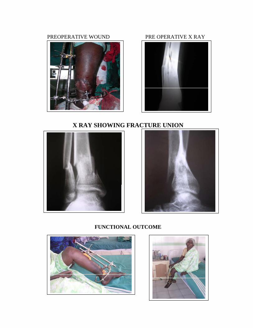
PREOPERATIVE WOUND PRE OPERATIVE X RAY
X RAY SHOWING FRACTURE UNION
FUNCTIONAL OUTCOME
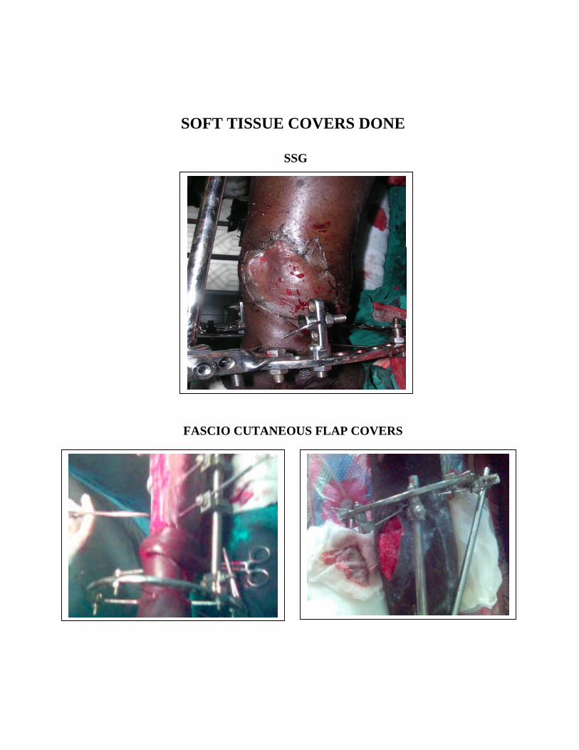
SOFT TISSUE COVERS DONE
SSG
FASCIO CUTANEOUS FLAP COVERS
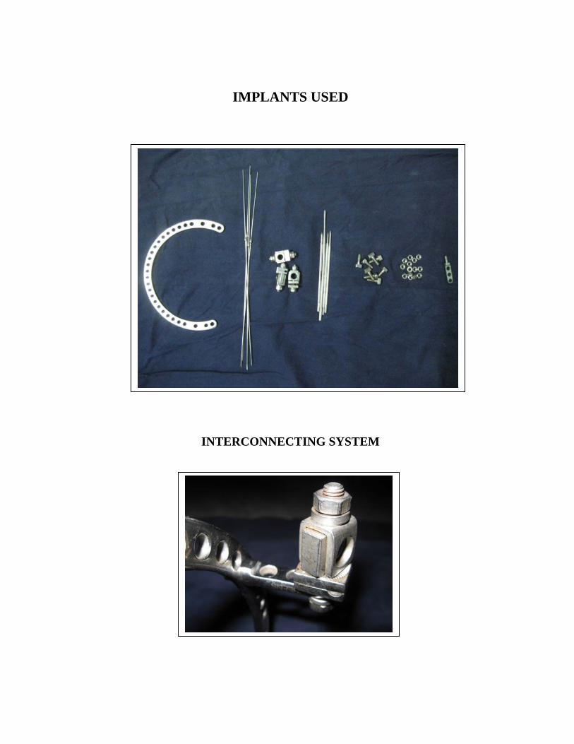
IMPLANTS USED
INTERCONNECTING SYSTEM

COMPOUND GRADE III B PROXIMAL TIBIA FRACTURE
PREOPERATIVE WOUND PREOPERATIVE X RAY
FASCIO CUTANEOUS FLAP POST OPERATIVE
X RAY
KNEE FLEXION REVIEW X RAY

PREOPERATIVE X RAY POST OPERATIVE X RAY
3 MONTHS REVIEW X RAY
ANKLE MOVEMENTS

COMPOUND GRADE III B DISTAL TIBIA FRACTURE
PREOPERATIVE WOUND
POST OPERATIVE X RAY FLAP COVER 4(M) REVIEW
RAY
FOLLOW UP

SIMPLE PROXIMAL TIBIA FRACTURE SCHATZKER TYPE - VI
PREOPERATIVE X RAY POST OP.
3 ½ MONTHS REVIEW X RAY
RANGE OF MOVEMENTS
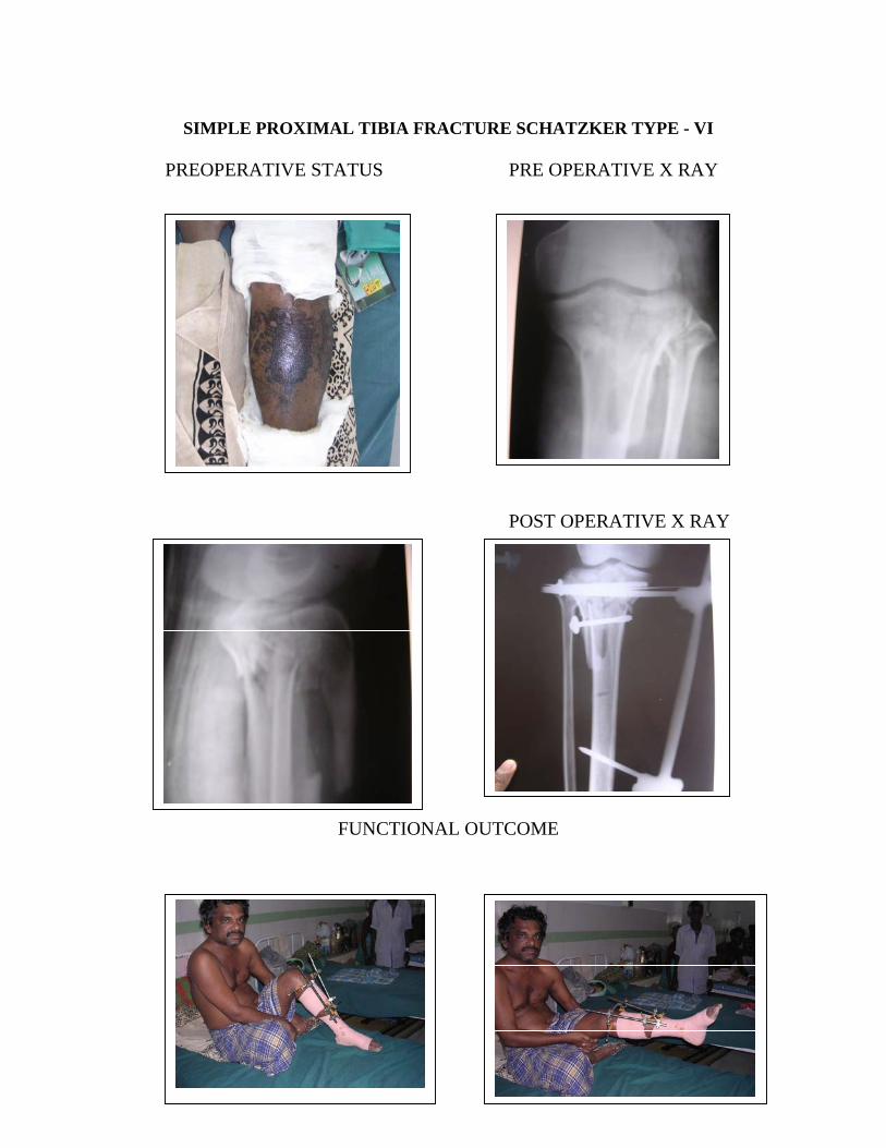
SIMPLE PROXIMAL TIBIA FRACTURE SCHATZKER TYPE - VI
PREOPERATIVE STATUS PRE OPERATIVE X RAY
POST OPERATIVE X RAY
FUNCTIONAL OUTCOME
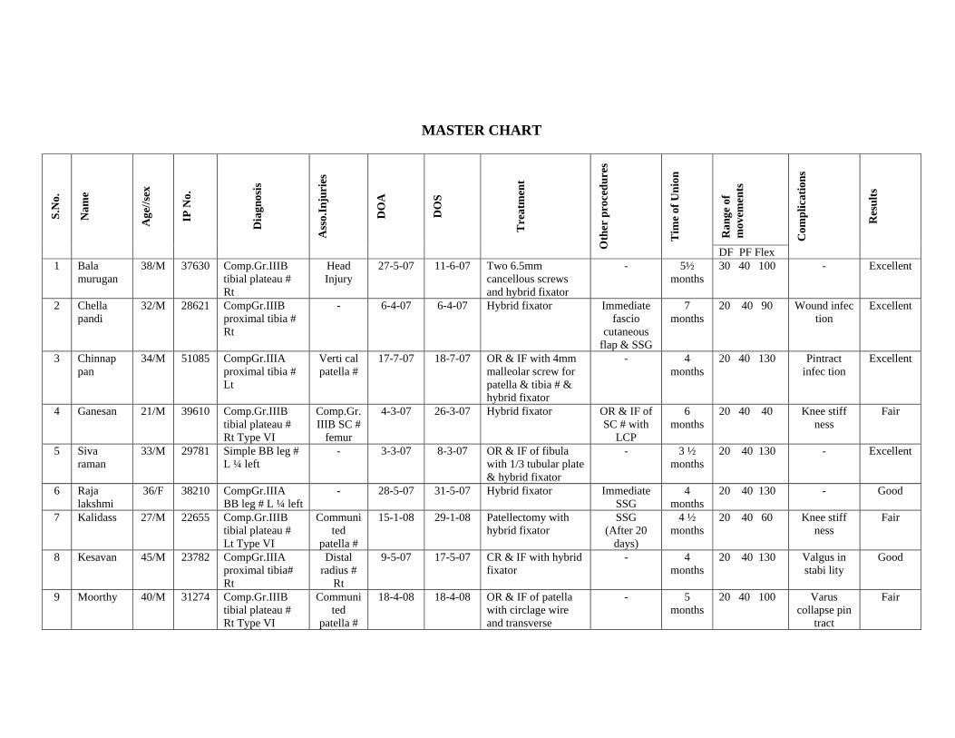
MASTER CHART
Ran
ge o
f m
ovem
ents
S.N
o.
Nam
e
Age
//sex
IP N
o.
D
iagn
osis
Ass
o.In
juri
es
DO
A
DO
S T
reat
men
t
Oth
er p
roce
dure
s
Tim
e of
Uni
on
DF PF Flex
Com
plic
atio
ns
Res
ults
1 Bala murugan
38/M 37630 Comp.Gr.IIIB tibial plateau # Rt
Head Injury
27-5-07 11-6-07 Two 6.5mm cancellous screws and hybrid fixator
- 5½ months
30 40 100 - Excellent
2 Chella pandi
32/M 28621 CompGr.IIIB proximal tibia # Rt
- 6-4-07 6-4-07 Hybrid fixator Immediate fascio
cutaneous flap & SSG
7 months
20 40 90 Wound infec tion
Excellent
3 Chinnap pan
34/M 51085 CompGr.IIIA proximal tibia # Lt
Verti cal patella #
17-7-07 18-7-07 OR & IF with 4mm malleolar screw for patella & tibia # & hybrid fixator
- 4 months
20 40 130 Pintract infec tion
Excellent
4 Ganesan 21/M 39610 Comp.Gr.IIIB tibial plateau # Rt Type VI
Comp.Gr.IIIB SC #
femur
4-3-07 26-3-07 Hybrid fixator OR & IF of SC # with
LCP
6 months
20 40 40 Knee stiff ness
Fair
5 Siva raman
33/M 29781 Simple BB leg # L ¼ left
- 3-3-07 8-3-07 OR & IF of fibula with 1/3 tubular plate & hybrid fixator
- 3 ½ months
20 40 130 - Excellent
6 Raja lakshmi
36/F 38210 CompGr.IIIA BB leg # L ¼ left
- 28-5-07 31-5-07 Hybrid fixator Immediate SSG
4 months
20 40 130 - Good
7 Kalidass 27/M 22655 Comp.Gr.IIIB tibial plateau # Lt Type VI
Communited
patella #
15-1-08 29-1-08 Patellectomy with hybrid fixator
SSG (After 20
days)
4 ½ months
20 40 60 Knee stiff ness
Fair
8 Kesavan 45/M 23782 CompGr.IIIA proximal tibia# Rt
Distal radius #
Rt
9-5-07 17-5-07 CR & IF with hybrid fixator
- 4 months
20 40 130 Valgus in stabi lity
Good
9 Moorthy 40/M 31274 Comp.Gr.IIIB tibial plateau # Rt Type VI
Communited
patella #
18-4-08 18-4-08 OR & IF of patella with circlage wire and transverse
- 5 months
20 40 100 Varus collapse pin
tract
Fair

malleolar screw & hybrid fixator
infection
10 Rathinam 36/M 29241 Simple BB leg # L 1/3 Rt
- 24-3-07 26-3-07 OR & IF of fibula with 1/3 tubular plate & hybrid fixator, 2 lag screw for tibia
- 3 ½ months
20 30 130 - Excellent
11 Alagar 22/M 30081 Simple BB leg # L ¼ left
- 1-6-07 4-6-07 CR & IF of # with hybrid fixator
- 4 months
20 40 130 5 degree varus
Good
12 Muru gesan
45/M 32817 CompGr.IIIB leg # L 1/3 Rt
- 22-6-07 22-6-07 K wire for fibula and hybrid fixator
BK amputation
- - Gross infection &
went for septi cemia
13 Neela vannan
38/M 36281 CompGr.IIIA BB segmental leg # L 1/3 Rt
- 8-9-07 9-9-07 CR & IF with hybrid fixator
Flap cover (25th day)
later converted to Ilizarov for segmental transport
8 months
10 20 120 - Good-fair
14 Sahul hameed
35/M 31181 Comp.Gr II BB leg # L 1/3 Lt
- 30-7-07 30-7-07 CR & IF with hybrid fixator
- 3 ½ months
10 40 130 Pin tract infection
Excellent to Good
15 Mani 36/M 28121 Simple tibial plateau # Lt Type VI
- 23-12-06 26-12-06 CR & IF with hybrid fixator & 2 cancellous screws
- 3 ½ months
20 40 130 Shorten ing 0.5 cm
Excellent
16 Kalai vanan
42/M 30012 Simple tibial plateau # Lt Type VI
- 6-2-07 12-2-07 CR & IF with hybrid fixator & 2 cancellous screws
- 4 months
20 40 110 Pin tract infection
Excellent
17 Paulraj 40/M 27181 Comp.Gr.IIIA tibial plateau # Rt Type VI
Commu nited
patella #
15-1-07 25-1-07 OR & IF of patella with TBW and hybrid fixator
Metal exit for patella
3 ½ months
20 40 100 Varus ang. 15 degrees Infection
shorten ing 1 cm
Good
18 Subra mani
51/M 21261 CompGr.IIIB BB leg # L 1/3 Rt
- 12-1-07 13-1-07 Hybrid fixator Flap cover 23rd day
3 ½ months
10 30 130 - Excellent