Embed Size (px)
Citation preview
Seedling Lethal1, a Pentatricopeptide Repeat ProteinLacking an E/E+ or DYW Domain in Arabidopsis, IsInvolved in Plastid Gene Expression and EarlyChloroplast Development1[C][W]
Young Jae Pyo2, Kwang-Chul Kwon3, Anna Kim, and Myeon Haeng Cho*
Department of Systems Biology, College of Biological Science and Biotechnology, Yonsei University, 120–749Seoul, Korea
ORCID IDs: 0000-0002-0339-0257 (Y.J.P.); 0000-0002-4037-1776 (K.-C.K.); 0000-0002-9824-899X (A.K.); 0000-0002-8392-2726 (M.H.C.).
Chloroplasts are the site of photosynthesis and the biosynthesis of essential metabolites, including amino acids, fatty acids, andsecondary metabolites. It is known that many seedling-lethal mutants are impaired in chloroplast function or development,indicating the development of functional chloroplast is essential for plant growth and development. Here, we isolated a noveltransfer DNA insertionmutant, dubbed sel1 (for seedling lethal1), that exhibited a pigment-defective and seedling-lethal phenotypewith a disrupted pentatricopeptide repeat (PPR) gene. Sequence analysis revealed that SEL1 is a member of the PLS subgroup,which is lacking known E/E+ or DYW domains at the C terminus, in the PLS subfamily of the PPR protein family containing aputative N-terminal transit peptide and 14 putative PPR or PPR-like motifs. Confocal microscopic analysis showed that the SEL1-green fluorescent protein fusion protein is localized in chloroplasts. Transmission electron microscopic analysis revealed that thesel1mutant is impaired in the etioplast, aswell as in chloroplast development. In sel1mutants, plastid-encoded proteins involved inphotosynthesis were rarely detected due to the lack of the corresponding transcripts. Furthermore, transcript profiles of plastidgenes revealed that, in sel1 mutants, the transcript levels of plastid-encoded RNA polymerase-dependent genes were greatlyreduced, but those of nuclear-encoded RNA polymerase-dependent genes were increased or not changed. Additionally, theRNA editing of two editing sites of the acetyl-CoA carboxylase beta subunit gene transcripts in the sel1 mutant was compromised,though it is not directly connectedwith the sel1mutant phenotype. Our results demonstrate that SEL1 is involved in the regulationof plastid gene expression required for normal chloroplast development.
Chloroplasts have their own genome and gene ex-pression machinery. The expression and regulation ofplastid genes are very different from that of nucleargenes, and the coordinated expression of nuclear andplastid genes is important for the development offunctional chloroplasts. Plastid genes are generally or-ganized in polycistronic transcription units. Therefore,they are transcribed as long primary transcripts, whichare extensively processed into mature transcripts byvarious posttranscriptional modifications, such as RNA
splicing, RNA editing, RNA cleavage, and RNA stabil-ity. Theseposttranscriptional regulations ofplastid geneexpression are mediated by many nuclear-encoded pro-teins (Barkan and Goldschmidt-Clermont, 2000).
Recently, many chloroplast-localized pentatricopep-tide repeat (PPR) proteins have emerged as primarynuclear factors that are involved in plastid gene ex-pression and RNA metabolism in higher plants. ThePPR proteins are characterized by a tandem array ofdegenerate 35-amino acid repeats, forming one of thelargest protein families that is particularly prevalent inhigher plants but not in other eukaryotes (Small andPeeters, 2000; Lurin et al., 2004; Schmitz-Linneweberand Small, 2008). Most of the PPR proteins are pre-dicted to be targeted in the chloroplasts ormitochondriaand involved in posttranscriptional regulation oforganellar gene expression by interacting with RNA.The PPR protein family is divided into P and PLS sub-families. The P subfamily usually contains only theclassic PPR (P) motifs. By contrast, the PLS subfamilycontains repeated blocks of PPR-like (P, L, and S)motifs,is specific to land plants (Lurin et al., 2004), and is alsoknown as the plant combinatorial and modular protein(PCMP) subfamily (Aubourg et al., 2000; Rivals et al.,2006). Based on the presence of different C-terminalmotifs, the PLS subfamily is further divided into thePLS, E, E+, and DYW subgroups (Lurin et al., 2004).
1 This work was supported by the Basic Science Research Programfrom the Korea National Research Foundation (grant nos. NRF–2010–0014284and NRF–2013R1A1A2009383 to M.H.C.).
2 Present address: Department of Molecular Bioscience,Universityof Texas, Austin, TX 78712.
3 Present address: Department of Biochemistry and Pathology,School of Dental Medicine, University of Pennsylvania, 240 South40th Street, 538 Levy Building, Philadelphia, PA 19104.
* Address correspondence to [email protected] author responsible for distribution of materials integral to the
findings presented in this article in accordance with the policy de-scribed in the Instructions for Authors (www.plantphysiol.org) is:Myeon Haeng Cho ([email protected]).
[C] Some figures in this article are displayed in color online but inblack and white in the print edition.
[W] The online version of this article contains Web-only data.www.plantphysiol.org/cgi/doi/10.1104/pp.113.227199
1844 Plant Physiology�, December 2013, Vol. 163, pp. 1844–1858, www.plantphysiol.org � 2013 American Society of Plant Biologists. All Rights Reserved. www.plantphysiol.orgon September 28, 2018 - Published by Downloaded from
Copyright © 2013 American Society of Plant Biologists. All rights reserved.
Recent studies have shown that PPR proteins aresequence-specific RNAbinding factors recruiting one ormore effector proteins to the target RNA and are in-volved in posttranscriptional regulation of organellargene expression (Schmitz-Linneweber and Small, 2008).Members of the P subfamily are involved in the tran-scription of plastid genes (Pfalz et al., 2006), RNAcleavage(Meierhoff et al., 2003; Nakamura et al., 2003), RNAstabilization (Beick et al., 2008; Pfalz et al., 2009), RNAsplicing (Schmitz-Linneweber et al., 2006; de Longevialleet al., 2008), and translation (Yamazaki et al., 2004;Schmitz-Linneweber et al., 2005; Cai et al., 2011).RNA editing is a posttranscriptional process that
alters specific cytidine-to-uridine (C-to-U) conversionin the chloroplast and mitochondrial RNA of higherplants (Shikanai, 2006; Bentolila et al., 2008; Stern et al.,2010). In Arabidopsis (Arabidopsis thaliana), at least 34sites are known to be edited in chloroplast transcripts(Chateigner-Boutin and Small, 2007), and more than500 sites are specifically modified in mitochondrialtranscripts (Bentolila et al., 2008; Takenaka et al., 2008).Thus far, more than 10 transfactors required for theediting of particular C targets in chloroplasts havebeen identified. They are all members of the E/E+ andDYW subgroups of the PLS subfamily of the PPRprotein family.Members of the E/E+ subgroupproteins,CHLORORESPIRATORY REDUCTION4 (CRR4),CRR21, CHLOROPLAST BIOGENESIS19 (CLB19), andORGANELLARTRANSCRIPTPROCESSING80 (OTP80),and members of the DYW subgroup proteins, includingCRR22, CRR28, YELLOW SEEDLING1 (YS1), EARLYCHLOROPLAST BIOGENESIS2 (AtECB2), PROTEINREQUIRED FOR accD RNA EDITING1 (AtECB2), andOTP82, have been shown to be required for the editingof corresponding targets such as NADH dehydrogenasesubunit4 (ndhB), ndhD, RNA polymerase alpha subunit(rpoA), acetyl-CoA carboxylase beta subunit gene (accD),and ndhG (Kotera et al., 2005; Okuda et al., 2006, 2007;Chateigner-Boutin et al., 2008; Hammani et al., 2009;Zhou et al., 2009). However, although the Arabidopsisgenome has six PLS subgroup genes in the PLS sub-family of the PPR protein family (Lurin et al., 2004), themolecular function of members of the PLS subgroupproteins has not yet been reported.The chloroplasts of higher plants contain two differ-
ent types of RNA polymerases: nuclear-encoded RNApolymerase (NEP) and plastid-encoded RNA poly-merase (PEP). NEP is a bacteriophage-type, singlesubunit RNApolymerase (Hedtke et al., 1997). PEP is aeubacterial-type, multisubunit RNA polymerase thatconsists of core subunits encoded by plastid genes(rpoA, rpoB, rpoC1, and rpoC2) and s factors encodedby nuclear genes (Hess and Börner, 1999). It has beensuggested that both NEP and PEP are responsible forthe transcription of distinct types of plastid genes(Allison et al., 1996; Hajdukiewicz et al., 1997). Class Igenes are predominantly transcribed by PEP, class IIgenes are transcribed by both NEP and PEP, and classIII genes are transcribed exclusively by NEP. WhereasNEP transcribes nonphotosynthetic housekeeping
genes, PEP transcribes photosynthesis-related genes(Hajdukiewicz et al., 1997; Swiatecka-Hagenbruchet al., 2007; Liere et al., 2011).
In addition to the core subunits, PEP complex con-tains many of the regulatory proteins encoded by nu-clear genes that are essential for plastid gene expression(Pfalz et al., 2006; Steiner et al., 2011).Most of the loss-of-function mutants for these regulatory proteins showseedling-lethal and albino or pale-green phenotype.Though there have been numerous studies on thefunction of the regulatory proteins in plastid gene ex-pression, the mechanism of the regulation has not yetbeen fully understood. Recent maize (Zea mays) nucle-oid proteome analysis has shown that PEP and itsregulatory proteins are found together with other mol-ecules in plastid nucleoids. The nucleoid is a very largecomplex and composed of plastid DNA, RNA, andvarious proteins. It is also the site of DNA regulation,transcription, and translation. For example, RNA mat-uration and ribosome assembly occur cotranscription-ally in association with nucleoids. The function of thenucleoid-enriched proteome is subject to qualitativechange from RNA metabolism to translation and ho-meostasis as proplastids convert to chloroplasts withassistance of regulatory proteins (Majeran et al., 2012).
In this study, we report the molecular characteriza-tion of seedling lethal1 (sel1) mutant, showing a pigment-defective and seedling-lethal phenotype. SEL1 encodesa novel chloroplast-localized PPR protein, which is amember of the PLS subgroup in the PLS subfamily of thePPR protein family. The mutation of SEL1 leads to de-fects in early chloroplast development. In the sel1 mu-tant, the transcription levels of PEP-dependent plastidgenes are decreased, but those of NEP-dependentplastid genes are increased or not changed. Therefore,our results suggest that SEL1 is involved in the regula-tion of plastid gene expression required for normalchloroplast development.
RESULTS
Isolation and Characterization of sel1 Mutant
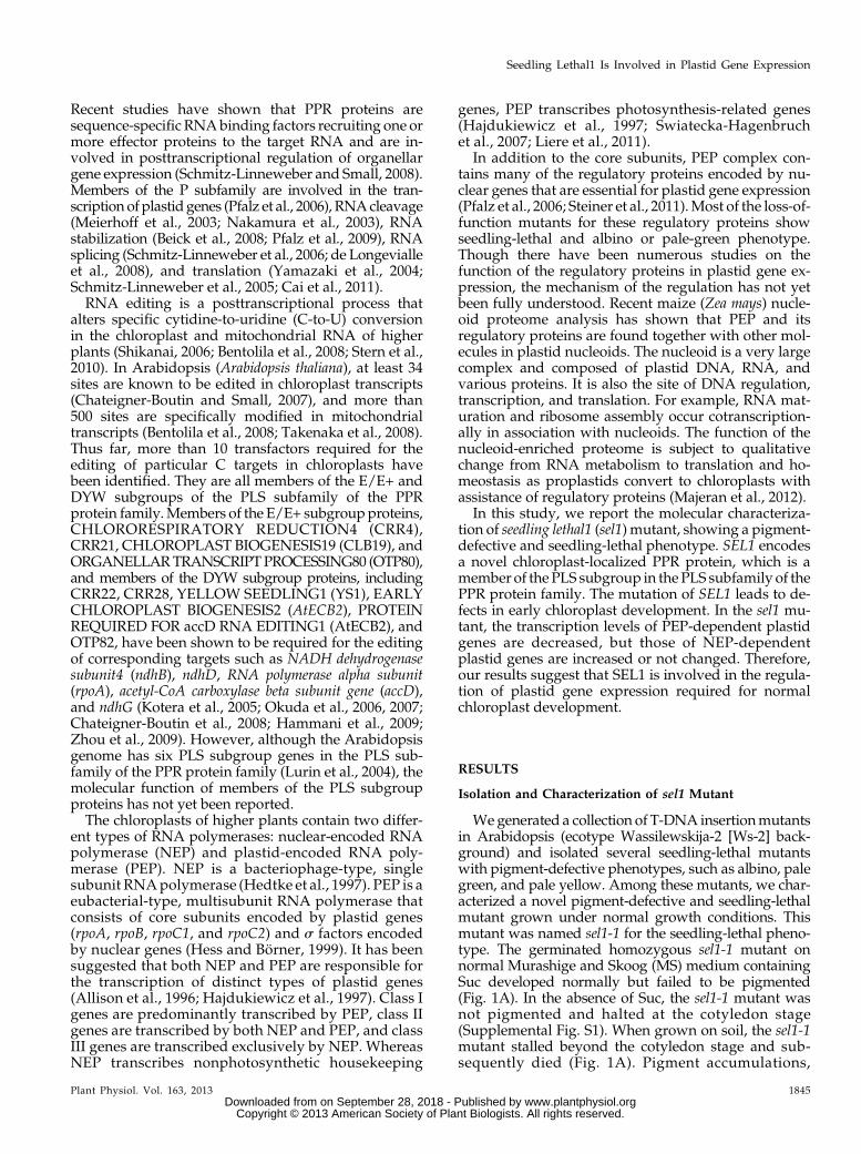
Wegenerated a collection of T-DNA insertionmutantsin Arabidopsis (ecotype Wassilewskija-2 [Ws-2] back-ground) and isolated several seedling-lethal mutantswith pigment-defective phenotypes, such as albino, palegreen, and pale yellow. Among these mutants, we char-acterized a novel pigment-defective and seedling-lethalmutant grown under normal growth conditions. Thismutant was named sel1-1 for the seedling-lethal pheno-type. The germinated homozygous sel1-1 mutant onnormal Murashige and Skoog (MS) medium containingSuc developed normally but failed to be pigmented(Fig. 1A). In the absence of Suc, the sel1-1 mutant wasnot pigmented and halted at the cotyledon stage(Supplemental Fig. S1). When grown on soil, the sel1-1mutant stalled beyond the cotyledon stage and sub-sequently died (Fig. 1A). Pigment accumulations,
Plant Physiol. Vol. 163, 2013 1845
Seedling Lethal1 Is Involved in Plastid Gene Expression
www.plantphysiol.orgon September 28, 2018 - Published by Downloaded from Copyright © 2013 American Society of Plant Biologists. All rights reserved.
including total chlorophyll and carotenoids, were com-pared between the wild type and sel1-1 mutant. Thecontents of total chlorophylls and carotenoids of thesel1-1 mutant were about 6 and 10 times lower, respec-tively, than those of thewild type (Supplemental Fig. S2).
Genetic analysis indicated that the pigment-defectiveand seedling-lethal phenotype of the sel1-1mutant wascontrolled by a recessive nuclear gene and cosegregatedwith the phosphinothricin resistance marker. Sequenceanalysis of the thermal asymmetric interlaced-PCRproduct revealed that the transfer DNA (T-DNA) wasinserted 467 bp downstream from the start codon of theAt4g18520 gene (Fig. 1B). Northern-blot analysis con-firmed that At4g18520 transcript was detected in the
wild type but not in the sel1-1 mutant (Fig. 1C). Theseresults indicate that disruption of the At4g18520 geneleads to a pigment-defective and seedling-lethal phe-notype of the sel1-1 mutant. To confirm that the sel1-1mutant phenotype was resulted from the T-DNA in-sertion in the At4g18520 gene, we obtained two inde-pendent T-DNA insertion lines for the At4g18520 genefrom the SAIL and SALK collections. Genotype andsequence analyses revealed that the position of theT-DNA insertion was 787 bp and 1,747 bp downstreamfrom the start codon of the At4g18520 gene inSAIL_793_F11 (sel1-2) and SALK_054374 (sel1-3), re-spectively (Fig. 1B). The phenotype of sel1-2 and sel1-3was identical to that of sel1-1 (Fig. 1A). Both the sel1-2
Figure 1. Isolation and characteri-zation of the sel1 mutants. A, Pig-ment-defective and seedling-lethalphenotypes of the sel1 mutants. Thewild-type (ecotypes Columbia andWs-2) and sel1 mutant alleles weregrown on MS medium with 1%(w/v) Suc or soil for 4 and 14 d,respectively. Bar = 1 mm. B, Genestructure of SEL1 (At4g18520). Theblack box represents an exon, andgray boxes represent the 59 and 39untranslated regions. The positionsof the T-DNA insertion in sel1-1,sel1-2 (SAIL_793_F11), and sel1-3(SALK_054374) are represented bytriangles. T-DNA is not drawn toscale. C, Northern-blot analysis ofthe SEL1 gene in wild-type (WT)and sel1 mutant alleles. Twentymicrograms of total RNA was iso-lated from 7-d-old seedlings grownon MS medium. 18S rRNA wasused as a loading control. D, Mo-lecular complementation of thesel1 mutant by pSEL1::SEL1 ge-nomic DNA. The top row showsthe 14-d-old wild type, sel1-2, andthe complemented line (Comp.).The bottom row shows northern-blotanalysis of SEL1 in the wild type,sel1-2, and the complemented line.18S rRNA was used as a loadingcontrol. [See online article for colorversion of this figure.]
1846 Plant Physiol. Vol. 163, 2013
Pyo et al.
www.plantphysiol.orgon September 28, 2018 - Published by Downloaded from Copyright © 2013 American Society of Plant Biologists. All rights reserved.
and sel1-3 mutants showed no SEL1 transcripts whenusing northern-blot analysis (Fig. 1C). Furthermore, weconfirmed that the pigment-defective and seedling-lethal phenotype of sel1 mutants was associated withthe T-DNA insertion into At4g18520 by molecularcomplementation. The genomic DNA of the At4g18520gene and its putative promoter region was introducedinto a heterozygous sel1-2 mutant. The subsequentgenotype, phenotype, and northern-blot analyses con-firmed that the introduction of At4g18520 genomicsequence restored the wild-type phenotype in thecomplemented lines (Fig. 1D). In addition,wegeneratedcomplemented transgenic plants expressing FLAG-tagged SEL1 protein under the control of the SEL1promoter. FLAG-tagged SEL1-complemented plantsalso grewnormally likewild-type plants. Thus, we usedthe FLAG-tagged SEL1-complemented plants for fur-ther studies. Taken together, these results revealed thatthe phenotypes observed in sel1mutants are caused by aloss of function of the At4g18520 gene.
SEL1 Gene Encodes a PPR protein That Is a Memberof the PLS Subgroup
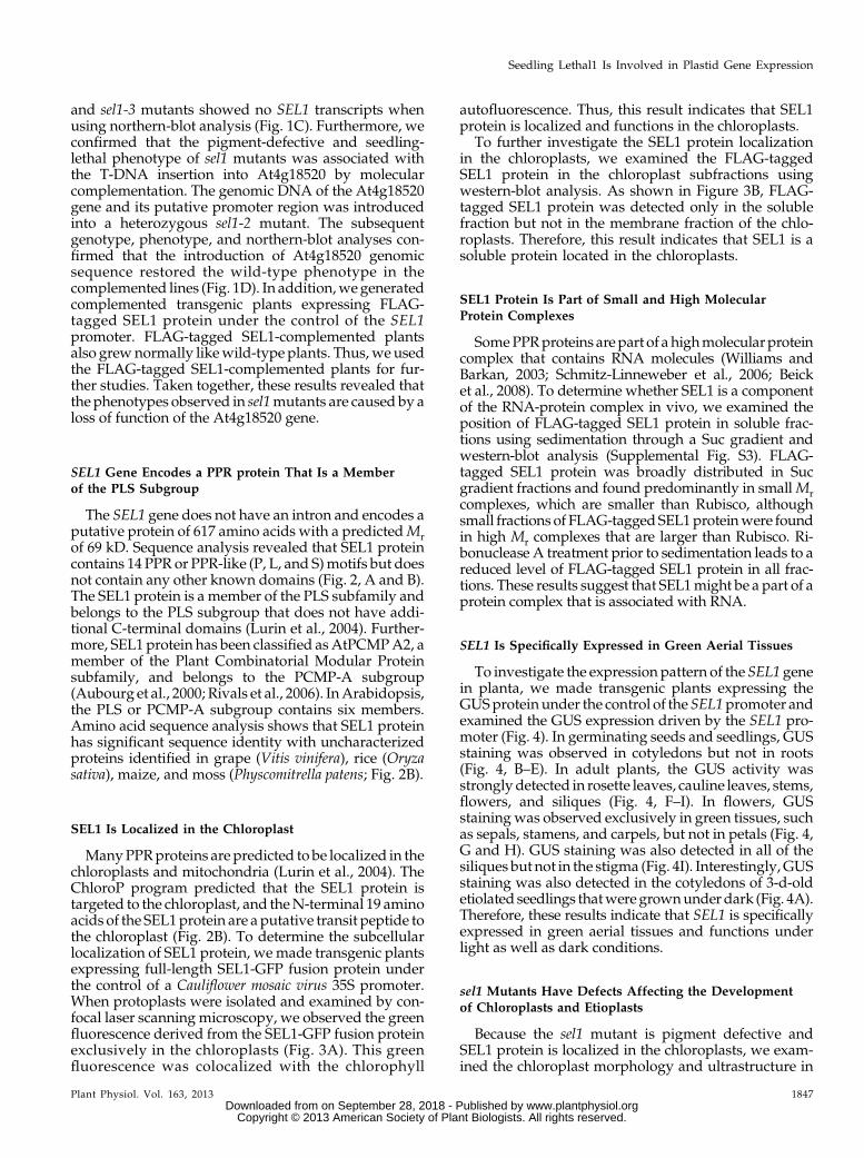
The SEL1 gene does not have an intron and encodes aputative protein of 617 amino acids with a predictedMrof 69 kD. Sequence analysis revealed that SEL1 proteincontains 14 PPR or PPR-like (P, L, and S)motifs but doesnot contain any other known domains (Fig. 2, A and B).The SEL1 protein is a member of the PLS subfamily andbelongs to the PLS subgroup that does not have addi-tional C-terminal domains (Lurin et al., 2004). Further-more, SEL1 protein has been classified asAtPCMPA2, amember of the Plant Combinatorial Modular Proteinsubfamily, and belongs to the PCMP-A subgroup(Aubourg et al., 2000; Rivals et al., 2006). InArabidopsis,the PLS or PCMP-A subgroup contains six members.Amino acid sequence analysis shows that SEL1 proteinhas significant sequence identity with uncharacterizedproteins identified in grape (Vitis vinifera), rice (Oryzasativa), maize, and moss (Physcomitrella patens; Fig. 2B).
SEL1 Is Localized in the Chloroplast
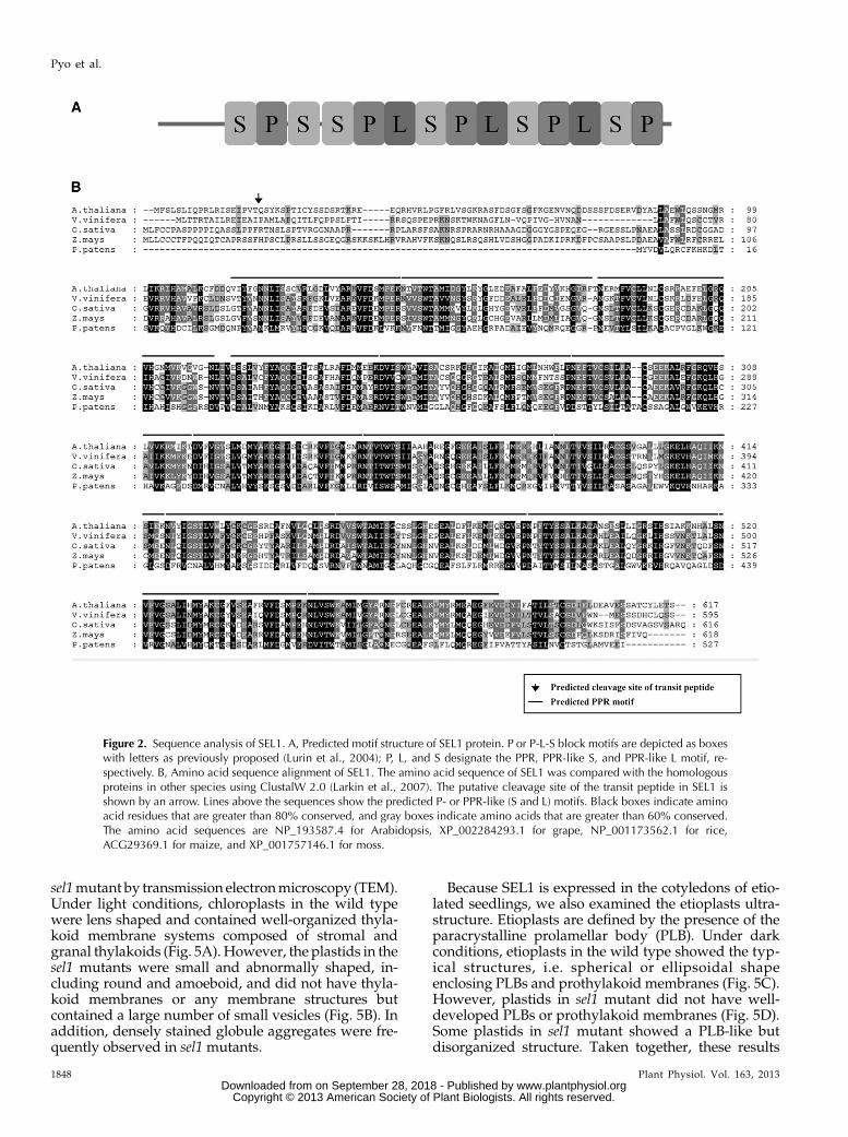
ManyPPRproteins are predicted to be localized in thechloroplasts and mitochondria (Lurin et al., 2004). TheChloroP program predicted that the SEL1 protein istargeted to the chloroplast, and theN-terminal 19 aminoacids of the SEL1protein are aputative transit peptide tothe chloroplast (Fig. 2B). To determine the subcellularlocalization of SEL1 protein, we made transgenic plantsexpressing full-length SEL1-GFP fusion protein underthe control of a Cauliflower mosaic virus 35S promoter.When protoplasts were isolated and examined by con-focal laser scanning microscopy, we observed the greenfluorescence derived from the SEL1-GFP fusion proteinexclusively in the chloroplasts (Fig. 3A). This greenfluorescence was colocalized with the chlorophyll
autofluorescence. Thus, this result indicates that SEL1protein is localized and functions in the chloroplasts.
To further investigate the SEL1 protein localizationin the chloroplasts, we examined the FLAG-taggedSEL1 protein in the chloroplast subfractions usingwestern-blot analysis. As shown in Figure 3B, FLAG-tagged SEL1 protein was detected only in the solublefraction but not in the membrane fraction of the chlo-roplasts. Therefore, this result indicates that SEL1 is asoluble protein located in the chloroplasts.
SEL1 Protein Is Part of Small and High MolecularProtein Complexes
SomePPRproteins are part of a highmolecularproteincomplex that contains RNA molecules (Williams andBarkan, 2003; Schmitz-Linneweber et al., 2006; Beicket al., 2008). To determine whether SEL1 is a componentof the RNA-protein complex in vivo, we examined theposition of FLAG-tagged SEL1 protein in soluble frac-tions using sedimentation through a Suc gradient andwestern-blot analysis (Supplemental Fig. S3). FLAG-tagged SEL1 protein was broadly distributed in Sucgradient fractions and found predominantly in smallMrcomplexes, which are smaller than Rubisco, althoughsmall fractions of FLAG-taggedSEL1proteinwere foundin high Mr complexes that are larger than Rubisco. Ri-bonuclease A treatment prior to sedimentation leads to areduced level of FLAG-tagged SEL1 protein in all frac-tions. These results suggest that SEL1might be a part of aprotein complex that is associated with RNA.
SEL1 Is Specifically Expressed in Green Aerial Tissues
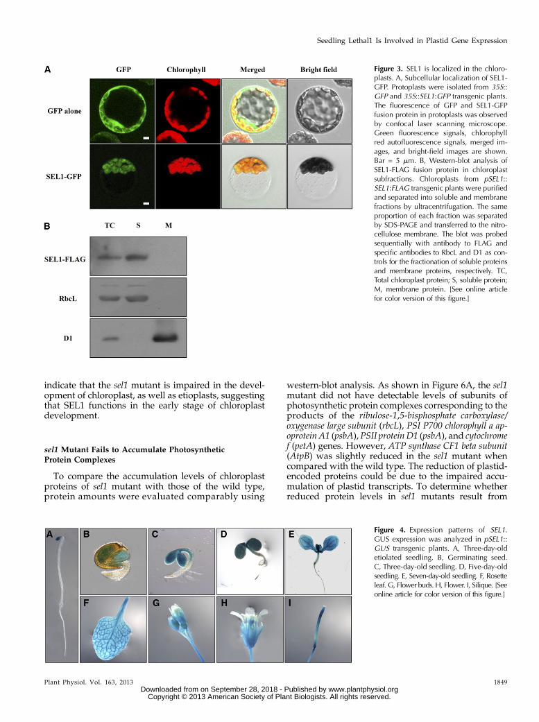
To investigate the expression pattern of the SEL1 genein planta, we made transgenic plants expressing theGUSprotein under the control of the SEL1promoter andexamined the GUS expression driven by the SEL1 pro-moter (Fig. 4). In germinating seeds and seedlings, GUSstaining was observed in cotyledons but not in roots(Fig. 4, B–E). In adult plants, the GUS activity wasstrongly detected in rosette leaves, cauline leaves, stems,flowers, and siliques (Fig. 4, F–I). In flowers, GUSstaining was observed exclusively in green tissues, suchas sepals, stamens, and carpels, but not in petals (Fig. 4,G and H). GUS staining was also detected in all of thesiliques but not in the stigma (Fig. 4I). Interestingly,GUSstaining was also detected in the cotyledons of 3-d-oldetiolated seedlings thatwere grownunder dark (Fig. 4A).Therefore, these results indicate that SEL1 is specificallyexpressed in green aerial tissues and functions underlight as well as dark conditions.
sel1 Mutants Have Defects Affecting the Developmentof Chloroplasts and Etioplasts
Because the sel1 mutant is pigment defective andSEL1 protein is localized in the chloroplasts, we exam-ined the chloroplast morphology and ultrastructure in
Plant Physiol. Vol. 163, 2013 1847
Seedling Lethal1 Is Involved in Plastid Gene Expression
www.plantphysiol.orgon September 28, 2018 - Published by Downloaded from Copyright © 2013 American Society of Plant Biologists. All rights reserved.
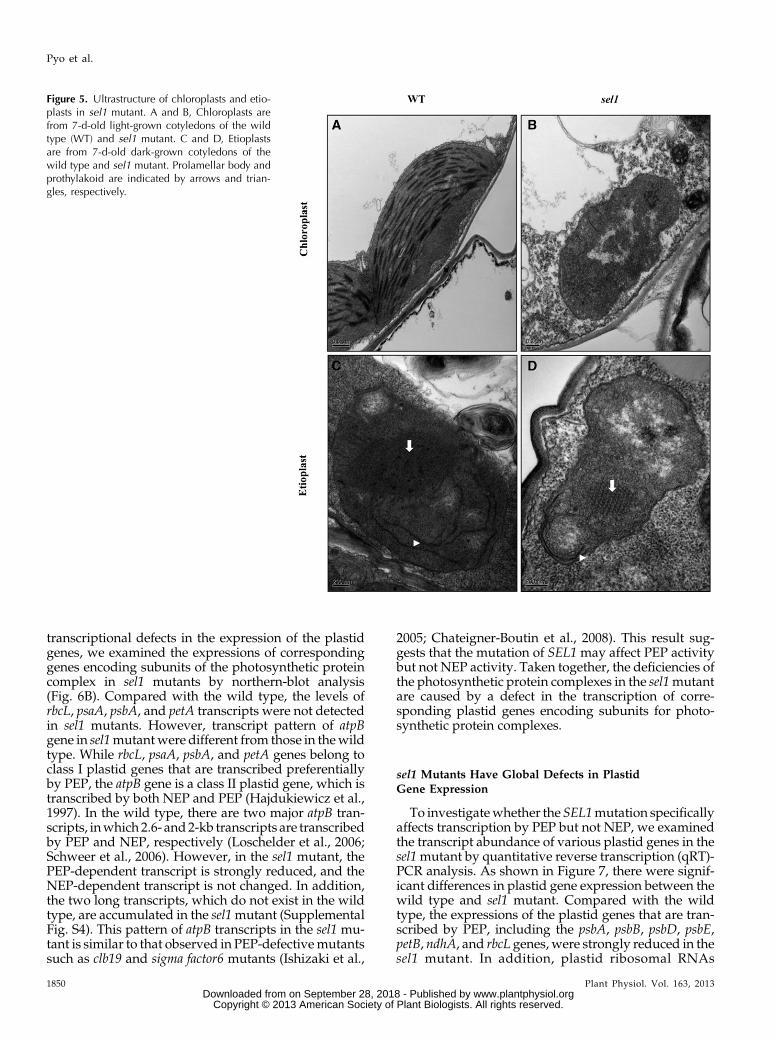
sel1mutant by transmission electronmicroscopy (TEM).Under light conditions, chloroplasts in the wild typewere lens shaped and contained well-organized thyla-koid membrane systems composed of stromal andgranal thylakoids (Fig. 5A). However, the plastids in thesel1 mutants were small and abnormally shaped, in-cluding round and amoeboid, and did not have thyla-koid membranes or any membrane structures butcontained a large number of small vesicles (Fig. 5B). Inaddition, densely stained globule aggregates were fre-quently observed in sel1 mutants.
Because SEL1 is expressed in the cotyledons of etio-lated seedlings, we also examined the etioplasts ultra-structure. Etioplasts are defined by the presence of theparacrystalline prolamellar body (PLB). Under darkconditions, etioplasts in the wild type showed the typ-ical structures, i.e. spherical or ellipsoidal shapeenclosing PLBs and prothylakoid membranes (Fig. 5C).However, plastids in sel1 mutant did not have well-developed PLBs or prothylakoid membranes (Fig. 5D).Some plastids in sel1 mutant showed a PLB-like butdisorganized structure. Taken together, these results
Figure 2. Sequence analysis of SEL1. A, Predicted motif structure of SEL1 protein. P or P-L-S block motifs are depicted as boxeswith letters as previously proposed (Lurin et al., 2004); P, L, and S designate the PPR, PPR-like S, and PPR-like L motif, re-spectively. B, Amino acid sequence alignment of SEL1. The amino acid sequence of SEL1 was compared with the homologousproteins in other species using ClustalW 2.0 (Larkin et al., 2007). The putative cleavage site of the transit peptide in SEL1 isshown by an arrow. Lines above the sequences show the predicted P- or PPR-like (S and L) motifs. Black boxes indicate aminoacid residues that are greater than 80% conserved, and gray boxes indicate amino acids that are greater than 60% conserved.The amino acid sequences are NP_193587.4 for Arabidopsis, XP_002284293.1 for grape, NP_001173562.1 for rice,ACG29369.1 for maize, and XP_001757146.1 for moss.
1848 Plant Physiol. Vol. 163, 2013
Pyo et al.
www.plantphysiol.orgon September 28, 2018 - Published by Downloaded from Copyright © 2013 American Society of Plant Biologists. All rights reserved.
indicate that the sel1 mutant is impaired in the devel-opment of chloroplast, as well as etioplasts, suggestingthat SEL1 functions in the early stage of chloroplastdevelopment.
sel1 Mutant Fails to Accumulate PhotosyntheticProtein Complexes
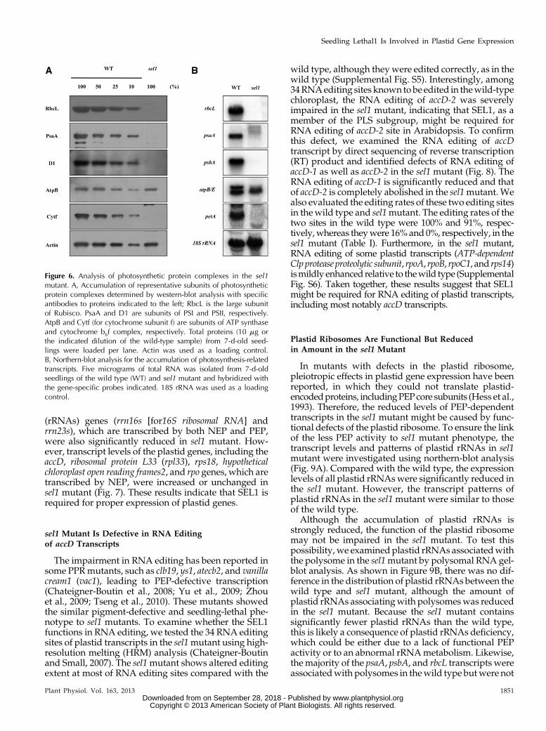
To compare the accumulation levels of chloroplastproteins of sel1 mutant with those of the wild type,protein amounts were evaluated comparably using
western-blot analysis. As shown in Figure 6A, the sel1mutant did not have detectable levels of subunits ofphotosynthetic protein complexes corresponding to theproducts of the ribulose-1,5-bisphosphate carboxylase/oxygenase large subunit (rbcL), PSI P700 chlorophyll a ap-oprotein A1 (psbA),PSII proteinD1 (psbA), and cytochromef (petA) genes. However, ATP synthase CF1 beta subunit(AtpB) was slightly reduced in the sel1 mutant whencompared with the wild type. The reduction of plastid-encoded proteins could be due to the impaired accu-mulation of plastid transcripts. To determine whetherreduced protein levels in sel1 mutants result from
Figure 4. Expression patterns of SEL1.GUS expression was analyzed in pSEL1::GUS transgenic plants. A, Three-day-oldetiolated seedling. B, Germinating seed.C, Three-day-old seedling. D, Five-day-oldseedling. E, Seven-day-old seedling. F, Rosetteleaf. G, Flower buds. H, Flower. I, Silique. [Seeonline article for color version of this figure.]
Figure 3. SEL1 is localized in the chloro-plasts. A, Subcellular localization of SEL1-GFP. Protoplasts were isolated from 35S::GFP and 35S::SEL1:GFP transgenic plants.The fluorescence of GFP and SEL1-GFPfusion protein in protoplasts was observedby confocal laser scanning microscope.Green fluorescence signals, chlorophyllred autofluorescence signals, merged im-ages, and bright-field images are shown.Bar = 5 mm. B, Western-blot analysis ofSEL1-FLAG fusion protein in chloroplastsubfractions. Chloroplasts from pSEL1::SEL1:FLAG transgenic plants were purifiedand separated into soluble and membranefractions by ultracentrifugation. The sameproportion of each fraction was separatedby SDS-PAGE and transferred to the nitro-cellulose membrane. The blot was probedsequentially with antibody to FLAG andspecific antibodies to RbcL and D1 as con-trols for the fractionation of soluble proteinsand membrane proteins, respectively. TC,Total chloroplast protein; S, soluble protein;M, membrane protein. [See online articlefor color version of this figure.]
Plant Physiol. Vol. 163, 2013 1849
Seedling Lethal1 Is Involved in Plastid Gene Expression
www.plantphysiol.orgon September 28, 2018 - Published by Downloaded from Copyright © 2013 American Society of Plant Biologists. All rights reserved.
transcriptional defects in the expression of the plastidgenes, we examined the expressions of correspondinggenes encoding subunits of the photosynthetic proteincomplex in sel1 mutants by northern-blot analysis(Fig. 6B). Compared with the wild type, the levels ofrbcL, psaA, psbA, and petA transcripts were not detectedin sel1 mutants. However, transcript pattern of atpBgene in sel1mutantwere different from those in thewildtype. While rbcL, psaA, psbA, and petA genes belong toclass I plastid genes that are transcribed preferentiallyby PEP, the atpB gene is a class II plastid gene, which istranscribed by both NEP and PEP (Hajdukiewicz et al.,1997). In the wild type, there are two major atpB tran-scripts, inwhich 2.6- and2-kb transcripts are transcribedby PEP and NEP, respectively (Loschelder et al., 2006;Schweer et al., 2006). However, in the sel1 mutant, thePEP-dependent transcript is strongly reduced, and theNEP-dependent transcript is not changed. In addition,the two long transcripts, which do not exist in the wildtype, are accumulated in the sel1mutant (SupplementalFig. S4). This pattern of atpB transcripts in the sel1 mu-tant is similar to that observed in PEP-defectivemutantssuch as clb19 and sigma factor6 mutants (Ishizaki et al.,
2005; Chateigner-Boutin et al., 2008). This result sug-gests that the mutation of SEL1 may affect PEP activitybut not NEP activity. Taken together, the deficiencies ofthe photosynthetic protein complexes in the sel1mutantare caused by a defect in the transcription of corre-sponding plastid genes encoding subunits for photo-synthetic protein complexes.
sel1 Mutants Have Global Defects in PlastidGene Expression
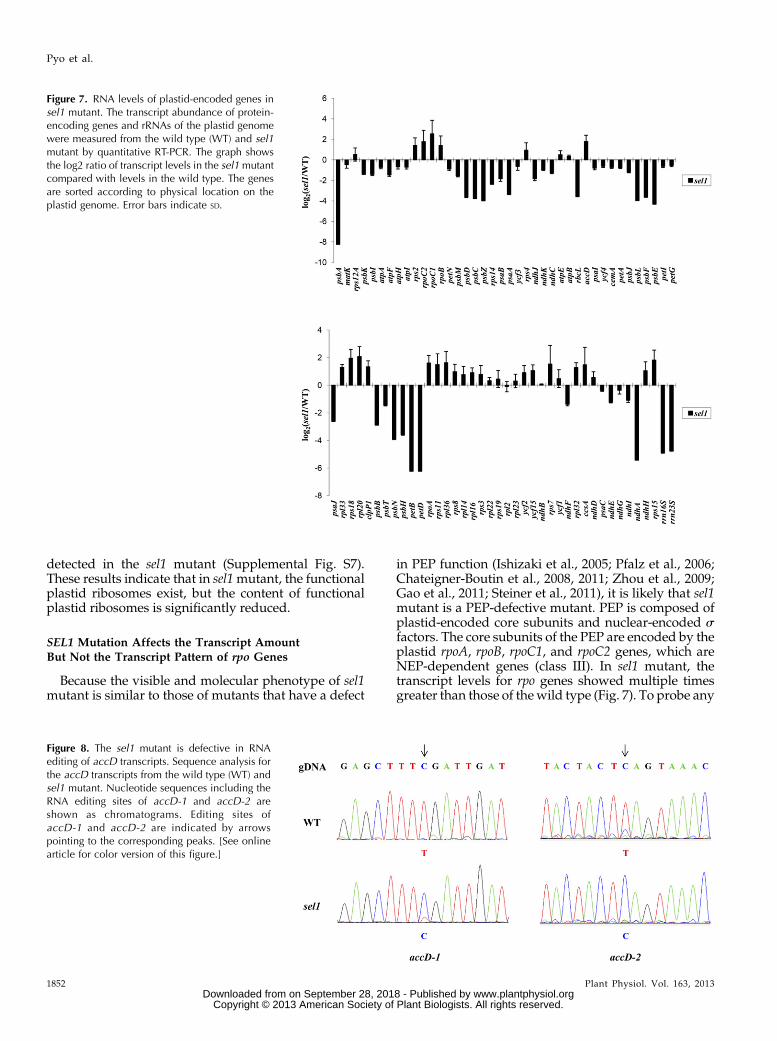
To investigatewhether the SEL1mutation specificallyaffects transcription by PEP but not NEP, we examinedthe transcript abundance of various plastid genes in thesel1mutant by quantitative reverse transcription (qRT)-PCR analysis. As shown in Figure 7, there were signif-icant differences in plastid gene expression between thewild type and sel1 mutant. Compared with the wildtype, the expressions of the plastid genes that are tran-scribed by PEP, including the psbA, psbB, psbD, psbE,petB, ndhA, and rbcL genes,were strongly reduced in thesel1 mutant. In addition, plastid ribosomal RNAs
Figure 5. Ultrastructure of chloroplasts and etio-plasts in sel1 mutant. A and B, Chloroplasts arefrom 7-d-old light-grown cotyledons of the wildtype (WT) and sel1 mutant. C and D, Etioplastsare from 7-d-old dark-grown cotyledons of thewild type and sel1 mutant. Prolamellar body andprothylakoid are indicated by arrows and trian-gles, respectively.
1850 Plant Physiol. Vol. 163, 2013
Pyo et al.
www.plantphysiol.orgon September 28, 2018 - Published by Downloaded from Copyright © 2013 American Society of Plant Biologists. All rights reserved.
(rRNAs) genes (rrn16s [for16S ribosomal RNA] andrrn23s), which are transcribed by both NEP and PEP,were also significantly reduced in sel1 mutant. How-ever, transcript levels of the plastid genes, including theaccD, ribosomal protein L33 (rpl33), rps18, hypotheticalchloroplast open reading frames2, and rpo genes, which aretranscribed by NEP, were increased or unchanged insel1 mutant (Fig. 7). These results indicate that SEL1 isrequired for proper expression of plastid genes.
sel1 Mutant Is Defective in RNA Editingof accD Transcripts
The impairment in RNA editing has been reported insome PPRmutants, such as clb19, ys1, atecb2, and vanillacream1 (vac1), leading to PEP-defective transcription(Chateigner-Boutin et al., 2008; Yu et al., 2009; Zhouet al., 2009; Tseng et al., 2010). These mutants showedthe similar pigment-defective and seedling-lethal phe-notype to sel1 mutants. To examine whether the SEL1functions in RNA editing, we tested the 34 RNA editingsites of plastid transcripts in the sel1mutant using high-resolution melting (HRM) analysis (Chateigner-Boutinand Small, 2007). The sel1mutant shows altered editingextent at most of RNA editing sites compared with the
wild type, although theywere edited correctly, as in thewild type (Supplemental Fig. S5). Interestingly, among34RNAediting sites known tobe edited in thewild-typechloroplast, the RNA editing of accD-2 was severelyimpaired in the sel1 mutant, indicating that SEL1, as amember of the PLS subgroup, might be required forRNA editing of accD-2 site in Arabidopsis. To confirmthis defect, we examined the RNA editing of accDtranscript by direct sequencing of reverse transcription(RT) product and identified defects of RNA editing ofaccD-1 as well as accD-2 in the sel1 mutant (Fig. 8). TheRNA editing of accD-1 is significantly reduced and thatof accD-2 is completely abolished in the sel1mutant. Wealso evaluated the editing rates of these two editing sitesin thewild type and sel1mutant. The editing rates of thetwo sites in the wild type were 100% and 91%, respec-tively,whereas theywere 16%and 0%, respectively, in thesel1 mutant (Table I). Furthermore, in the sel1 mutant,RNA editing of some plastid transcripts (ATP-dependentClp protease proteolytic subunit, rpoA, rpoB, rpoC1, and rps14)ismildly enhanced relative to thewild type (SupplementalFig. S6). Taken together, these results suggest that SEL1might be required for RNA editing of plastid transcripts,including most notably accD transcripts.
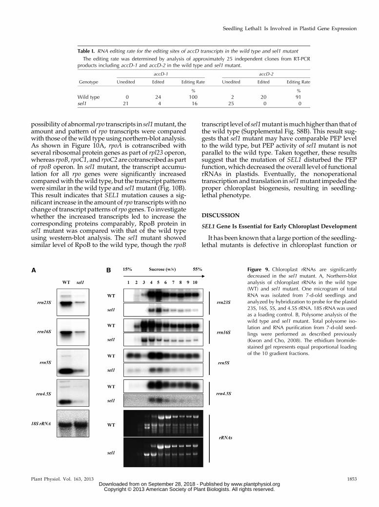
Plastid Ribosomes Are Functional But Reducedin Amount in the sel1 Mutant
In mutants with defects in the plastid ribosome,pleiotropic effects in plastid gene expression have beenreported, in which they could not translate plastid-encodedproteins, includingPEPcore subunits (Hess et al.,1993). Therefore, the reduced levels of PEP-dependenttranscripts in the sel1 mutant might be caused by func-tional defects of the plastid ribosome. To ensure the linkof the less PEP activity to sel1 mutant phenotype, thetranscript levels and patterns of plastid rRNAs in sel1mutant were investigated using northern-blot analysis(Fig. 9A). Compared with the wild type, the expressionlevels of all plastid rRNAswere significantly reduced inthe sel1 mutant. However, the transcript patterns ofplastid rRNAs in the sel1 mutant were similar to thoseof the wild type.
Although the accumulation of plastid rRNAs isstrongly reduced, the function of the plastid ribosomemay not be impaired in the sel1 mutant. To test thispossibility,we examined plastid rRNAs associatedwiththe polysome in the sel1mutant by polysomal RNA gel-blot analysis. As shown in Figure 9B, there was no dif-ference in the distribution of plastid rRNAs between thewild type and sel1 mutant, although the amount ofplastid rRNAs associatingwith polysomeswas reducedin the sel1 mutant. Because the sel1 mutant containssignificantly fewer plastid rRNAs than the wild type,this is likely a consequence of plastid rRNAs deficiency,which could be either due to a lack of functional PEPactivity or to an abnormal rRNAmetabolism. Likewise,the majority of the psaA, psbA, and rbcL transcripts wereassociatedwith polysomes in thewild type butwere not
Figure 6. Analysis of photosynthetic protein complexes in the sel1mutant. A, Accumulation of representative subunits of photosyntheticprotein complexes determined by western-blot analysis with specificantibodies to proteins indicated to the left; RbcL is the large subunitof Rubisco. PsaA and D1 are subunits of PSI and PSII, respectively.AtpB and Cytf (for cytochrome subunit f) are subunits of ATP synthaseand cytochrome b6f complex, respectively. Total proteins (10 mg orthe indicated dilution of the wild-type sample) from 7-d-old seed-lings were loaded per lane. Actin was used as a loading control.B, Northern-blot analysis for the accumulation of photosynthesis-relatedtranscripts. Five micrograms of total RNA was isolated from 7-d-oldseedlings of the wild type (WT) and sel1 mutant and hybridized withthe gene-specific probes indicated. 18S rRNA was used as a loadingcontrol.
Plant Physiol. Vol. 163, 2013 1851
Seedling Lethal1 Is Involved in Plastid Gene Expression
www.plantphysiol.orgon September 28, 2018 - Published by Downloaded from Copyright © 2013 American Society of Plant Biologists. All rights reserved.
detected in the sel1 mutant (Supplemental Fig. S7).These results indicate that in sel1mutant, the functionalplastid ribosomes exist, but the content of functionalplastid ribosomes is significantly reduced.
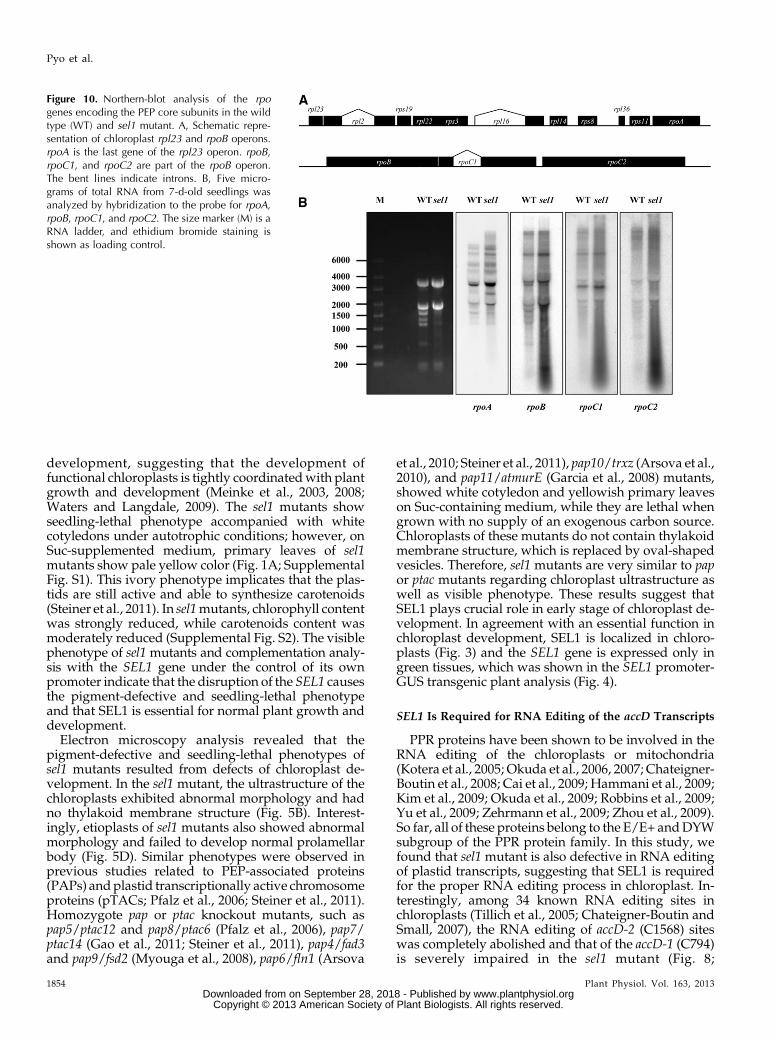
SEL1 Mutation Affects the Transcript AmountBut Not the Transcript Pattern of rpo Genes
Because the visible and molecular phenotype of sel1mutant is similar to those of mutants that have a defect
in PEP function (Ishizaki et al., 2005; Pfalz et al., 2006;Chateigner-Boutin et al., 2008, 2011; Zhou et al., 2009;Gao et al., 2011; Steiner et al., 2011), it is likely that sel1mutant is a PEP-defective mutant. PEP is composed ofplastid-encoded core subunits and nuclear-encoded sfactors. The core subunits of the PEP are encoded by theplastid rpoA, rpoB, rpoC1, and rpoC2 genes, which areNEP-dependent genes (class III). In sel1 mutant, thetranscript levels for rpo genes showed multiple timesgreater than those of thewild type (Fig. 7). To probe any
Figure 8. The sel1 mutant is defective in RNAediting of accD transcripts. Sequence analysis forthe accD transcripts from the wild type (WT) andsel1 mutant. Nucleotide sequences including theRNA editing sites of accD-1 and accD-2 areshown as chromatograms. Editing sites ofaccD-1 and accD-2 are indicated by arrowspointing to the corresponding peaks. [See onlinearticle for color version of this figure.]
Figure 7. RNA levels of plastid-encoded genes insel1 mutant. The transcript abundance of protein-encoding genes and rRNAs of the plastid genomewere measured from the wild type (WT) and sel1mutant by quantitative RT-PCR. The graph showsthe log2 ratio of transcript levels in the sel1mutantcompared with levels in the wild type. The genesare sorted according to physical location on theplastid genome. Error bars indicate SD.
1852 Plant Physiol. Vol. 163, 2013
Pyo et al.
www.plantphysiol.orgon September 28, 2018 - Published by Downloaded from Copyright © 2013 American Society of Plant Biologists. All rights reserved.
possibility of abnormal rpo transcripts in sel1mutant, theamount and pattern of rpo transcripts were comparedwith those of thewild type using northern-blot analysis.As shown in Figure 10A, rpoA is cotranscribed withseveral ribosomal protein genes as part of rpl23 operon,whereas rpoB, rpoC1, and rpoC2 are cotranscribed as partof rpoB operon. In sel1 mutant, the transcript accumu-lation for all rpo genes were significantly increasedcomparedwith thewild type, but the transcript patternswere similar in the wild type and sel1mutant (Fig. 10B).This result indicates that SEL1 mutation causes a sig-nificant increase in the amount of rpo transcriptswith nochange of transcript patterns of rpo genes. To investigatewhether the increased transcripts led to increase thecorresponding proteins comparably, RpoB protein insel1 mutant was compared with that of the wild typeusing western-blot analysis. The sel1 mutant showedsimilar level of RpoB to the wild type, though the rpoB
transcript level of sel1mutant ismuchhigher than that ofthe wild type (Supplemental Fig. S8B). This result sug-gests that sel1 mutant may have comparable PEP levelto the wild type, but PEP activity of sel1 mutant is notparallel to the wild type. Taken together, these resultssuggest that the mutation of SEL1 disturbed the PEPfunction, which decreased the overall level of functionalrRNAs in plastids. Eventually, the nonoperationaltranscription and translation in sel1mutant impeded theproper chloroplast biogenesis, resulting in seedling-lethal phenotype.
DISCUSSION
SEL1 Gene Is Essential for Early Chloroplast Development
It has been known that a large portion of the seedling-lethal mutants is defective in chloroplast function or
Figure 9. Chloroplast rRNAs are significantlydecreased in the sel1 mutant. A, Northern-blotanalysis of chloroplast rRNAs in the wild type(WT) and sel1 mutant. One microgram of totalRNA was isolated from 7-d-old seedlings andanalyzed by hybridization to probe for the plastid23S, 16S, 5S, and 4.5S rRNA. 18S rRNAwas usedas a loading control. B, Polysome analysis of thewild type and sel1 mutant. Total polysome iso-lation and RNA purification from 7-d-old seed-lings were performed as described previously(Kwon and Cho, 2008). The ethidium bromide-stained gel represents equal proportional loadingof the 10 gradient fractions.
Table I. RNA editing rate for the editing sites of accD transcripts in the wild type and sel1 mutant
The editing rate was determined by analysis of approximately 25 independent clones from RT-PCRproducts including accD-1 and accD-2 in the wild type and sel1 mutant.
accD-1 accD-2
Genotype Unedited Edited Editing Rate Unedited Edited Editing Rate
% %
Wild type 0 24 100 2 20 91sel1 21 4 16 25 0 0
Plant Physiol. Vol. 163, 2013 1853
Seedling Lethal1 Is Involved in Plastid Gene Expression
www.plantphysiol.orgon September 28, 2018 - Published by Downloaded from Copyright © 2013 American Society of Plant Biologists. All rights reserved.
development, suggesting that the development offunctional chloroplasts is tightly coordinatedwith plantgrowth and development (Meinke et al., 2003, 2008;Waters and Langdale, 2009). The sel1 mutants showseedling-lethal phenotype accompanied with whitecotyledons under autotrophic conditions; however, onSuc-supplemented medium, primary leaves of sel1mutants show pale yellow color (Fig. 1A; SupplementalFig. S1). This ivory phenotype implicates that the plas-tids are still active and able to synthesize carotenoids(Steiner et al., 2011). In sel1mutants, chlorophyll contentwas strongly reduced, while carotenoids content wasmoderately reduced (Supplemental Fig. S2). The visiblephenotype of sel1 mutants and complementation analy-sis with the SEL1 gene under the control of its ownpromoter indicate that the disruption of the SEL1 causesthe pigment-defective and seedling-lethal phenotypeand that SEL1 is essential for normal plant growth anddevelopment.
Electron microscopy analysis revealed that thepigment-defective and seedling-lethal phenotypes ofsel1 mutants resulted from defects of chloroplast de-velopment. In the sel1 mutant, the ultrastructure of thechloroplasts exhibited abnormal morphology and hadno thylakoid membrane structure (Fig. 5B). Interest-ingly, etioplasts of sel1 mutants also showed abnormalmorphology and failed to develop normal prolamellarbody (Fig. 5D). Similar phenotypes were observed inprevious studies related to PEP-associated proteins(PAPs) andplastid transcriptionally active chromosomeproteins (pTACs; Pfalz et al., 2006; Steiner et al., 2011).Homozygote pap or ptac knockout mutants, such aspap5/ptac12 and pap8/ptac6 (Pfalz et al., 2006), pap7/ptac14 (Gao et al., 2011; Steiner et al., 2011), pap4/fad3and pap9/fsd2 (Myouga et al., 2008), pap6/fln1 (Arsova
et al., 2010; Steiner et al., 2011), pap10/trxz (Arsova et al.,2010), and pap11/atmurE (Garcia et al., 2008) mutants,showed white cotyledon and yellowish primary leaveson Suc-containing medium, while they are lethal whengrown with no supply of an exogenous carbon source.Chloroplasts of these mutants do not contain thylakoidmembrane structure, which is replaced by oval-shapedvesicles. Therefore, sel1 mutants are very similar to papor ptac mutants regarding chloroplast ultrastructure aswell as visible phenotype. These results suggest thatSEL1 plays crucial role in early stage of chloroplast de-velopment. In agreement with an essential function inchloroplast development, SEL1 is localized in chloro-plasts (Fig. 3) and the SEL1 gene is expressed only ingreen tissues, which was shown in the SEL1 promoter-GUS transgenic plant analysis (Fig. 4).
SEL1 Is Required for RNA Editing of the accD Transcripts
PPR proteins have been shown to be involved in theRNA editing of the chloroplasts or mitochondria(Kotera et al., 2005; Okuda et al., 2006, 2007; Chateigner-Boutin et al., 2008; Cai et al., 2009; Hammani et al., 2009;Kim et al., 2009; Okuda et al., 2009; Robbins et al., 2009;Yu et al., 2009; Zehrmann et al., 2009; Zhou et al., 2009).So far, all of these proteins belong to the E/E+ andDYWsubgroup of the PPR protein family. In this study, wefound that sel1 mutant is also defective in RNA editingof plastid transcripts, suggesting that SEL1 is requiredfor the proper RNA editing process in chloroplast. In-terestingly, among 34 known RNA editing sites inchloroplasts (Tillich et al., 2005; Chateigner-Boutin andSmall, 2007), the RNA editing of accD-2 (C1568) siteswas completely abolished and that of the accD-1 (C794)is severely impaired in the sel1 mutant (Fig. 8;
Figure 10. Northern-blot analysis of the rpogenes encoding the PEP core subunits in the wildtype (WT) and sel1 mutant. A, Schematic repre-sentation of chloroplast rpl23 and rpoB operons.rpoA is the last gene of the rpl23 operon. rpoB,rpoC1, and rpoC2 are part of the rpoB operon.The bent lines indicate introns. B, Five micro-grams of total RNA from 7-d-old seedlings wasanalyzed by hybridization to the probe for rpoA,rpoB, rpoC1, and rpoC2. The size marker (M) is aRNA ladder, and ethidium bromide staining isshown as loading control.
1854 Plant Physiol. Vol. 163, 2013
Pyo et al.
www.plantphysiol.orgon September 28, 2018 - Published by Downloaded from Copyright © 2013 American Society of Plant Biologists. All rights reserved.
Supplemental Fig. S5). The RNA editing defects in sel1mutant are similar to those in atecb2 and vac1 mutants,which are independent mutant alleles of AtECB2/VAC1 encoding a DYW subgroup PPR protein that isrequired for the editing of accD and ndhF transcripts (Yuet al., 2009; Tseng et al., 2010). However, it has beenreported that the loss of RNA editing at the accD-1(C794) site was not associated with a pigment-defectiveand seedling-lethal phenotype; because Protein Requiredfor accD RNA Editing1 mutant showed wild type-likephenotype, even the RNA editing of the accD-1 (C794)site is completely abolished (Robbins et al., 2009).Therefore, the editing defect in the sel1 mutant is un-likely to be a primary effect by the loss of SEL1 protein.The decreased editing extent is probably caused by thegreatly increased level of accD transcripts in sel1mutantas shown in Figure 7 and Supplemental Figure S8A. Toomany transcripts may saturate the editing activity andconsequently leads to lower editing extent as shown inprevious studies on the relation between the transcriptabundance and the level of editing extent (Lu andHanson, 1992). However, we cannot exclude the possi-bility that SEL1 is involved in RNA editing process ofaccD transcript, because the editing events on the ATP-dependent Clp protease proteolytic subunit, rpoA, rpoB,rpoC1, and rps14 transcripts are not significantly af-fected (Supplemental Fig. S5), although they are highlyexpressed in the sel1 mutant compared with the wildtype (Figs. 7 and 10).
SEL1 Plays an Important Role in the Regulationof Plastid Gene Expression
Analysis of plastid gene expression revealed that, insel1 mutants, the transcript levels of PEP-dependentgenes were dramatically reduced, whereas those ofNEP-dependent genes were increased or unchanged(Fig. 7), suggesting that sel1mutant is severely impairedin PEP activity and SEL1 plays an important role in theregulation of plastid gene expression. Thus, it seemslikely that the early arrest of chloroplast development insel1 mutants is due to the reduced PEP activity. Al-though the sel1 mutant shows many similarities in mo-lecular phenotypes, including transcript pattern andaccumulation, and expression profiles of plastid genesto other PEP-related mutants (Pfalz et al., 2006;Chateigner-Boutin et al., 2008, 2011; Zhou et al., 2009;Steiner et al., 2011), the molecular function of SEL1currently remains elusive. However, it is obvious thatSEL1 is required for the proper expression of plastidgenes. From the recent study, maize ortholog of SEL1has been identified in nucleoid-enriched protein frac-tions (Majeran et al., 2012). Thus, it is possible that SEL1might be involved in the control of PEP activity as acomponent of nucleoid proteins like pTACs or PAPs,and required for the proper plastid gene expression andnormal chloroplast development. In addition to theglobal reduction of PEP-dependent transcripts, severallines of evidence suggest that sel1mutant is defective in
PEP activity.Whenwe investigated the accumulation ofplastid-encoded proteins in sel1 mutants, the accumu-lation of Rubisco and thylakoid membrane proteincomplexes, except for ATP synthase, was severely de-creased or not detected in the sel1mutant due to the lackof corresponding transcripts (Fig. 6A). The amount ofPEP-dependent transcript of atpB decreased (upperband, approximately 2.6 kb) in theRNAgel blot (Fig. 6B;Supplemental Fig. S4), even though the level of AtpBprotein did not change significantly. As seen in Figure5B, the existence of mitochondria was observed in sel1mutant. Thereby, the amount of AtpB protein could beoverestimated due to the cross reactivity of antibodyusedagainst themitochondrialATPase (KwonandCho,2008). The amount of NEP-dependent transcript of atpBis not significantly altered and novel 4.8-kb transcript,which is not detected in the wild type, accumulated insel1 mutant (Fig. 6B; Supplemental Fig. S4). A similartranscription pattern was also observed in the sigmafactor6 (Loschelder et al., 2006; Schweer et al., 2006) andother PEP-defective mutants, such as ptac2/ptac6/ptac12/ptac14 (Pfalz et al., 2006; Gao et al., 2011), clb19(Chateigner-Boutin et al., 2008), delayed green1 (Chi et al.,2008), and ys1 (Zhou et al., 2009).
In terms of the molecular phenotypes, sel1 mutantsshow the similarity to those of mutants with defects inplastid ribosome aswell (Bang et al., 2012). In ribosome-deficient mutants, the expression of PEP-dependentgenes is decreased, whereas that of NEP-dependentgenes is increased. In sel1mutant, steady-state levels ofall plastid rRNAs were significantly decreased (Fig. 9).This may lead to a global defect in the expression ofplastid genes due to the lack of functional plastid ribo-somes, which causes the level of PEP to be much lessthan required to synthesize plastid proteins controlledby PEP. In sel1mutant, protein levels of AccD and RpoBare not significantly changed, even though their tran-scripts are highly up-regulated (Figs. 7 and 10B;Supplemental Fig. S8). Thus, reduced translation of accDand rpoB could be a consequence of the decreased levelof plastid rRNAs. Global defects in plastid gene ex-pression in sel1 mutants are similar to those of plastidribosome-deficient mutants, suggesting that the SEL1may be involved in the regulation of plastid ribosomeassembly or function. So, the loss of SEL1 leads tooverall declines of all PEP-dependent transcriptionscaused by the reduced translation of PEP. However, wecould not rule out the possibility that the reduction inthe plastid ribosome is also caused by defects of PEPactivity.
In sel1mutant, the expression of PEP-dependent genesis decreased, whereas that of NEP-dependent genes isincreased. This increase in NEP-dependent transcripts insel1 can be indirect and possibly explained by a recentstudy on the division of labor between PEP and NEPduring plastid development and in mature chloroplastsusing the primary transcriptome analysis in barley(Hordeum vulgare; Zhelyazkova et al., 2012). In this study,authors found that the extremely high number of pro-moters, at least 222 NEP promoters, can be used inwhite
Plant Physiol. Vol. 163, 2013 1855
Seedling Lethal1 Is Involved in Plastid Gene Expression
www.plantphysiol.orgon September 28, 2018 - Published by Downloaded from Copyright © 2013 American Society of Plant Biologists. All rights reserved.
leaves in the absence of PEP and that NEP is able togenerate virtually the same mRNAs as PEP in severalgenes, suggesting that activation of transcription byNEPcanbe a compensatorymechanism for the absence ofPEPin higher plants.
In this work, we report a novel pigment-defectiveand seedling-lethal mutant, sel1mutant, that disruptedgenes encoding a member of the PLS subgroup of thePPR protein family. Our results demonstrate that SEL1is required for the proper plastid gene expression andthus essential for normal chloroplast development. Todiscover the more detailed molecular function of SEL1,the identification of target RNA and interacting pro-teins of SEL1 in the future should be helpful to providethe insight into its function in plastid gene expressionand chloroplast development.
MATERIALS AND METHODS
Plant Materials and Growth Conditions
Seeds were surface sterilized and incubated at 4°C for 3 d to synchronizegermination and plated on MS medium (Duchefa Biochemie) containing 1%(w/v) Suc and 0.7% (w/v) agar in a growth chamber at 22°C under 16-h-light/8-h-dark condition. Plants were grown in soil (Sunshine Mix #1; SunGro)in a growth room at 22°C under continuous light. The sel1-1 mutant wasisolated from T-DNA insertion mutant lines, which are generated in Ws-2background with simple T-DNA vector pDAP101 (Sessions et al., 2002),whereas seeds of sel1-2 (SAIL_793_F11) and sel1-3 (SALK_054374) wereobtained from the Arabidopsis Biological Resource Center.
Complementation Analysis
For the complementation test of the sel1 mutant, the 2,629-bp wild-typegenomic DNA fragment of the At4g18520 gene, containing 645 bp of a pu-tative promoter sequence and 130 bp of 39 untranslated region, was amplifiedusing Pfu-X polymerase (SolGent) and gene-specific primers. After verificationby sequencing, the amplified fragment was cloned into a pBIN19 binary vectorand subsequently introduced into heterozygous sel1-3 mutant plants using theAgrobacterium tumefaciens-mediated floral dip method (Clough and Bent,1998). For the generation of FLAG-tagged SEL1 complementation line, DNAencoding three copies of FLAG was amplified and cloned into pCB308 binaryvector (Xiang et al., 1999), producing pCB308-FLAG vector. The SEL1 genomicDNA without stop codon, including 645-bp upstream region from ATG, wasamplified and cloned into pCB308-FLAG vector and subsequently introducedinto heterozygous sel1-2 mutant plants.
Northern-Blot Analysis
Total RNA was extracted from 7-d-old seedlings using the easy-BLUE TotalRNA Extraction Kit (iNtRON Biotechnology), separated onMOPS-formaldehydeagarose gel, and transferred to a nitrocellulose membrane (Whatman). The32P-labeled probes were prepared using a DecaLabel DNA Labeling Kit (Fer-mentas). Prehybridization, hybridization, washes, and signal detection wereperformed as described previously (Kwon and Cho, 2008).
Subcellular Localization of GFP Fusion Proteinsin Protoplasts
Analysis of the subcellular localization of GFP fusion proteins in protoplastswas performed as described previously (Kwon and Cho, 2008).
Chloroplast Isolation and Fractionation
Chloroplasts were isolated and separated into soluble and membranefractions as described previously (Kwon and Cho, 2008).
Histochemical GUS Staining
Tissues were incubated in cold 90% (v/v) acetone and placed in a stainingbuffer (50 mM sodium phosphate buffer, pH 7.2, 0.2% Triton-X, 2 mM potassiumferrocyanide, 2 mM potassium ferricyanide, and 2 mM 5-bromo-4-chloro-3-indolyl-b-glucuronic acid). After a vacuum infiltration, samples were incubatedovernight at 37°C. GUS-stained tissues were cleared with an ethanol series andfixed in formaldehyde-acetic acid solution (50% [v/v] ethanol, 10% [v/v] aceticacid, and 5% [v/v] formaldehyde). Following fixation, samples were stored in70% (v/v) ethanol and examined under the dissecting microscope.
TEM Analysis
TEManalysiswas performed as describedpreviously (Kwon andCho, 2008),with minor modification. For analysis of the etioplast ultrastructure, wild-typeand sel1 mutant seedlings were grown on MS agar plates in the dark for 7 d.Under dim green light, cotyledons of the wild type were directly detached fromseedlings and then immediately soaked in fixation solution. For the sel1mutant,cotyledons were detached and soaked in numbered 1.5-mL tubes containingfixation solution. The genotype of the sample was determined by PCR using theremaining part of the seedling. The detached cotyledons in the correspondingtubes confirmed that homozygous sel1 mutant was used for TEM analysis.
Western-Blot Analysis
Total proteins were isolated from 7-d-old seedlings as described previously(Barkan, 1998), separated by SDS-PAGE, transferred to polyvinylidenedifluoride membrane, immunoblotted with various antibodies, and detectedusing the enhanced chemiluminescence kit (Santa Cruz Biotechnology). An-tibodies were obtained from Agrisera (RbcL, PsaA, D1 protein of PSII, AtpB,and cytochrome subunit f) and from Uniplastomic (AccD and RpoB).
Analysis of Plastid Transcript Abundance
Total RNA was extracted from 7-d-old wild-type and sel1 mutant seedlingsby using the NucleoSpin RNA Plant Kit (Macherey-Nagel). DNA-free RNA(2 mg) was then reverse transcribed with random hexamers by using aRevertAid First-Strand cDNA Synthesis Kit (Fermentas). Quantitative real-time PCR was carried out in an IQ5 Light Cycler (Bio-Rad) using SYBR Pre-mix Ex Taq II (Takara). The relative quantification of gene expression datawere analyzed using the IQ5 Optical System Software (Bio-Rad). The data setwas normalized using cytosolic 18S rRNA as a reference and setting the me-dian value to 0. The primer sequences used in quantitative RT-PCR analysisare described in Okuda et al. (2009).
Analysis of RNA Editing
HRM analysis was performed as previously described (Chateigner-Boutinand Small, 2007; Okuda et al., 2009). Genomic DNA for HRM analysis wasisolated with plant I genomic DNA (iNtRON Biotechnology). RNA isolationand RT were performed as above. The sequence including the accD RNAediting sites (accD-1 and accD-2) from the wild type and sel1 mutant wasamplified by PCR with gene-specific primer pairs, and then the RT-PCRproducts were sequenced directly or cloned into a T-blunt vector using aT-blunt PCR Cloning Kit (SolGent). Plasmids prepared from 25 independentcolonies of each sample were sequenced to determine the RNA editing effi-ciency of accD-1 and accD-2.
Sequence data from this article can be found in the GenBank/EMBL datalibraries under accession number At4g18520.
Supplemental Data
The following materials are available in the online version of this article.
Supplemental Figure S1.
Supplemental Figure S2. The content of chlorophyll and carotenoids insel1 mutant.
Supplemental Figure S3. Sucrose gradient sedimentation analysis of SEL1-containing particles in chloroplast.
1856 Plant Physiol. Vol. 163, 2013
Pyo et al.
www.plantphysiol.orgon September 28, 2018 - Published by Downloaded from Copyright © 2013 American Society of Plant Biologists. All rights reserved.
Supplemental Figure S4. The transcript accumulation and pattern of atpBgene in sel1 mutant.
Supplemental Figure S5. High-resolution melting analysis of plastid RNAediting in sel1 mutant.
Supplemental Figure S6. RNA editing of plastid transcripts in sel1 mutant.
Supplemental Figure S7. Polysome association of photosynthesis-relatedmRNAs in sel1 mutant.
Supplemental Figure S8. Analysis of the plastid-encoded genes in sel1mutant.
ACKNOWLEDGMENTS
We thank Allen Sessions (Syngenta Biotechnology, Torrey Mesa ResearchInstitute) and Syngenta Biotechnology for providing the pDAP101 vector.
Received September 2, 2013; accepted October 16, 2013; published October 21,2013.
LITERATURE CITED
Allison LA, Simon LD, Maliga P (1996) Deletion of rpoB reveals a seconddistinct transcription system in plastids of higher plants. EMBO J 15:2802–2809
Arsova B, Hoja U, Wimmelbacher M, Greiner E, Ustün S, Melzer M,Petersen K, Lein W, Börnke F (2010) Plastidial thioredoxin z interactswith two fructokinase-like proteins in a thiol-dependent manner: evi-dence for an essential role in chloroplast development in Arabidopsis andNicotiana benthamiana. Plant Cell 22: 1498–1515
Aubourg S, Boudet N, Kreis M, Lecharny A (2000) In Arabidopsis thaliana,1% of the genome codes for a novel protein family unique to plants. PlantMol Biol 42: 603–613
Bang WY, Chen J, Jeong IS, Kim SW, Kim CW, Jung HS, Lee KH,Kweon HS, Yoko I, Shiina T, et al (2012) Functional characterizationof ObgC in ribosome biogenesis during chloroplast development.Plant J 71:122–134
Barkan A (1998) Approaches to investigating nuclear genes that function inchloroplast biogenesis in land plants. Photosynthesis. Molecular Biologyof Energy Capture 297: 38–57
Barkan A, Goldschmidt-Clermont M (2000) Participation of nuclear genesin chloroplast gene expression. Biochimie 82: 559–572
Beick S, Schmitz-Linneweber C, Williams-Carrier R, Jensen B, Barkan A(2008) The pentatricopeptide repeat protein PPR5 stabilizes a specifictRNA precursor in maize chloroplasts. Mol Cell Biol 28: 5337–5347
Bentolila S, Elliott LE, Hanson MR (2008) Genetic architecture of mito-chondrial editing in Arabidopsis thaliana. Genetics 178: 1693–1708
Cai W, Ji D, Peng L, Guo J, Ma J, Zou M, Lu C, Zhang L (2009) LPA66 isrequired for editing psbF chloroplast transcripts in Arabidopsis. PlantPhysiol 150: 1260–1271
Cai W, Okuda K, Peng L, Shikanai T (2011) PROTON GRADIENT REG-ULATION 3 recognizes multiple targets with limited similarity and mediatestranslation and RNA stabilization in plastids. Plant J 67: 318–327
Chateigner-Boutin AL, des Francs-Small CC, Delannoy E, Kahlau S, TanzSK, de Longevialle AF, Fujii S, Small I (2011) OTP70 is a penta-tricopeptide repeat protein of the E subgroup involved in splicing of theplastid transcript rpoC1. Plant J 65: 532–542
Chateigner-Boutin AL, Ramos-Vega M, Guevara-García A, Andrés C, dela Luz Gutiérrez-Nava M, Cantero A, Delannoy E, Jiménez LF, LurinC, Small I, et al (2008) CLB19, a pentatricopeptide repeat protein re-quired for editing of rpoA and clpP chloroplast transcripts. Plant J 56:590–602
Chateigner-Boutin AL, Small I (2007) A rapid high-throughput method forthe detection and quantification of RNA editing based on high-resolutionmelting of amplicons. Nucleic Acids Res 35: e114
Chi W, Ma J, Zhang D, Guo J, Chen F, Lu C, Zhang L (2008) The pen-tratricopeptide repeat protein DELAYED GREENING1 is involved inthe regulation of early chloroplast development and chloroplast geneexpression in Arabidopsis. Plant Physiol 147: 573–584
Clough SJ, Bent AF (1998) Floral dip: a simplified method for Agrobacterium-mediated transformation of Arabidopsis thaliana. Plant J 16: 735–743
de Longevialle AF, Hendrickson L, Taylor NL, Delannoy E, Lurin C,Badger M, Millar AH, Small I (2008) The pentatricopeptide repeat geneOTP51 with two LAGLIDADG motifs is required for the cis-splicing ofplastid ycf3 intron 2 in Arabidopsis thaliana. Plant J 56: 157–168
Gao ZP, Yu QB, Zhao TT, Ma Q, Chen GX, Yang ZN (2011) A functionalcomponent of the transcriptionally active chromosome complex, ArabidopsispTAC14, interacts with pTAC12/HEMERA and regulates plastid gene ex-pression. Plant Physiol 157: 1733–1745
Garcia M, Myouga F, Takechi K, Sato H, Nabeshima K, Nagata N, TakioS, Shinozaki K, Takano H (2008) An Arabidopsis homolog of the bac-terial peptidoglycan synthesis enzyme MurE has an essential role in chlo-roplast development. Plant J 53: 924–934
Hajdukiewicz PT, Allison LA, Maliga P (1997) The two RNA polymerasesencoded by the nuclear and the plastid compartments transcribe distinctgroups of genes in tobacco plastids. EMBO J 16: 4041–4048
Hammani K, Okuda K, Tanz SK, Chateigner-Boutin AL, Shikanai T, Small I(2009) A study of new Arabidopsis chloroplast RNA editing mutants revealsgeneral features of editing factors and their target sites. Plant Cell 21: 3686–3699
Hedtke B, Börner T, Weihe A (1997) Mitochondrial and chloroplast phage-type RNA polymerases in Arabidopsis. Science 277: 809–811
Hess WR, Börner T (1999) Organellar RNA polymerases of higher plants.Int Rev Cytol 190: 1–59
Hess WR, Prombona A, Fieder B, Subramanian AR, Börner T (1993)Chloroplast rps15 and the rpoB/C1/C2 gene cluster are strongly tran-scribed in ribosome-deficient plastids: evidence for a functioning non-chloroplast-encoded RNA polymerase. EMBO J 12: 563–571
Ishizaki Y, Tsunoyama Y, Hatano K, Ando K, Kato K, Shinmyo A, KoboriM, Takeba G, Nakahira Y, Shiina T (2005) A nuclear-encoded s factor,Arabidopsis SIG6, recognizes s-70 type chloroplast promoters and regulatesearly chloroplast development in cotyledons. Plant J 42: 133–144
Kim SR, Yang JI, Moon S, Ryu CH, An K, Kim KM, Yim J, An G (2009)Rice OGR1 encodes a pentatricopeptide repeat-DYW protein and is es-sential for RNA editing in mitochondria. Plant J 59: 738–749
Kotera E, Tasaka M, Shikanai T (2005) A pentatricopeptide repeat proteinis essential for RNA editing in chloroplasts. Nature 433: 326–330
Kwon KC, Cho MH (2008) Deletion of the chloroplast-localized AtTerCgene product in Arabidopsis thaliana leads to loss of the thylakoid membraneand to seedling lethality. Plant J 55: 428–442
Larkin MA, Blackshields G, Brown NP, Chenna R, McGettigan PA,McWilliam H, Valentin F, Wallace IM, Wilm A, Lopez R, et al (2007)Clustal W and Clustal X version 2.0. Bioinformatics 23: 2947–2948
Liere K, Weihe A, Börner T (2011) The transcription machineries of plantmitochondria and chloroplasts: composition, function, and regulation.J Plant Physiol 168: 1345–1360
Loschelder H, Schweer J, Link B, Link G (2006) Dual temporal role ofplastid sigma factor 6 in Arabidopsis development. Plant Physiol 142:642–650
Lu B, Hanson MR (1992) A single nuclear gene specifies the abundance andextent of RNA editing of a plant mitochondrial transcript. Nucleic AcidsRes 20: 5699–5703
Lurin C, Andrés C, Aubourg S, Bellaoui M, Bitton F, Bruyère C, CabocheM, Debast C, Gualberto J, Hoffmann B, et al (2004) Genome-wideanalysis of Arabidopsis pentatricopeptide repeat proteins reveals theiressential role in organelle biogenesis. Plant Cell 16: 2089–2103
Majeran W, Friso G, Asakura Y, Qu X, Huang M, Ponnala L, Watkins KP,Barkan A, van Wijk KJ (2012) Nucleoid-enriched proteomes in devel-oping plastids and chloroplasts from maize leaves: a new conceptualframework for nucleoid functions. Plant Physiol 158: 156–189
Meierhoff K, Felder S, Nakamura T, Bechtold N, Schuster G (2003)HCF152, an Arabidopsis RNA binding pentatricopeptide repeat proteininvolved Sucrose-dependent growth phenotype of sel1 mutant. in theprocessing of chloroplast psbB-psbT-psbH-petB-petD RNAs. Plant Cell15: 1480–1495
Meinke D, Muralla R, Sweeney C, Dickerman A (2008) Identifying es-sential genes in Arabidopsis thaliana. Trends Plant Sci 13: 483–491
Meinke DW, Meinke LK, Showalter TC, Schissel AM, Mueller LA, Tzafrir I(2003) A sequence-based map of Arabidopsis genes with mutant phenotypes.Plant Physiol 131: 409–418
Myouga F, Hosoda C, Umezawa T, Iizumi H, Kuromori T, Motohashi R,Shono Y, Nagata N, Ikeuchi M, Shinozaki K (2008) A heterocomplex ofiron superoxide dismutases defends chloroplast nucleoids against oxi-dative stress and is essential for chloroplast development in Arabidopsis.Plant Cell 20: 3148–3162
Seedling Lethal1 Is Involved in Plastid Gene Expression
Plant Physiol. Vol. 163, 2013 1857 www.plantphysiol.orgon September 28, 2018 - Published by Downloaded from
Copyright © 2013 American Society of Plant Biologists. All rights reserved.
Nakamura T, Meierhoff K, Westhoff P, Schuster G (2003) RNA-bindingproperties of HCF152, an Arabidopsis PPR protein involved in theprocessing of chloroplast RNA. Eur J Biochem 270: 4070–4081
Okuda K, Chateigner-Boutin AL, Nakamura T, Delannoy E, Sugita M,Myouga F, Motohashi R, Shinozaki K, Small I, Shikanai T (2009)Pentatricopeptide repeat proteins with the DYW motif have distinct molec-ular functions in RNA editing and RNA cleavage in Arabidopsis chloroplasts.Plant Cell 21: 146–156
Okuda K, Myouga F, Motohashi R, Shinozaki K, Shikanai T (2007) Conserveddomain structure of pentatricopeptide repeat proteins involved in chloroplastRNA editing. Proc Natl Acad Sci USA 104: 8178–8183
Okuda K, Nakamura T, Sugita M, Shimizu T, Shikanai T (2006) A pen-tatricopeptide repeat protein is a site recognition factor in chloroplastRNA editing. J Biol Chem 281: 37661–37667
Pfalz J, Bayraktar OA, Prikryl J, Barkan A (2009) Site-specific binding of aPPR protein defines and stabilizes 59 and 39 mRNA termini in chloro-plasts. EMBO J 28: 2042–2052
Pfalz J, Liere K, Kandlbinder A, Dietz KJ, Oelmüller R (2006) pTAC2, -6, and-12 are components of the transcriptionally active plastid chromosome thatare required for plastid gene expression. Plant Cell 18: 176–197
Rivals E, Bruyère C, Toffano-Nioche C, Lecharny A (2006) Formation of theArabidopsis pentatricopeptide repeat family. Plant Physiol 141: 825–839
Robbins JC, Heller WP, Hanson MR (2009) A comparative genomics ap-proach identifies a PPR-DYW protein that is essential for C-to-U editingof the Arabidopsis chloroplast accD transcript. RNA 15: 1142–1153
Schmitz-Linneweber C, Small I (2008) Pentatricopeptide repeat proteins: asocket set for organelle gene expression. Trends Plant Sci 13: 663–670
Schmitz-Linneweber C, Williams-Carrier R, Barkan A (2005) RNA im-munoprecipitation and microarray analysis show a chloroplast Penta-tricopeptide repeat protein to be associated with the 59 region of mRNAswhose translation it activates. Plant Cell 17: 2791–2804
Schmitz-Linneweber C, Williams-Carrier RE, Williams-Voelker PM, Kroeger TS,Vichas A, Barkan A (2006) A pentatricopeptide repeat protein facilitates thetrans-splicing of the maize chloroplast rps12 pre-mRNA. Plant Cell 18: 2650–2663
Schweer J, Loschelder H, Link G (2006) A promoter switch that can rescuea plant sigma factor mutant. FEBS Lett 580: 6617–6622
Sessions A, Burke E, Presting G, Aux G, McElver J, Patton D, Dietrich B,Ho P, Bacwaden J, Ko C, et al (2002) A high-throughput Arabidopsisreverse genetics system. Plant Cell 14: 2985–2994
Shikanai T (2006) RNA editing in plant organelles: machinery, physio-logical function and evolution. Cell Mol Life Sci 63: 698–708
Small ID, Peeters N (2000) The PPR motif - a TPR-related motif prevalentin plant organellar proteins. Trends Biochem Sci 25: 46–47
Steiner S, Schröter Y, Pfalz J, Pfannschmidt T (2011) Identification of es-sential subunits in the plastid-encoded RNA polymerase complex revealsbuilding blocks for proper plastid development. Plant Physiol 157: 1043–1055
Stern DB, Goldschmidt-Clermont M, Hanson MR (2010) Chloroplast RNAmetabolism. Annu Rev Plant Biol 61: 125–155
Swiatecka-Hagenbruch M, Liere K, Börner T (2007) High diversity ofplastidial promoters in Arabidopsis thaliana. Mol Genet Genomics 277:725–734
Takenaka M, Verbitskiy D, van der Merwe JA, Zehrmann A, Brennicke A(2008) The process of RNA editing in plant mitochondria. Mitochon-drion 8: 35–46
Tillich M, Funk HT, Schmitz-Linneweber C, Poltnigg P, Sabater B, Martin M,Maier RM (2005) Editing of plastid RNA in Arabidopsis thaliana ecotypes.Plant J 43: 708–715
Tseng CC, Sung TY, Li YC, Hsu SJ, Lin CL, Hsieh MH (2010) Editing ofaccD and ndhF chloroplast transcripts is partially affected in the Arabidopsisvanilla cream1 mutant. Plant Mol Biol 73: 309–323
Waters MT, Langdale JA (2009) The making of a chloroplast. EMBO J 28:2861–2873
Williams PM, Barkan A (2003) A chloroplast-localized PPR protein re-quired for plastid ribosome accumulation. Plant J 36: 675–686
Xiang C, Han P, Lutziger I, Wang K, Oliver DJ (1999) A mini binary vectorseries for plant transformation. Plant Mol Biol 40: 711–717
Yamazaki H, Tasaka M, Shikanai T (2004) PPR motifs of the nucleus-encoded factor, PGR3, function in the selective and distinct steps ofchloroplast gene expression in Arabidopsis. Plant J 38: 152–163
Yu QB, Jiang Y, Chong K, Yang ZN (2009) AtECB2, a pentatricopeptiderepeat protein, is required for chloroplast transcript accD RNA editingand early chloroplast biogenesis in Arabidopsis thaliana. Plant J 59: 1011–1023
Zehrmann A, Verbitskiy D, van der Merwe JA, Brennicke A, Takenaka M(2009) A DYW domain-containing pentatricopeptide repeat protein isrequired for RNA editing at multiple sites in mitochondria of Arabidopsisthaliana. Plant Cell 21: 558–567
Zhelyazkova P, Sharma CM, Förstner KU, Liere K, Vogel J, Börner T(2012) The primary transcriptome of barley chloroplasts: numerous noncodingRNAs and the dominating role of the plastid-encoded RNA polymerase. PlantCell 24: 123–136
Zhou W, Cheng Y, Yap A, Chateigner-Boutin AL, Delannoy E, HammaniK, Small I, Huang J (2009) The Arabidopsis gene YS1 encoding a DYWprotein is required for editing of rpoB transcripts and the rapid devel-opment of chloroplasts during early growth. Plant J 58: 82–96
1858 Plant Physiol. Vol. 163, 2013
Pyo et al.
www.plantphysiol.orgon September 28, 2018 - Published by Downloaded from Copyright © 2013 American Society of Plant Biologists. All rights reserved.


![Formation of the Arabidopsis Pentatricopeptide · Genome Analysis Formation of the Arabidopsis Pentatricopeptide Repeat Family1[W] Eric Rivals, Cle´mence Bruye`re, Claire Toffano-Nioche,](https://img.pdfslide.us/doc/110x75/5c34a76309d3f2fd288c16ca/formation-of-the-arabidopsis-pentatricopeptide-genome-analysis-formation-of.jpg)