Embed Size (px)
Citation preview
Section 2:
Signal Transmission Between the Neurons

Neurotransmission
1.Chemical synapse (Classical Synapse)
– Predominates in the vertebrate nervous system
2.Non-synaptic chemical transmission
3.Electrical synapse
– Via specialized gap junctions
– Does occur, but rare in vertebrate NS
– Astrocytes can communicate via gap junctions
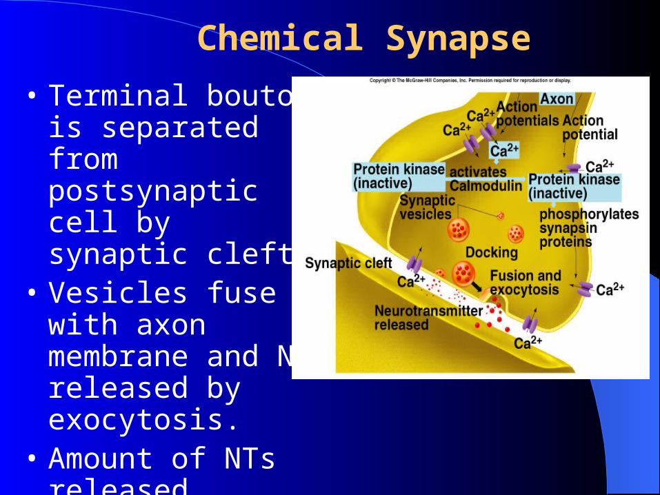
Chemical Synapse
• Terminal bouton is separated from postsynaptic cell by synaptic cleft.
• Vesicles fuse with axon membrane and NT released by exocytosis.
• Amount of NTs released depends upon frequency of AP.
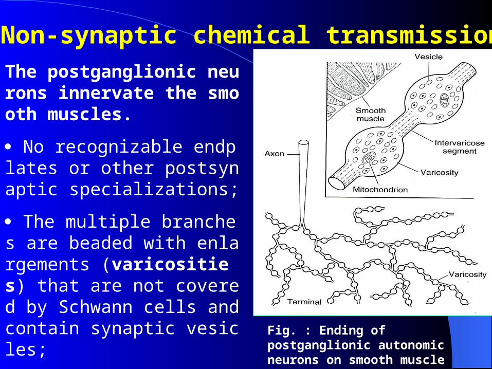
Non-synaptic chemical transmission
The postganglionic neurons innervate the smooth muscles.
No recognizable endplates or other postsynaptic specializations;
The multiple branches are beaded with enlargements (varicosities) that are not covered by Schwann cells and contain synaptic vesicles;
Fig. : Ending of postganglionic autonomic neurons on smooth muscle
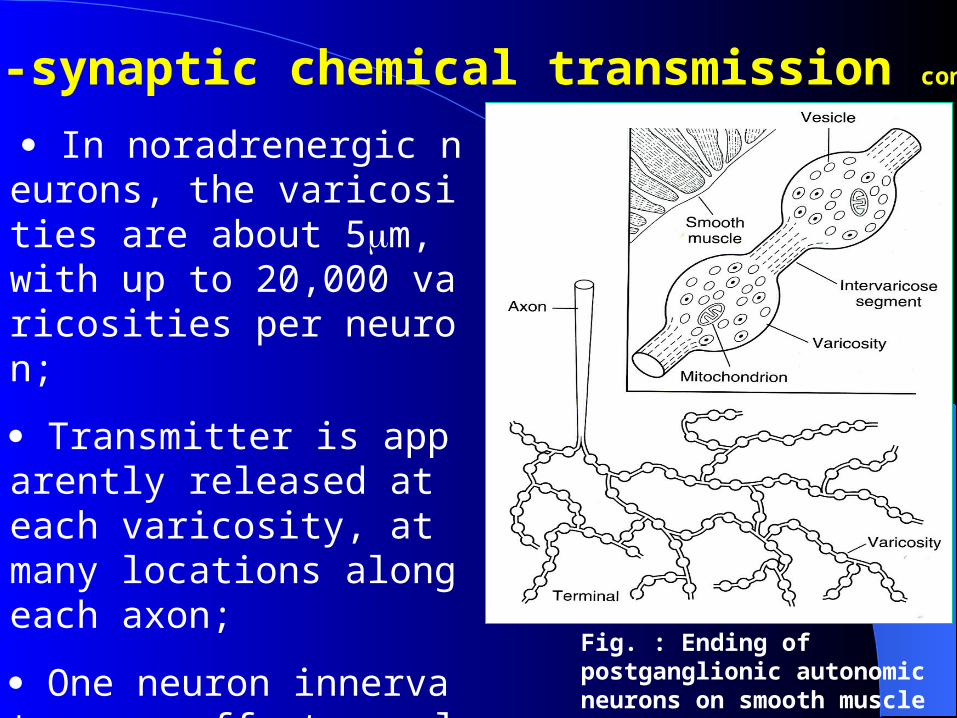
In noradrenergic neurons, the varicosities are about 5m, with up to 20,000 varicosities per neuron;
Transmitter is apparently released at each varicosity, at many locations along each axon;
One neuron innervate many effector cells.
Fig. : Ending of postganglionic autonomic neurons on smooth muscle
Non-synaptic chemical transmission continued
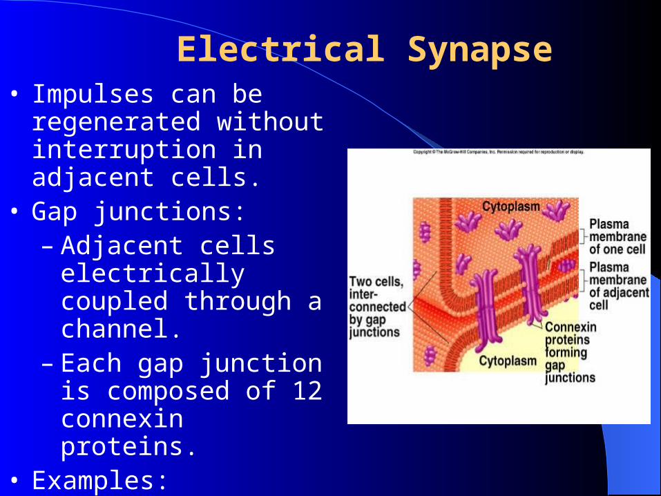
Electrical Synapse• Impulses can be regenerated
without interruption in adjacent cells.
• Gap junctions:– Adjacent cells electrically
coupled through a channel.
– Each gap junction is composed of 12 connexin proteins.
• Examples:– Smooth and cardiac
muscles, brain, and glial cells.
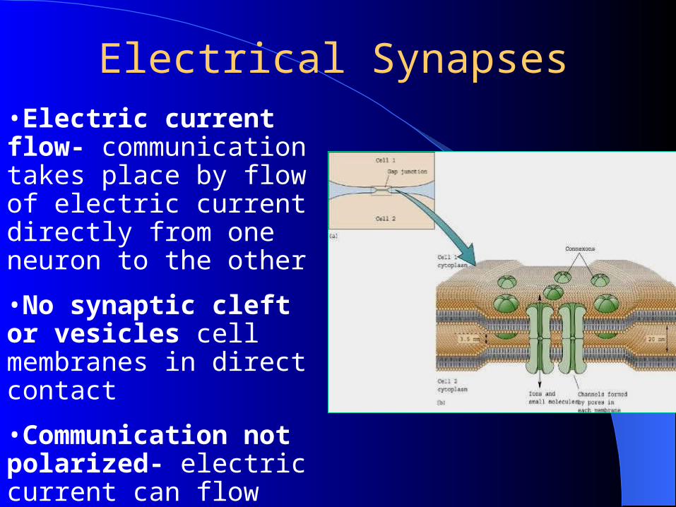
•Electric current flow- communication takes place by flow of electric current directly from one neuron to the other
•No synaptic cleft or vesicles cell membranes in direct contact
•Communication not polarized- electric current can flow between cells in either direction
Electrical Synapses
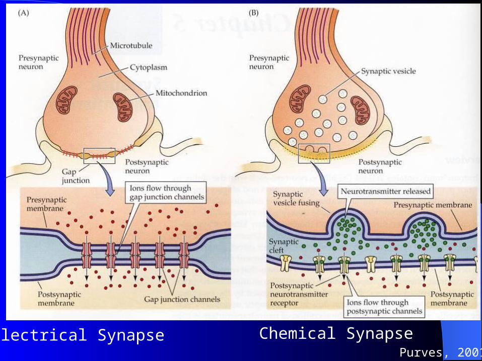
Electrical Synapse Chemical SynapsePurves, 2001
I The Chemical Synapse and Signal Transmission
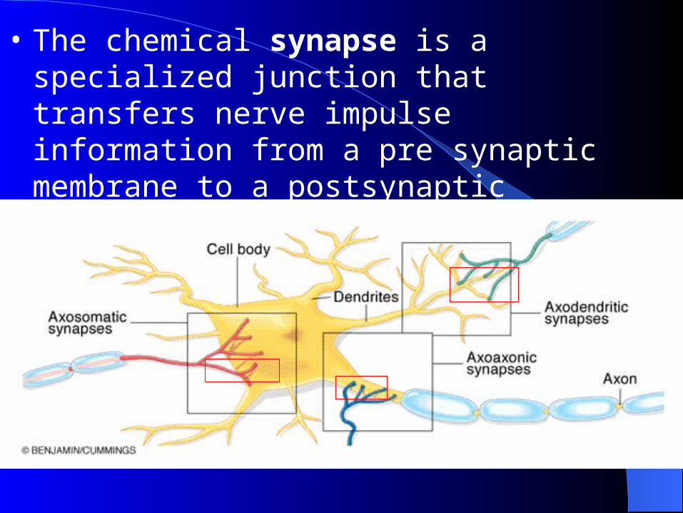
• The chemical synapse is a specialized junction that transfers nerve impulse information from a pre synaptic membrane to a postsynaptic membrane using neurotransmitters and enzymes
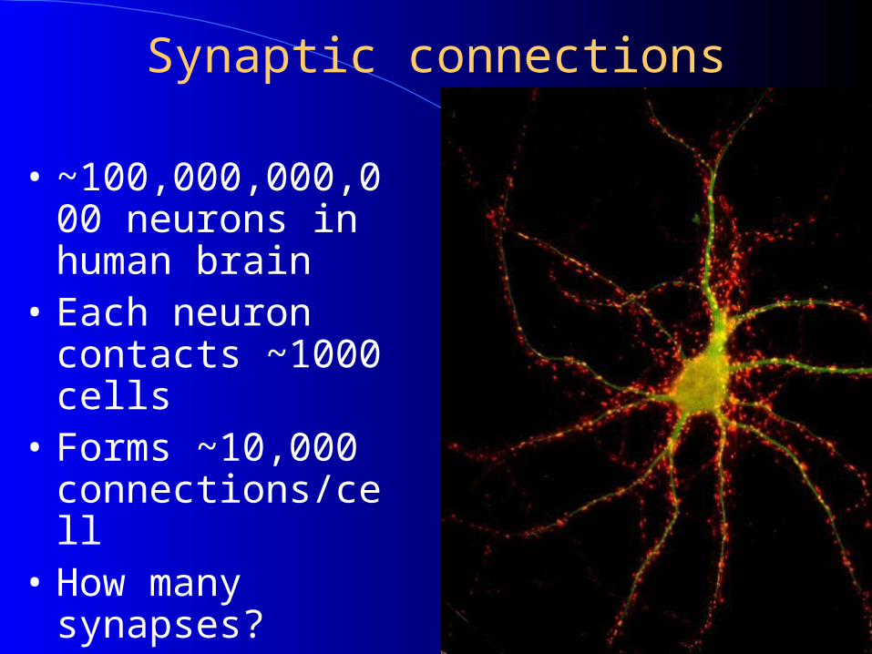
Synaptic connections
• ~100,000,000,000 neurons in human brain
• Each neuron contacts ~1000 cells
• Forms ~10,000 connections/cell
• How many synapses?
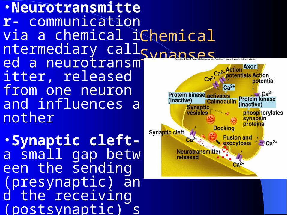
•Neurotransmitter- communication via a chemical intermediary called a neurotransmitter, released from one neuron and influences another
•Synaptic cleft- a small gap between the sending (presynaptic) and the receiving (postsynaptic) site
Chemical Synapses
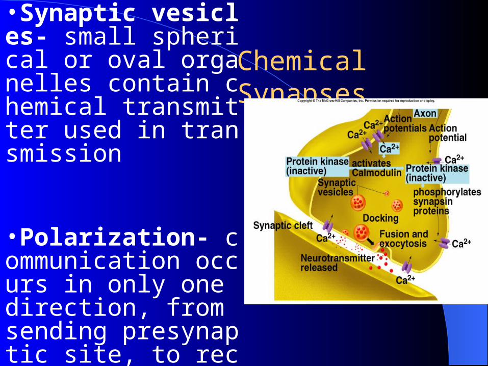
•Synaptic vesicles- small spherical or oval organelles contain chemical transmitter used in transmission
•Polarization- communication occurs in only one direction, from sending presynaptic site, to receiving postsynaptic site
Chemical Synapses
• Precursor transport
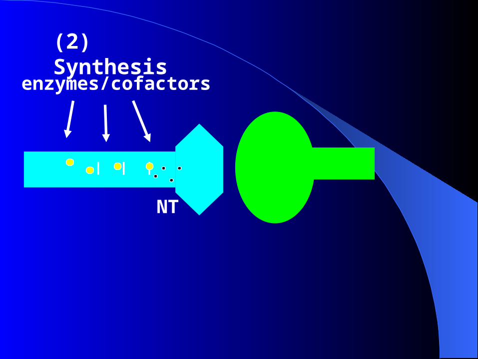
• NT synthesis
• Storage
• Release
• Activation
• Termination ~diffusion, degradation, uptake, autoreceptors
1. Synaptic Transmission Model
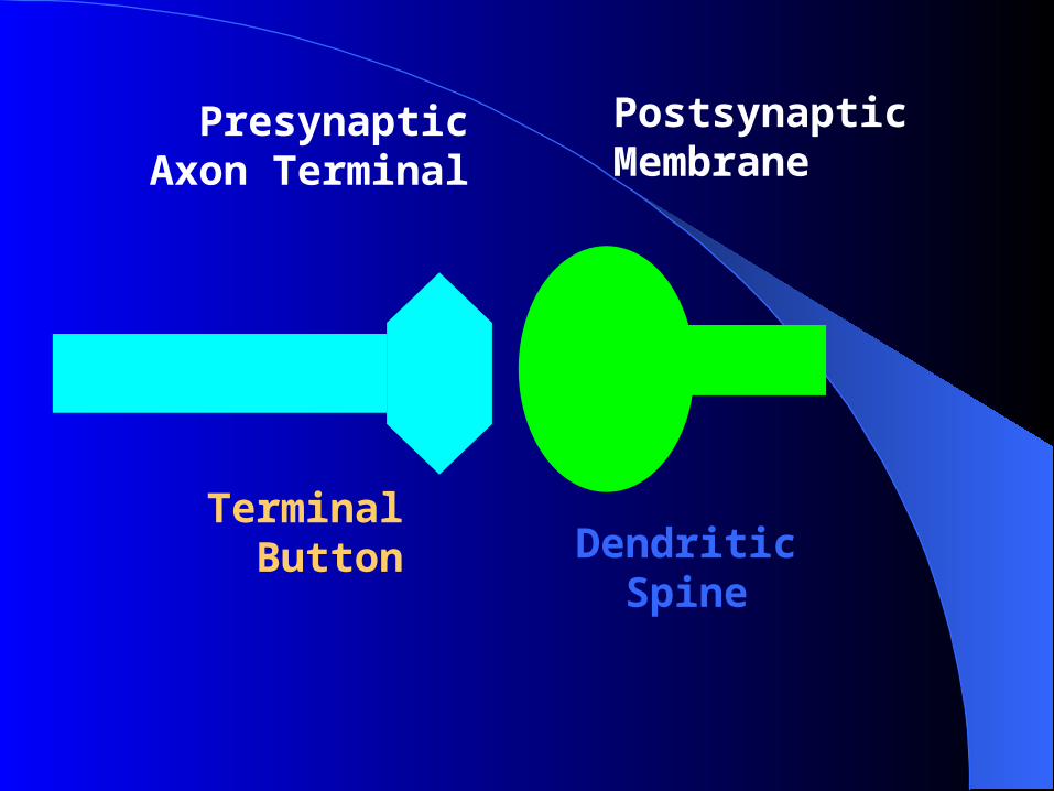
PresynapticAxon Terminal
PostsynapticMembrane
Terminal Button Dendritic
Spine
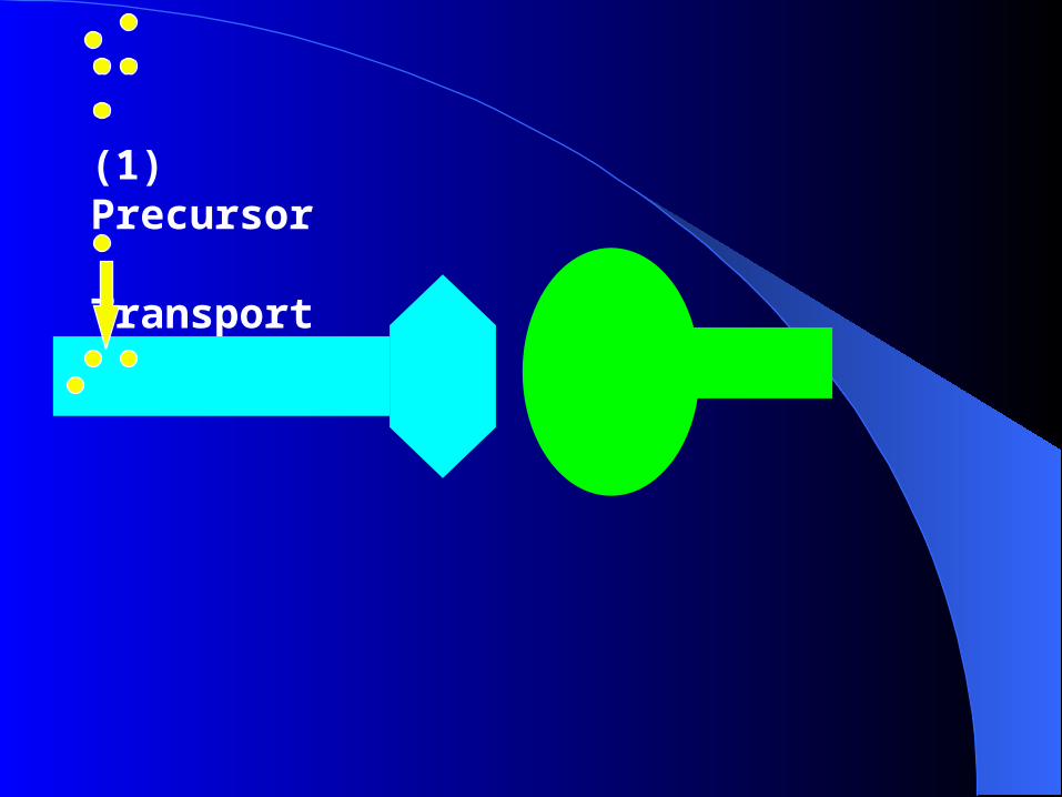
(1) Precursor Transport
_ _ _
NT
(2) Synthesis
enzymes/cofactors
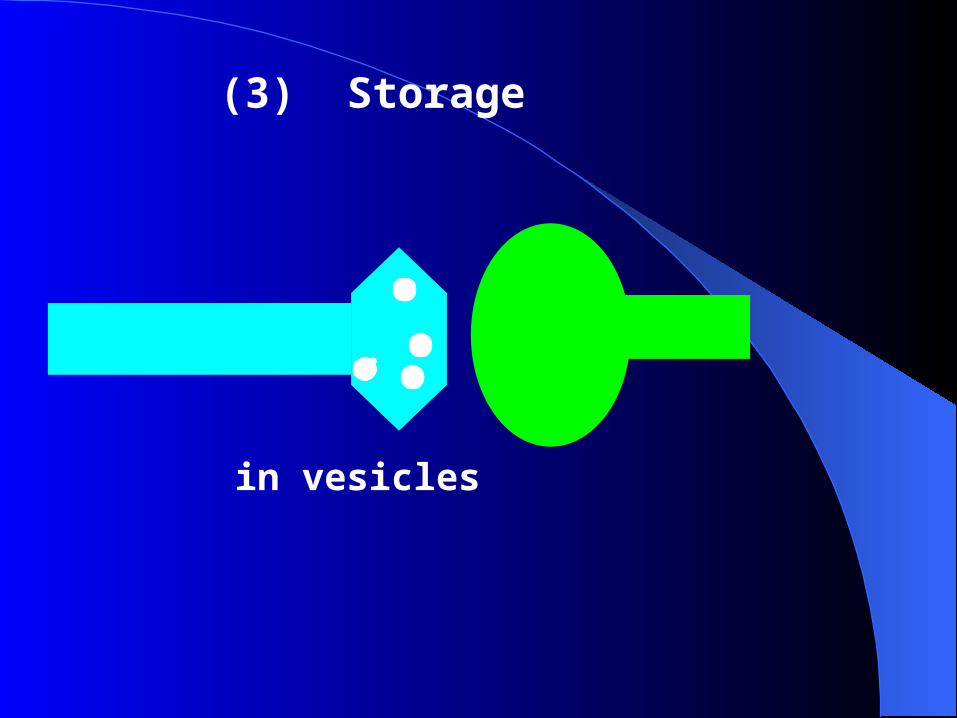
(3) Storage
in vesicles
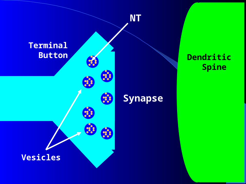
Synapse
Vesicles
NT
Terminal Button Dendritic
Spine
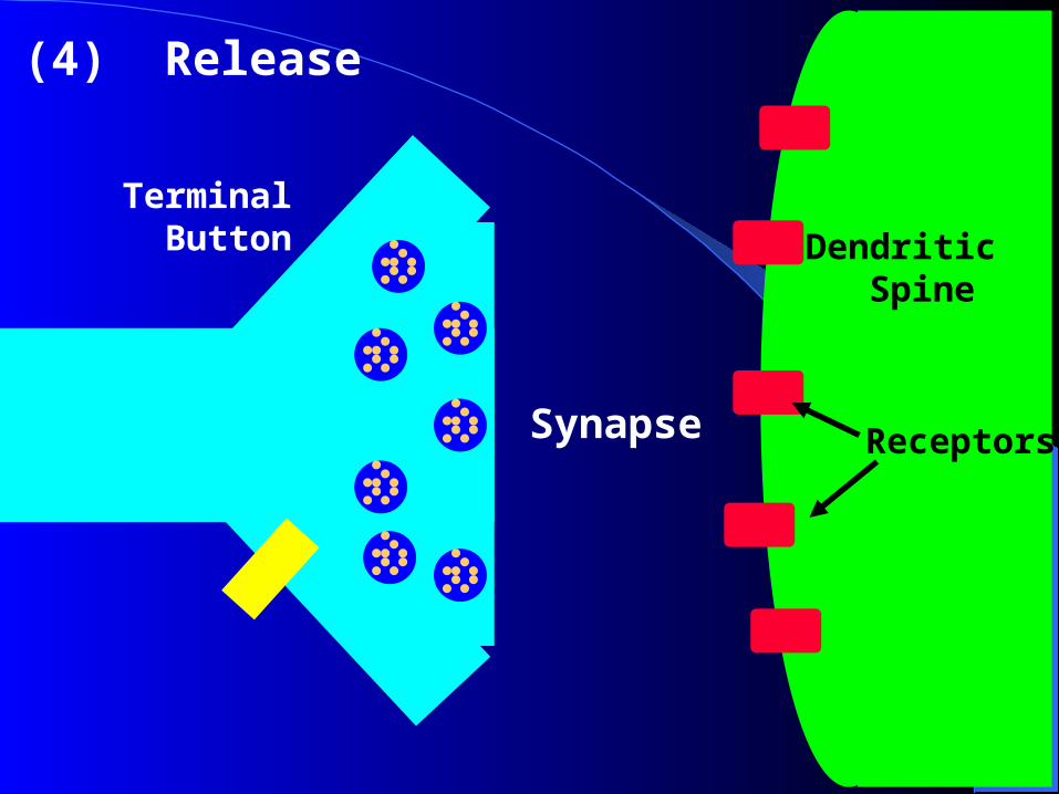
Synapse
Terminal Button Dendritic
Spine
(4) Release
Receptors

Synapse
Terminal Button Dendritic
Spine
AP
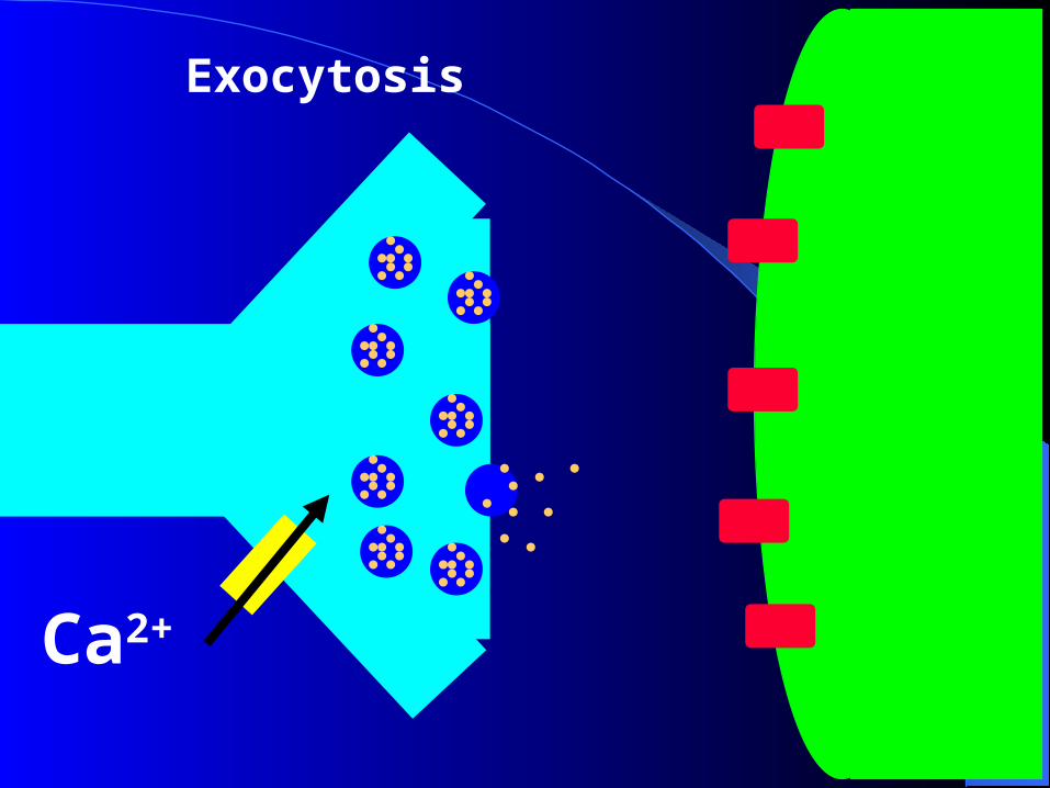
Ca2+
Exocytosis

Each vesicle contains one quanta of neurotransmitter (approximately 5000 molecules) – quanta release
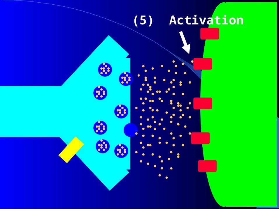
(5) Activation
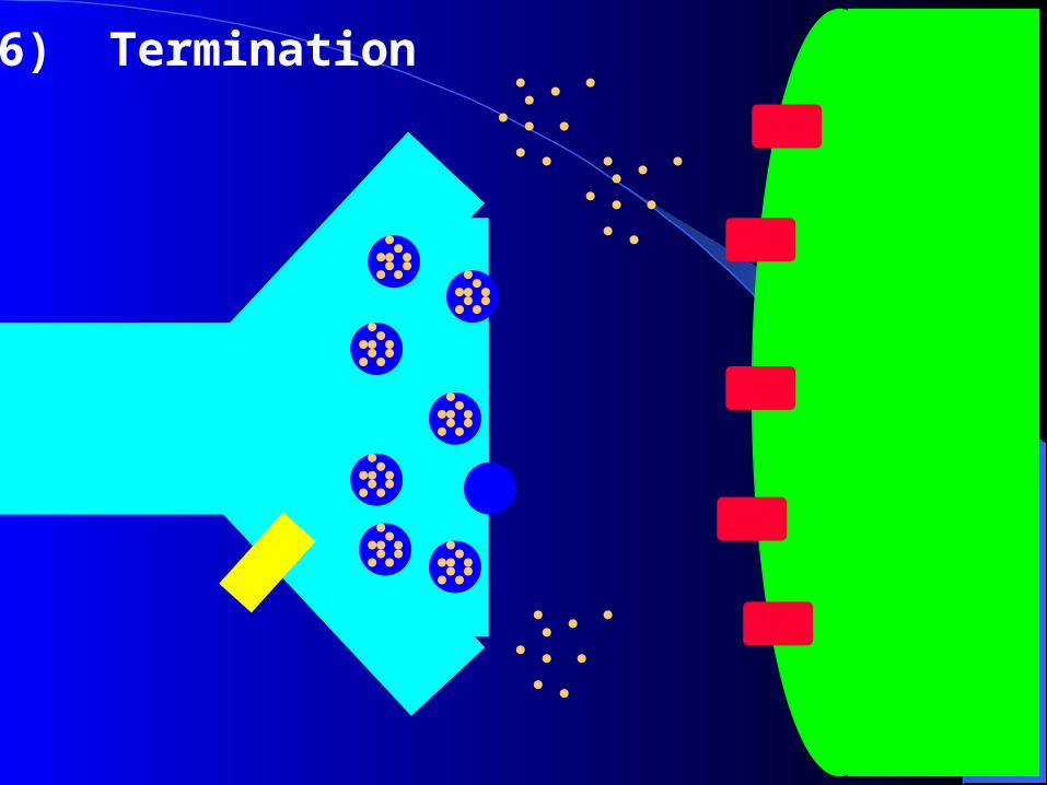
(6) Termination
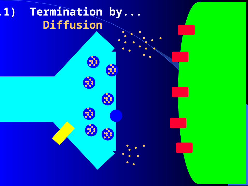
(6.1) Termination by... Diffusion
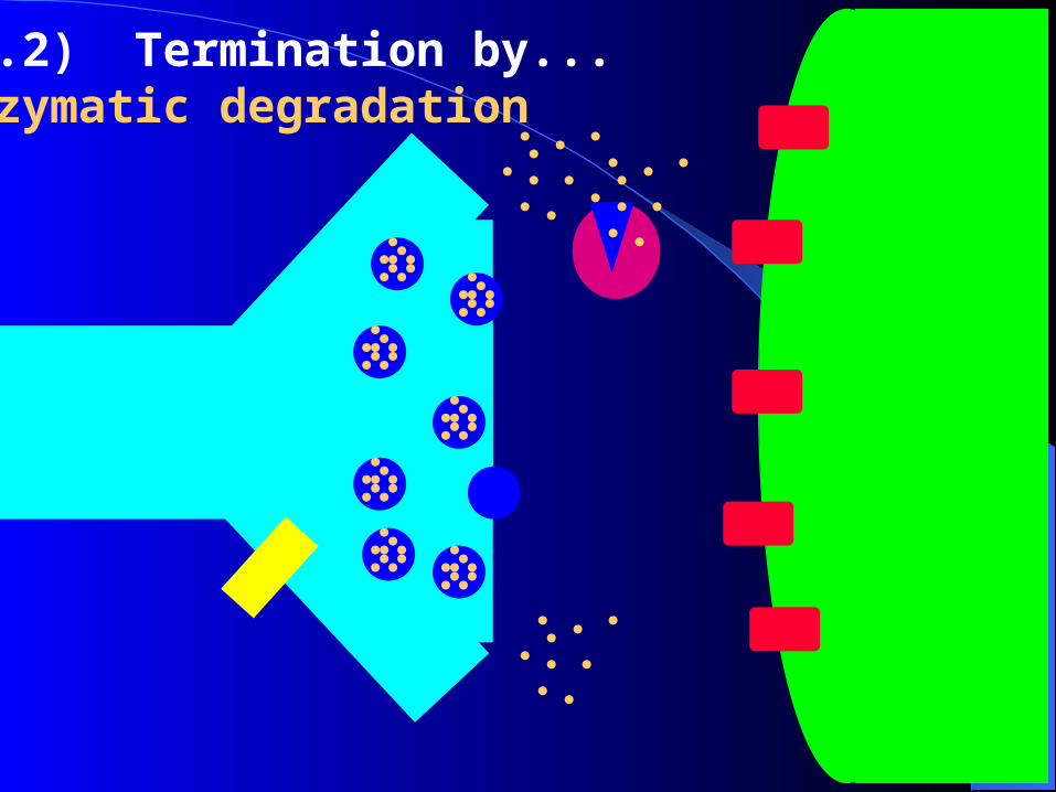
(6.2) Termination by...Enzymatic degradation
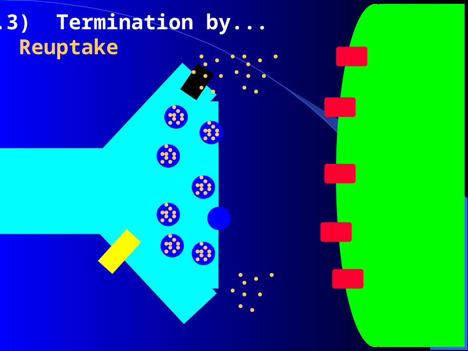
(6.3) Termination by... Reuptake
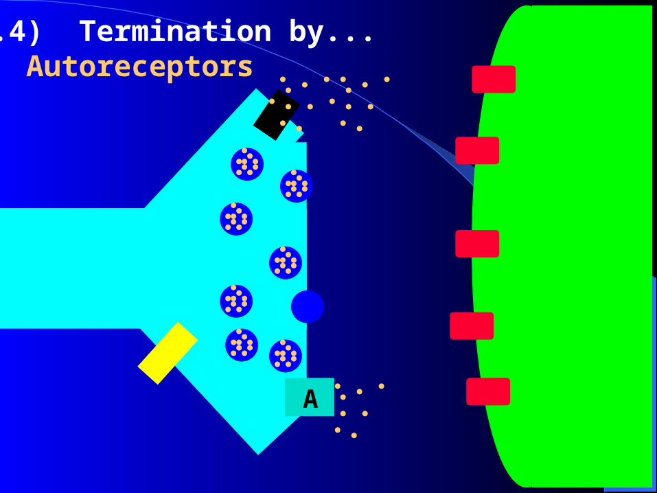
(6.4) Termination by... Autoreceptors
A
Autoreceptors
• On presynaptic terminal
• Binds NT
same as postsynaptic receptors
different receptor subtype
• Decreases NT release & synthesis
• Metabotropic receptors
Synaptic Transmission
• AP travels down axon to bouton.• VG Ca2+ channels open.
– Ca2+ enters bouton down concentration gradient.
– Inward diffusion triggers rapid fusion of synaptic vesicles and release of NTs.
• Ca2+ activates calmodulin, which activates protein kinase.
• Protein kinase phosphorylates synapsins.– Synapsins aid in the fusion of synaptic vesicles.
Synaptic Transmission (continued)
• NTs are released and diffuse across synaptic cleft.
• NT (ligand) binds to specific receptor proteins in postsynaptic cell membrane.
• Chemically-regulated gated ion channels open.– EPSP: depolarization.– IPSP: hyperpolarization.
• Neurotransmitter inactivated to end transmission.

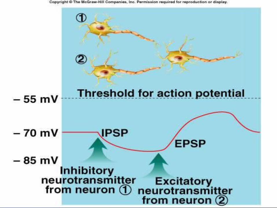
2 EPSP and IPSP
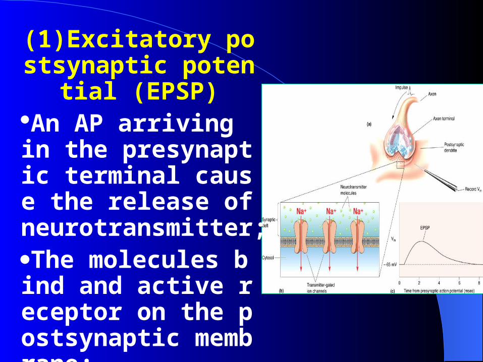
(1)Excitatory postsynaptic potential (E
PSP)An AP arriving in the presynaptic terminal cause the release of neurotransmitter;The molecules bind and active receptor on the postsynaptic membrane;
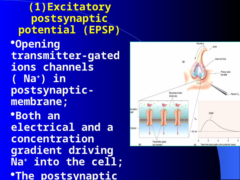
(1)Excitatory postsynaptic
potential (EPSP)Opening transmitter-gated ions channels ( Na+) in postsynaptic- membrane;Both an electrical and a concentration gradient driving Na+ into the cell; The postsynaptic membrane will become depolarized(EPSP).

EPSP• No threshold.• Decreases resting
membrane potential.– Closer to threshold.
• Graded in magnitude.
• Have no refractory period.
• Can summate.
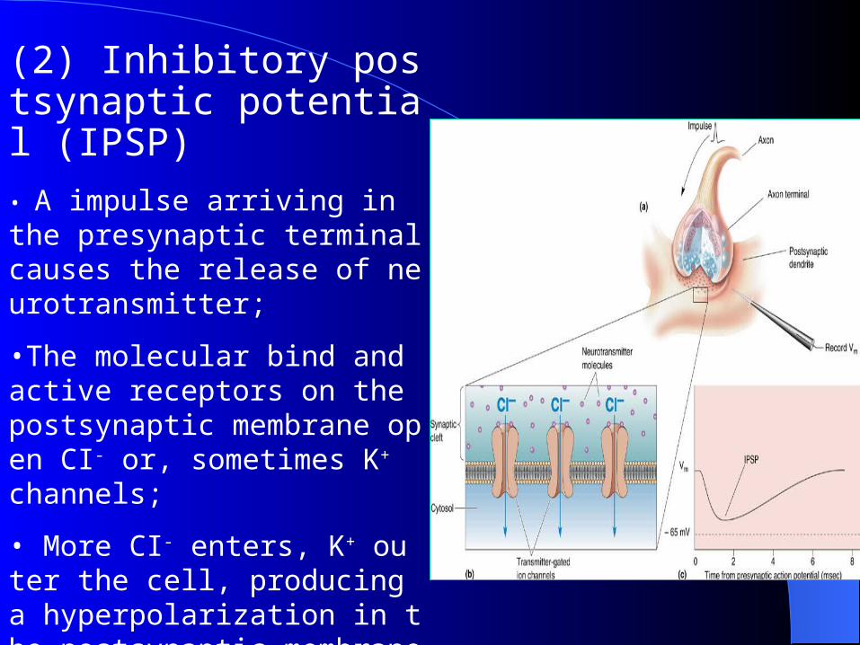
(2) Inhibitory postsynaptic potential (IPSP)
• A impulse arriving in the presynaptic terminal causes the release of neurotransmitter;
•The molecular bind and active receptors on the postsynaptic membrane open CI- or, sometimes K+ channels;
• More CI- enters, K+ outer the cell, producing a hyperpolarization in the postsynaptic membrane.
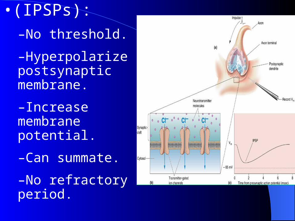
•(IPSPs):–No threshold.
–Hyperpolarize postsynaptic membrane.
–Increase membrane potential.
–Can summate.
–No refractory period.
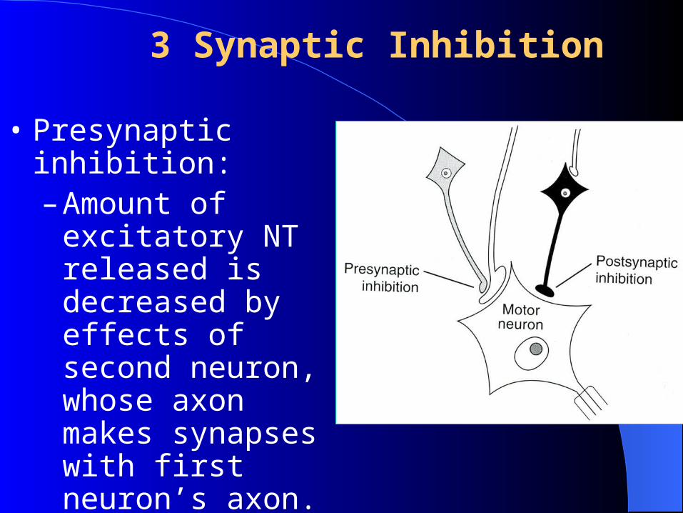
3 Synaptic Inhibition
• Presynaptic inhibition:– Amount of
excitatory NT released is decreased by effects of second neuron, whose axon makes synapses with first neuron’s axon.
• Postsynaptic inhibition
Concept: effect of inhibitory synapses on
the postsynaptic membrane. Mechanism: IPSP, inhibitory interneuron Types:
Afferent collateral inhibition( reciprocal
inhibition)
Recurrent inhibition.
(1) Postsynaptic inhibition
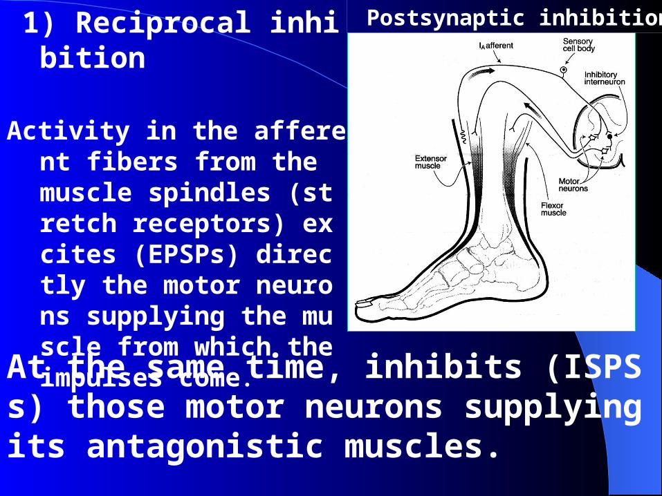
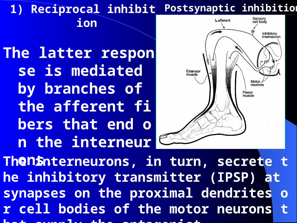
1) Reciprocal inhibition
Activity in the afferent fibers from the muscle spindles (stretch receptors) excites (EPSPs) directly the motor neurons supplying the muscle from which the impulses come.
Postsynaptic inhibition
At the same time, inhibits (ISPSs) those motor neurons supplying its antagonistic muscles.
1) Reciprocal inhibition
The latter response is mediated by branches of the afferent fibers that end on the interneurons.
Postsynaptic inhibition
The interneurons, in turn, secrete the inhibitory transmitter (IPSP) at synapses on the proximal dendrites or cell bodies of the motor neurons that supply the antagonist.
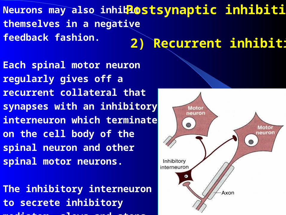
Neurons may also inhibit
themselves in a negative feedback
fashion.
Each spinal motor neuron regularly
gives off a recurrent collateral that
synapses with an inhibitory
interneuron which terminates on
the cell body of the spinal neuron
and other spinal motor neurons.
The inhibitory interneuron to
secrete inhibitory mediator, slows
and stops the discharge of the
motor neuron.
2) Recurrent inhibition
Postsynaptic inhibition
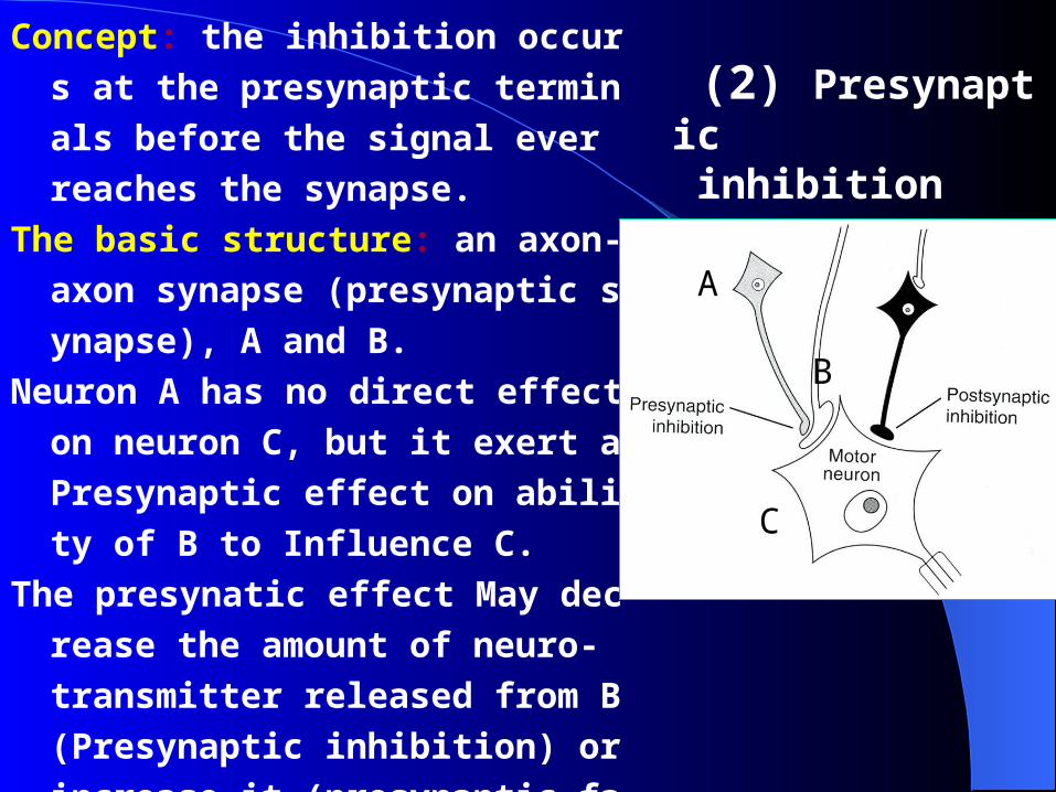
Concept: the inhibition occurs at the
presynaptic terminals before the s
ignal ever reaches the synapse.
The basic structure: an axon-axon s
ynapse (presynaptic synapse), A a
nd B.
Neuron A has no direct effect on neu
ron C, but it exert a Presynaptic ef
fect on ability of B to Influence C.
The presynatic effect May decrease t
he amount of neuro- transmitter re
leased from B (Presynaptic inhibiti
on) or increase it (presynaptic faci
litation).
(2) Presynaptic inhibition
AAB
C
A
C
B
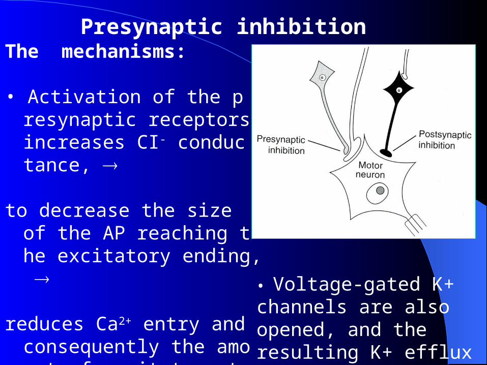
The mechanisms:
• Activation of the presynaptic receptors increases CI- conductance,
to decrease the size of the AP reaching the excitatory ending,
reduces Ca2+ entry and consequently the amount of excitatory transmitter decreased.
Presynaptic inhibition
• Voltage-gated K+ channels are also opened, and the resulting K+ efflux also decreases the Ca2+ influx.
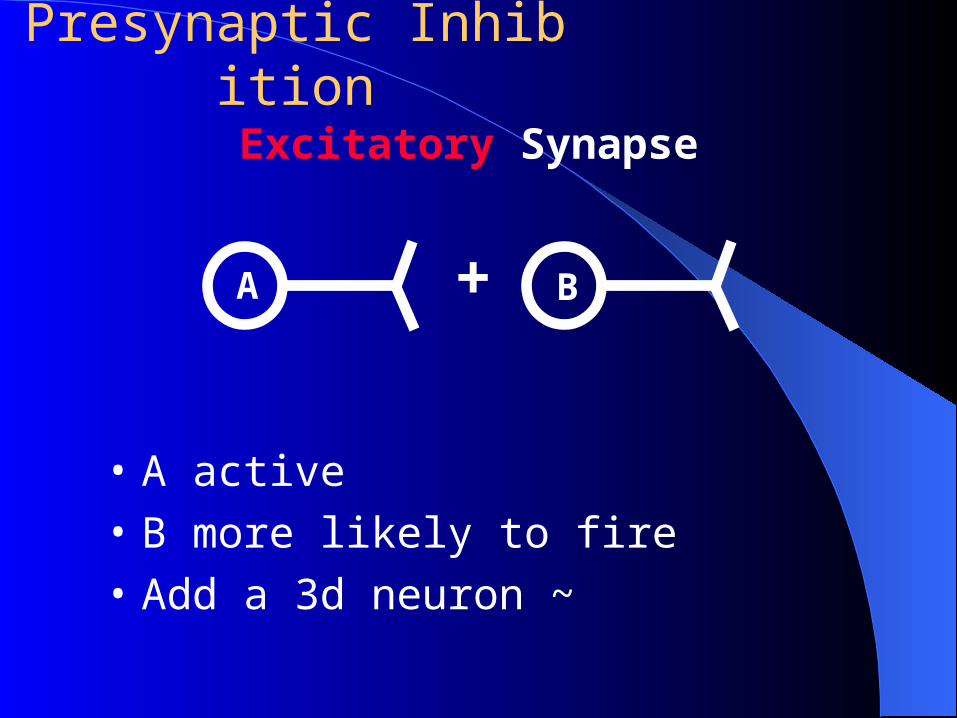
Excitatory Synapse
• A active
• B more likely to fire
• Add a 3d neuron ~
A B+
Presynaptic Inhibition
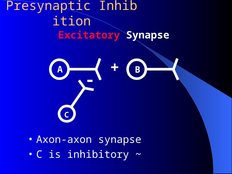
Excitatory Synapse
• Axon-axon synapse
• C is inhibitory ~
A B+
Presynaptic Inhibition
C
-
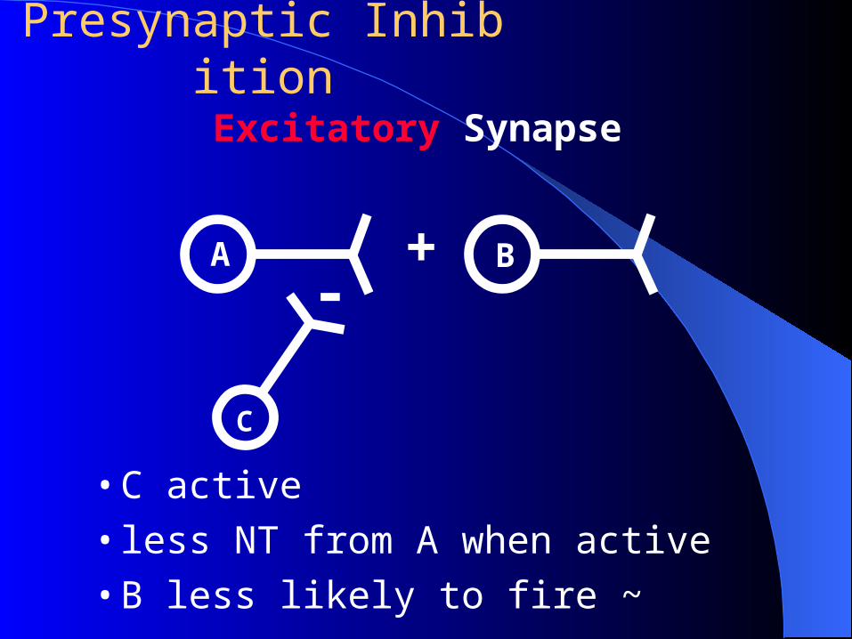
Excitatory Synapse
A B+
Presynaptic Inhibition
C
-
• C active
• less NT from A when active
• B less likely to fire ~
4 Synaptic Facilitation: Presynaptic and Postsynaptic
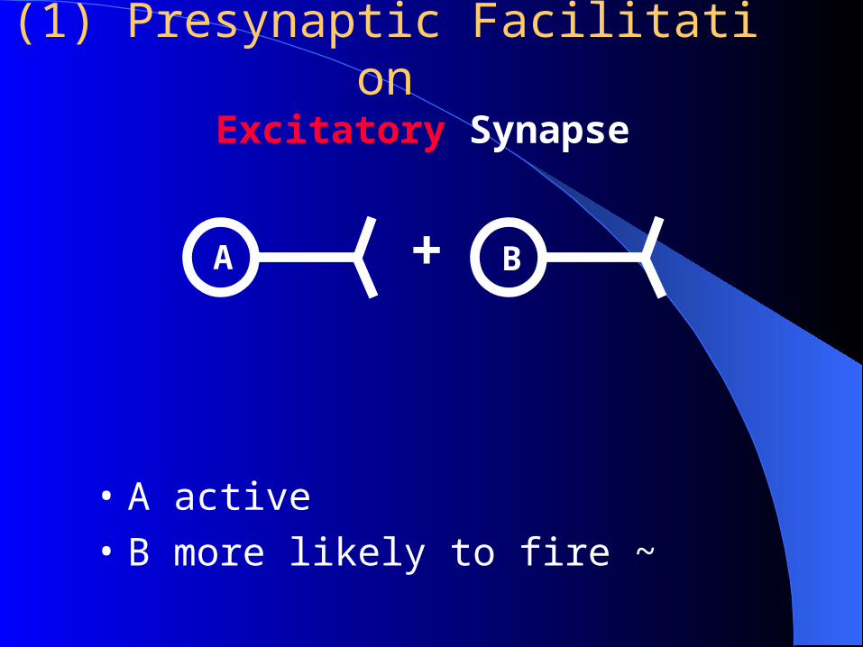
Excitatory Synapse
• A active
• B more likely to fire ~
A B+
(1) Presynaptic Facilitation
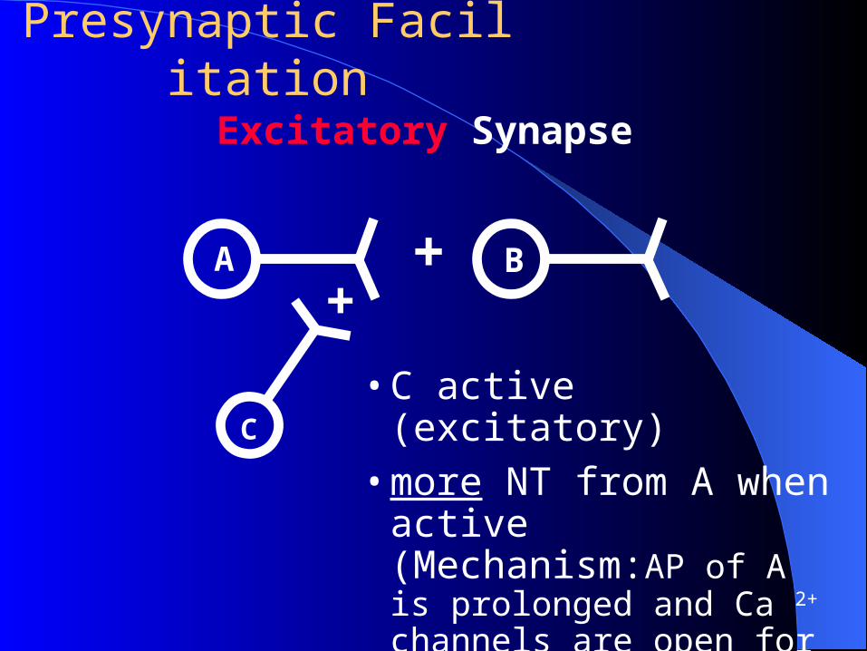
Excitatory Synapse
A B+
Presynaptic Facilitation
C
+
• C active (excitatory)• more NT from A when
active (Mechanism:AP of A is prolonged and Ca 2+ channels are open for a longer period.)
• B more likely to fire ~
(2) Postsynaptic facilitation: neuron that has been partially depolarized is more likely to undergo AP.
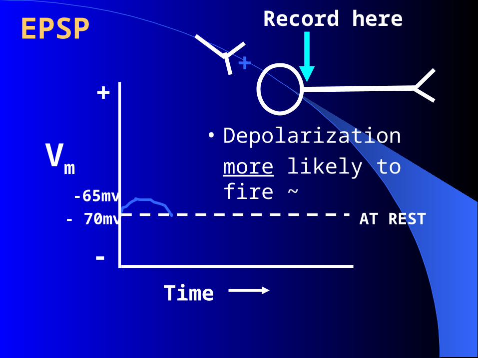
-65mv
- 70mv AT REST
Vm
Time
EPSP
+
-
• Depolarization
more likely to fire ~
Record here
+
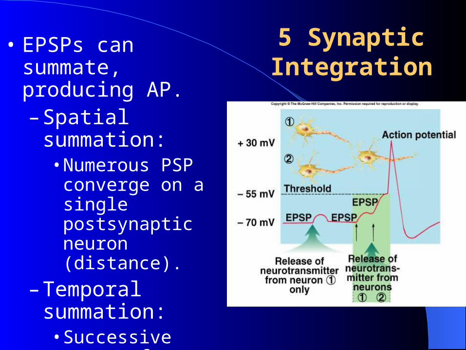
5 Synaptic Integration
• EPSPs can summate, producing AP.– Spatial summation:
• Numerous PSP converge on a single postsynaptic neuron (distance).
– Temporal summation: • Successive waves of
neurotransmitter release (time).
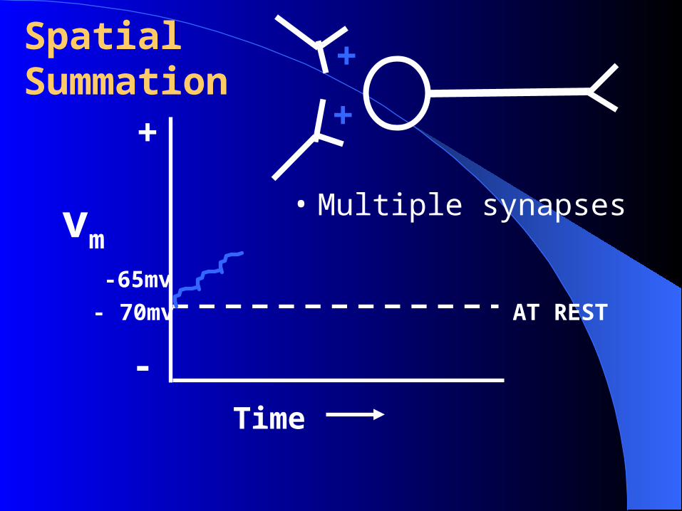
(1) Spatial Summation
• The accumulation of neurotransmitter in the synapse due the combined activity of several presynaptic neurons entering the Area (Space) of a Convergent Synapse.
• A space (spatial) dependent process.
-65mv
- 70mv AT REST
vm
Time
+
-
SpatialSummation +
• Multiple synapses
+
(2) Temporal Summation
• The accumulation of neurotransmitters in a synapse due to the rapid activity of a presynaptic neuron over a given Time period.
• Occurs in a Divergent Synapse. (explain later)
• Is a Time (Temporal) dependent process.
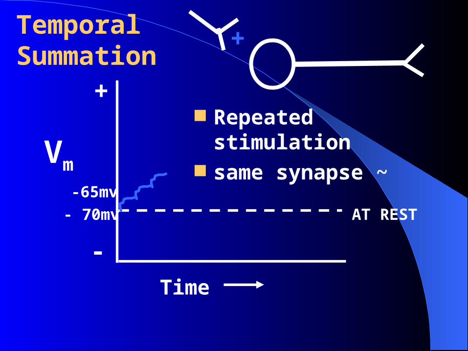
-65mv
- 70mv AT REST
Vm
Time
+
-
TemporalSummation
+
Repeated stimulation same synapse ~
(3) EPSPs & IPSPs summate
• CANCEL EACH OTHER
• Net stimulation – EPSPs + IPSPs = net effects ~
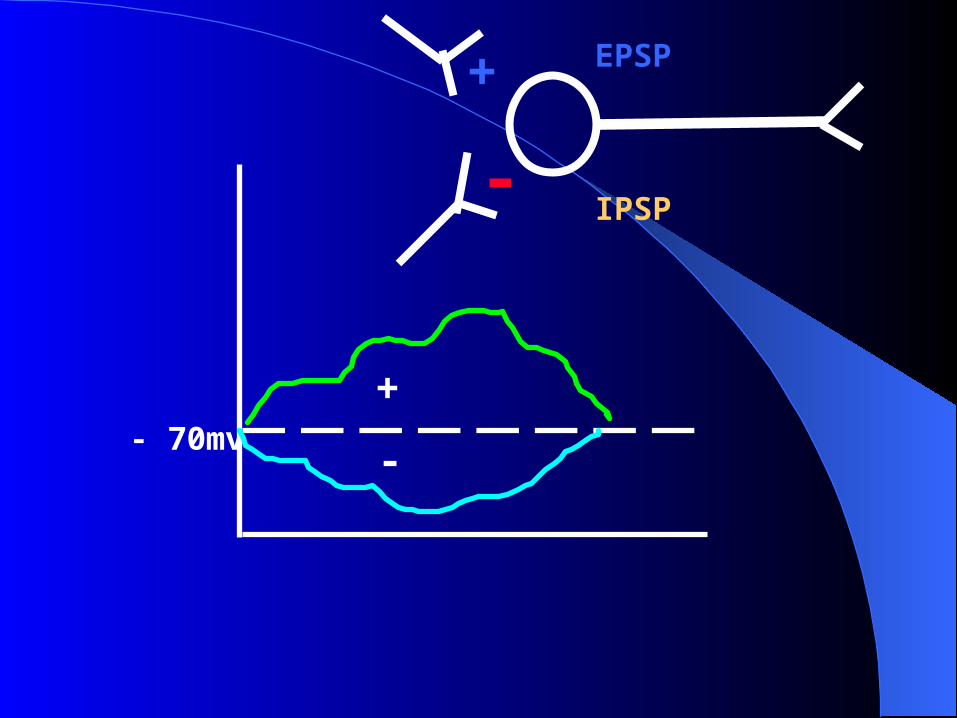
- 70mv
+
-
-
EPSP
IPSP
+
6. Divergent and Convergent Synapse
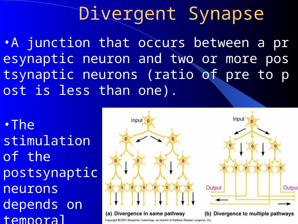
Divergent Synapse
•A junction that occurs between a presynaptic neuron and two or more postsynaptic neurons (ratio of pre to post is less than one).
•The stimulation of the postsynaptic neurons depends on temporal summation).
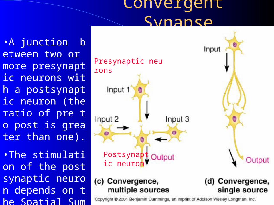
Convergent Synapse
Presynaptic neurons
Postsynaptic neuron
•A junction between two or more presynaptic neurons with a postsynaptic neuron (the ratio of pre to post is greater than one).
•The stimulation of the postsynaptic neuron depends on the Spatial Summation.
II Neurotransmitters and receptors
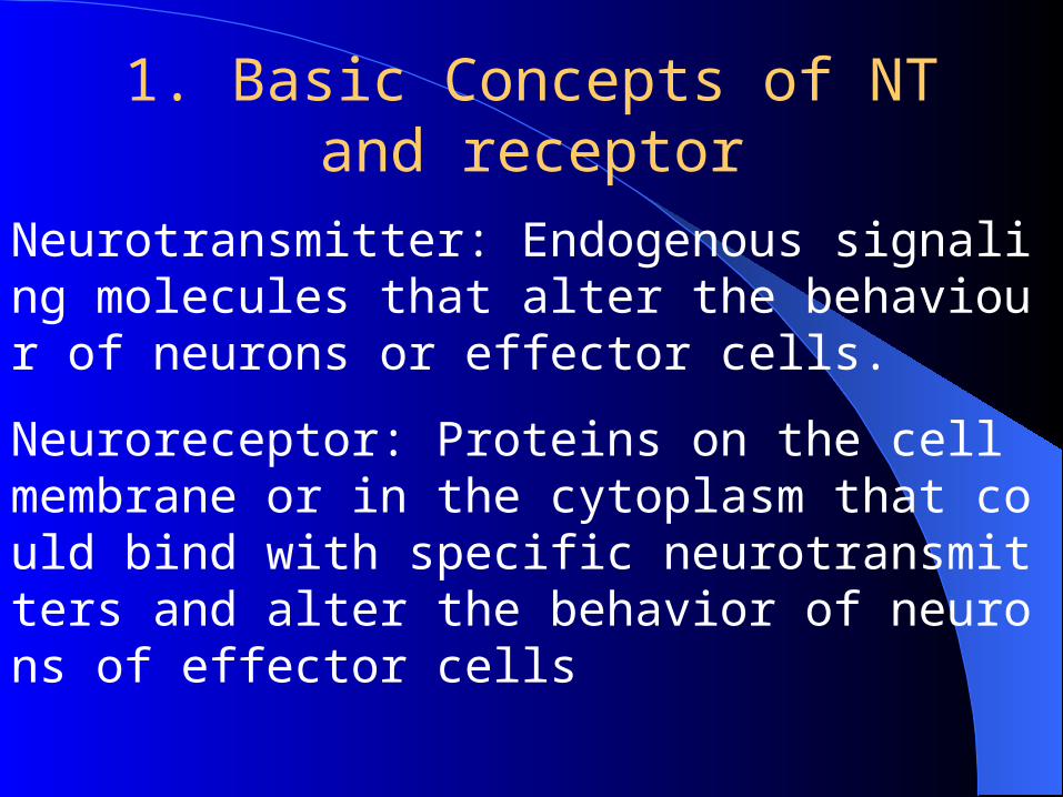
1. Basic Concepts of NT and receptor
Neurotransmitter: Endogenous signaling molecules that alter the behaviour of neurons or effector cells.
Neuroreceptor: Proteins on the cell membrane or in the cytoplasm that could bind with specific neurotransmitters and alter the behavior of neurons of effector cells
•Vast array of molecules serve as neurotransmitters
•The properties of the transmitter do not determine its effects on the postsynaptic cells
•The properties of the receptor determine whether a transmitter is excitatory or inhibitory
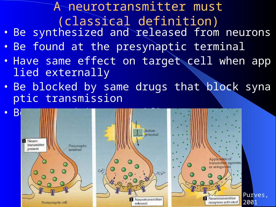
A neurotransmitter must (classical definition)
• Be synthesized and released from neurons• Be found at the presynaptic terminal• Have same effect on target cell when applied externally• Be blocked by same drugs that block synaptic transmission• Be removed in a specific way
Purves, 2001
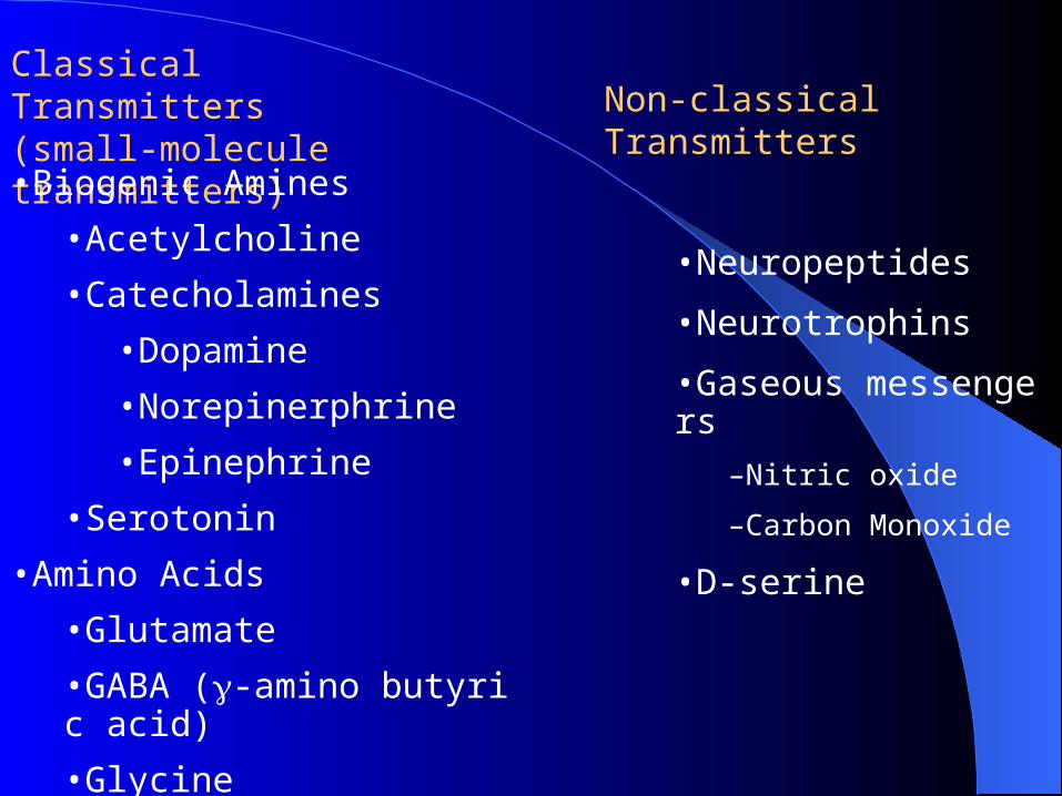
Classical Transmitters (small-molecule transmitters)•Biogenic Amines
•Acetylcholine
•Catecholamines
•Dopamine
•Norepinerphrine
•Epinephrine
•Serotonin
•Amino Acids
•Glutamate
•GABA (-amino butyric acid)
•Glycine
•Neuropeptides
•Neurotrophins
•Gaseous messengers
–Nitric oxide
–Carbon Monoxide
•D-serine
Non-classical Transmitters
Agonist
A substance that mimics a specific neurotransmitter,
is able to attach to that neurotransmitter's receptor
and thereby produces the same action that the neurotransmitter usually produces.
Drugs are often designed as receptor agonists to treat a variety of diseases and disorders when the original chemical substance is missing or depleted.
Antagonist
Drugs that bind to but do not activate neuroreceptors,
thereby blocking the actions of neurotransmitters or the neuroreceptor agonists.
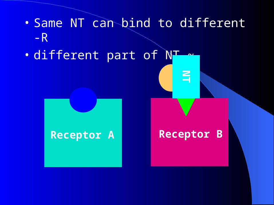
Receptor BReceptor A
• Same NT can bind to different -R
• different part of NT ~
NT
NT
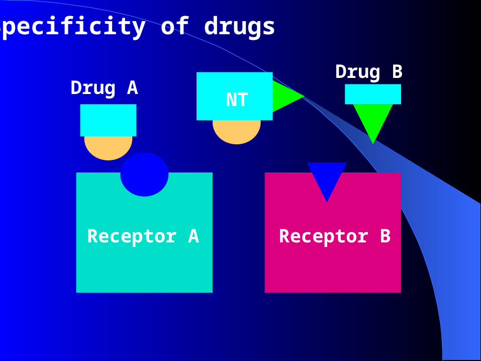
Specificity of drugs
Drug ADrug B
Receptor BReceptor A
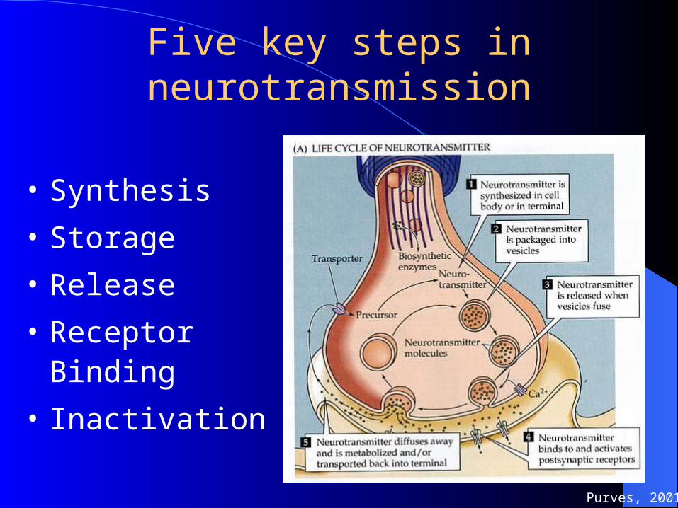
Five key steps in neurotransmission
• Synthesis
• Storage
• Release
• Receptor Binding
• Inactivation
Purves, 2001
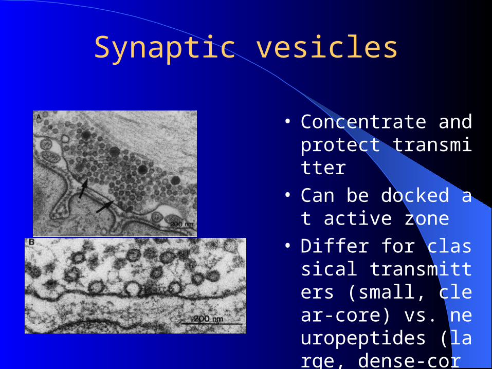
Synaptic vesicles
• Concentrate and protect transmitter
• Can be docked at active zone
• Differ for classical transmitters (small, clear-core) vs. neuropeptides (large, dense-core)
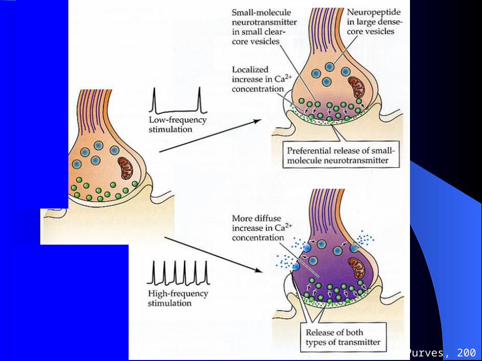
Neurotransmitter Co-existence (Dale principle)Some neurons in both the PNS and CNS produce both a classical neurotransmitter (ACh or a catecholamine) and a polypeptide neurotransmitter.
They are contained in different synaptic vesicles that can be distinguished using the electron microscope.
The neuron can thus release either the classical neurotransmitter or the polypeptide neurotransmitter under different conditions.
Purves, 2001
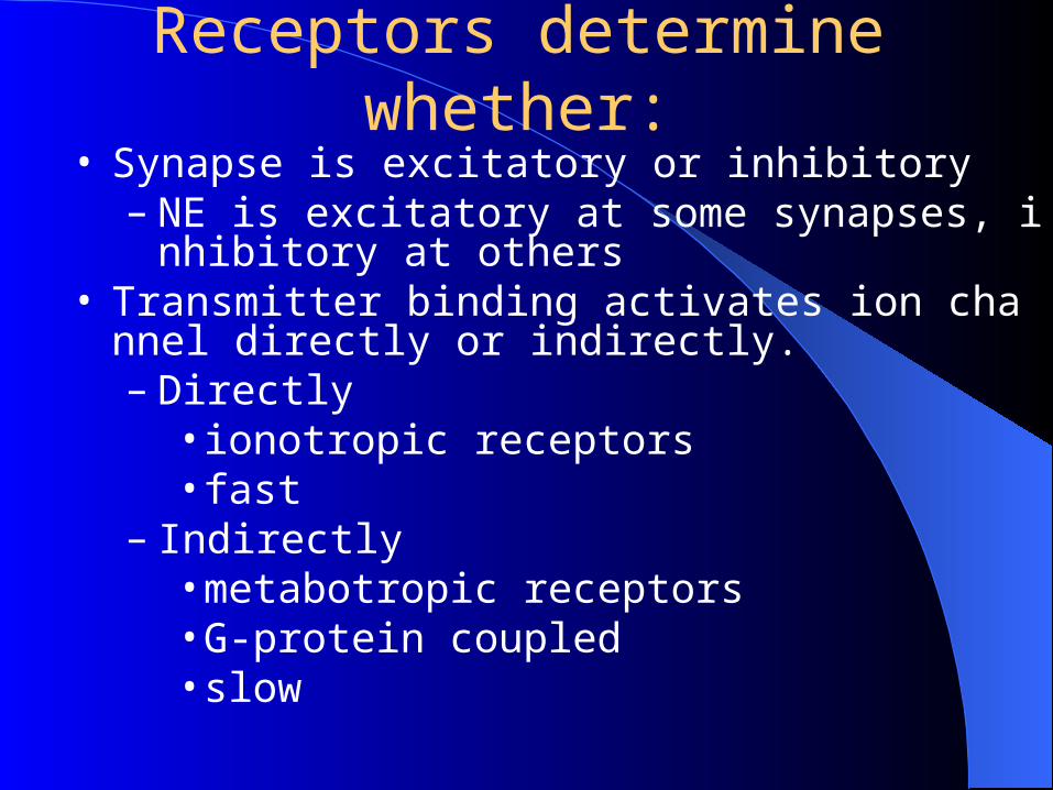
Receptors determine whether:• Synapse is excitatory or inhibitory
– NE is excitatory at some synapses, inhibitory at others
• Transmitter binding activates ion channel directly or indirectly.– Directly
• ionotropic receptors• fast
– Indirectly• metabotropic receptors• G-protein coupled• slow
2. Receptor Activation
• Ionotropic channel– directly controls channel– fast
• Metabotropic channel– second messenger systems– receptor indirectly controls channel ~
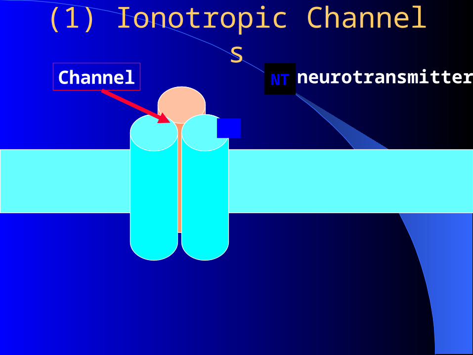
(1) Ionotropic ChannelsneurotransmitterNTChannel
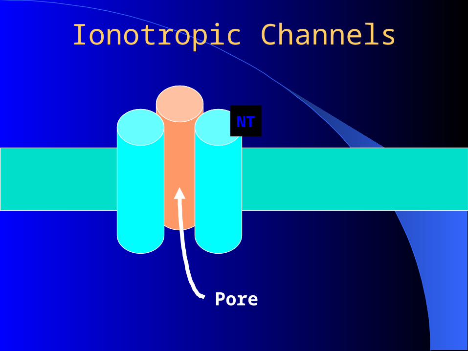
Ionotropic Channels
NT
Pore
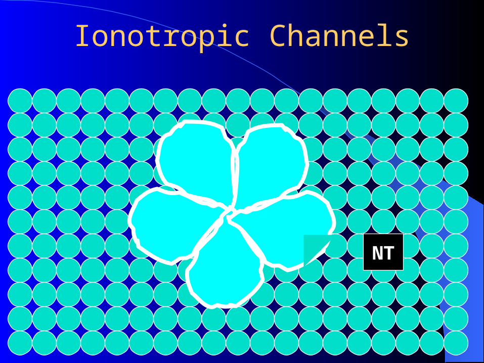
Ionotropic Channels
NT
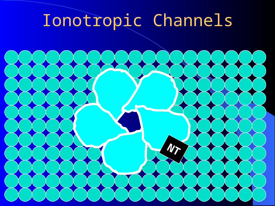
Ionotropic Channels
NT
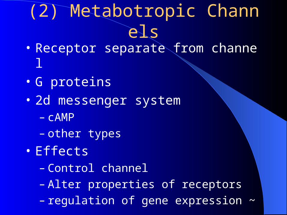
(2) Metabotropic Channels
• Receptor separate from channel
• G proteins
• 2d messenger system– cAMP– other types
• Effects– Control channel– Alter properties of receptors– regulation of gene expression ~
(2.1) G protein: direct control
• NT is 1st messenger
• G protein binds to channel– opens or closes– relatively fast ~
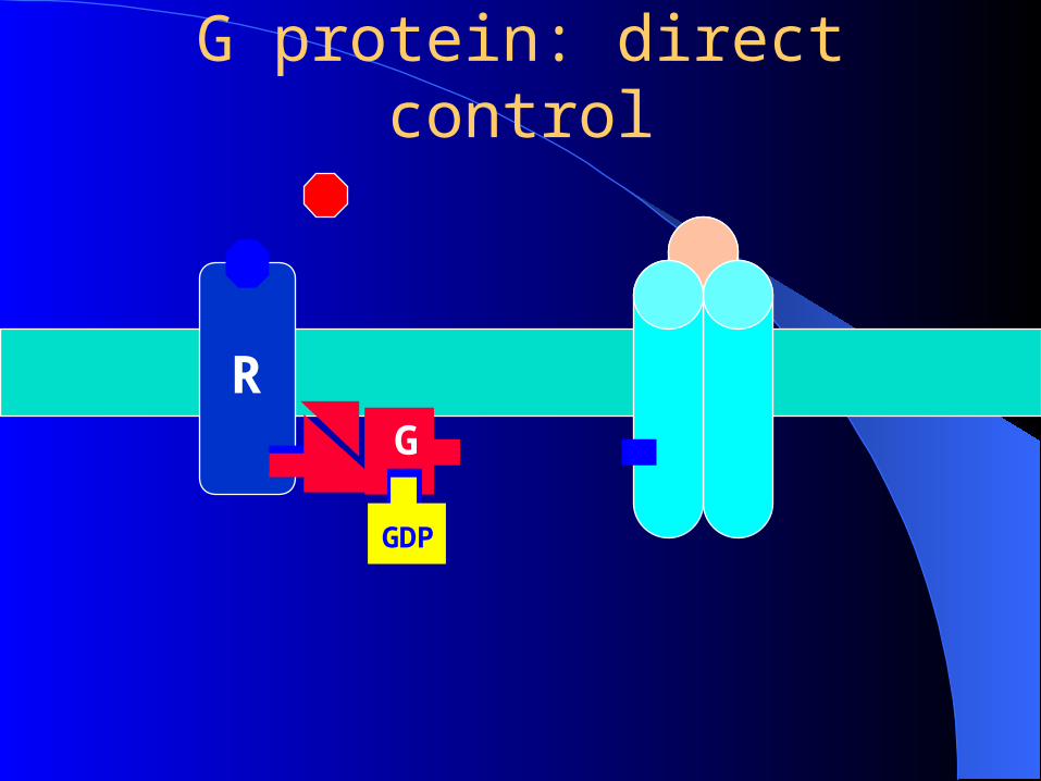
G protein: direct control
RG
GDP
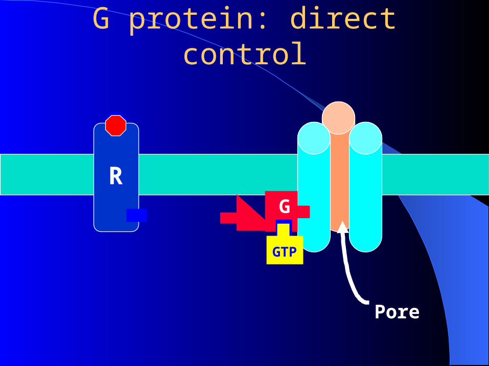
G protein: direct control
RG
GTP
Pore
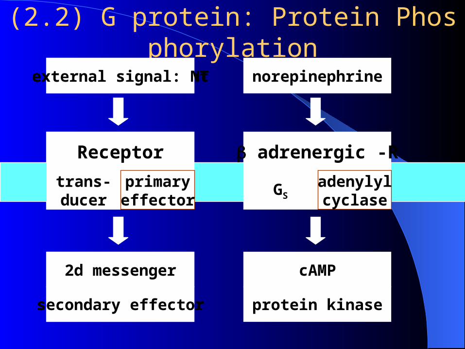
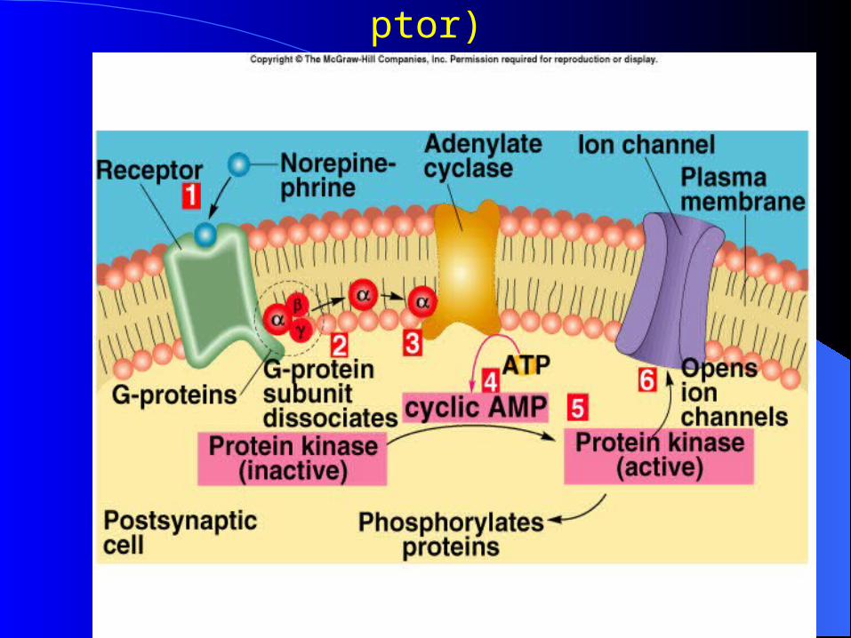
(2.2) G protein: Protein Phosphorylation
Receptor
trans-ducer
primaryeffector
external signal: nt
2d messenger
secondary effector
Receptor
trans-ducer
primaryeffector
external signal: NT
2d messenger
secondary effector
GS
norepinephrine
cAMP
protein kinase
adrenergic -R
adenylylcyclase
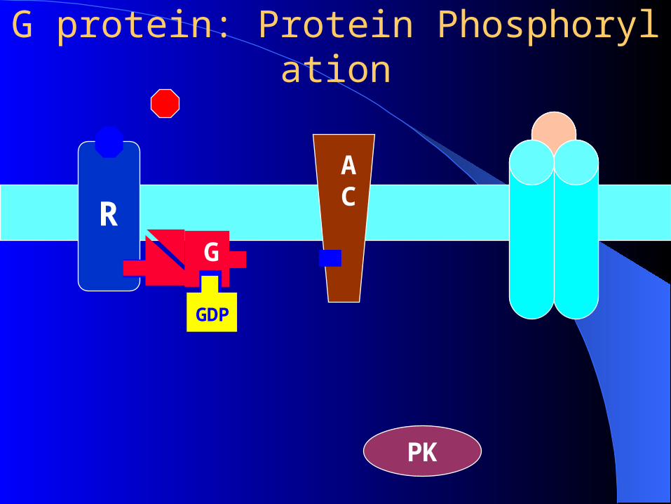
G protein: Protein Phosphorylation
RG
GDP
AC
PK

G protein: Protein Phosphorylation
R
AC
PK
G
GTPATP
cAMP
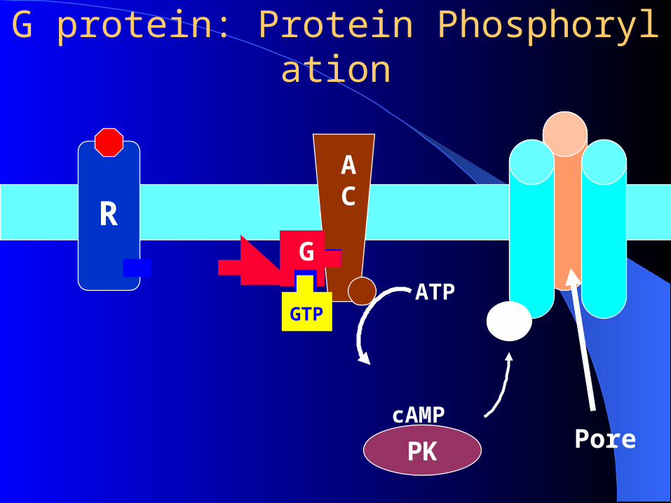
G protein: Protein Phosphorylation
R
AC
PK
G
GTPATP
cAMP
P
Pore
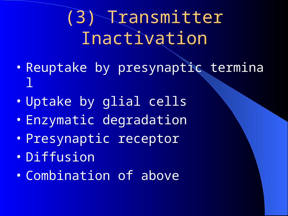
(3) Transmitter Inactivation
• Reuptake by presynaptic terminal
• Uptake by glial cells
• Enzymatic degradation
• Presynaptic receptor
• Diffusion
• Combination of above
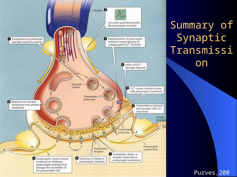
Summary of Synaptic
Transmission
Purves,2001
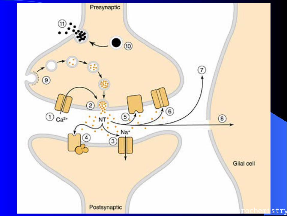
Basic Neurochemistry
3. Some Important Transmitters
(1) Acetylcholine (ACh) as NT
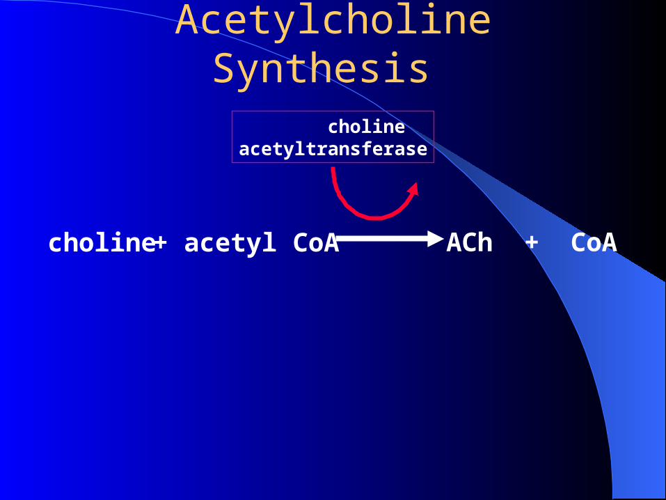
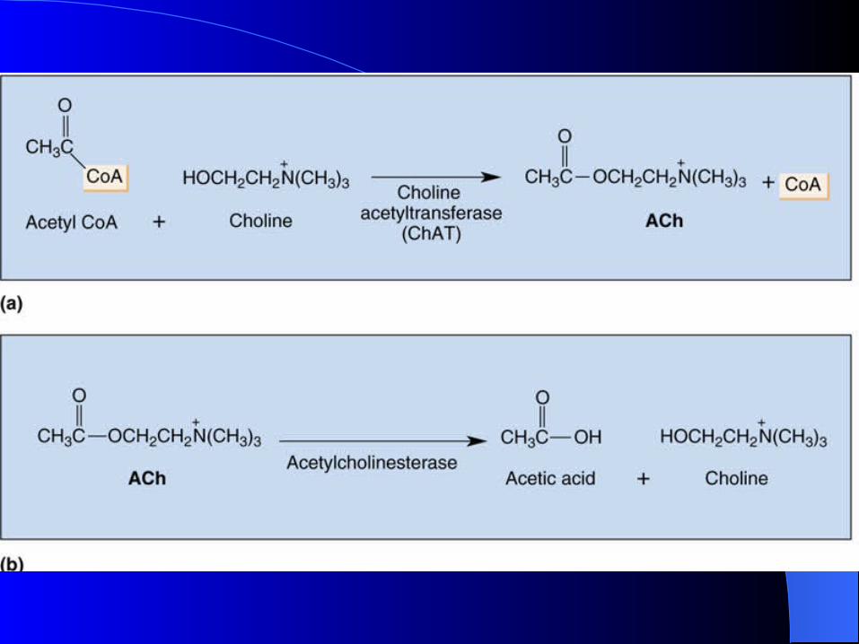
Acetylcholine Synthesis
choline + acetyl CoA ACh + CoA
cholineacetyltransferase
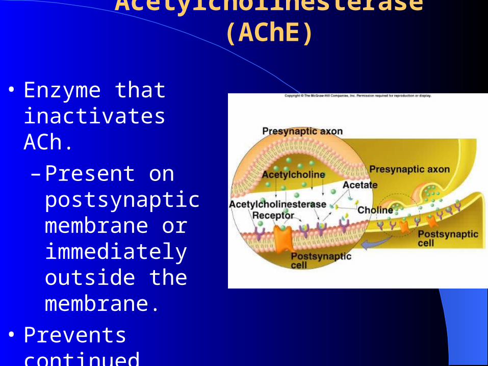
Acetylcholinesterase (AChE)
• Enzyme that inactivates ACh.
– Present on postsynaptic membrane or immediately outside the membrane.
• Prevents continued stimulation.
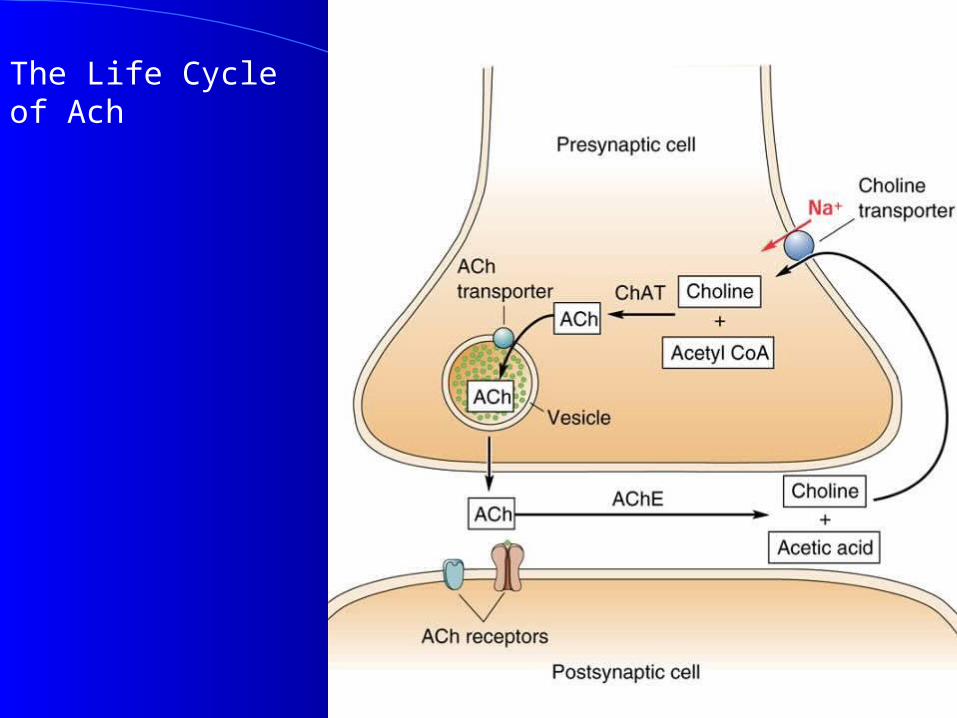
The Life Cycle of Ach
Ach - Distribution
• Peripheral N.S.• Excites somatic skeletal muscle (neuro-muscular jun
ction)• Autonomic NS
Ganglia
Parasympathetic NS--- Neuroeffector junction
Few sympathetic NS – Neuroeffector junction
• Central N.S. - widespreadHippocampus
Hypothalamus ~
•ACh is both an excitatory and inhibitory NT, depending on organ involved.
–Causes the opening of chemical gated ion channels.
•Nicotinic ACh receptors:
–Found in autonomic ganglia (N1) and skeletal muscle fibers (N2).
•Muscarinic ACh receptors:
–Found in the plasma membrane of smooth and cardiac muscle cells, and in cells of particular glands .
Ach Receptors
Acetylcholine Neurotransmission
• “Nicotinic” subtype Receptor:– Membrane Channel for Na+ and K+
– Opens on ligand binding– Depolarization of target (neuron, muscle)– Stimulated by Nicotine, etc.– Blocked by Curare, etc.– Motor endplate (somatic) (N2), – all autonomic ganglia, hormone
producing cells of adrenal medulla (N1)
Acetylcholine Neurotransmission
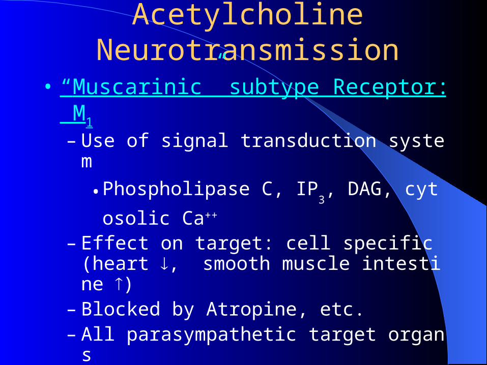
• “Muscarinic” subtype Receptor: M1
– Use of signal transduction system
• Phospholipase C, IP3, DAG, cytosolic Ca++
– Effect on target: cell specific (heart , smooth muscle intestine )
– Blocked by Atropine, etc.– All parasympathetic target organs– Some sympathetic targets (endocrine sweat glan
ds, skeletal muscle blood vessels - dilation)
Acetylcholine Neurotransmission
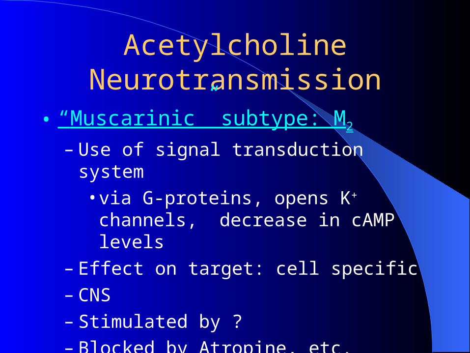
• “Muscarinic” subtype: M2
– Use of signal transduction system• via G-proteins, opens K+ channels, decrease
in cAMP levels– Effect on target: cell specific– CNS – Stimulated by ?– Blocked by Atropine, etc.
Cholinergic Agonists
• Direct– Muscarine – Nicotine
• Indirect– AChE Inhibitors ~
Cholinergic Antagonists
• Direct
Nicotinic - Curare
Muscarinic - Atropine
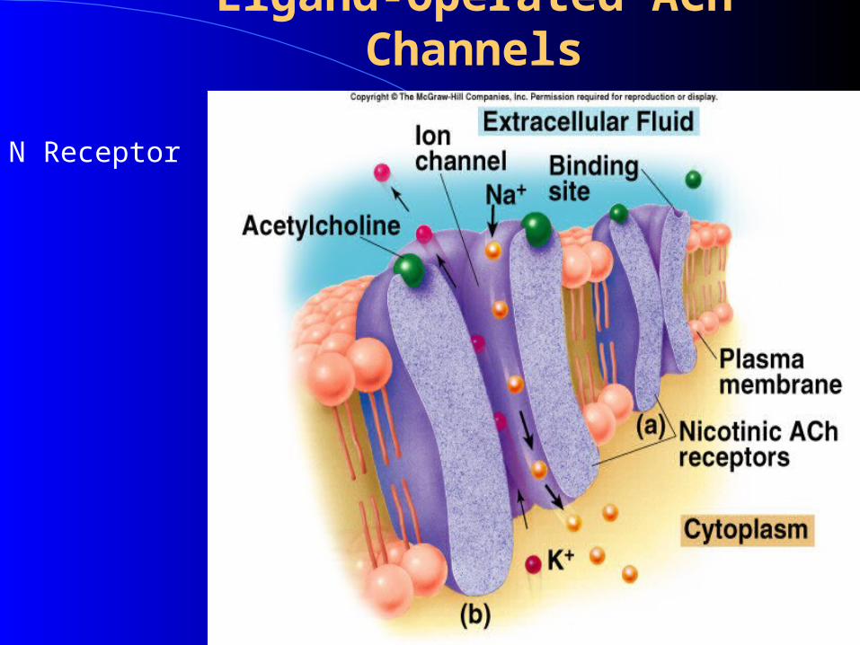
Ligand-Operated ACh Channels
N Receptor
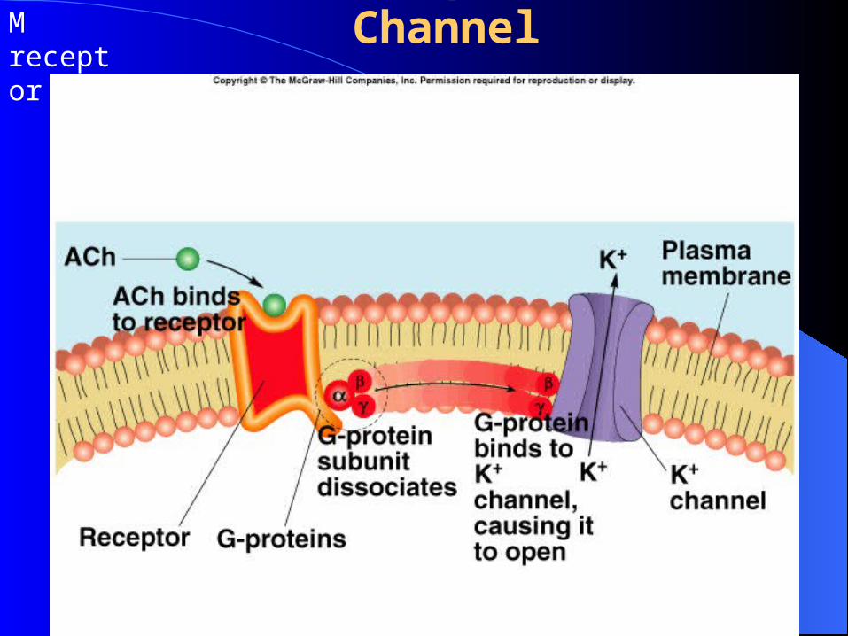
G Protein-Operated ACh ChannelM receptor
(2) Monoamines as NT
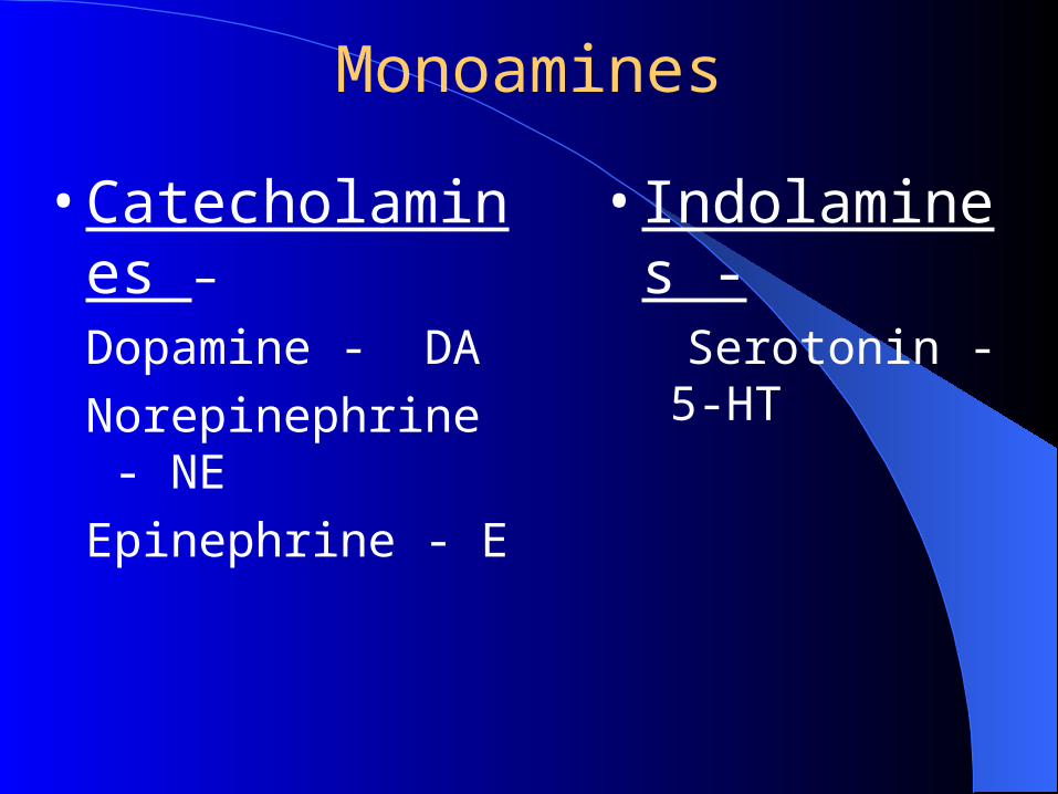
Monoamines
• Catecholamines –
Dopamine - DA
Norepinephrine - NE
Epinephrine - E
• Indolamines - Serotonin - 5-HT
Mechanism of Action ( receptor)
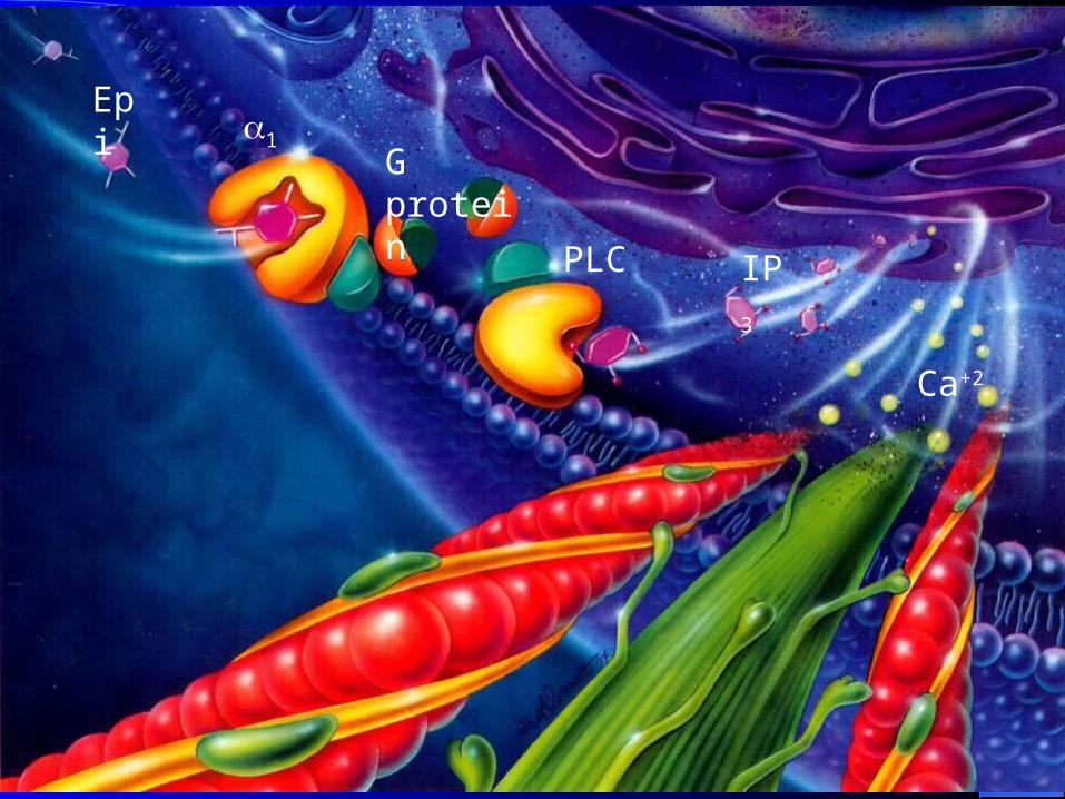
Epi1
G protein
PLC IP3
Ca+2
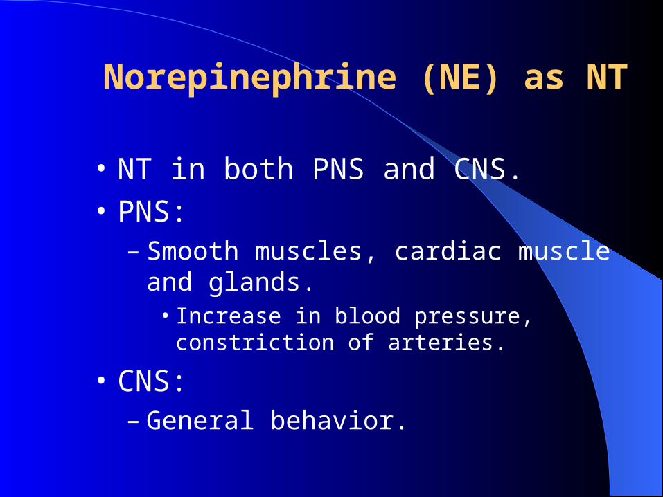
Norepinephrine (NE) as NT
• NT in both PNS and CNS.
• PNS: – Smooth muscles, cardiac muscle and glands.
• Increase in blood pressure, constriction of arteries.
• CNS:– General behavior.
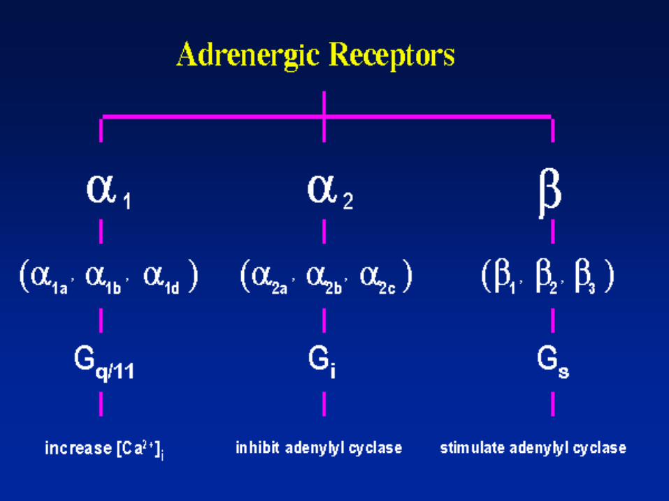
Adrenergic Neurotransmission
1 Receptor– Stimulated by NE, E, – blood vessels of skin, mucosa, abdominal
viscera, kidneys, salivary glands – vasoconstriction, sphincter constriction, pupil
dilation
Adrenergic Neurotransmission2 Receptor
– stimulated by, NE, E, …..– Membrane of adrenergic axon terminals (pre-sy
naptic receptors), platelets– inhibition of NE release (autoreceptor), – promotes blood clotting, pancreas decreased ins
ulin secretion
Adrenergic Neurotransmission
• 1 receptor– stimulated by E, ….– Mainly heart muscle cells, – increased heart rate and strength
Adrenergic Neurotransmission
• 2 receptor– stimulated by E ..– Lungs, most other sympathetic organs, blood
vessels serving the heart (coronary vessels),– dilation of bronchioles & blood vessels
(coronary vessels), relaxation of smooth muscle in GI tract and pregnant uterus
Adrenergic Neurotransmission
• 3 receptor– stimulated by E, …. – Adipose tissue, – stimulation of lipolysis
(3) Amino Acids as NT
• Glutamate acid and aspartate acid:– Excitatory Amino Acid (EAA)
• gamma-amino-butyric acid (GABA) and glycine:– Inhibitory AA
(4) Polypeptides as NT
• CCK:– Promote satiety following meals.
• Substance P:– Major NT in sensations of pain.
(5) Monoxide Gas: NO and CO
• Nitric Oxide (NO)– Exerts its effects by stimulation of cGMP.– Involved in memory and learning. – Smooth muscle relaxation.
• Carbon monoxide (CO):– Stimulate production of cGMP within neurons.– Promotes odor adaptation in olfactory neurons.– May be involved in neuroendocrine regulation in
hypothalamus.