Embed Size (px)
Citation preview

NAP 3Report and findings of the 3rd National Audit
Project of the Royal College of Anaesthetists
Clinical reviews:Clinical reviews by complication typeClinical reviews by indication
Section 2

NAP 3Report and findings of the 3rd National Audit Project of the Royal College of Anaesthetists
48

NAP 3Report and findings of the 3rd National Audit
Project of the Royal College of Anaesthetists
49
Clinical reviews by complication typeChapter 6Cord ischaemia
from the internal iliac artery and serves the area of the conus medullaris. The radicular arteries also form a plexus within the pia mater, but there are no arterial anastomoses within the cord.
Clinical syndromesThe main patterns of ischaemic injury to the cord are either a global infarction injury, spinal stroke, or more limited lesions related to specific arterial occlusions. The anterior spinal artery supplies the anterior two thirds of the cord and, as an end artery, is at risk of damage from a number of causes, so giving rise to the anterior spinal artery syndrome. The characteristic symptoms are motor weakness and loss of bowel and bladder function, with some loss of spinothalamic tract function (pinprick and temperature sensation) because the motor and spinothalamic tracts are within the anterior two thirds of the cord. The dorsal columns, transmitting proprioception and sensation are largely spared, although they can be affected in some cases, especially in the acute phase. The initial period of spinal shock with flaccid motor paralysis is usually followed by some return of muscle tone with varying leg flexor muscle weakness and increased tendon reflexes. Unilateral infarction, with a partial Brown-Sequard syndrome is possible, and another variant is conus medullaris infarction
HeadlineSpinal cord infarction is one of the most devastating neurological complications encountered after thoracic, abdominal and pelvic surgery, although it is fortunately rare. Central Neuraxial block (CNB) used as part of the anaesthetic technique may be implicated, but it can be difficult to decide whether the injury has occurred as a result of the block or is due to other perioperative factors. Six cases of spinal cord ischaemia were reported, but two were excluded from incidence calculations due to a lack of evidence that the CNB had been a contributory factor. All four of the included cases had a poor outcome, with permanent motor and/or sensory dysfunction resulting from spinal cord infarction.
What we know already
Anatomical backgroundThe blood supply of the spinal cord is complex, but one anterior, and two posterior, arteries run along its whole length, fed by radicular arteries entering the vertebral canal at each intervertebral foramen. One of these radicular arteries (usually on the left) in the low thoracic or high lumbar region (the artery of Adamkiewicz) is larger than the others and provides a large proportion of the blood supply to the anterior spinal artery in that area. The variable artery of Desproges-Gotteron arises
Barrie Fischer
Chapter 6: Spinal cord ischaemia
with expert comment fromMax Damian

NAP 3Report and findings of the 3rd National Audit Project of the Royal College of Anaesthetists
50which causes ‘saddle’ anaesthesia and sphincter paralysis with variable distal weakness. The posterior spinal artery syndrome typically causes prominent proprioceptive sensory loss with a variable degree of motor and sphincter involvement. It is rare in comparison with anterior spinal artery syndrome.
Aetiology and pathogenesisSpinal cord blood flow depends on perfusion pressure: arterial pressure minus both tissue and venous pressures. A decrease in arterial pressure or increases in the other two can reduce perfusion pressure below critical levels to cause ischaemic damage and infarction within the cord. Patients with arterial atheroma will, inevitably, have an increased risk of impaired perfusion compared to those without atheroma. Other factors implicated in spinal cord ischaemia include systemic hypotension, surgical procedures involving aortic cross clamping, retroperitoneal or paravertebral dissection, spinal surgery, Diabetes mellitus, cigarette smoking and cocaine misuse.1,2 Thoracotomy is a recognised risk because of the possibility of embolisation or surgical injury involving one or more radicular arteries.3 Extrinsic cord compression by spinal canal tumour, prolapsed intervertebral disc or epidural
haematoma has also been reported to cause cord ischaemia. Case reports highlight the risks of surgical positioning especially the use of prolonged hyperlordosis (‘jack-knife’ position) favoured by surgeons undertaking major pelvic surgery.4–6
The role of CNBCase reports also implicate epidural block as a risk factor,7–9 with three concerns relating to this. The first is causative (and equally affects spinal anaesthesia), namely that inadequately managed sympathetic nerve block can lead to severe hypotension and cord ischaemia. However, there are no data defining either the threshold pressure or its duration for increasing the risk of spinal cord injury. Perfusion pressure is critical, but while CNB can influence the supply side (mean arterial pressure), venous drainage is more influenced by surgical positioning and local patient factors such as decreased spinal canal compliance (excess epidural adipose tissue, spinal canal stenosis) and decreased venous drainage through the Azygos system. Thus simply maintaining an adequate arterial pressure may not be sufficient to prevent the anterior spinal artery syndrome developing
The second concern is that rapid injection of fluid into the epidural space causes a transient increase in both epidural and CSF pressures, but even with relatively large volumes this dissipates rapidly.10–11 Whether such increases are clinically important (particularly in the presence of spinal stenosis or epidural fibrosis) or relevant during epidural infusions is unknown.
Finally, there is concern that the recognition of a possible problem in the postoperative period may be delayed because the neurological signs and symptoms of cord ischaemia are wrongly attributed to continuing epidural infusion or to the delayed offset of a spinal anaesthetic.9
Prevention and managementThere is very limited scope for the anaesthetist in reducing the likelihood of cord ischaemia
Elderly patients receiving postoperative epidural analgesia were the group most at risk of spinal cord ischaemia
Clinical reviews by complication type
Chapter 6Cord ischaemia

NAP 3Major complications of
central neuraxial block in the UK
51
and one severe respiratory impairment) but only one had documented atheromatous disease and one hypertension.
Perioperative hypotension was reported in only two cases.
Presentation was with weak legs in all cases. An epidural infusion was used to provide postoperative analgesia in four patients for up to four days. In three of these patients and the patient with a single shot caudal, dense motor weakness (one had only significant numbness) was noted in the legs at an early stage. In two cases, leg weakness was noted to improve when the epidural infusion was stopped or reduced on the first postoperative day, but worsened again when the infusion was restarted. Diagnosis was rapid after the caudal-associated
occurring, other than being clear that the indication for the CNB is appropriate, ensuring that the circulation (particularly the arterial blood pressure) is managed properly throughout, and persuading the surgeon to avoid positioning the patient in extreme extension.
Early diagnosis will, unlike the situation with haematoma and abscess, have little impact on outcome so it is not a specifically relevant issue here. To a degree this is because there is no definitive treatment for established spinal cord ischaemia other than surgical intervention when extrinsic compression is thought to be the precipitating cause. Further, the capacity of MRI to demonstrate cord infarction itself is limited.12
Case reviewA total of six patients with spinal cord ischaemia were reported to the project. One case followed CNB performed outwith the time limits of the audit. One very elderly patient with ischaemic heart disease made a full recovery from a perioperative spinal block and then, at least 12 hours later, developed sudden leg weakness due to spinal cord ischaemia. There had been brief hypotension per-operatively but none postoperatively. MRI scan showed a lesion consistent with ischaemia in the upper/mid thoracic region. This case was considered to be an incidental spinal stroke and judged not caused by CNB. Both these cases were therefore excluded from incidence calculations but the former case is included in the review of clinical features.
Of the five patients with CNB-associated spinal cord ischaemia all occurred after perioperative CNB (four thoracic epidurals and one caudal all performed by consultants). Two patients were elderly, two middle aged and one young. Four patients were judged to be ASA 3 or above including the young patient who was ASA 4. All patients except one underwent elective major surgery and all had significant co-morbidities (two cancer, two use of corticosteroids, one diabetes mellitus, one end stage renal failure
Case 1A middle aged patient received a low thoracic epidural for major thoraco-abdominal surgery. Motor weakness was noted on the first postoperative day and the epidural infusion was stopped, with some return of motor power on day 1. The infusion was restarted due to difficult pain control and the weakness continued until the patient was reviewed on day 4. There was some sparing of proprioception but persistent weakness and sensory loss. An MRI on day 5 was assessed as normal except for minor signal changes in the low thoracic area of the spinal cord. Cord ischaemia was thought to be the most likely cause, with surgical positioning implicated as a causative factor.
At six months the patient remained wheelchair dependant and paraplegic. Clinical features included a thoracic sensory level, sparing of proprioception and considerable neuropathic pain and dysaesthesia.
The case was included in the pessimistic interpretation of permanent harm from CNB but excluded on optimistic analysis. Outcome was judged to be permanent paraplegia.
Clinical reviews by complication typeChapter 6Cord ischaemia

NAP 3Report and findings of the 3rd National Audit Project of the Royal College of Anaesthetists
52
case but took up to three days in each of the other cases.
MRI scan was performed in all cases within 24 hours of a major complication being suspected. In all cases the MRI scan excluded cord compression but in only one did it show definitive signs of spinal cord ischaemia. Two patients has spinal stenosis.
One patient died within three weeks of surgery, but from causes unrelated to the spinal cord ischaemia. There was limited recovery of function in three patients after a period of rehabilitation, but persistent motor and sensory deficit in the other, and all four surviving patients remained unable to walk unaided at six months.
Quantitative aspectsFour cases of spinal cord ischaemia were included in the audit and all lead to permanent harm. The incidence is therefore 4 in 707,425 or approximately 1 in 170,000 (0.57 in 100,000 cases, 95% confidence interval 0–1.5). As all cases were excluded on optimistic
interpretation (the link between the CNB and the ischaemia being merely assumed) the optimisitic incidence is 0 (95% CI 0–0.5).
As all cases occurred after perioperative epidural the pessimistic incidence in this group is 4 in 97, 925 or approximately 1 in 24,500 (4.1 in 100,000, 95% CI 1.1-10.6).
CommentThe diagnosis of spinal cord ischaemia is mainly one of exclusion (haematoma, abscess and direct spinal cord injury) because there may be no diagnostic findings on MRI. Difficulty arises in trying to ascertain whether any episodes of perioperative hypotension are relevant. Given the rarity of the condition and the possibility that it can occur in the absence of CNB, it is difficult to draw any firm conclusions about the risks of spinal cord ischaemia and CNB. The cases reported to this project appear to confirm that spinal cord ischaemia associated with CNB is very rare, but there is no way of determining the possible role of the regional block in the subsequent development of cord ischaemia.
Case 2A middle-aged very unfit patient received a high thoracic epidural for lung surgery and an initial bolus of 0.5% bupivacaine was administered in theatre. There was no per-operative hypotension. On admission to recovery, there was an episode of severe hypotension requiring extensive treatment. Four hours after arrival in recovery the patient complained of weak legs and examination confirmed dense bilateral motor and sensory block (‘like a total spinal’). It was not clear if motor weakness was present before this. An epidural infusion was started when postoperative pain was reported, but continuing weak legs lead to several anaesthetic reviews. The
epidural infusion was continued for 48 hours. On the third postoperative day a MRI showed no spinal cord injury but noted extensive osteoporosis and lumbar spinal stenosis. Cord ischaemia was diagnosed and the patient treated conservatively, with partial recovery following a period of rehabilitation. The patient, who was barely able to walk pre-operatively, remained unable to walk unsupported. The review panel considered that thoracic surgery might be a confounding or contributory factor in this case.
The case was included in the pessimistic interpretation of permanent harm from CNB but excluded on optimistic analysis. Outcome was judged to be permanent paraplegia.
Clinical reviews by complication type
Chapter 6Cord ischaemia

NAP 3Major complications of
central neuraxial block in the UK
53
Prolonged and severe hypotension risks cord hypoperfusion, but critical thresholds for either cannot be defined. Active avoidance and effective management of perioperative hypotension will minimise risk, particularly for patients with risk factors for cord ischaemia. This requires strategies to prevent, identify and manage hypotension in all patients receiving epidural infusions, especially the elderly and those known to have hypertension or vascular disease.
An elderly patient undergoing major pelvic surgery in the hyperlordotic position in whom a perioperative epidural is used includes most of the recognised risk factors. Careful planning and communication with the surgeon should help to minimise the duration and impact of these risks.
The four cases reported during the data collection period all received an epidural as part of their perioperative management. There is no equivalent data collection for cases of cord ischaemia occurring in patients who have received general anaesthesia without epidural. It is therefore not possible to comment on the relative risks of cord ischaemia happening in association with a CNB compared to a general anaesthetic alone.
In several cases weak legs were assumed, for several days, to be due to epidural local anaesthetic, despite the epidural being placed in the thoracic level. In addition when patients were reviewed, and epidural infusions temporarily stopped, it appears that recurrence of leg weakness on restarting the infusion did not lead to further review or investigation. The reality is that in the case of spinal cord ischaemia these omissions would have little impact on outcome, but such inaction does prevent detection of treatable complications (vertebral canal haematoma and abscess) and may lead to avoidable harm. This topic is discussed further in Chapter 15: Management of dense motor block following CNB or during continuous epidural analgesia.
Learning pointsThe incidence of spinal cord ischaemia is low◆◆
Patients most at risk tend to be elderly and/◆◆
or infirm and undergoing major surgery.
Epidural infusion can complicate the early ◆◆
diagnosis of spinal cord ischaemia if clear policies are not followed (see Chapter 15: Management of dense motor block following CNB or during continuous epidural analgesia)
The data reported to the project do not allow ◆◆
us to state with certainly whether the CNB performed before the development of spinal ischaemia was causative or co-incident.
Hypotension is likely to be causative or ◆◆
contributary and should be prevented, diagnosed early and treated promptly.
Case 3A young, unfit patient who was normally dialysis dependant and who’s normal systolic blood pressure was <100 mmHg underwent minor surgery. The patient also had a pre-existing undefined neurological condition and other co-morbidities. Immediately postoperatively pain was impossible to control with systemic analgesia and several hours later a caudal epidural was uneventfully placed by a consultant. The patient was hypotensive before the caudal but this worsened considerably after it. An unexpectedly high block developed over the next two hours. Various diagnoses were considered including spinal cord ischaemia. MRI performed on day 2 (and day 24) was normal. The patient made a partial neurological recovery over the next few days but this was incomplete. Follow-up was incomplete but at one month motor weakness persisted.
The case was not certainly one of spinal cord ischaemia but it was included as such. Final outcome was pessimistically judged as paraplegia.
As the case occured outside the time limits of the audit it was excluded from incidence calculations.
Clinical reviews by complication typeChapter 6Cord ischaemia

NAP 3Report and findings of the 3rd National Audit Project of the Royal College of Anaesthetists
54
In all reported cases, there was inappropriately ◆◆
dense motor and/or sensory loss in the lower limbs. Thoracic epidural blockade should provide segmental blockade of the chest and abdomen, with minimal spread to the lumbar nerve roots. Therefore dense motor block of the legs should always be considered as a warning sign and the patient reviewed closely (see Chapter 15).
In two cases the epidural infusion was ◆◆
stopped, but restarted when lower limb power had only returned partially, leading to a delay in diagnosis. This was also observed in patients who presented with vertebral canal haematoma (see Chapter 7: Vertebral canal haematoma and Chapter 15).
In any circumstances where spinal cord ◆◆
ischaemia (or other major neurological complication) is being considered a senior opinion should be sought with a view to urgent MRI scanning. Decisions should involve both anaesthetists and neurologists. Although cord ischaemia has limited potential for recovery and no specific treatment, it is important to investigate without delay to exclude other causes of spinal cord injury that may be treatable if diagnosed in their early stages (i.e. abscess and haematoma).
MRI scans may show no changes in the ◆◆
spinal cord, particularly early in the evolution of the condition.
The prognosis of patients with spinal cord ◆◆
ischaemia was universally poor in this series, though disability was less at six months than at presentation.
ReferencesCheshire WP et al. Spinal cord infarction: etiology and 1 outcome. Neurology 1996;47:321–330.
Weidauer S et al. Spinal cord infarction: MR imaging 2 and clinical features in 16 cases. Neuroradiology 2002;44:851–857.
Raz A et al. Spinal cord ischemia following 3 thoracotomy without epidural anesthesia. Can J Anaesth 2006;6:551–555.
Cheney FW et al. Nerve injury associated with 4 anesthesia. A closed claims analysis. Anesthesiology 1999;90:461–470.
Beloeil H et al. Bilateral lower limb hypoesthesia after 5 radical prostatectomy in the hyperlordotic position under general anesthesia. Can J Anaesth 2003;7:653–656.
Amoiridis G et al. Spinal cord infarction after surgery 6 in the hyperlordotic position. Anesthesiology 1996;84:228–230.
Urquhart-Hay D. Paraplegia following epidural 7 analgesia. Anaesthesia 1969;24:461–470.
Yoshida S, Nitta Y, Oda K. Anterior spinal artery 8 syndrome after minimally invasive direct coronary artery bypass grafting under general combined epidural anesthesia. Jpn J Thorac Cardiovasc Surg 2005;53:230–233.
Linz SM et al. Spinal artery syndrome masked by 9 postoperative epidural analgesia. Can J Anaesth 1997;44:1178–1181/
Usubiaga JE et al. Effect of saline injections on epidural 10 and subarachnoid space pressures and relation to post-spinal anesthesia headache. Anesth Analg 1967;46:293–296.
Ramsay M, Roberts C. Epidural injection does cause an 11 increase in CSF pressure. Anesth Analg 1991; 73: 668
Novy J et al. Spinal cord ischaemia: clinical and 12 imaging patterns, pathogenesis and outcomes in 27 patients. Archives of Neurology 2006;63:1113–1120.
Clinical reviews by complication type
Chapter 6Cord ischaemia

NAP 3Report and findings of the 3rd National Audit
Project of the Royal College of Anaesthetists
55
Clinical reviews by complication typeChapter 7Haematoma
Chapter 7: Vertebral canal haematoma
HeadlineEight cases of vertebral canal haematoma (VCH) were reported, including two patients not meeting the audit’s inclusion criteria and one making a full recovery. Therefore five cases of VCH were included in calculations of incidence of permanent harm. All eight cases were reviewed for leaning points: all were associated with postoperative epidural block, seven in patients older than 70 years and five in women. Seven patients had received drugs affecting coagulation, but technical difficulty with the block was an obvious factor in only one. Delay in diagnosis occurred in four because of a failure to appreciate the significance of leg weakness or numbness, and other, organisational factors delayed management as well. Only one patient made a complete recovery, reaction to the early features of the haematoma being very prompt.
What we know alreadyOf all the complications of regional anaesthesia, VCH is, perhaps, the most feared because paraplegia will result if it is not diagnosed and treated within 12 hours.
Spontaneous VCHVCH is a rare condition which occurs ‘spontaneously’, a review of 13 cases from one centre estimating the incidence to be one per
million of the population per year.1 Four of these 13 patients had received anticoagulant drug therapy and five had sustained minor trauma, but no risk factors were apparent in the remaining four. Another review of spontaneous cases found that 25% were associated with a clotting ‘disorder’: drug induced, acquired or congenital.2 Disorders of coagulation have long been considered to contraindicate central nerve block (CNB) techniques, although reviews of the literature performed some years ago found more reports of spontaneous cases than the numbers which give rise to concerns about anaesthetic practice.2,3
VCH associated with CNBThe factors associated with VCH occurring after CNB were best identified in a review of case reports published between 1906 and 1994.4 Of 61 patients, 42 were identified as having a ‘disorder’ of coagulation. In 30, a heparin-type drug had been administered, and a variety of factors were identified in the other 12: chronic alcohol abuse, chronic renal failure, and therapy with antiplatelet or other anticoagulant drugs. Four patients had obvious anatomical abnormalities affecting the spinal cord or column. There was also a high incidence of problems with the block, this being technically
Professor Tony Wildsmith
Dr Tim CookDr Nick Scott

NAP 3Report and findings of the 3rd National Audit Project of the Royal College of Anaesthetists
56
difficult in 15, bloody in 15 and requiring multiple punctures in 12. A spinal anaesthetic had been administered in 15, with the other 46 having an epidural, a catheter being inserted in 32 of these. Taking these last two points together with the first does imply that both tissue disruption (‘trauma’) and coagulation impairment are implicated in causation. A final observation of note from this review was that the haematoma developed immediately after catheter removal in 15, nine of these patients receiving therapeutic amounts of heparin at the time. In a separate series of 40 VCH 50% were considered to have occurred at the time of epidural catheter removal.5 The association with epidural catheters raises concern about obstetric practice, but there was only one
VCH in an audit of 505,000 women receiving CNB for delivery in the UK,6 and a more recent metaanalysis put the incidence in obstetric patients at 1 in 168,000.7
The rarity of VCH and the clear implications from the cases described in the literature allowed the provision of straightforward advice to clinicians on using CNB in patients receiving drugs having an affect on coagulation.8 However, the introduction of Low Molecular Weight Heparins (LMWH), which should have been ‘safer’ for CNB use than unfractionated heparin,9 resulted in an increase in concern. This was due to an increase in the incidence of VCH in the USA, was related only to enoxaparin (with an incidence of 1 in 14,000), and was not mirrored in Europe (incidence much less than 1 in 1,000,000).10 Eventually, the major factor was found to be a trans-Atlantic difference in the dosage of enoxaparin, although many important lessons were learned (often re-learned) from review of the cases:9–11 the elderly (especially females) are at particular risk, probably because slower metabolism results in drug accumulation; combinations of drugs are often synergistic in their effect on coagulation; poor communication can lead to problems; epidural block is associated with a higher incidence than spinal anaesthesia; and epidural catheter removal is a time of high risk. One important new lesson from these cases was that patients with perioperative VCH do not present with the classic feature of severe radicular back pain, but lower limb weakness or numbness.
The natural reaction to such problems is to take an extreme position, either to avoid CNB use in patients who are to receive these drugs, or to deny the patients effective thromboembolic prophylaxis. However, individual patients may not be well served by such extremes, and there are good sources of information available to guide practice in this area.12,13 These should be
Lumbar vertebral canal haematoma
Clinical reviews by complication type
Chapter 7Haematoma

NAP 3Major complications of
central neuraxial block in the UK
57
used as the basis for local hospital guidelines which must not only advise anaesthetists on their decision making and clinical practices, but also provide information for the other staff, surgical and nursing, who are involved in perioperative care of these patients. Such guidelines should be updated regularly in the light of local experience and new information in the literature, and particularly to take into account the challenges presented by the development of new antiplatelet drugs14 and changes in guidance on perioperative thromboprophylaxis.15
A major issue leading to permanent patient harm in the past has been delay in the diagnosis and or drainage of a haematoma, this being the subject of a recent review.16 The safe management of CNB must include the capability to detect and treat rare, but major, complications rapidly and the two broad requirements for this are that:
1 The guideline documents mentioned above must include both advice on monitoring the patients for early signs of problems and a reporting system for seeking anaesthetic input.
2 The definitive investigation, magnetic resonance imaging (MRI), and expert neurological advice must both be available.
Case reviewEight cases of VCH were reported, but three were excluded from calculation of the incidence of permanent harm: one was outwith the time period of the project; one occurred in a non-NHS hospital; and one patient made a full recovery from a small haematoma. All eight patients have been reviewed for learning points, but perhaps the most notable factor was that each one had an epidural catheter inserted for the management of postoperative pain. Not one VCH was reported after approximately
Case 1An elderly patient who normally took warfarin for atrial fibrillation underwent pelvic surgery for malignancy. Warfarin was stopped three days before surgery and daily enoxaparin was substituted. The INR was mildly prolonged. A low thoracic epidural was inserted without complication by a consultant anaesthetist and an epidural infusion continued for 48 hours postoperatively. The epidural catheter was removed eight hours prior to restarting warfarin, while enoxaparin was continued. Eight hours later the patient reported back pain, and motor weakness in one leg (power 3/5) was recorded. A junior surgeon assessed the patient but no further action was taken for more than 12 hours. An anaesthetic consultant reviewed the patient and decided that, despite marked right lower leg paresis
and reduced sensation, the persisting unilateral symptoms were unlikely to be due to epidural haematoma. Symptoms persisted and MRI scan was performed more than 12 hours later, confirming vertebral canal haematoma. At this time the INR was very prolonged. The patient was treated with vitamin K and referred to a neurosurgical centre for urgent spinal decompression. Transfer was delayed for several days due to lack of available beds at this tertiary centre (and several others centres also contacted). Decompression occurred seven days after onset of neurological symptoms. Six months later there was some recovery, but the patient remained unable to mobilize without assistance.
The case was included in both pessimistic and optimistic calculations of incidence of permanent harm.
Clinical reviews by complication typeChapter 7Haematoma

NAP 3Report and findings of the 3rd National Audit Project of the Royal College of Anaesthetists
58
360,000 spinal anaesthetics, or in over 300,000 obstetric, 40,000 chronic pain or 20,000 paediatric patients. The other features of the eight patients are as follows:
Seven were over 70 years of age, the other ◆◆
over 50 years, and five were female;
Seven had significant co-morbidities, ◆◆
including atheromatous disease in five, and six patients were undergoing surgery for malignancy;
They all underwent elective surgery, major in ◆◆
seven and intermediate in one; and
Seven were reported to have received ◆◆
drugs interfering with coagulation (LMWH or aspirin) at the time of epidural catheter insertion and removal. Two received warfarin postoperatively.
Technical difficulty (implying trauma) does not seem to have been a general issue in the performance of the epidurals:
All were performed by career grade staff, ◆◆
six of them consultants, but the aseptic technique was incomplete in half (see Chapter 8: Vertebral Canal Abscess);
Five were inserted in the thoracic region (one ◆◆
‘high’, three ‘mid’ and one ‘low’) and three were lumbar (two for lower limb surgery, but one for gastrectomy);
Six were sited at the first attempt, one ◆◆
required two attempts and one three attempts. In this last instant blood was later aspirated from the catheter which was re-sited in the early postoperative period.
All eight patients received a continuous infusion of local anaesthetic (with or without opioid), and evidence of the VCH appeared in the early postoperative period, the latest presenting four days after surgery (one day after catheter removal). Other features noted were:
Three (possibly four) presented, and seem to ◆◆
have occurred, after removal of the epidural catheter;
After their first appearance, symptoms ◆◆
progressed rapidly in all patients;
Seven patients (five of them with a thoracic ◆◆
catheter) presented with leg weakness (unilateral in two), three with sensory symptoms; and
Only two patients complained of back pain.◆◆
Delay in clinical diagnosis occurred in four of the seven cases in which this could be assessed:
In two patients leg weakness led to ◆◆
suspicions of a complication so the epidural infusion was stopped. Motor function recovered partially and the infusion was restarted without any apparent increase in surveillance. Profound motor block recurred and did not raise further concern; there was delay in diagnosis of greater than 24 hours and the outcome was poor in both patients (e.g. case 1);
In one of the two patients with unilateral leg ◆◆
weakness the one sided nature of symptoms delayed diagnosis considerably;
Delay also occurred in several cases when ◆◆
motor weakness was referred (out of hours) to (non-anaesthetic) junior staff who did not appreciate its significance so that anaesthetic
Case 2An elderly, but healthy patient taking regular aspirin underwent upper abdominal surgery with a lumbar epidural placed uneventfully. No information was provided on the use of perioperative thromboembolic prophylaxis, but the patient was noted to be ‘oozy’ during surgery although coagulation tests were normal (before and afterwards). On the first postoperative day the acute pain team noted increasing motor block and some ‘ooze’ at both the epidural and venepuncture sites. The epidural infusion was stopped and an urgent MRI (performed six 6 hours later) showed a small epidural haematoma without compression. Fresh frozen plasma was given empirically, laminectomy was not performed and the patient made a full recovery.
Clinical reviews by complication type
Chapter 7Haematoma

NAP 3Major complications of
central neuraxial block in the UK
59
staff were not informed until the following day; and
Senior anaesthetists made the error of ◆◆
ignoring inappropriate or profound motor weakness on occasion.
Organisational issues led to further delays, there being instances of inability to obtain a senior neurological opinion promptly, unavailability of MRI imaging out of hours or at weekends, and lack of a bed at the tertiary referral centre. At its worst, delay led to decompressive surgery being performed seven days after the onset of symptoms and left the patient with permanent deficit (Case 1). In direct contrast, immediate reaction by an acute pain team to the very early features of a haematoma resulted in prompt diagnosis and treatment (Case 2), this being the only case of haematoma reported to the audit from which the patient made a complete recovery.
Quantitative aspectsThe incidence of VCH in this audit was 6 in 707,425 CNB (0.85 per 100,000, 95% confidence interval 0–1.8 per 100,000, 1 in 117,000), with permanent neurological deficit occurring in 5 in 707,425 on a pessimistic interpretation of the data (0.7 per 100,000, 95% CI 0–1.7 per 100,000, 1 in 140,000). Four of the five cases were also included on optimistic interpretation.
However, all the VCHs occurred in patients receiving a perioperative epidural so the incidence of permanent harm in that group was 5 in 97,925 (5.1 per 100,000, 95% CI 1.7–11.9, 1 in 19,500,).
Although no reports of VCH after CNB for other indications were received, there are relevant reports in the literature.
CommentThe absence of VCH after >360,000 spinal injections is reassuring, as is its absence after all CNBs inserted for obstetric, chronic pain and paediatric indications. However, these zero
numerators do not imply that there is no risk in these circumstances, and readers are referred to the chapter on quantitative aspects for clarification (see Chapter 5: Discussion in Section 1 – Quantitative results).
Conversely, the occurrence of six VCH after approximately 100,000 perioperative epidurals is a concern, particularly because all occurred after elective surgery and diagnosis was frequently delayed despite the appearance of recognised clinical features in all cases. The outcome of patients reported to this project as developing VCH was particularly bad, with five of six left with permanent impairment of mobility and sensation. A developing epidural haematoma is a clinical emergency requiring immediate recognition, investigation and treatment.
Co-administration or mistiming of drugs which interfere with coagulation at the time of CNB performance or epidural catheter removal is a well recognised risk factor for VCH. In April
Traumatic CNB is a risk factor for vertebral canal haematoma
Clinical reviews by complication typeChapter 7Haematoma

NAP 3Report and findings of the 3rd National Audit Project of the Royal College of Anaesthetists
60
2007 the National Institute for Health and Clinical Excellence (NICE) issued guidance on the prevention of thromboembolic disease.15 This recommends formal risk assessment in all surgical patients and the use of perioperative low molecular weight heparin (or fondaparinux) for those identified as at increase risk and also for all orthopaedic patients. Those at ‘increased risk’ include all over 60 and patients with cancer, heart or lung disease. NICE also advocates the use of regional anaesthesia to reduce the risk of thromboembolism. The likely increase in the use of thromboprophylaxis, and of longer acting drugs (e.g. fondaparinux) suggest that extra vigilance with CNB, perhaps including a re-appraisal of the indications as well as strict adherence to protocols, will be required to avoid an increase in VCH. The same is implied by the greater use of new, long-acting anti-platelet drugs such as clopidogrel in the management of percutaneous angioplasty and cerebrovascular disease.14
Five of the eight VCHs occurred after a thoracic level epidural block, and it seems likely, from clinical indications alone, that fewer thoracic level blocks are inserted than lumbar in the UK. Thus, the figures could be taken to imply a greater incidence of VCH after thoracic block, especially as insertion at that level is more difficult technically and might result in more tissue ‘trauma’, although this was so in only one patient reported here. A small haematoma in the thoracic epidural space will lead, fairly quickly, to spinal cord compression whereas
displacement of the greater volume of CSF might ‘buffer’ the effect initially at lumbar level. However, each of these points is somewhat speculative and the number of cases is very small. There may be other, confounding factors in patients who require thoracic epidurals, the obvious ones being that all of the cases (lumbar and thoracic) occurred in elderly patients undergoing high risk surgery. The difficulty is, of course, that the rarity of the complication makes it quite impossible to study such factors objectively.
The incidence of VCH (reported here and in the literature) is greater after epidural than spinal block, and this would support a general assumption that needle size is a factor, although there is little, if any, specific evidence on this. A larger gauge needle will cause more tissue disruption and appear to increase the risk of bleeding, but the issue is complicated by the insertion of a catheter technique on most occasions when an epidural is used. A 16G needle was used in six of the cases described here, and it was unspecified in two. Whether the use of a smaller gauge needle and catheter system (e.g. 18G) would reduce the incidence of VCH is also something which would be almost impossible to prove. Further, what little circumstantial evidence there is implies that catheter insertion is the more important factor.17
As is already noted above, and considered elsewhere in this report in regard to other complications, the safe use of CNB (particularly epidural infusions) requires high quality postoperative monitoring of patients. This must include the ability to detect and respond to specific features (progressive weakness and sensory disturbance) in the lower limbs, the clinical data presented here providing further evidence of the necessity for this. Early involvement of senior, experienced clinicians is essential. (see Chapter 15: Management of dense motor block following CNB or during continuous epidural analgesia).
Drugs that interfere with coagulation increase the risk of vertebral canal haematoma
Clinical reviews by complication type
Chapter 7Haematoma

NAP 3Major complications of
central neuraxial block in the UK
61
Learning pointsA developing VCH is a clinical emergency requiring urgent investigation and treatment if patient harm is to be minimised. It is rare and can occur in any CNB setting, but most cases are associated with the use of postoperative epidural analgesia. While the patients reviewed here have not provided any new insights, their details certainly reinforce much that is known already:
Overall, the incidence of VCH is small. In all ◆◆
patients receiving CNB the point estimate of the incidence of permanent harm was approximately 1 in 140,000, and 1 in 20,000 after perioperative epidural block.
All reports of VCH occurred during ◆◆
postoperative epidural infusions, but VCH was not restricted to procedures which were difficult, traumatic or performed by trainees: indeed these were all infrequent;
All patients, except one, who developed ◆◆
VCH also received drugs interfering with the coagulation process. This is a recognised risk factor for VCH and increasing use of such drugs requires careful consideration of the decision to use CNB and its timing. Clear policies on the combination of CNB with thromboprophylaxis should be available at hospital level to guide practice;
Most cases occur in elderly, high risk surgical ◆◆
patients in whom slow drug metabolism may lead to greater than usual effects on coagulation, so reduced dose (or frequency of administration) may be appropriate;
VCH after CNB rarely presents with the classic ◆◆
feature of intense back pain, neurological deficit in the legs being more common. Too often this is (and was in the cases described here) assumed to relate to the effects of local anaesthetic administration. Inappropriate motor weakness, even when unilateral, requires urgent assessment and if appropriate investigation to exclude VCH (see Chapter 15: Management of dense
motor block following CNB or during continuous epidural analgesia);
Early diagnosis requires that epidural ◆◆
analgesic regimens minimise the degree of lower limb nerve block so that the early features of VCH can be better identified;
Staff responsible for the immediate ◆◆
supervision of patients must be made aware of the potential significance of lower limb block and have clear referral instructions so that senior anaesthetic review is quickly available: and
VCH patients, as a group, made the poorest ◆◆
recovery of all those reviewed. The speed of onset and limited time available for intervention require early detection and prompt treatment to prevent permanent harm. When VCH is suspected it must be treated as a limb/life–threatening emergency.
ReferencesHoltas S, Heiling M, Lonntoft M. Spontaneous spinal 1 epidural haematoma: findings at MR imaging and clinical correlation. Radiology 1996;199:409–413.
Groen RJ, Ponssen H. The spontaneous spinal epidural 2 haematoma. A study of the etiology. J Neurolog Science 1990;98:121–138.
Schmidt A, Nolte H. Subdural and epidural 3 haematomas following spinal, epidural or caudal anaesthesia. Anaesthesist 1992;41:276–284.
Clinical reviews by complication typeChapter 7Haematoma
Evacuation of a particularly large acute spinal haematoma

NAP 3Report and findings of the 3rd National Audit Project of the Royal College of Anaesthetists
62
Vandermeulen EP, Van Aken H, Vermylen J. 4 Anticoagulants and spinal-epidural anesthesia. Anesth Analg 1994;79:1165–1177.
Miyazaki M et al. Spinal epidural hematoma after 5 removal of an epidural catheter: case report and review of the literature. J Spinal Cord Disorders and Technology 2005;18:547–551.
Scott DB, Hibbard BM. Serious non-fatal complications 6 associated with extradural block in obstetric practice. Br J Anaesth 1990;64:537–541.
Ruppen W, et al. Incidence of epidural hematoma, 7 infection, and neurologic injury in obstetric patients with epidural analgesia/anesthesia. Anesthesiology 2006;105:394–399.
Wildsmith JAW, McClure JH. Editorial: Anticoagulant 8 drugs and central nerve block. Anaesthesia 1991;46:613–14.
Checketts MR, Wildsmith JAW. Central nerve block 9 and thromboprophylaxis – is there a problem? Br J Anaesth 1999;82:164–167.
Tryba M, Wedel DJ. Central neuraxial block and low 10 molecular weight heparin (enoxaparine): Lessons learned from different dosage regimens in two contienents. Acta Anaesth Scand 1997;41:100–103.
Wysowski DK et al. Spinal and epidural hematoma 11 and low-molecular-weight-heparin. New Engl J Med 1998;338:1774–1775.
http://www.asra.com/consensus-statements/2.html 12
http://www.sign.ac.uk/guidelines/fulltext/62/index.13 html
Checketts MR, Wildsmith JAW. Regional block and DVT 14 prophylaxis. Continuing Educ Anaesth Crit Care Pain 2004;4:48–51.
Venous thromboembolism; reducing the risk of 15 venous thromboembolism (deep vein thrombosis and pulmonary embolism) in inpatients undergoing surgery. NICE clinical guideline 46. National Institute for Health and Clinical Excellence http://www.nice.org.uk/nicemedia/pdf/CG046NICEguideline.pdf.
Meikle J et al. Detection and management of epidural 16 haematomas related to anaesthesia in the UK: a national survey of current practice. Br J Anaesth 2008;101:400–404.
Horlocker TT, Wedel DJ. Anticoagulation and neuraxial 17 block: historical perspective, anesthetic implications, and risk management. Regional Anesth Pain Med 1998;23(Suppl 2):129–134.
Clinical reviews by complication type
Chapter 7Haematoma

NAP 3Report and findings of the 3rd National Audit
Project of the Royal College of Anaesthetists
63
Clinical reviews by complication typeChapter 8Abscess
Professor Tony Wildsmith
Chapter 8: Vertebral canal abscess
and urgent treatment if permanent disability is to be avoided. It occurs ‘spontaneously’, accounting for 0.2–1.2 of every 10,000 hospital admissions and has some well identified risk factors.6
Compromised immunity: Diabetes ◆◆
mellitus (the major risk factor), malignancy, pregnancy, HIV infection, alcoholism/cirrhosis and immuno-suppressive therapy (including cortico-steroids).
Disruption of the vertebral canal: trauma and ◆◆
instrumentation may lead to a haematoma which provides ideal conditions for bacterial growth.
A source of infection: usually ◆◆
haematogenous, but local spread is possible.
Combinations of factors obviously increase ◆◆
the risk and an extremely wide range of organisms has been isolated from abscesses.6
Vertebral canal abscess associated with CNBThe risk factors for epidural abscess related to CNB fall into the same categories, but with some specific aspects to be considered:
Immunity: All of the factors affecting ◆◆
immunity may be seen in patients who receive CNB, but repeated epidural injection of cortico-steroids in chronic pain states adds another group.7
HeadlineSeventeen vertebral canal abscesses were notified although in two the procedure was performed outwith the time limits of the project. The majority of patients had risk factors for the development of an abscess, with prolonged epidural catheterisation being prominent. Presentation was often atypical. Those patients who had signs of local sepsis at the site of the epidural catheter insertion had better outcomes than those who did not, but the significance of this is unclear. Seven of the 15 patients meeting the inclusion criteria made a documented full recovery, but eight did not although some degree of recovery occurred in most during the six months of follow-up. In five of these eight patients an optimistic interpretation of events would suggest that they also recovered.
What we know alreadyFor many years epidural abscess was viewed as almost a theoretical complication of central neuraxial block (CNB),1 with much more attention being focussed on the risk of vertebral canal haematoma.2 However, occasional case reports and, more pressingly, the appearance of some case series3–5 prompted re-evaluation and review.1
Spontaneous vertebral canal abscessEpidural abscess is a rare, but serious medical emergency which requires prompt diagnosis

NAP 3Report and findings of the 3rd National Audit Project of the Royal College of Anaesthetists
64
Disruption: CNB obviously disrupts the ◆◆
vertebral canal and technical difficulty may make it more likely that a haematoma is produced as a nidus for infection, especially if a drug affecting coagulation has been used for thromboprophylaxis. Technical difficulty may also make it more difficult to maintain a strict aseptic technique and so increase the risk of contamination. The needle track provides a pathway for the entry of organisms, and the pathway is kept open if a catheter is inserted. How long such catheters should be left in situ is a matter for debate, but studies quoting a low incidence of epidural infection relate to catheterisation for a maximum of 48 hours.8,9
Source of infection: Although the need for ◆◆
a full aseptic technique might seem self-evident, this does not mean that it is always used even though current professional advice advocates it quite definitively.10
Case reviewThere were 20 reports of a patient developing an epidural abscess after a CNB although two were outwith the time frame of the audit. Another three patients were, on review, found to have primarily local infection at an epidural catheter insertion site and, while there was some hint of central spread, neither abscess nor neurological features developed. One patient developed discitis (but no abscess) which presented four months after a perioperative epidural. After some consideration this case has been included in the abscess group as diagnosis, management and learning points are similar. Therefore 15 patients met inclusion criteria for epidural abscess in the audit period and only these were used in the calculation of incidences of permanent harm. Seven of these 15 patients were documented as making a full recovery. The indications for CNB were: perioperative (including acute pain) patients, 13 (six with permanent harm); obstetric patient, one (with permanent harm); chronic pain patient, one (with permanent harm). Of the
Case 1A patient in late middle-age on long-term steroids had been in hospital for four weeks with pneumonia, bronchiectasis and severe back pain due to vertebral collapse. Opioid analgesia led to respiratory arrest. After extensive discussions the patient was transferred to ICU and an epidural block was instituted with good effect, but leg weakness developed within 24 hours. This persisted on day two, in spite of a reduced concentration of local anaesthetic, and a clear sensory level had developed on day 3. An MRI scan showed an epidural abscess, but the patient refused surgical drainage. Antibiotic therapy, while improving the markers of infection, did not result in any neurological improvement. The patient was discharged from hospital, wheelchair bound, at six months and died shortly thereafter. The features appeared so soon after institution of the epidural as to raise the possibility that the abscess (or perhaps a haematoma) was already present. Alternatively, because there was no surgical confirmation of an abscess, the neurological features might have been a consequence of the pre-existing vertebral collapse. This case was included in the pessimistic incidence of permanent harm, and recorded as an indirect death, but excluded from the optimistic incidence of permanent harm.
Clinical reviews by complication type
Chapter 8Abscess
Multiple attempts at CNB may risk loss of sterility
NAP 3Major complications of
central neuraxial block in the UK
65
perioperative patients ten underwent major surgery (seven elective, three emergency) and three received an epidural for pain relief only (pancreatitis, fractured ribs, vertebral collapse one each).
The details of all 20 patients, particularly the 17 with abscesses, have been reviewed in the search for learning points, both positive and negative, but only the 15 who were within the prospectively defined limits of the audit were used in the calculation of incidence.
The demographics of the 17 were as follows:
7 female, 10 male;◆◆
4 aged 19–50, 13 over 50 years;◆◆
14 epidural catheters (7 mid-thoracic, 3 low ◆◆
thoracic, 4 lumbar of which one involved a combined spinal epidural technique [CSE]), 2 spinal; 1 caudal (without catheter).
Presence of risk factorsMany of the risk factors outlined above were identified positively in the 17 patients who developed an abscess. These were:
Compromised immunity – 12 patients◆◆ : Diabetes mellitus, 4; Malignancy, 4; Immuno-suppressive therapy, 3; Chronic pancreatitis, 2; IV drug abuse, 1; and Pregnancy, 1.
Anti-thrombotic drug therapy – 7 patients◆◆ : Low molecular weight heparin (LMWH), 4; LMWH and non-steriodal anti-inflammatory drug (NSAID), 1; Aspirin & NSAID, 1; and Aspirin and Clopidogrel, 1.
Traumatic procedure (> 2 attempts) – 1 ◆◆
patient: 8 attempts.
Source of infection – 6 patients◆◆ : 4 on antibiotics at the time of the block, 2 not; the organism causing the primary infection was obtained from the epidural abscess in only one (and that in spite of appropriate antibiotic therapy).
Failure of aseptic technique – 5 patients◆◆ : no face mask, 2; no fenestrated drape, 2; neither of these precautions, 1. The wound
dressings used at the catheter entry point ◆◆
were quite varied and there was insufficient information gathered to make any useful comment on these.
Duration of epidural catheterisation◆◆ : 1 or 2 days, 3 patients; 3 or 4 days, 5 patients; 5 or more days, 8 patients; and unspecified, 1 patient.
Although there were no obvious patterns or combinations, there were no risk factors in only four patients, one or two factors in five patients, and three or four factors in eight.
DiagnosisThe classic presentation of an epidural abscess is of back pain, systemic features of infection and progressive loss of neural control of the lower half of the body, but the clinical presentation of the 17 patients reported here was inconsistent with that. Back pain was recorded as an early feature in only nine patients, pyrexia or other
Case 2A patient in late middle-age (with hypertension) underwent a knee replacement under an entirely blameless spinal anaesthetic. Six weeks later the patient presented with low back pain and pyrexia, but no neurological features. An MRI scan showed a lumbar epidural abscess which was drained at laminectomy and the patient made a good recovery. However nine days later the patient developed sudden onset tetraplegia and respiratory failure. The cervical spinal cord was described as ‘normal’ on further MRI scanning, but there was no resolution of features during the next six months. This patient’s initial recovery from the abscess was ‘complete’ and there does not seem to be any direct connection between it and the subsequent tetraplegia which might have been due to a spinal stroke.
The case was included in the pessimistic incidence of permanent harm, and recorded as a paraplegia, but excluded from the optimistic incidence of permanent harm.
Clinical reviews by complication typeChapter 8Abscess

NAP 3Report and findings of the 3rd National Audit Project of the Royal College of Anaesthetists
66
clinical features of sepsis in nine, meningism in three, sensory or motor deficit in the legs in four and raised white cell count or C-reactive protein in seven. Poor clinical record keeping and poor reporting of information may both be relevant, but the over-riding impression is of partial and incomplete syndromes, this demonstrating the need for a high index of suspicion for epidural abscess in a patient with any of these features. Seven of the abscesses presented within a week of the institution of the block (the earliest on day two) and another three during the second week, with the longest intervals being six weeks and four months (two patients). Unfortunately this information was not provided in three patients.
An observation of note is that, of the 17 reports of epidural abscess that were received, the nine who made a complete recovery, all had some feature of infection (redness, swelling or pus) noted at the injection site. In addition, the three patients with only subcutaneous infection made a full recovery. In seven of the eight patients who suffered permanent harm there was a clear statement that there was no external evidence of an infection. Staphylococcus aureus was the infecting organism in seven patients, but no other organism was reported more than once in the other ten.
The prevention of permanent harm due to epidural abscess requires that both diagnosis and treatment are instituted as soon as possible, but delay can occur at three stages: considering the possibility clinically; arranging definitive diagnosis by MRI scanning; and then seeking a neurosurgical opinion for advice on treatment. Delayed clinical diagnosis was a factor in eight patients: two in the sub-group who suffered permanent harm (both had back pain with leg symptoms) and six in the group who made a full recovery. In two of these six, the primary presentations were with systemic features of infection and no localising factors so the delay is, to a degree, understandable. However, in the other four patients the delay was in reporting the infection at the injection site to the anaesthetist, but (fortuitously?) all four of these patients required only conservative treatment for their abscesses. Once the possibility of an abscess had been raised, both MRI scanning and neurosurgical opinion were obtained readily except in one case where the scanner was broken, this leading to a 24 hour delay. No delays were reported in arranging laminectomy and surgical drainage if this was thought necessary.
Treatment and outcomeOf the 15 patients meeting project inclusion criteria seven made a documented full recovery. The other eight developed permanent harm if their features are interpreted pessimistically, although the number reduces to three on optimistic interpretation. The final deficit in the eight patients who did not make a complete recovery were: ‘indirect’ death, two patients; tetraplegia, one patient; motor weakness, four patients; and sensory symptoms only, one patient. Even in these patients there was some degree of recovery in the six months of follow up, but three were left with significant lower limb motor deficit.
Traditional teaching is that an epidural abscess requires surgical drainage and prolonged antibiotic therapy although a more conservative
Clinical reviews by complication type
Chapter 8Abscess
Staphylococcus aureus is the commonest infective organism in vertebral canal abscess

NAP 3Major complications of
central neuraxial block in the UK
67
approach involving prolonged systemic antibiotic therapy has developed in recent years.1 This is reflected in this series of reports with only two of the seven patients who later made complete recoveries undergoing laminectomy. Of the eight patients left with permanent disability three underwent laminectomy, but one refused surgery and another was considered to have an abscess too extensive to be amenable to operative treatment. It might be thought that the remaining three patients should have had surgical drainage if they suffered ‘permanent’ disability, but the situation has to be qualified in each case. One elderly patient, who had developed a sacral abscess without neurological features after a caudal block, died from a primary cardiac arrest while in intensive care for a hospital acquired pneumonia. In the other two patients complete recovery was anticipated, but had not been achieved at six months and, for the purpose of this review, residual deficit at six months has been graded as ‘permanent’ and so they must be included in this group.
In addition to the three patients just mentioned, another two of the eight patients who suffered permanent harm were excluded for the calculation of the ‘optimistic’ incidence of permanent harm. An initial reaction might be that all eight should be so included, but the specific (and often complex) situation of each patient has to be taken into account. As noted, it was anticipated that two of them would make a complete recovery, but this had not occurred at six months, and the patient with the caudal abscess developed his pneumonia from ‘unrelated problems’, although it is possible to construct an argument that the abscess should be considered an indirect cause of his death. The other two who were excluded for the ‘optimistic’ calculation were even more complex and are described briefly in boxes as Cases 1 and 2.
In both of theses cases there are features which support the application of the maxim that ‘association does not prove causation’, an important factor in the whole project.
Quantitative aspectsThere were 15 epidural abscesses meeting inclusion criteria (i.e. in the NHS and correct diagnosis). The incidence of epidural abscess in the whole population of the project is 15 in 707,425 or approximately 1 in 47,000 (2.1 in 100,000, 95% confidence interval, 1.2–3.5). Seven patients made a documented full recovery. With a pessimistic interpretation the incidence of permanent harm from abscess is 8 in 707,425, approximately 1 in 88,000 (1.3 in 100,000, 95% CI 1–2.3). The incidence of paraplegia (again on pessimistic interpretation) is 3 in 707,425 or 1 in 236,000 (0.42 in 100,000, 95% CI 0–1.2).
Most abscesses occurred in the perioperative group: in total there were 13, of which 6 (3 epidural, 2 spinal, 1 CSE) suffered permanent harm (pessimistic interpretation). Therefore the incidence of abscess in the perioperative group is 13 in 312,450 or 1 in 24,000 (4.2 in 100,000, 95% CI 2.2–7.2) and the incidence of permanent harm from abscess following perioperative CNB (pessimistic interpretation) is 6 in 312,450 or
Clinical reviews by complication typeChapter 8Abscess
Lumbar vertebral canal abscess (CT scan) with skin marker at the level the epidural was placed
NAP 3Report and findings of the 3rd National Audit Project of the Royal College of Anaesthetists
68
1 in 52,000 (1.9 in 100,000, 95% CI 1–4.2). The incidence of abscess following perioperative epidural was 10 in 92,925 or 1 in 9,800 (10.2 in 100,000, 95% CI 4.9–18.8) and of permanent harm (pessimistic interpretation) 3 in 97,925 or 1 in 33,000 (3.1 in 100,000, 95% CI 1–9.0).
CommentsThe overall clinical features of this group of patients are much as might be expected from information already in the literature.1 The majority were in the sixth or later decades of life and there was a high incidence of risk factors, although it is surprising, even disappointing, that less than half of a group of patients who were at high risk of thrombo-embolic disease had not received pharmacological prophylaxis. It is of some interest that all those patients who made full recoveries from vertebral canal abscesses had features of infection at the catheter entry point.
Unfortunately, such visible evidence did not always lead to early diagnosis so that cannot be the explanation for the lack of permanent harm in this sub-group. It is speculative, but the observation raises the possibility that the
infection was ‘spreading’ out along the needle/catheter track and reducing the build up of pressure within the vertebral canal.
Six of the 17 patients had a systemic bacterial infection at the time of the insertion of an epidural catheter, yet traditionally this has been said to contra-indicate the use of a central block technique. However, it is noteworthy that in only one patient was the same organism responsible for the epidural abscess. It is also very important to recognise the quandary faced by the clinicians. This is well seen in Case 1 above; he was in severe pain and had already suffered a respiratory arrest due to systemic opioid therapy. What other option was available? Of greater concern is that there was clear evidence that a full aseptic technique had not been used in six patients, and no information on this was provided in another, this several years after definitive professional advice had been published.10
The great majority (14 of 17) of abscesses occurred in patients in whom an epidural catheter was inserted. An 18G needle was used in three, and a 16G in eleven, both being much larger than the needles used for spinal anaesthesia today and implying a greater degree of tissue ‘disruption’. This disruption would be increased by the passage of the catheter which would then maintain an open track along which bacteria could spread. It is thus perhaps not surprising that the incidence of abscess was greater after epidural block than spinal. As noted in the introduction, what evidence there is indicates that the lowest incidence of abscess after epidural block is associated with catheters removed within 48 hours, but this period was exceeded in the great majority (13 of 17) of patients considered here. However, the clinical indication (e.g. very severe pain due to pancreatitis or rib fractures) may persist for much longer than 48 hours and justify the extended period of cannulation. Until much more evidence on the incidence of abscess formation with duration of epidural analgesia is available it is impossible to make strictures
Clinical reviews by complication type
Chapter 8Abscess
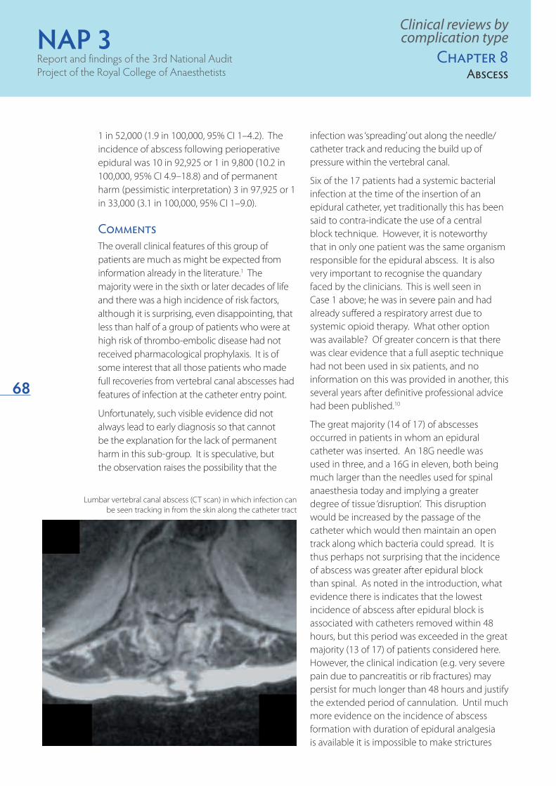
Lumbar vertebral canal abscess (CT scan) in which infection can be seen tracking in from the skin along the catheter tract

NAP 3Major complications of
central neuraxial block in the UK
69
reports. Ugeskrift for laeger 2000;162:5640–5641.
Yin KS, Wang C, Lucero Y. Myelopathy secondary to 5 spinal epidural abscess: case reports and a review. J Spinal Cord Med 1998;21:348–354.
Reihsaus E, Waldbaur H, Seeling W. Spinal epidural 6 abscess: a meta-analysis of 915 patients. Neurosurg Review 2000;23:175–204.
Bromage PR. Spinal extradural abscess: pursuit of 7 vigilance. Br J Anaesth 1993;70:471–473.
Ready LB et al. Postoperative epidural morphine is safe 8 on surgical wards. Anesthesiology 1991:75:452–456.
Schug SA, Torrie JJ. Safety assessment of postoperative 9 pain management by an acute pain service. Pain 1993;55:387–391.
Association of Anaesthetists of Great Britain and 10 Ireland. Infection Control in Anaesthesia. AAGBI, London 2002.
on the ‘maximum’ time over which a catheter may be used. Use for the shortest appropriate period, with daily review of ongoing necessity, seems a sensible minimum guideline.
Learning pointsApart from the apparent association between the presence of superficial evidence of infection and a good outcome, nothing new was learned about vertebral canal abscess, but there is further evidence for issues raised previously:
Vertebral canal abscess may present in very ◆◆
different ways, including with only systemic evidence of infection, so a high level of suspicion is required.
Delay in diagnosis, rather than in subsequent ◆◆
treatment, continues to occur.
A significant proportion of anaesthetists are ◆◆
still not using a full aseptic technique for CNB.
Epidural analgesia may, for good reasons, be ◆◆
required in patients with a number of risk factors for the development of an abscess. These factors may not contraindicate the technique, but should prompt particularly close monitoring of the patient, especially when catheterisation is prolonged beyond 48 hours.
Because an abscess may not present until ◆◆
after discharge from hospital, indeed sometimes several weeks or months later, there is merit in the suggestion that patients should be provided with a letter indicating what features might develop.1 An example is shown in Appendix 2.
ReferencesGrewal S, Hocking G, Wildsmith JAW. Epidural Abscess. 1 Br J Anaesth 2006;96:292–302.
American Society of Regional Anesthesia: Consensus 2 statements on central nerve block and anticoagulation. Reg Anesth Pain Med 1998;23:Supplement 2.
Phillips JM et al. Epidural abscess complicating 3 insertion of epidural catheters. Br J Anaesth 2002;89:778–782.
Wang LP, Hauerberg J, Schmidt JF. Epidural Abscess 4 after epidural catheterization. Frequency and case
Clinical reviews by complication typeChapter 8Abscess

NAP 3Report and findings of the 3rd National Audit Project of the Royal College of Anaesthetists
70

NAP 3Report and findings of the 3rd National Audit
Project of the Royal College of Anaesthetists
71
Clinical reviews by complication typeChapter 9Meningitis
Dr Iain Christie
Chapter 9: Infective meningitis
HeadlineThree cases of bacterial meningitis associated with neuraxial block (one spinal, one epidural and one CSE) were identified during the project. Two occurred in the perioperative setting and one in obstetrics with diagnosis and treatment being prompt in each case. All three patients made a full recovery and so were excluded from calculations of the incidence of permanent harm. Another three patients were reported as having meningitis (one bacterial and two ‘aseptic’), but the evidence was so weak that they were excluded from further consideration.
What we know alreadyIn the last 50 years almost 200 cases of post dural puncture meningitis (PDPM: i.e. meningitis after spinal anaesthesia or diagnostic lumbar puncture) have been reported, including three deaths with around 70% of these cases following anaesthetic procedures.1
Meningitis after central neuraxial block (CNB) is very rare, probably less than 1 in 50,000,1,2 based on retrospective data from other European countries, but this may not reflect UK practice. The risk factors include immuno-compromise (diabetes, steroid therapy, malignancy, alcoholism, HIV infection, IV drug abuse and pregnancy), sepsis and prolonged duration of neuraxial catheterisation, with the bacterial source being exogenous (e.g. contaminated
equipment, and solutions, poor aseptic technique) or endogenous (local or systemic sepsis).3 Interestingly, the pathogenesis appears to be almost technique specific. In most reported cases of meningitis complicating epidural analgesia the causative organism is a skin commensal (e.g. Staphylococcus), suggesting spread along the epidural catheter tract.2,4 After spinal anaesthesia or diagnostic lumbar puncture nasopharyngeal commensals (e.g. Streptococcus) are most often identified, an observation suggesting a causative role for droplet spread from the operator’s airway, with direct inoculation of the organism into the CSF by the spinal needle.1,2 It is increasingly

NAP 3Report and findings of the 3rd National Audit Project of the Royal College of Anaesthetists
72
difficult, therefore, to support the argument against wearing surgical facemasks during spinal anaesthesia.5 Consequently organisations on both sides of the Atlantic now recommend maximal barrier precautions for all neuraxial procedures.3,6,7 Endogenous infection may be associated with bacteraemia so that blood vessel damage during needle or catheter insertion will lead to organisms gaining access to the CSF. The American Society of Regional Anesthesia has recommended that CNB in patients with systemic sepsis should only be performed after appropriate antibiotic therapy has been started.3 The association between duration of epidural catheterisation and risk of vertebral canal abscess is presumed but not proven8 and discussed further in Chapter 8: Vertebral Canal Abscess. Whether such extrapolated evidence is relevant to meningitis is not known.
Chlorhexidine is the antiseptic solution of choice for regional anaesthesia.6 It has a faster onset, greater bactericidal activity and longer duration of action than povidone iodine.
While prevention is crucial, prompt diagnosis and treatment of meningitis reduce morbidity and mortality. Delay can lead to neurological injury,3 and a review of 179 cases after spinal
anaesthesia reported three deaths.2 Meningitis after dural puncture usually presents with severe headache, but the onset of other typical features (e.g. nuchal rigidity, photophobia, pyrexia) may be delayed.2,3 Thus initial differentiation from post dural puncture headache may be difficult and a high index of suspicion is required if treatment is to be started promptly. In contrast, the clinical features of patients developing meningitis after epidural block are usually more typical and diagnosis more straightforward.3,8
Before the advent of disposable equipment chemical (aseptic) meningitis was not unknown after spinal anaesthesia, with contamination with chemical antiseptics or detergents, high concentrations of drug and extremes of solution pH all being blamed.9 Presentation is usually within 24 hours of the procedure with clinical features and CSF findings both typical of bacterial meningitis. Differentiation relies on bacteriological studies of blood and CSF, with antibiotics recommended until the results are available. Outcome is usually good.
Case reviewSix cases of bacterial meningitis were reported, but only three patients met the audit criteria, the other three being excluded because there was little or no evidence to support the diagnosis. In these excluded cases, symptoms were variable and delayed, occurring up to 10 days after the block and without other major clinical features. Lumbar puncture was performed in only one and the results (very minor increase in white cell count, normal protein concentration) did not support a diagnosis of bacterial meningitis. ‘Aseptic’ meningitis was considered a possible diagnosis in two of these excluded patients although the evidence for even this was weak. One patient received an intrathecal catheter after an accidental dural puncture during labour, and then a series of epidural blood patches. An MRI performed because of persistent headache was reported as showing leptomeningitis and a neurologist diagnosed chemical meningitis.
Scrupulous asceptic technique is mandatory for all CNB
Clinical reviews by complication type
Chapter 9Meningitis

NAP 3Major complications of
central neuraxial block in the UK
73
Each of these three patients made a rapid and uneventful recovery and none is considered further.
Two of the cases of bacterial meningitis occurred in the perioperative setting and one in obstetrics. No patient had evidence of pre-existing local or systemic infection and only one had a risk factor (diabetes) for immuno-suppression. The skin was disinfected with chlorhexidine in alcohol in each case.
None of the block procedures was entirely straightforward:
A spinal (Case 1) involved four attempts with ◆◆
the same needle, raising the possibility of an unnoticed breakdown in aseptic technique, repeated passes having been shown not to increase the risk of bacterial contamination of the needle provided the sterile field is maintained.10
A CSE for labour analgesia was followed by a ◆◆
spinal for delivery (Case 2). This patient also had, in effect, multiple procedures, but there are no incidence data to indicate whether this is a frequent occurrence or whether this particular sequence increases the risk of infective sequelae.
An epidural catheter (Case 3) was left in ◆◆
place for nine days. As was noted in Chapter 8: Vertebral Canal Abscess there is no definitive evidence regarding the risk of prolonged catheterisation, but in this patient there were signs of inflammation at the insertion site before meningitis developed.
All three patients in this series presented fairly typically with a combination of pyrexia, headache, meningism and confusion, and the diagnosis was made promptly on the basis of lumbar puncture: CSF showed typical findings in each case, but an organism was identified in only one (E. coli in the epidural associated case). Antibiotics were commenced swiftly in each patient and they all made a rapid and full recovery – there were no neurological sequelae.
Case 1
An elderly patient underwent spinal anaesthesia for joint replacement surgery. The patient had no risk factors for immunocompromise. The spinal was difficult and four attempts were made, but it was otherwise uneventful. Less than 12 hours later the patient developed headaches, vomiting, pyrexia and neck stiffness. At lumbar puncture the CSF was cloudy and showed a raised white cell count, high protein and low glucose. Meningitis was diagnosed and the patient was treated with ceftriaxone and vancomycin for two weeks. No organisms were seen or grown from the CSF. The patient was transferred to critical care, but was well enough to return to the ward the next day and made a full recovery within the next four weeks.
The case was included in the audit, but excluded from calculations of incidence of permanent injury.
Clinical reviews by complication typeChapter 9Meningitis
Several organisms including Streptococcus are implicated in meningitis after CNB

NAP 3Report and findings of the 3rd National Audit Project of the Royal College of Anaesthetists
74
Quantitative aspectsThree cases of bacterial meningitis were reported to the project, giving an overall risk (in this series) of less than 1 in 200,000 CNB. The very small numerators mean the confidence intervals are more relevant than point estimates.
The project incidences of bacterial meningitis were as follows:
following perioperative epidural analgesia*: ◆◆
95% Confidence interval 0–4.9 in 100,000
following perioperative spinal anaesthesia**: ◆◆
95% CI 0–2.7 in 100,000
following obstetric spinal anaesthesia***: ◆◆
95% CI 0–3.5 in 100,000
These figures should be treated with caution as confidence intervals are wide. Similarly those clinical indications where meningitis did not occur cannot be assumed to be free of this risk. While limitations on the validity of the project numerator data are dealt with elsewhere in this report, these figures are still reassuring.
[*epidural here includes all adult perioperative epidurals and CSEs and their complications.**spinal here includes all adult perioperative spinals and CSEs and their complications.***spinal here includes all obstetric epidurals and CSEs and their complications.]
CommentThe data from this project confirms that meningitis after neuraxial procedures is rare. With over 700,000 neuraxial blocks and over 360,000 spinal blocks performed in the audit year11 the incidence of confirmed bacterial meningitis was considerably lower than 1 in 100,000 after such procedures. Outcome should be favourable provided diagnosis is early and management prompt. However, despite the positive findings of this project, it would be wise to avoid complacency: in the three cases in the literature of death following spinal anaesthesia each patient was a healthy parturient.1
Case 3A patient with Diabetes mellitus underwent below knee amputation under general anaesthesia. An epidural catheter was inserted for post operative analgesia and was reviewed daily. On day six the patient developed a surgical wound infection, but the epidural was continued. The wound infection required debridement and critical care admission. On the ninth day the patient became confused, pyrexial and developed neck stiffness. The epidural site was found to be inflamed and the epidural catheter was removed. Neuraxial infection was suspected and an MRI scan demonstrated meningeal inflammation, but no epidural abscess. A lumbar puncture was performed and E. coli isolated from CSF. Antibiotics were commenced and the patient went on to make a full neurological recovery. The case was included in the audit but excluded from incidence calculations because of full recovery.
Clinical reviews by complication type
Chapter 9Meningitis
Case 2A healthy parturient had an uneventful CSE for analgesia in labour. Subsequently she required a Caesarean section and a spinal anaesthetic was administered because the epidural was inadequate. Both blocks were uneventful and were performed by a registrar using a full aseptic technique. After the Caesarean section the patient’s behaviour became inappropriate, but the results of an initial lumbar puncture and CT scan were normal. She was transferred to a tertiary centre where MRI was normal, but a repeat lumbar puncture showed low CSF glucose and raised white cell count. A diagnosis of bacterial meningitis was made and treatment with antibiotics was started. There was no growth from the CSF. She made a full recovery. The case was included in the audit, but excluded from calculations of incidence of permanent injury.

NAP 3Major complications of
central neuraxial block in the UK
75
Learning pointsMeningitis is a rare complication of CNB and ◆◆
in this series had an estimated incidence of less than 1 in 200,000. Prompt treatment led to full resolution in all reported cases.
Where multiple attempts are required for ◆◆
spinal anaesthesia it is essential that asepsis is maintained scrupulously.
Presentation may be atypical and it may be ◆◆
difficult initially to differentiate from a post dural puncture headache.
Suspicion of infective meningitis should ◆◆
prompt early diagnostic lumbar puncture and full laboratory examination of CSF.
Meningitis may occur after epidural as well as ◆◆
subarachnoid block.
Poor aseptic technique has been implicated ◆◆
in a number of cases after diagnostic lumbar puncture/spinal anaesthesia despite its absence in this series. A full aseptic technique should be used for all
Clinical reviews by complication typeChapter 9Meningitis
Chlorhexidine in alcohol is the skin preparation of choice for CNB
CNB. Chlorhexidine in alcohol is the skin preparation solution of choice.
In patients with systemic sepsis it has been ◆◆
recommended that antibiotics should be administered before performing CNB.
ReferencesBaer E. Post-Dural Puncture Bacterial Meningitis. 1 Anesthesiology 2006;105:381–393.
Moen V, Dahlgren N, Irestedt L. Severe Neurological 2 Complications after Central Neuraxial Blockade in Sweden 1990–1999. Anesthesiology 2004;101:950–959.
Wedel D, Horlocker T. Regional anesthesia in the 3 febrile or infected patient. Reg Anesth Pain Med 2006;31:324–334.
Christie I, McCabe S. Major complications of epidural 4 analgesia after surgery: results of a six-year survey. Anaesthesia 2007;62:335–341.
Hepner D. Gloved and masked – will gowns be next? 5 Anesthesiology 2006;105:241–243.
Hebl J. The importance and implications of aseptic 6 techniques during regional anesthesia. Reg Anesth Pain Med 2006;31:311–323.
Association of Anaesthetists of Great Britain and 7 Ireland. Infection Control in Anaesthesia. AAGBI, London 2002
Grewal S, Hocking G, Wildsmith JAW. Epidural 8 Abscesses. Br J Anaesth 2006;96:292–302.
Lee JA, Atkinson RS. A Synopsis of Anaesthesia, 6th 9 Edition. John Wright & Sons Ltd, Bristol 1968.
Orlikowski C, Majedi P, Keil A. Bacterial contamination 10 of epidural needles after multiple skin passes. Br J Anaesth 2002;89:922–924.
Cook TM, Mihai R, Wildsmith JAW. A national census 11 of central neuraxial block in the UK: results of the snapshot phase of the Third National Audit Project of the Royal College of Anaesthetists. Anaesthesia 2008,63:143–146.

NAP 3Report and findings of the 3rd National Audit Project of the Royal College of Anaesthetists
76

NAP 3Report and findings of the 3rd National Audit
Project of the Royal College of Anaesthetists
77
Chapter 10: Other nerve and spinal cord injury
populations. In general terms, the overall risk of permanent neurological injury after CNB is greatest in the elderly patient with co-morbidity undergoing major surgery, whereas the risk is extremely low in the obstetric population.2 Further, most studies record all causes of nerve injury including that due to haematoma, infection and ischaemia so the incidence of permanent traumatic nerve and spinal cord injury associated with CNB is unknown.
Although they are seldom as severe or as persistent as complications causing cord compression, injury of peripheral nerves (neuropathy), nerve roots (radiculopathy) and even the spinal cord itself occurring soon after CNB cause considerable concern. They must be differentiated from spinal cord compression so early neuroimaging may be needed to exclude vertebral canal haematoma or abscess, or spinal cord infarction. Exceptionally, nerve and spinal cord injury may cause long term disability and pain, or even symptoms with delayed onset such as spinal arachnoiditis; for accurate diagnosis and prognostic assessment most cases will require detailed and expert electrophysiological examination and interpretation.
CausationTraumatic injury associated with CNB most commonly involves injury to a nerve root close
HeadlineEighteen patients with nerve or spinal cord injury (not attributable to vertebral canal haematoma, neuraxial infection or ischaemia) were notified. Four were excluded for lack of anaesthetic causation or being outwith the reporting period. All but one of the remaining 14 cases was judged to be caused by physical injury from needle or catheter. Seven made a documented full recovery within six months, leaving seven cases of permanent harm (i.e. duration greater than six months) attributable to CNB. In six cases of traumatic injury, the final injury included motor weakness in four and only sensory dysfunction in two.
One patient developed paraplegia as a result of arachnoiditis. All seven cases were included in the calculation of incidence of permanent harm, interpreted pessimistically, but only four if interpreted optimistically.
What we know alreadyNerve injury as a result of CNB is rare and usually temporary in nature with large retrospective studies of permanent nerve injury from all causes producing figures ranging from 1 in 1,000 to 1 in 1,000,000.1 This variation may be explained by the difficulties of data collection inherent in any investigation of very rare events, the different methodologies of data collection and the different risks in different patient
Barrie Fischer
with expert comment fromMax Damian
Clinical reviews by complication typeChapter 10Nerve and cord injury

NAP 3Report and findings of the 3rd National Audit Project of the Royal College of Anaesthetists
78
to the site of instrumentation. Depending on the exact site, there is a mixed clinical picture of numbness and muscle weakness in the distribution of the affected nerve. Any resultant pain may include neuropathic features and can be difficult to treat. Associated autonomic nerve dysfunction may also result in altered temperature perception, loss of normal vascular homeostasis and visceral organ dysfunction. The most likely cause of injury is direct trauma caused by needle or catheter during CNB. Less commonly, drug or chemical injury may be implicated. However, there is often, considerable initial uncertainty as to whether the injury is caused by the CNB or factors such as surgical positioning, the operation, the pathology under treatment, or pre-existing conditions (e.g. Diabetes mellitus, spinal stenosis).1
Cranial nerve palsies after dural puncture should also be classified as peripheral neuropathies, although these are usually temporary.3
Adhesive arachnoiditis is a very rare, serious complication of CNB, often with delayed presentation and a confusing clinical picture. Establishing causation may be difficult in hindsight.4,5 Guillain-Barre syndrome is another
very rare cause of multi-radiculopathy, after epidural block, attributed to a delayed immune reaction to the procedure.6
Thus the exact mechanism of nerve injury is not always apparent. Traditionally, neurological disease is said to predispose to further nerve injury (the ‘double-crush’ phenomenon)7. A recent review found no increase in neurological injury when CNB was performed in patients with pre-existing neurological disorders8 although the same authors did report an increased incidence in patients with diabetes.9 This highlights the importance of appropriate pre-operative screening and accurate risk assessment.
Nerve injury due to procedural trauma during CNB is usually (but not invariably) associated with pain or paraesthesia radiating along the affected nerve. This has led to the generally accepted advice that CNBs should be performed on conscious or only lightly sedated patients whenever possible, so that patients may report pain or other symptoms. However, CNBs are still performed on anaesthetised patients, especially children, and at least one large study showed no increased risk of nerve damage in anaesthetised adults having a lumbar epidural.10 Probably the most common avoidable cause of direct trauma is the insertion of a spinal needle at too cephalad a level within the vertebral column. Either the needle or the injection of drug solution damages the conus medullaris.11
Investigation and diagnosisWhen abnormal neurology, and in particular nerve injury, is present after CNB, non-anaesthetists may assume that the CNB was the cause. However as there are multiple other potential factors this assumption should be challenged. Localisation of the site of injury is important in determining whether CNB was causally involved and consideration of the pattern of clinical features will help determine whether the injury is at nerve, root or cord level.
Clinical reviews by complication type
Chapter 10Nerve and cord injury
Failure to identify the correct spinal level risks injury to the lower spinal cord

NAP 3Major complications of
central neuraxial block in the UK
79
Clinical signs may be subtle and, initially, residual effects of regional anaesthesia may complicate assessment. If signs and symptoms of nerve root injury are apparent following regression of CNB early neurological consultation is advised. The patient should be reviewed by both anaesthetist and neurologist, ideally together. A full, detailed history, careful physical examination and neurological investigation are essential if the true nature and site of injury are to be accurately diagnosed.
Injury from other perioperative causes such as surgical trauma, tourniquets, limb positioning and hyperextension/traction injury should be considered and if possible excluded. Damage to specific peripheral nerves (which form outside the vertebral canal) makes it less likely that the CNB is causative. Other surgical or unrelated causes (such as birth injury) must be actively considered in parallel with causes associated with anaesthetic technique. It is notable that nerve damage in obstetric cases is not infrequently judged, on balance, to result from an obstetric cause (pressure on the lumbo-sacral plexus during vaginal delivery or instrumental injury) rather than CNB.12
Electrophysiological studies are needed in most cases, in order to precisely determine the distribution of nerve injury. Nerve conduction studies can also provide an estimate of the percentage of axonal loss and hence of the chances of recovery. However these should not be performed too early as spontaneous electromyogram (EMG) activity, the hallmark of axonal injury, takes at least two weeks to develop after nerve injury. Neurophysiological studies can be difficult to interpret and are likely to be of limited or no diagnostic benefit unless accompanied by a full history and clinical findings, so that the correct nerves are tested with the appropriate test and the results can be interpreted by an experienced neurophysiologist.
Case reviewAccurate analysis of the nerve injury data is difficult. Although only 18 cases were reported it is likely that the project did not capture all cases. It was never the intention of the project to identify all minor cases of neuropathy/radiculopathy following CNB. The project did not seek notification of minor nerve injuries or those that resolved fully. Whether the project also missed cases that should have been included is speculation, but we accept the possibility.
Case 1A young patient requested a spinal anaesthetic for day case minor lower limb surgery, having previously experienced side effects after general anaesthesia. Although a general anaesthetic was advised, the patient declined and a spinal was agreed. The patient experienced pain on spinal needle insertion, which persisted, so the needle was removed. A second attempt was uneventful: motor and partial sensory weakness developed. The spinal was inadequate for surgery, so a general anaesthetic was administered; on recovering from anaesthesia the patient complained of perianal numbness and severe abdominal pain. The patient was discharged home later that day but returned a few days later because of persisting numbness and urinary retention. An MRI at that time was normal, but a CT scan several weeks later showed a communicating hydrocephalus, requiring the insertion of a ventriculo-peritoneal shunt. Despite this, symptoms worsened and the patient developed lower limb weakness and became dependent on a wheelchair. A subsequent MRI showed severe generalised arachnoiditis. The cause of this major complication was not apparent.
The case was included in the incidence calculations for permanent injury and paraplegia, both pessimistically and optimistically.
Clinical reviews by complication typeChapter 10Nerve and cord injury

NAP 3Report and findings of the 3rd National Audit Project of the Royal College of Anaesthetists
80Of the 18 reported cases, four were excluded as the nerve injury was judged as due to a non-anaesthetic cause. Where the mechanism of injury was judged to be due to non-anaesthetic causes, surgical instrument damage and/or surgical positioning were the most likely causes. Of the remaining 14 cases seven made a documented full recovery, with two of these being rather minor even at presentation. Seven cases were judged pessimistically to have suffered permanent harm, with only three included if judgement was optimistic.
Of the 14 cases fully reviewed and followed-up the indications for CNB were perioperative ten, obstetric three and chronic pain one. Eight followed spinal anaesthesia, five epidural and one CSE. Of the 14 cases four were judged to be caused by direct injury of the spinal cord or conus (three epidurals, one spinal block) and two of these led to permanent harm.
Of the seven cases judged pessimistically to have permanent injury, two were performed for obstetric indications and five perioperative. The injuries followed three epidural, three
spinals and one combined spinal epidural. The obstetric cases are discussed in more detail in Chapter 16: Obstetrics. Of the five perioperative cases three were male and two were female. Only one was older than 70yrs (ASA 3) the others were all ASA 1 and all underwent elective major surgery except one who requested a spinal for a minor day surgery procedure (see Case 1). Technical difficulties are a recognised risk factor for the subsequent development of direct nerve injury and were reported in four of the cases; paraesthesiae occurred with one spinal, pain radiating along a nerve occurred with another spinal, there was a single epidural dural tap and there were multiple attempts at a spinal block in one case.
Of the seven pessimistically judged permanent injuries two resulted in sensory deficit only, four motor weakness and one resulted in paraplegia. The four patients excluded on optimistic grounds improved during the six months of follow-up or gave good reason to assume that the injury would ultimately resolve beyond the end of the project. Prolonged follow up was difficult and so final outcome was not always documented. However there are important lessons to learn from some of the cases.
There is no clear basis to explain the onset of such severe complications in case 1. Although the patient experienced typical nerve root pain with the first spinal needle insertion, the pattern of injury (delayed onset severe arachnoiditis) would imply that a wrong drug or a contaminant was injected but there is no evidence for this. The uneventful second attempt failed to produce a clinically adequate spinal block but there is no direct link between this and the subsequent development of hydrocephalus and arachnoiditis. There is no evidence of pre-existing neurological disease or any other explanation for such a severe adverse outcome, which remains unexplained. The topic of arachnoiditis following CNB has been reviewed previously13 with the finding that numerous causes may be implicated,
Case 2An elderly patient received a thoracic epidural for intermediate abdominal surgery. On a previous occasion the patient had undergone a difficult lumbar epidural but an uneventful thoracic epidural. On this occasion, he suffered a dural tap and immediately complained of bilateral nerve pain, followed by sensori-motor loss in the right leg. The epidural was abandoned, general anaesthesia was induced and surgery proceeded as planned. Following surgery the sensori-motor symptoms persisted in the right leg with loss of calf and ankle sensation including proprioception and extremity weakness.
The case was included in the incidence calculations for permanent injury both pessimistically and optimistically.
Clinical reviews by complication type
Chapter 10Nerve and cord injury

NAP 3Major complications of
central neuraxial block in the UK
81
which as well as injection of the wrong drug or preservatives include trauma and abscess. The authors advocated prompt steroid and NSAID treatment, but the evidence base for this is unclear.
Case 2 demonstrates a direct link between a complication of the epidural and the development of a permanent neuropathy. Dural tap is a common complication of epidural anaesthesia but rarely causes significant nerve injury. From the limited data available, there appear to have been no additional risk factors (e.g. bleeding, multiple attempts, catheter insertion) – the injury was immediate and apparently permanent.
In case 3, in the light of two identical patterns of epidural effect and an unsuspected spinal abnormality, the question of whether the first epidural (asleep) or the second epidural (awake), both of which were technically without any concern, contributed more to the onset of the neuropathy is impossible to answer. It is tempting to suggest that the injury is likely to have occurred during the first (asleep) epidural as the second (awake) was not associated with any paraesthesia, but this is speculative. Previous cases of direct injury to the spinal cord during procedures performed awake and without paraesthesia have been reported.14,15 As with other cases in this section, it raises a number of important points, which are not always easy to address in clinical practice.
Quantitative aspectsThere were seven cases of nerve or spinal cord injury leading to permanent harm after CNB, judged pessimistically. With a denominator of 707,425 the incidence of permanent harm is close to 1 in 100,000 (0.99 in 100,000 95% confidence interval 0–2 in 100,000). The optimistic incidence of permanent harm is 1 in 234,000 (0.42 in 100,000, (95% CI 0–1.2)
Of the seven, three were after spinal anaesthesia (denominator 324,950: incidence 1 in 108,000,
0.92 in 100,000, 95% CI 0–2.7), three after epidural block (denominator 293,050: incidence 1 in 98,000, 1.02 in 100,000, 95% CI 1–3) and one after CSE (denominator 41,875: incidence 1 in 41,875, 2.4 in 100,000, 95% CI 1–13.3).
CommentThe cases reported to the project as nerve injuries were a combination of CNB induced neuropathy and radiculopathy but also included several that were likely unrelated to the co-
Case 3A middle-aged patient received a mid-thoracic epidural for major abdominal surgery. The epidural was performed after general anaesthesia had been induced. No problems occurred during epidural insertion. Postoperatively, the epidural was judged to be ineffective, with unilateral spread and inadequate pain control, so the catheter was removed and a second epidural was re-inserted at the same interspace, with the patient awake. The second epidural was technically uneventful but showed a similar unilateral pattern of spread and ineffective pain control. The epidural catheter was removed three days postoperatively and the block was slow to regress with visceral and sensory dysfunction, leaving permanent bilateral dysaesthesia of both legs. An early MRI revealed an unsuspected central disc prolapse at the same level as the epidural insertions, which probably accounted for the unequal distribution of the block and the sensory nerve injury. The disc protrusion pushed the cord posteriorly and reduced the size of the epidural space. There was MRI evidence of injury to the posterior cord at this level. The patient’s condition improved considerably over time but was left with permanent (non-disabling) mild dysaesthesia affecting part of both legs.
The case was included in the incidence calculations for permanent injury both pessimistically and optimistically.
Clinical reviews by complication typeChapter 10Nerve and cord injury

NAP 3Report and findings of the 3rd National Audit Project of the Royal College of Anaesthetists
82
incident CNB. The CNB-associated injuries included several cases of direct injury to the spinal cord or conus medullaris as well as injuries to nerve root and peripheral nerves.
In contrast to many of the other injuries considered in this project nerve injury occurred most frequently in young healthy patients and was equally frequently seen after spinal as epidural blockade.
The classic description of nerve injury caused by a difficult procedure was not frequently seen. Pain at the time of the procedure occurred in six of the 11 cases where its presence or absence was reliably reported.
Permanent nerve injury is a rare event in association with CNB. The striking feature of this project’s data is that of the 14 cases initially included, the majority showed either complete resolution of symptoms or a marked improvement in symptoms during follow-up which in most caases was limited to six months. As a minimum seven of 14 (50%) made a full recovery and with the exception of the patient with arachnoiditis all patients made substantial improvement during the follow-up period. With a longer period of follow-up, and more consistent reporting it would be possible to be more certain of the outcome in those patients judged to have suffered permanent harm.
Patients in whom symptoms (bilateral and severe paraesthesia) and investigations suggested or indicated that spinal cord injury had occurred generally fared less well than those with nerve or nerve root injury. Spinal cord injury was likely in four of 13 patients with initial signs of direct nerve injury and two of these had definite permanent injury, while of the ten apparent direct nerve and nerve root injuries none had definite permanent harm (both based on optimistic assessments).
The incidence of nerve injury after CSE was twice that after spinal or epidural procedures. The incidence of paraesthesia after needle through needle (NTN) CSE is reported to be
high16 but correlation with subsequent nerve injury after CSE has not been demonstrated. The CSE technique used in the cases reported to this project was not stated, though it is known that NTN technique is the most widely practiced technique in the UK.17 While it is certainly possible that our observed increased incidence of complications is a statistical quirk it is plausible that it represents a real increase in harm.
Although the overall incidence of permanent neuropathy/radiculopathy may be reassuring, there are several important learning points relevant to minimising the risks of serious neurological injury as a CNB.
Learning pointsWhen significant procedural problems ◆◆
(severe or sustained paraesthesiae) occur during performance of CNB for elective surgery, it is unwise to continue with surgery. Serious consideration should be given to postponing surgery so that the consequences of the adverse event can be monitored and investigated more rapidly. However whether progressing to general anaesthesia and surgery constitute a further risk to the development of nerve or spinal injury is speculation.
Previous failure or difficulty with CNB should be ◆◆
regarded as a risk factor for future problems.
Current data is inadequate to be certain ◆◆
whether a distinction can be drawn between localised, non painful paraesthesiae and paraesthesiae which radiate along a nerve distribution and/or are painful but several permanent injuries were associated with the latter. Further research may illuminate this.
The issue of whether CNB should only be ◆◆
performed on conscious or lightly sedated patients remains unresolved.
Clinical reviews by complication type
Chapter 10Nerve and cord injury

NAP 3Major complications of
central neuraxial block in the UK
83
Referencesde Seze M-P et al. Severe and long-lasting 1 complications of the nerve root and spinal cord after central neuraxial blockade. Anesth Analg 2007;104:975–979.
Moen V, Dalhgren N, Irestedt L. Severe neurological 2 complications after neuraxial blockade in Sweden 1990–1999. Anesthesiology 2004;101:950–959.
Day CJ, Schutt LE. Auditory, ocular, and facial 3 complications of central neural block. A review of possible mechanisms. Reg Anaesth 1996;21:197–201.
Rice I, Wee MY, Thomson K. Obstetric epidurals 4 and chronic adhesive arachnoiditis. Br J Anaesth 2004;92:109–120.
Aldrete JA et al. Exacerbation of preexisting 5 neurological deficits by neuraxial anesthesia: report of 7 cases. J Clin Anesth 2005;4:304–313.
Flores-Barragán JM et al. Guillain-Barre syndrome as 6 a complication of epidural anaesthesia. Revista de Neurolgica 2006;42:631–632.
Osterman AL. The double crush syndrome. 7 Orthop Clin N Am 1988;19:147–155.
Hebl JR, Horlocker TT, Schroeder DR. Neuraxial 8 anesthesia and analgesia in patients with preexisting central nervous system disorders. Anesth Analg 2006;103:223–228.
Hebl JR et al. Neurologic complications after neuraxial 9 anaesthesia or analgesia in patients with pre-existing peripheral sensorimotor neuropathy or diabetic polyneuropathy. Reg Anesth 2006,103:1294–1299.
Horlocker TT et al. Small risk of serious neurologic 10 complications related to lumbar epidural catheter placement in anesthetised patients. Anesth Analg 2003;96:1547–1552.
Reynolds F. Damage to the conus medullaris following 11 spinal anaesthesia. Anaesthesia 2001;56:235–247.
Wong CA et al. Incidence of postpartum lumbosacral 12 spine and lower extremity nerve injuries. Obstet Gynecol 2003;101:279–288.
Aldrete JA. Neurologic deficits and arachnoiditis 13 following neuroaxial anesthesia. Acta Anaesthesiol Scand 2003;47:3–12.
Tsui BC, Armstrong K. Can direct spinal cord injury 14 occur without paresthesia? A report of delayed spinal cord injury after epidural placement in an awake patient. Anesth Analg 2005;101:1212–1214.
1Wilkinson PA, Valentine A, Gibbs JM. Intrinsic spinal 15 cord lesions complicating epidural anaesthesia and analgesia: report of three cases. J Neurol Neurosurg Psych 2002;72:537–539.
Ahn HJ, Choi DH, Kim CS. Paraesthesia during the 16 needle-through-needle and the double segment technique for combined spinal epidural anaesthesia. Anaesthesia 2006;61:634–638.
Blanchard H, Cook TM. Use of combined spinal-17 epidural by obstetric anaesthetists. Anaesthesia 2004;59:922–923.
Clinical reviews by complication typeChapter 10Nerve and cord injury

NAP 3Report and findings of the 3rd National Audit Project of the Royal College of Anaesthetists
84

NAP 3Report and findings of the 3rd National Audit
Project of the Royal College of Anaesthetists
85
HeadlineThere were 11 cases of wrong route administration of drugs reported: nine were genuine ‘wrong route errors’ (six cases of inadvertent administration of bupivacaine intravenously, three cases of vasopressors being given epidurally) and two were not (epidural catheter misplacement or migration leading to intravenous drug administration). One error led to death and eight to no harm. Five of six inadvertent intravenous administrations of bupivacaine occurred in an obstetric setting. After review only one case was considered to meet audit inclusion criteria as having led to permanent harm.
What we know alreadyWrong route errors refer to those incidents where a drug, usually one intended for infusion, has been administered into the wrong body compartment. The classic error involves switch between the intravenous and epidural routes. Commonly, the term is understood to imply misconnection, but it can also be used to describe an error arising when the connection has been made correctly, but the line to which the connection has been made has come to lie in the wrong compartment, for example the epidural catheter that has entered a blood vessel.
Wrong route errors have had a high profile in recent years, both in the medical literature and the popular press. The death of a teenager in Nottingham following inadvertent administration of vincristine into the subarachnoid space received widespread international publicity, especially when it became clear that this was the 15th such death in the UK, with five occurring in the previous 10 years.1 The apparent failure of the NHS to learn from such incidents led to the formation of the National Patient Safety Agency (NPSA). In
Chapter 11: Wrong route administration
Dr David Bogod
Clinical reviews by complication typeChapter 11Wrong route
Wrong route – a predictable error?

NAP 3Report and findings of the 3rd National Audit Project of the Royal College of Anaesthetists
86
another widely publicised incident, a child died intra-operatively when an anaesthetist gave a large air bolus intravenously instead of down a gastric tube.2
The wrong route errors with the greatest potential for harm appear to be those where a drug is erroneously administered into the subarachnoid space or where large volumes of local anaesthetic intended for epidural administration are given intravenously. The erroneous administration of drugs epidurally appears less likely to cause harm because the meninges offer some degree of protection to the spinal cord. Inadvertent intravenous administration of drugs intended for the subarachnoid space also has a lower potential for harm as the drug volumes and doses are small.
The most recent report of the Confidential Enquiries into Maternal Death highlighted the case of a woman given (by a midwife) a fatal intravenous infusion of a bupivacaine solution intended for epidural use.3 After this death and two others, the NPSA reported that they had received notification of 346 incidents involving epidural infusions and injections in the 18 months from January 2005, leading them to issue guidance regarding preparation, storage and administration of epidural solutions.4 A national survey carried out while this guidance was still in draft form showed that one in four maternity units had experienced a wrong route error related to the use of similar systems for intravenous and regional drug administration.5
The NPSA’s recommendations were in large part about segregating local anaesthetics and making packaging and administration sets more obviously distinguishable from intravenous fluids. As well as implementing these changes, there are three alternative strategies to minimising the frequency and consequences of neuraxial/intravenous crossovers – arguably the commonest wrong route error.
Disaster preventionThe first approach is to prevent wrong route errors. The obvious technical solution would be to design mutually incompatible epidural and intravenous connectors. This would theoretically provide protection even in the face of failure of vigilance and would echo the similar solution devised to deal with cross-over anaesthetic pipeline errors in the mid-20th century. Several non-interchangeable connectors have been devised and some have reached the stage of bench-top and clinical trials,6 but it seems that international standardisation issues may be preventing progress on this front. (See Appendix 1)
Secondly, if wrong route errors cannot be totally prevented, perhaps the potential harm caused when a drug switch occurs can be minimised. The local anaesthetic toxicity that arises from the intravenous-epidural crossover error causes refractory cardiac arrhythmias which have, in the past, been very difficult to resolve. One approach is to routinely use less cardiotoxic drugs. In this respect, both ropivacaine and l-bupivacaine have theoretical advantages over (racemic) bupivacaine. Routine avoidance of racemic bupivacaine during large volume blocks or local anaesthetic infusion has theoretical benefits, but in view of the large doses of drug often delivered in fatal cases it is not certain that this theoretical benefit would be a reality. Similarly, limiting the available bag size and concentration will reduce the total dose of local anaesthetic infused and hence the toxicity. The use of a smaller bag (e.g. 250 ml) of a size unique to epidural infusion may further reduce the potential for confusion with intravenous preparations, most commonly in 500 or 1000 ml bags.
Thirdly when systemic toxicity occurs it must be treated promptly and aggressively if the patient is to recover. A successful outcome requires control of both central nervous and cardiovascular effects using standard
Clinical reviews by complication type
Chapter 11Wrong route

NAP 3Major complications of
central neuraxial block in the UK
87
techniques. However, the rapid infusion of lipid solutions, usually used as a component of parenteral nutrition, may augment resuscitation.7 This technique, termed ‘lipid rescue’, has been widely promoted and is the subject of promising reports. At present this is an unlicensed indication and any such use should be reported to the website set up to monitor its impact.8
Case reviewSix out of eleven cases of wrong route administration reported to the project occurred in obstetric patients, and five in the perioperative setting.
In two perioperative patients epidural catheters were found to have been placed intravenously (e.g. Case 1) although it is not clear whether they were malpositioned originally or migrated subsequently. In both patients the absence of evidence of a block contributed to identification of the problem. While such incidents are potentially serious, in the absence of permanent harm they do not fall within the remit of this project and are not considered further.
There were three reports, one obstetric and two perioperative, of metaraminol being administered by an anaesthetist into an epidural catheter during surgery. Volumes administered ranged from 5 ml to 10 ml. Mild hypertension was reported in the larger administration in an awake obstetric patient, but there were no persisting untoward sequelae.
There were six reports of inadvertent connection of an epidural infusion to a venous line, five of them during labour, Case 2 being typical. In all the obstetric cases where information is available, the infusions contained opioid and 0.1% bupivacaine. Four of the misconnections were made by midwives, one by an intensive care nurse and one by a trainee anaesthetist, the error usually being noticed by someone else. The perioperative wrong route error occurred in a patient on a high
dependency unit (Case 3) and the patient died, but none of the obstetric patients came to any harm despite one infusion running for three hours before the misconnection was discovered.
Quantitative analysisWrong route error was numerically the third most frequent complication in this series, after abscess and nerve injury. The data do not allow calculation of an incidence of wrong route errors. Indeed, it is likely that other wrong route events which have caused no harm – the most
Case 1A patient undergoing spinal fusion had an epidural catheter placed under direct vision by the surgeon. Blood present in the catheter initially cleared on flushing. A bolus dose of bupivacaine and fentanyl was given in the recovery unit, and followed by an infusion. Although there was no measurable block after 30 minutes, the patient was comfortable and anaesthetic review was sought because of peri-oral tingling. The inadvertent venous placement was confirmed by free aspiration of blood. A total dose of 53 mg of bupivacaine had been administered, but it was not certain whether all of this had been intravenous. There were no sequelae.
Case 2A fit healthy parturient had an epidural inserted by a trainee anaesthetist and a test dose of local anaesthetic was administered correctly. However, the epidural infusion of bupivacaine and fentanyl was attached to the intravenous line by a midwife and the infusion started. The anaesthetist returned when the patient was in pain, noted the error, explained events to the patient and re-established analgesia. No harm came to the patient.
The case was excluded from incidence calculation due to absence of patient harm.
Clinical reviews by complication typeChapter 11Wrong route
NAP 3Report and findings of the 3rd National Audit Project of the Royal College of Anaesthetists
88
common outcome – have not been reported. What is less likely is that wrong route errors associated with harm have not been reported.
The census phase of the project estimated that approximately 335,000 epidurals (all epidurals and CSEs) are performed in the UK each year, of which approximately 45% are obstetric and 42% perioperative.9 Six of the wrong route errors occurred in the obstetric setting and three were perioperative. This, and the observation that five of six cases of intravenous bupivacaine administration occurred in obstetrics, raises
the possibility that obstetrics is an area of particularly high risk for this complication.
Of note, in five of the six cases of intravenous bupivacaine administration, the error was made by a non-anaesthetist.
We received no reports of wrong route errors associated with spinal anaesthesia. We estimate that approximately 365,000 spinals (all spinals and CSEs) are performed in the UK each year.
CommentIt is important not to infer too much from these small numbers, but the evidence presented here suggests that misconnection errors are still occurring in spite of significant publicity. Although this project was primarily about complications leading to permanent harm all episodes of misconnection error were sought. It is an area where relative under-reporting and under-recognition may well have occurred and relate, as in the five obstetric cases described above, to the error being detected relatively quickly and the outcome usually benign. However, the potential for disaster is clearly apparent, and it is probably only the low concentrations of bupivacaine and slow infusion rates which protected these patients from serious morbidity or death. This protection is far from guaranteed and Case 3 highlights just how hazardous such misconnections can be.3
It may be that the prevailing conditions in obstetric units predispose to this sort of incident. Much of the work is outside normal working hours, and the workload can be unpredictably and suddenly intense. There are multiple changes of intravenous and epidural infusions, often performed by multiple personnel, and the opportunities for error and miscommunication may well be greater in such an environment. While the importance of carefully reading the labels on infusion bags and drug ampoules cannot be over-emphasised, and while labelling, storage and protocol-driven controls as recommended by the NPSA can reduce risk, it will not be
Case 3An elderly patient with ischaemic heart disease and chronic obstructive airway disease (ASA grade 4), underwent total knee replacement. A CSE technique was used and the surgery was performed uneventfully under spinal anaesthesia. Postoperatively, in the High Dependency Unit, 12 ml of 0.125 % bupivacaine was administered epidurally and resulted in hypotension. Intravenous colloid was prescribed, but the bag of bupivacaine and fentanyl, checked and ready for the epidural infusion, was inadvertently connected to the intravenous line and administered rapidly. The patient quickly developed seizures and then pulseless electrical activity progressing to asystole. The misconnection was noticed about one minute after the fits had started and the infusion was stopped, but 250-300 ml of 0.125 % bupivacaine + fentanyl 2 µg/ml had been given. In an attempt to reverse the toxic effects of the bupivacaine, 1000 ml of total parenteral nutrition was given (Intralipid being unavailable), but this was unsuccessful, as were other prolonged attempts at resuscitation, and the patient died.
The case was included in the audit and incidence of permanent harms, both pessimistically and optimistically. The death was considered a direct death.
Clinical reviews by complication type
Chapter 11Wrong route

NAP 3Major complications of
central neuraxial block in the UK
89
eliminated without mechanical solutions based on non-interchangeable connections. Two such systems are, according to the authors of the above survey reported by Jones,5 under trial by the Department of Health. In the meantime the use of an infusion system which is clearly different (from bag to patient) to that used for intravenous fluid should be considered.
The drive to develop non-interchangeable connectors started as a consequence of harm caused by chemotherapy drugs inadvertently administered intrathecally. These events were unrelated to anaesthetic practice. This project received no reports of the wrong drug being injected intrathecally despite more than 360,000 spinals being performed in the audit period. Wrong route errors in anaesthetic practice are far more associated with epidural block and this should be considered when preventative strategies are developed.
The death of the patient described in Case 3 acts as a reminder that elderly patients with chronic disease have far lower physiological reserve than healthy young parturients, but also that outcome is very dependent upon the speed of the infusion and the concentration of the local anaesthetic. This case also conveys a salutary message that, if a non-interchangeable connection solution is to be found, it must encompass the ‘spike’ connection between infusion bag and giving set. The exact make-up of the total parenteral nutrition used for resuscitation is not known, but it is speculative to wonder whether full ‘lipid rescue’ would have reversed the malignant dysrrhythmia in this tragic case.
Several of these wrong route errors occurred as a result of treatment for the hypotension induced by the epidural itself. All the drugs intended for intravenous use which were given epidurally were vasopressors, and the bupivacaine which led to death in Case 3 was mistaken for a plasma expander. The tendency of epidurals to decrease blood pressure may result in staff acting rapidly to give drugs to
counter the effect, and the cases in this chapter suggest that this may be a high-risk time for cross-over mistakes to occur.
Epidural ‘test doses’ have been advocated and used for decades to check for inadvertent placement of epidural catheters in either the spinal (subarachnoid) space or an epidural blood vessel. The latter is more difficult to detect than the former, and intravascular placement is often picked up by a combination of failure of the block, signs of systemic local anaesthetic and aspiration of blood from the catheter. Even a correctly placed epidural catheter can ‘migrate’ later into a blood vessel. Examples of these lessons were again evident in these cases.
Learning pointsWrong route errors involving intravenous ◆◆
administration of local anaesthetic intended for epidural use were the commonest type in
Clinical reviews by complication typeChapter 11Wrong route
Two designs considered for reducing the risk of wrong route errors during CNB (Photographs courtesy of the Centre for Evidence Based Purchasing, from: ‘Non-luer connections for the use in administration of spinal injections’, CEP 07013, October 2007)

NAP 3Report and findings of the 3rd National Audit Project of the Royal College of Anaesthetists
90
this series. There were no reported episodes of the wrong drug being administered intrathecally.
Many such cases have benign outcomes, ◆◆
particularly when slow infusions of relatively low concentrations of local anaesthetic are inadvertently given intravenously in healthy young patients. However, even in this group of patients there is potential risk of serious morbidity or death.
Protocols which use physical separation, ◆◆
special labelling of bags, unique bag sizes, infusion systems and colour-coding of lines are important and may have some impact upon the frequency of such errors, but are not 100% effective. The impact of related factors such as drug packaging, bag labelling and even choice of drug names needs further critical appraisal.
The oft-repeated mantra of ‘read the label’ is ◆◆
not the whole solution, but if all labels were read more carefully, most of these wrong route errors would be eliminated.
Technical solutions, such as non-◆◆
interchangeable connections, should be pursued with vigour, but must encompass the whole system from fluid reservoir to patient. They should only be introduced after careful assessment that they themselves do not introduce problems as a result of ‘unintended consequences’.
There is an increasing body of evidence ◆◆
that the use of ‘lipid rescue’ is an effective
addition to standard management of local anaesthetic toxicity. The Association of Anaesthetists of Great Britain and Ireland has published helpful recommendations about its availability and training in its use.10
Treatment for epidural-induced hypotension ◆◆
involves the use of intravenous drugs and/or plasma expanders, and these may be needed with some degree of urgency. This, along with the unpredictable, out-of-hours workload of the maternity unit or critical care areas and frequent changes of staff caring for patients, creates an environment in which wrong-route errors may be more likely to occur. Protocols for checking drugs should be followed meticulously in such circumstances.
Referenceshttp://www.guardian.co.uk/society/2001/apr/19/1 health1
http://news.bbc.co.uk/2/hi/uk_news/wales/6196731.2 stm
http://www.guardian.co.uk/society/2008/feb/05/nhs.3 health1
National Patient Safety Agency. Patient Safety Alert 21: 4 Safer practice with epidural injections and infusions. http://www.npsa.nhs.uk/nrls/alerts-and-directives/alerts/epidural-injections-and-infusions/
Jones R, Swales HA, Lyons GR. A national survey of 5 safe practice with epidural analgesia in obstetric units. Anaesthesia 2008;63:516–519.
Lanigan C et al. Improving patient safety with a 6 dedicated neuraxial connector – the CorrectInject© System. Anaesthesia 2007;62:305–306.
Picard J, Meek T. Lipid emulsion to treat overdose of 7 local anaesthetic: the gift of the glob. Anaesthesia 2007;61:107–109.
http://lipidrescue.squarespace.com/8
Cook T, Mihai R, Wildsmith J. A national census 9 of central neuraxial block in the UK: results of the snapshot phase of the Third National Audit Project of the Royal College of Anaesthetists. Anaesthesia 2008;63:143–146.
Guidelines for the Management of Severe Local 10 anaesthetic toxicity. AAGBI, London 2007 (http://www.aagbi.org/publications/guidelines/docs/latoxicity07.pdf ).
Clinical reviews by complication type
Chapter 11Wrong route

NAP 3Report and findings of the 3rd National Audit
Project of the Royal College of Anaesthetists
91
Dr David Counsell
HeadlineThree cases of fatal cardiovascular collapse related to central neuraxial block (CNB) were reported. Two were intra-operative deaths during spinal anaesthesia in high risk patients. The other occurred in the post operative period in association with an accidental dural puncture during a combined spinal epidural (CSE) technique. The reported cases raise concerns more about patient management than specific consequences of CNB.
What we know alreadyThe cardiovascular effects of CNB with local anaesthetic drugs are well known.1 The unwanted, but unavoidable block of the sympathetic outflow leads to vasodilatation, the degree depending on the upper extent of local anaesthetic spread. If that spread extends to the upper thoracic dermatomes (above T5), the cardio-accelerator nerves are blocked as well, and this adds negative chronotropic and inotropic effects to extensive vasodilatation. The cardiovascular effects can be marked, especially if vagal stimulation is added to sympathetic block. Spinal anaesthesia is usually considered to be accompanied by more profound sympathetic block of faster onset, and perhaps greater extent, than epidural injection. The onset can be very quick, producing a rapid decrease in blood pressure and organ perfusion, and the effects will be compounded by any
hypovolaemia. Inappropriate pre-operative fluid restriction and the patho-physiological consequences of sepsis, haemorrhage and fluid or electrolyte loss can all exaggerate the cardiovascular changes by reducing blood volume.
In younger patients the cardiovascular response is increased vasoconstriction in body areas unaffected by the CNB in an attempt to moderate the degree of hypotension. However, the extent of these protective responses may be reduced by age, autonomic neuropathy (e.g. diabetes) and drugs (e.g. beta-blockers). The anaesthetist’s response to progressive hypotension may include head-down tilt to maintain venous return, and administration of fluids and or vasoconstrictor drugs to normalise the situation. Many regimens using combinations of these agents are employed in attempts to prevent hypotension.2,3 One group recognised to be at particular risk are those with known or occult ischaemic heart disease in whom an abrupt decrease in blood pressure may reduce cardiac perfusion, particularly in the left ventricle, and produce ischaemia. This may then start a spiral of further hypotension and myocardial ischaemia leading to sudden death if not corrected quickly or, better still, avoided in the first place. Patients with aortic stenosis are also particularly at risk. Though these patients clearly stand out as being at-risk, cardiovascular
Chapter 12: Cardiovascular collapse
Clinical reviews by complication typeChapter 12CVS collapse

NAP 3Report and findings of the 3rd National Audit Project of the Royal College of Anaesthetists
92
collapse requiring cardiopulmonary resuscitation or leading to death is also reported in young healthy patients.4,5
The use of CNB, including spinal anaesthesia, in high risk patients, including those with ischaemic heart disease, is held to be of benefit in reducing morbidity and mortality.6 Fatal cardiovascular collapse is only one of the complications to be balanced against this claim, but there are no figures available on its incidence in UK practice. A large prospective study from France reported 26 cardiorespiratory arrests (six fatal) in 40,640 patients undergoing spinal anaesthesia, and three non-fatal arrests in 30,413 patients receiving epidural block.7 This equates to one cardiac arrest in 1,563 (and one death in 6,773) patients undergoing spinal anaesthesia, and one cardiac arrest in 10,137 epidurals. Comparing these figures with the incidence of cardiac arrest (3 in 10,000) reported
during all types of anaesthesia (including spinal) for non-cardiac surgery in a single centre suggests that spinal anaesthesia is a higher risk procedure.8 However, this may simply reflect a tendency to use spinal anaesthesia, perceived as a safer technique, in high risk patients with subsequent greater mortality.
Case review Six reports of cardiovascular collapse were received. The criterion for reporting this event was patient death, but three of the six survived after a brief admission to a critical care area and made a full recovery. One of these reports was also from outside the audit period and the three were excluded from the calculations of permanent harm, but brief mention may be informative:
An elderly patient collapsed after an ◆◆
uneventful caudal for back pain. The cause was unclear, but may have been a profound vaso-vagal attack (see Chapter 17: case 2).
An elderly patient had a thoracic epidural ◆◆
catheter placed. Immediately after the first bolus of local anaesthetic, general anaesthesia was induced. This was followed by profound bradycardia, hypotension and then pulseless electrical activity. The clinicians reporting interpreted this as a case of total spinal block.
A woman undergoing a spinal anaesthetic ◆◆
for Caesarean section, after an epidural for labour, developed a high block leading to cardiovascular collapse requiring vasoconstrictors and ventilation.
Each case illustrates a different point. Vagal overactivity can be the cause of cardiovascular collapse, general anaesthesia should not be induced immediately after institution of CNB and care must be exercised when superimposing one form of CNB on another.
Two of the three fatalities were intra-operative and associated with spinal anaesthesia. One occurred 12 minutes after the insertion of a
Case 1An elderly patient presented for surgery for a bowel tumour invading the bladder. The patient was known to have liver metastases and also other co-morbiditities including diabetes and peripheral vascular disease. Surgery was performed under a spinal block managed by two senior house officer anaesthetists. Surgery was prolonged and during the second hour of surgery the patient had prolonged hypotension (systolic 80–90 mmHg). Fluid replacement was only 1000 ml despite significant blood loss. After two hours of surgery hypotension worsened, there was evidence of cardiac ischaemia and the patient’s clinical condition worsened to cardiac arrest. Initial resuscitation was successful, but was then followed by further deterioration, asystolic cardiac arrest and death. The case was included in both pessimistic and optimistic incidences of permanent harm and death was considered a direct complication of CNB.
Clinical reviews by complication type
Chapter 12CVS collapse

NAP 3Major complications of
central neuraxial block in the UK
93
spinal in an elderly, emergency ASA 3 patient. Whilst the cause of death seems, plausibly at least, due to the spinal, lack of detail in the report and non-compliance with subsequent follow-up requests, make it impossible to exclude other causes of sudden death (e.g. pulmonary embolus, anaphylaxis). For this reason this case is only included in incidence calculations after pessimistic interpretation. The other death during spinal anaesthesia is described as Case 1.
This report raises several questions, not least why such surgery was being undertaken in the first place given the diagnosis of disseminated carcinoma. Notwithstanding that, this was clearly a difficult and complex case requiring senior anaesthetic input from the outset. More aggressive management of the circulation may well have avoided myocardial ischaemia, cardiovascular collapse and death.
The third death was postoperative in a patient who had received an intended CSE (Case 2).
Clearly this was a case where things had not gone to plan from the outset, and where greater care and more vigilant observation, particularly on the ward, should have avoided disaster. It was not apparent from the report whether an aspiration test was performed on the epidural catheter prior to the infusion being started and the patient returned to the ward. It is plausible that the ‘epidural’ catheter was placed intrathecally from the outset, but even if this is not the case, dural puncture during CSE increases the possibility of the passage of drugs into the CSF.9 When, as in this case, the dural puncture is due to a large bore needle this risk increases. Whether the collapse was due to respiratory depression from fentanyl or hypotension from local anaesthetic is unclear, but both are possible. The use of a CSE (or the presence of an inadvertent dural puncture) should be clearly identified when handing over a patient and the epidural component of
CSE should be ‘tested’ before responsibility is delegated by the anaesthetist.
Of note, there were no reports of unheralded, sudden asystolic cardiac arrest occurring during spinal anaesthesia as have been reported previously from both the USA and France.4,5,7
Quantitative aspectsOverall the ‘pessimistic’ incidence of death due to cardiovascular collapse after spinal injection (excluding CSE) in all groups is 0.62 in 100,000 (95% confidence interval 0–2.2 in 100,000) and after adult, non-obstetric surgery the ‘pessimistic’ incidence is 2 in 189,000 or 1.1 in 100,000 (95% confidence interval 1.0–3.8 in 100,000).
Case 2
During the epidural component of a planned CSE in an elderly patient there was an inadvertent dural puncture. Subarachnoid injection was administered via the Touhy needle and the epidural catheter was re-sited at the same spinal interspace. The operative course was uneventful with no hypotension or vasoconstrictors use, and the patient was returned to the ward with an epidural infusion of bupivacaine 0.1% and fentanyl 2 mcg/ml at 10 ml/hr. Observations were stable in recovery, and initially on the ward also, but the patient suffered a cardiac arrest five hours, later, no observations having been recorded in the previous three hours. Cardiovascular resuscitation was successful and the patient was transferred to intensive care where it was noted that CSF could be aspirated freely from the epidural catheter. The patient remained unconscious and died some time later after active support was withdrawn.
The death was included in both pessimistic and optimistic calculations and was considered to be a direct complication of CNB.
Clinical reviews by complication typeChapter 12CVS collapse

NAP 3Report and findings of the 3rd National Audit Project of the Royal College of Anaesthetists
94
CommentThe very low incidence of fatal cardiovascular collapse reported here is at odds with the French experience (approximately 1 in 100,000 versus 1 in 6,800),7 and is explicable in several ways: differences in clinical practice; case ‘clustering’; different definitions in data collection; and under reporting. Much of the interest and publicity for this project was concentrated on neurological injuries, and the importance of fatal cardiovascular collapse to the project may have been understated and hence under-reported. In addition professional embarrassment may have limited reporting particularly if a sense of responsibility for the adverse event is more evident than is the case for other complications such as an epidural haematoma. However, attempts to validate these results (see Section 1) by cross-checking with other data bases did not identify any other cases. It is possible that some cases were dealt with at local level, death having occurred not unexpectedly in an elderly high risk patient. The death of a healthy young patient might be expected to receive greater attention, but no such case has come to light, even through the medico-legal organisations.
The cases reported here show clearly that there is no room for complacency when elderly, high risk patients undergo spinal anaesthesia. It may be ‘simpler’ than general anaesthesia, but it should not be considered intrinsically safer. In
more than one of the above cases it seems that hypotension was either undetected or ignored for so long that it led to cardiac arrest. The need for adequate monitoring and senior anaesthetic input to ensure active management of the circulation during surgery cannot be overstated. Similarly, patients need careful monitoring in the early postoperative period and the care of Case 2 is a particular concern when standard advice is that observations should be made hourly for at least four hours.10 Whilst it is easy to be critical here, a ‘there but for the grace of god’ approach might be more appropriate because the reliability of patient observation on the wards should be an area of concern to all. Staffing levels sufficient to provide the necessary standard of care are essential, but the individuals need to be trained to the requisite standard as well, and they must know when (and how) to obtain anaesthetic advice.
Case 2 illustrates the need for greater caution when CNB does not go as planned because complications are both more likely and less predictable. The presence of a larger than expected dural puncture is an example and should have led to more careful observation in an appropriate environment. Whether this is on a high dependency unit or ward is a matter for local agreement, but the real needs are explicit communication to staff of the problem and appropriate follow-up by an anaesthetist. The need for continued vigilance in ensuring high standards of postoperative observation cannot be overstated. The NPSA recently published Safer care for the acutely ill patient: learning from serious incidents,11 detailing the conclusions of review of over 1800 serious incidents and deaths notified to the National Reporting and Learning System. The reviewers concluded that more than 500 potentially avoidable deaths occurred, 64 of them considered to be due to failure to detect or respond to patient deterioration, as with the case described here.
Many of these issues have been highlighted previously by the National Confidential Enquiry into Patient Outcome and Death in their 2001
Clinical reviews by complication type
Chapter 12CVS collapse

NAP 3Major complications of
central neuraxial block in the UK
95
report12 which, although supporting the use of CNB, warns of its dangers. Specifically mentioned are the problems of hypotension made worse by dehydration and sepsis; the need for caution regarding the dose of local anaesthetic used in patients at risk of hypotension; the need for an ‘appropriate and timely response (to hypotension) especially for those patients who have a co-existing disease such that hypotension is potentially harmful’ and the need for appropriate training in this regard for trainee anaesthetists undertaking CNB techniques. The problems associated with aortic stenosis are also considered at length.
Learning pointsThe six cases described illustrate the multi-◆◆
factorial nature of cardiovascular collapse during CNB.
CNB, particularly spinal anaesthesia, ◆◆
is associated with rapid changes in cardiovascular status. While these can be anticipated, they may be unexpectedly severe in some patients and have the potential to progress, particularly in the elderly and unfit.
The circulation must be managed actively ◆◆
throughout the period of CNB to prevent both further cardiovascular deterioration and other complications of hypotension such as spinal cord ischaemia (see Chapter 6: Spinal Cord Ischaemia).
Appropriate training in management of the ◆◆
circulation is a necessity for all anaesthetists undertaking CNB techniques.
CNB should only be performed in an ◆◆
environment where circulatory support with intravenous fluid and vasopressor drugs is available and the practitioners are experienced enough to use these.
Continuous CNB used on wards requires the ◆◆
same standards of care.
Monitoring of all patients after CNB should ◆◆
be frequent and performed by those with the knowledge and authority to ensure
abnormalities are acted upon promptly. This applies equally in theatre and on the wards when infusion techniques are used.
When CNB techniques do not go entirely ◆◆
to plan the risk of complications is likely to increase and their nature may change. This demands clear communication between those caring for these patients and increased levels of surveillance.
ReferencesSalinas FV. Sueda LA, Liu, SS. Physiology of spinal 1 anaesthesia and practical suggestions for successful spinal anaesthesia. Clin Anesthesiol 2003;17:289–303,
Pollard, J B. Cardiac arrest during spinal anesthesia: 2 common mechanisms and strategies for prevention. Anesth Analg 2001;92:252–256.
Emmett RS et al. Techniques for preventing 3 hypotension during spinal anaesthesia for caesarean section. Cochrane Database of Systematic Reviews 2001;4:CD002251.
Caplan RA et al. Unexpected cardiac arrest during 4 spinal anesthesia: a closed claims analysis of predisposing factors. Anesthesiology 1988;68:5–11.
Geffin B, Shapiro L. Sinus bradycardia and asystole 5 during spinal and epidural anesthesia: a report of 13 cases. J Clin Anesth 1998;10:278–285.
Wheatley RG, Schug SA, Watson D. Safety and efficacy 6 of postoperative epidural analgesia. Br J Anaesth 2001;87:47–61.
Auroy Y et al. Serious complications related to regional 7 anesthesia. Anesthesiology 1997;87:479–486.
Chopra V, Bovill JG, Spierdijk J. Accidents, near 8 accidents and complications during anaesthesia: a retrospective analysis of a 10-year period in a teaching hospital. Anaesthesia 1990;45:3–6.
Vartis A, Collier CB, Gatt SP. Potential intrathecal 9 leakage of solutions injected into the epidural space following combined spinal epidural anaesthesia. Anaesth Intens Care 1998;26:256–261.
Working Party of the Commission on the Provision of 10 Surgical Services. Pain after surgery. The Royal College of Surgeons and the College of Anaesthetists, London 1990.
The fifth report from the Patient Safety Observatory. 11 Safer care for the acutely ill patient: learning from serious incidents. National Patient Safety Agency 2007.
Changing the way we operate. 2001 report from 12 The National Confidential Enquiry Into Patient Outcome and Death. See http://www.ncepod.org.uk/pdf/2001/01s06.pdf
Clinical reviews by complication typeChapter 12CVS collapse

NAP 3Report and findings of the 3rd National Audit Project of the Royal College of Anaesthetists
96

NAP 3Report and findings of the 3rd National Audit
Project of the Royal College of Anaesthetists
97
Clinical reviews by complication typeChapter 13Miscellany
Chapter 13: Miscellaneous complications
Dr David Counsell
in high doses or if lipid insoluble drugs are used. The physical characteristic of the opioid determine the uptake of the opioid by the lipid rich spinal cord.3 If the drug is insoluble the uptake is poor and more cephalad spread of
HeadlineAlthough nine miscellaneous reports were received most were of a minor nature and therefore excluded. Only three warrant further consideration. One case of respiratory arrest in the recovery area followed the administration of a large subarachnoid dose of diamorphine. This was a knowledge based error. The only two cases where permanent harm may have been caused were due to subdural haematomas associated with CSF leakage following dural puncture, one following an obstetric spinal and one following dural tap during a failed epidural.
What we know alreadyIn addition to the widely reported complications of central neuraxial block (CNB) are those that occur even more infrequently. These include rare sequelae of dural puncture and complications associated with the use of novel subarachnoid drugs.
Several unusual neurological complications are most described in association with spinal anaesthesia or inadvertent dural puncture during epidural insertion. These include persistent lesions of several cranial nerves,1 and intracerebral bleeds2 all thought to be due to cerebro-spinal fluid (CSF) loss producing a fall in CSF pressure with consequent tension on intracerebral structures.
The use of spinal opioids has the potential to produce respiratory depression particularly
Subdural haematoma is an infrequent complication of spinal anaesthesia

98
NAP 3Report and findings of the 3rd National Audit Project of the Royal College of Anaesthetists
the drug in the CSF occurs, thereby affecting higher brain functions such as respiratory drive. Morphine is the opioid classically associated with cephalad spread4,5 but all intrathecal opioids have the potential to lead to respiratory depression in overdose.6
The spinal injection of alpha-2 agonists lead to blockade of re-uptake of noradrenaline in the
spinal cord. This augments descending pain inhibitory pathways thereby increasing the activity of these pathways and in turn analgesia. As with opioids there is no clear guidance on the use of these drugs via the spinal route and use of both opioids and alpha-2 antagonists is ‘off license’ when administered via the central neuraxis.
Case reviewNine miscellaneous complications were reported. Six were of a minor nature leading to no long term harm, for example a particularly problematic post dural puncture headache and a broken epidural catheter. Of three cases meeting inclusion criteria one made a full recovery but is of interest and warrants further consideration (Case 1).
The full details of this case are incomplete but one cannot help question the need for both spinal and general anaesthetic in this patient and also to question the dose of intrathecal diamorphine used for routine surgery in a patient who was expected to return to a general ward. This case demonstrates the delay in onset of respiratory depression that may occur with intrathecal opioids and the prolonged duration of that respiratory depression. Extended monitoring may be required and if necessary further doses of naloxone. Doses of this magnitude would appear to be excessive.
The remaining two cases were both of (cerebral) subdural haematomas, one following spinal anaesthesia for an operative delivery and one following a failed epidural, with dural puncture, for planned renal surgery (Case 2). The obstetric case was included in only pessimistic incidence calculations as causation and the extent of recovery was not fully documented. Residual urinary problems were possibly, but not definitely, due to the neurological complication of CNB. The spinal required multiple attempts (four) which may have led to greater CSF leakage.
In case 2 it is unclear from the report what attempts if any were made to reduce this
Case 1A middle aged patient with hypertension and respiratory disease underwent spinal anaesthesia for major orthopaedic surgery. A spinal anaesthetic was performed with 2mgs of diamorphine added to bupivacaine. This was in line with normal practice for the consultant anaesthetist concerned. A general anaesthetic was also administered. At the end of the operation the patient was transferred to the recovery area where respiratory arrests occurred on two occasions despite intravenous naloxone. The patient was subsequently transferred to the High Dependency Unit and made a full recovery.
The case was included in review of cases but excluded from calculations of incidence of permanent harm.
Clinical reviews by complication type
Chapter 13Miscellany

NAP 3Major complications of
central neuraxial block in the UK
99
Case 2A middle aged patient with ischaemic heart disease, hypertension and respiratory problems presented for nephrectomy. Blood pressure ranged from 190/135 to 160/100 in the 24 hours before surgery and was recorded at 210/125 in the anaesthetic room despite sedation with 2 mgs of midazolam. This was thought to be ‘white coat’ hypertension as when the operation was delayed the patient’s blood pressure fell to 180/105. The patient returned to the anaesthetic room and an attempt was made to insert a thoracic epidural. This resulted in an inadvertent, mid-thoracic dural puncture after which surgery was abandoned. At least 20mls of CSF were aspirated to confirm the dural puncture. Blood pressure remained high postoperatively despite medical interventions. Headache with nausea
and vomiting became problematic overnight and a diagnosis of post dural puncture headache was made the following morning, which was treated conservatively with fluids and analgesics. 24 hours later the patient collapsed and rapidly became unresponsive with fixed pupils. Following intubation on ICU the patient underwent urgent CT scan which showed a cerebral subdural haematoma. Urgent transfer to the local neurosurgical unit and craniotomy followed. Operative findings were of arterial bleeding in the supramarginal gyrus. The patient made a good if protracted recovery but was left with some neurological impairment.
The case was included in the pessimistic incidence of permanent harm. As the latest report implied full recovery the case was excluded from optimistic incidence of permanent harm.
patient’s blood pressure before surgery. Medication is not recorded but was clearly inadequate. It is tempting to implicate the extreme hypertension at least in part for this complication although that is contrary to the opinion of a neurologist at the tertiary centre who blamed only low CSF pressure. The presentation, with a subdural arterial rather than venous bleed, was unusual and likely due in part to the poorly controlled hypertension. The necessity to withdraw such a large amount of CSF to confirm dural puncture must be questioned but it is unlikely, considering the time scale, that this contributed to the development of the subdural bleed in this case.
Quantitiative aspectsThe small number of cases in this section does not merit useful quantitative analysis.
Comment A single case of respiratory arrest following spinal opioid serves only to remind users that use of inappropriate drugs or doses may risk this delayed, but potentially fatal complication.
Subdural haematoma though not common, does occur following uncomplicated spinal anaesthesia. Fortunately the young patient involved in this audit made a good recovery and there is doubt that ongoing bladder problems are due to the subdural. Multiple attempts at spinal injection may be a factor in the aetiology of this case (see also Chapter 16: Obstetrics, page 119).
The other subdural following dural puncture with a Touhy needle was doubtless as a result of the loss of CSF. It appears likely that uncontrolled hypertension was also a factor
Clinical reviews by complication typeChapter 13Miscellany

NAP 3Report and findings of the 3rd National Audit Project of the Royal College of Anaesthetists
100
in precipitating this event. No reports were received of cranial nerve injuries associated with CNB. This may be due to failure to relate the nerve injury to the block or failure to report as the publicity for the project did not make specific mention of these rare complications.
Learning PointsSubdural haematoma is a recognised ◆◆
complication of CNB due to CSF loss.
Multiple attempts at dural puncture may ◆◆
increase the leakage of CSF.
Uncontrolled hypertension and a significant ◆◆
dural leak may interact to increase the rare complication of subdural haematoma after spinal anaesthesia or inadvertent dural puncture.
The aspiration of CSF when inadvertent dural ◆◆
puncture occurs is both unnecessary and ill advised.
Atypical or persistent headache after CNB ◆◆
should lead to investigation to exclude subdural haematoma which has the potential to lead to permanent harm.
ReferencesDay CJ, Schutt LE. Auditory, ocular, and facial 1 complications of central neural block. A review of possible mechanisms. Reg Anaesth 1996;21:197–201.
Reynolds F. Dural puncture headache. Avoid the first 2 but treat the second. Br Med J 1993;306:874–875.
McQuay HJ et al. Intrathecal opioids, potency and 3 lipophilicity. Pain 1989;36:111–115.
Davies GK, Tolhurst-Cleaver CL, James TL. Respiratory 4 depression after intrathecal narcotics. Anaesthesia 1980;35:1080–1083.
Hampton WA et al. Cisternal cerebrospinal fluid 5 concentrations of morphine following intrathecal and epidural administration in the baboon. Anaesth Intens Care 1987;15:445–450.
Etches RC, Sandler AN, Daley MD. Respiratory 6 depression and spinal opioids. Can J Anaesth 1989;36:165–185.
Clinical reviews by complication type
Chapter 13Miscellany

NAP 3Report and findings of the 3rd National Audit
Project of the Royal College of Anaesthetists
101
Clinical reviews by indicationChapter 14Perioperative
Dr David Counsell
Chapter 14: Complications after Perioperative CNB
HeadlineThe census phase of this project estimates that around 310,000 central neuraxial blocks (CNBs) are performed annually in the NHS for adult perioperative (non-obstetric) indications. This group includes CNB performed for non-operated acute pain management (e.g. fractured ribs, pancreatitis). The CNB comprise 189,000 spinals, 98,000 epidurals, 9,000 caudals and 16,500 combined spinal epidurals [CSEs]).1 Perioperative CNB accounted for more than 80% of complications reported to the project. The pessimistically interpreted incidence of permanent injury or death following all perioperative CNB is 8.0 in 100,000 (95% confidence interval 5.2–11.8) or 1 in 12,500. Interpreted pessimistically epidurals are responsible, for permanent injury or death in 1 in 5,800 cases (17 in 100,000, 95% CI 10–28) and CSEs 1 in 5,500 cases (18 in 100,000, 95% CI 3.7–53). Incidences interpreted optimistically are approximately half of the pessimistic incidences. In this series spinal and caudal blocks were less frequently followed by complications than epidural and CSE, though whether this is due to inherent safety or case mix is not possible to determine.
What we know already Spinal anaesthesia was first performed by August Bier on his brave colleague Hildebrandt in 1898 using cocaine as the local anaesthetic.2
Since then it has become an increasingly important technique, though its popularity has waned at times for instance after reports of severe complications such as the infamous Woolley and Roe case in 1954.3 Cauda equina syndrome due to the use of hyperbaric local anaesthetic solutions particularly in combination with intrathecal catheters was briefly a concern until a change in practices reduced its occurrence.4 Important complications of spinal anaesthesia include traumatic nerve injury, vertebral canal haematoma, neuraxial infections, cardiovascular collapse and its sequelae. Arachnoiditis and cauda equina syndrome now appear to be rare complications, but have not been eliminated.

NAP 3Report and findings of the 3rd National Audit Project of the Royal College of Anaesthetists
102
Concerns remains over the risk of injection of the wrong drugs into the subarachnoid space with devastating consequences. Conversely improvements in needle technology have notably reduced the incidence of post dural puncture headache leading to a further increase in the use of spinal anaesthetics for younger people, for example in obstetric practice. The expansion of drugs available for spinal analgesia (e.g. opioids and alpha2 antagonists) has enabled prolongation of spinal blockade and analgesia, but brings with it the risk of new complications such as respiratory depression.
Epidural blockade was first described by Sicard and Cathelin in 1901.5 The use of epidural catheters was pioneered by teams led by Hingson and Touhy in the 1940s.6,7 For many years obstetric analgesia and anaesthesia was the main arena for epidural techniques but this changed following the publication of ‘Pain after Surgery’ in 1991 which revolutionised acute pain management and promoted the use of epidural analgesia in the postoperative period.8 With this expansion, new risks became evident as epidurals, including those containing opioids, were increasingly used for prolonged periods postoperatively – even in high risk patients and in emergency surgery where systemic infection may be present.
CSE techniques have been developed in recent years potentially allowing the benefits
of both excellent anaesthesia and prolonged postoperative analgesia. However, by definition CSE techniques also combine the complication risks of spinals and epidurals while at the same time producing a situation where drugs are being infused into the epidural space in the presence of a dural puncture: a circumstance suggested to increase the risk of side effects.9
Caudal epidural blocks give excellent anaesthesia or analgesia of the perineum. The site of injection, being potentially contaminated, might be assumed to increase the risk of infection if appropriate precautions are not taken. Abscess or haematoma formation is less prone to cause cord compression as the likely site of accumulation is well below the cauda equina.
The complications of perioperative CNB are the same as after CNB for any other indication but use in this clinical setting may increase the risk of infective complications (pre-existing or developing systemic infection, surgery induced immuno-suppression, prolonged use of epidural catheters on the general ward) haematoma (co-incident medication, use of chemo-thromboprophylaxis) spinal cord ischaemia and cardiovascular collapse (perioperative hypovolaemia and haemorrhage).10
Several previous reports have identified perioperative CNB as associated with a greater incidence of major complications than when performed for other indications.11–14 This is particularly so for epidural techniques.11–14 Despite the continued popularity of perioperative CNB we have, until now, no knowledge of the number of procedures performed each year in the UK or the incidence of major complications that follow.
The use of CNB in the perioperative period is widely believed to be of benefit to some groups of patients and operations and this is reviewed in Chapter 2: Potential benefits of central neuraxial block. Suffice it to say that reductions in pain and cardiovascular, respiratory gastrointestinal and thromboembolic morbidity have been demonstrated as well as reductions
Clinical reviews by indicationChapter 14
Perioperative

NAP 3Major complications of
central neuraxial block in the UK
103
in length of hospital stay.15–20 In some groups mortality benefit has been reported but this is not a consistent finding and remains a subject of controversy.21–24 The quality and power of many of the randomised controlled trials included in these reviews has however been brought into question.24–26
Multi-professional UK guidelines for the safe management of perioperative epidurals were produced in 2004,27 though it is unknown to what extent these are currently followed.
Case ReviewThe census phase of this project estimated that approximately 310,000 CNBs are performed annually in the NHS for adult perioperative (non-obstetric) indications: This group includes CNB performed for non-operated acute pain management (e.g. fractured ribs, pancreatitis). The CNB comprise 189,000 spinals, 98,000 epidurals, 9,000 caudals and 16,500 CSEs.1 Unfortunately the census data for the perioperative group is subject to the highest levels of uncertainty as only 83% of the data submitted were classified as ‘accurate’ (for all other groups this was >90%).
Perioperative CNB is approximately 44% of CNB for all indications, but accounted for more than 80% of complications reported to the project, whether interpretation of the cases is pessimistic or optimistic.
A total of 64 complications were reported in the perioperative category. Of these 22 cases were excluded due to wrong diagnosis, the CNB being performed outwith the qualifying dates or occurring in a non NHS hospital. Of the remaining 42, a full recovery was documented during follow-up in 27 who were therefore excluded from calculations of incidence of permanent injury. There were 25 perioperative cases of permanent injury interpreted pessimistically, and 13 interpreted optimistically.
Of the four CNB techniques the major complication rate is higher after epidural and CSE techniques.
CaudalCaudal block had a very low incidence of complications with only one case reported in the perioperative group. This fell outside the reporting period, and so was not included in the incidence calculations, but is worthy of comment as the outcome was paralysis due to spinal ischaemia. In this complex case a caudal block was placed three hours after surgery because of uncontrolled pain. During anaesthesia and in the recovery area there were episodes of severe hypotension. Causation was difficult to determine: there was no record that the patient was neurologically intact prior to the caudal block and it is quite possible that neurological injury preceded it (see Chapter 6: case 3).
SpinalsThe number of cases of permanent harm after almost 190,000 perioperative spinal blocks in isolation was low. There were eleven cases of harm involving perioperative spinal block that met inclusion criteria. Six made a documented full recovery and were therefore excluded from all incidence calculations. Of the remaining five all were included on pessimistic interpretation as having permanent injury but reduced to three on optimistic interpretation. These three cases were one death due to cardiovascular collapse, one paraplegia due to arachnoiditis (see Chapter 10: Other nerve and spinal cord injury, case 1) and one assumed motor deficit due to vertebral canal abscess. The two cases only included on pessimistic interpretation were one patient who was recovering from a lumbar abscess, neurologically intact, when rendered tetraplegic by a high spinal cord infarction (see Chapter 8: Vertebral Canal Abscess, case 2) and a patient who had a fatal cardiac arrest but limited reported details made causation speculative.
A further case was reported to the project but excluded. This patient suffered a spinal cord infarction 12 hours after full recovery from an
Clinical reviews by indicationChapter 14Perioperative

NAP 3Report and findings of the 3rd National Audit Project of the Royal College of Anaesthetists
104
Two of the above cases presented after discharge from hospital, one (arachnoiditis) three days later and one (abscess) six weeks later. These cases underline the need for patients to be given clear discharge advice regarding the development of neurological symptoms following CNB. An example is shown in Appendix 2.
In the case of arachnoiditis where a spinal block was performed for a day case procedure it is clear from the report that something unusual was happening as perianal numbness and abdominal pain were present following an inadequate surgical spinal block. It is unclear why this patient was allowed home as the circumstances indicate that a neurological deficit was still present at the time of discharge. It would seem sensible to suggest that full resolution of spinal block should be confirmed before discharge in day case patients.
Overall the cause of this patient’s severe and disabling arachnoiditis remains undetermined. However chlorhexidine has been implicated as producing a chemical arachnoiditis in another similar case, but this is not proven and was a diagnosis of exclusion.28 The reporting hospital for this case made sensible changes in response to this event. Free liquid chlorhexidine for skin preparation was abandoned in preference for chlorhexidine ‘sticks’. Chlorhexidine spray applied by an assistant is another alternative method of avoiding the use of free solution. Allowing the skin preparation to dry before needling would also seem to be a sensible precaution.
Combined spinal epidural (CSE)The CSE subgroup demonstrated the highest incidence of complications. There were four cases meeting audit criteria that were included in the incidence calculations. Two (a fatal cardiovascular collapse and fatal administration of intravenous bupivacaine) were included on both pessimistic and optimistic interpretation. The other two cases (one discitis with abscess
operative spinal block. The consensus among the review panel was that this could not be blamed on the earlier spinal anaesthetic despite some moderate, though not prolonged, perioperative hypotension.
Case 1An elderly patient who normally took warfarin for atrial fibrillation underwent pelvic surgery for malignancy. Warfarin was stopped three days before surgery and daily enoxaparin was substituted. The INR was mildly prolonged. A low thoracic epidural was inserted without complication by a consultant anaesthetist and an epidural infusion continued for 48 hours postoperatively. The epidural catheter was removed eight hours prior to restarting warfarin, while enoxaparin was continued. Eight hours later the patient reported back pain, and motor weakness in one leg (power 3/5) was recorded. A junior surgeon assessed the patient but no further action was taken for more than 12 hours. An anaesthetic consultant reviewed the patient and decided that, despite marked right lower leg paresis and reduced sensation, the persisting unilateral symptoms were unlikely to be due to epidural haematoma. Symptoms persisted and MRI scan was performed more than 12 hours later, confirming vertebral canal haematoma. At this time the INR was very prolonged. The patient was treated with vitamin K and referred to a neurosurgical centre for urgent spinal decompression. Transfer was delayed for several days due to lack of available beds at this tertiary centre (and several others centres also contacted). Decompression occurred seven days after onset of neurological symptoms. Six months later there was some recovery, but the patient remained unable to mobilize without assistance.
The case was included in both pessimistic and optimistic calculations of incidence of permanent harm.
Clinical reviews by indicationChapter 14
Perioperative

NAP 3Major complications of
central neuraxial block in the UK
105
Complication Cases meeting audit criteria
Excluded due to documented full recovery
Permanent harm on pessimistic interpretation (optimistic interpretation)
Paraplegia or death on pessimistic interpretation (optimistic interpretation)
Vertebral canal abscess 10 7 3 (1) 1 (0)
Vertebral canal haematoma 6 1 5 (4) 1 (1)
Spinal cord ischaemia 4 0 4 (0) 4 (0)
Other nerve or spinal cord injury
4 1 3 (2) 0 (0)
Subdural haematoma (cerebral)
1 0 1 (1) 0 (0)
Meningitis 1 1 0 (0) 0 (0)
Cardiovascular collapse 1 1 0 (0) 0 (0)
Table 1Perioperative complications within the audit period, from NHS hospitals
formation presenting four months after surgery and one nerve injury in which surgical causes were strongly suspected) were only included on pessimistic interpretation.
The two fatal complications were clearly related to CNB and raise concerns over management. In one case cardiorespiratory arrest occurred on the ward several hours postoperatively: monitoring had been inadequate. This case and aspects of monitoring are discussed further in Chapter 12: Cardiovascular Collapse case 2. In the other case a rapid infusion of bupivacaine (instead of colloid) was given intravenously in response to CNB induced hypotension leading to cardiac arrest (see Chapter 11: Wrong Route Administration, Case 3 and commentary).
One case of vertebral canal haematoma leading to probable paraplegia was also reported after CSE but occurred outwith the audit dates.
EpiduralsEpidural block was the group in which the most complications were reported; of the 42 cases of complications after perioperative CNB that met audit criteria 27 were associated with epidural block. In eleven full recovery or absence of injury led to the cases being excluded from incidence calculation.
The 27 cases are summarised in table 1.
Of the 16 patients with permanent harm assessed pessimistically there were five vertebral canal haematomas, four cases of spinal cord ischaemia, three vertebral canal abscesses, three other nerve injuries and one case of cerebral subdural haematoma after an (attempted) epidural. These cases are also summarised in table 1. Of the 16 cases only eight were included on optimistic interpretation (four haematoma, two spinal cord injuries, one abscess and the subdural haematoma).
Vertebral canal abscess after perioperative epidural was reported in nine cases and there was one case of discitis included in this group. Six patients underwent laminectomy. Four patients made a documented full recovery with conservative treatment and three after decompression. Interpreted pessimistically three patients were left with permanent harm and only one if interpretation is optimistic. Several presented with abscesses a week or more after insertion of the epidural catheter, four following discharge from hospital.
There were six cases of epidural haematoma following perioperative epidural that met audit criteria. Four underwent decompressive laminectomy. Four or five, depending on interpretation, were left with permanent harm.
Clinical reviews by indicationChapter 14Perioperative

NAP 3Report and findings of the 3rd National Audit Project of the Royal College of Anaesthetists
106
One case is of particular interest (see Chapter 7: Vertebral Canal Haematoma) as it has features of significant concern.
The perioperative management of the anticoagulants in this patient raises concerns. The INR was prolonged at the time of epidural insertion, the warfarin having been stopped for only three days. The timing of enoxaparin administration is unclear but its use in the presence of an existing raised INR is perhaps ill-advised. Restarting warfarin while still giving enoxaparin on the day the catheter was removed is similarly ill-advised given the variable response to warfarin, evident here from the INR of >5 only 36 hours later. The first signs of weakness at 15 hours were not reported to an anaesthetist and when they were the unilateral signs were misinterpreted as not consistent with vertebral canal haematoma. In all a delay exceeding 48 hours occurred before MRI scanning was performed. Once the correct diagnosis was made, further delays ensued due to lack of beds in the local neurosurgical unit. Decompressive laminectomy was finally performed seven days after initial symptoms.
Spinal cord ischaemia was reported in four patients following perioperative epidural and all led to permanent harm. This complication is described further in Chapter 6: Spinal Cord Ischaemia.
Quantitative aspects The census phase of this project estimates that around 310,000 adult perioperative CNBs (including CNB for non-operated acute pain management) are performed annually in the UK NHS. This is 44% of CNB performed for all indications. More than 80% of complications reported to the project occurred after perioperative CNB.
The pessimistically interpreted incidence of permanent injury or death following all perioperative CNB is 8.0 in 100,000 (95% confidence interval 5.2–11.8) or 1 in 12,500 and on optimistic interpretation reduces to 4.2 in 100,000 (95% confidence interval 2–7) or 1 in 24,000.
Perioperative epidurals comprise approximately one in seven CNB in the UK but lead to a little over half of all cases of permanent harm, however judged. It is important not to infer from this that perioperative epidural block is therefore inappropriate: it is entirely possible that all the excess risk is accounted for by case mix variation (i.e. the patients receiving perioperative epidurals are higher risk than other patients receiving other perioperative or non-peri-operartive CNB). Similarly, the data do not allow interpretation of the potential benefits of perioperative epidurals. Interpreted pessimistically epidurals are responsible, for permanent injury or death in 1 in 5,800 cases (17 in 100,000, 95% CI 10–28) while optimistic interpretation reduces the incidence to 1 in 12,200 cases (8 in 100,000, 95% CI 4–16).
Perioperative CSE, similar to epidural, is associated with a risk of permanent harm on pessimistic interpretation of 1 in 5,500 cases (18 in 100,000, 95% CI 3.7–53) and on optimistic interpretation 1 in 8,300 (12 in 100,000, 95% CI 1–44). Of note, as these figures are based on an annual activity estimate of fewer than 17,000 CSEs, the confidence intervals are wide (and thus the reliability of the point estimates is low).
In this series perioperative spinal and caudal blocks were less frequently followed by
Clinical reviews by indicationChapter 14
Perioperative
Superficial abscess in patient about to undergo laminectomy and drainage of deeper vertebral canalabscess

NAP 3Major complications of
central neuraxial block in the UK
107
complications than epidural and CSE, though whether this is due to inherent safety or case mix is not possible to determine. The incidence of permanent harm after spinal anaesthesia in isolation was pessimistically 2.6 per 100,000 (95% confidence interval 0–6.2 in 100,000) or 1 in 37,800) and optimistically 1.6 in 100,000 (95% CI 0–5, 1 in 63,000). If inaccuracy in reported deaths from cardiovascular collapse existed the reliability of these figures would be reduced.
The incidence of permanent harm after a perioperative caudal was zero with a pessimistic confidence interval of 0–41 in 100,000.
The incidence of laminectomy after a perioperative CNB was 11.2 in 100,000 (95% CI 6–20). See Chapter 5 for discussion of this.
CommentInterpretation of incidence data from this project must be considered with some caution as approximately one in six of the census returns for perioperative indications were estimates. Nevertheless this is the most comprehensive project of its kind in the UK to date and is unlikely to be repeated in the foreseeable future.
The perioperative group accounts for approximately 45% all CNB in this series but over 80% of cases of permanent harm. Most complications occurred after epidurals and CSE. Both perioperative epidural and CSE techniques involve insertion of an epidural catheter and most likely are used to provide analgesia over a number of days, commonly while nursed in the general ward environment. Specific to CSE (and inadvertent dural puncture during epidural block) the presence of both an epidural catheter and a dural puncture has the potential to change the distribution of drugs between the epidural and subarachnoid spaces, with unintended consequences. Whilst the use of spinal and caudal block is generally limited to the immediate perioperative period, the management of epidural and CSE block spans the intraoperative and postoperative periods. Each period is therefore discussed separately
Preoperative preparation and insertion of the epidural/spinalThe cases of vertebral canal haematoma reported to this project all occurred in the perioperative setting and reinforce the fact that this complication has a very poor outcome usually leading to major permanent harm. There was evidence of delay in management due to lack of recognition of warning signs both by nursing staff and doctors. Antiplatelet drugs, particularly aspirin and clopidogrel and anticoagulants such as warfarin are frequently encountered in patients presenting for surgery, often in combination. Unless stopped several days before surgery these increase the risk of vertebral canal haematoma particularly when used in combination.
Whether they also increase the risk of small haematomas leading to abscess formation cannot be determined from the current data, but is plausible. Drugs used to minimise the risk of thromboembolic disease and complications are used with increasing frequency. Newer drugs with more prolonged action are likely to become more widely available. If the risk of neuraxial bleeding is to be minimised, timing of perioperative CNB must take account of the prior or planned administration of these drugs (and vice versa). Local practice should be guided by published or locally agreed protocols. This topic is discussed in detail in Chapter 7: Vertebral Canal Haematoma.
Vertebral canal abscess remains an important cause of permanent harm after perioperative CNB. Ten cases were considered within the audit and there were also cases reported from outside the NHS and outwith the project dates. While most patients recovered, vertebral canal abscess may lead to severe permanent harm. Though the cases we reviewed showed no causal association between gaps in aseptic technique and subsequent abscess, such gaps were seen in many of the other cases reported to the project with unrelated complications Full asepsis during the insertion of CNB is mandatory and should include the use of full scrub, hat, mask, gown, gloves and suitable drapes to
Clinical reviews by indicationChapter 14Perioperative

NAP 3Report and findings of the 3rd National Audit Project of the Royal College of Anaesthetists
108
produce a stable sterile field. Chlorhexidine is the skin preparation of choice and it should be allowed to dry fully. This is required to both enable it to work effectively and reduce the possibility of nerve injury through contamination and chemical irritation. Vertebral canal abscess and meningitis are considered in Chapter 8 and Chapter 9.
Several cases of neurological damage, though not always permanent, were reported in which pain or dysaesthesia occurred during performance of CNB. The clear, though not new, message is that when such symptoms occur further attempts should cease. In some cases the symptoms appeared to occur because the CNS was particularly at risk (e.g. displaced posteriorly by prolapsed intervertebral disk, see Chapter 10 case 3 ). Consideration should therefore be given to further attempts being performed at a different site. If symptoms are recurrent, persistent, severe or bilateral, continuation with CNB (or progression to surgery) appears ill advised except in circumstances of absolute necessity (Chapter 10 case 2). Patients who have experienced such symptoms should be actively followed up to exclude nerve injury. Patients who are anaesthetised cannot report such symptoms during CNB.
The profile and consequences of neurological injuries of this type and advice on their investigation and management is further considered in Chapter 10: Other Nerve and Spinal Cord Injuries.
The majority of cases reported to the project were after CNB in awake patients, However the census phase of the project did not determine the proportions or distributions of CNBs inserted awake or asleep therefore no inference can be made regarding the importance or otherwise of this issue.
Intraoperative care The incidence of death directly due to CNB is lower in this series than in other studies
suggesting either a genuinely lower incidence or raising the possibility of under-reporting. This issue is discussed in more detail in Chapter 12: Cardiovascular Collapse, where the importance of maintaining adequate blood pressure and circulation during CNB, and in particular spinal anaesthesia, is emphasised.
Spinal cord ischaemia is a devastating complication though the relationship between it and CNB is incompletely defined. Hypotension is an obvious cause but the degree and duration of hypotension required to produce ischaemia in an ‘at risk patient’ is unknown and will likely vary widely between patients depending upon un-measurable parameters such as the integrity of the spinal vasculature. In the cases of perioperative spinal cord ischaemia reported in this series hypotension was not a notable feature. Notwithstanding this, hypotension may lead to spinal cord ischaemia and, if untreated, deteriorate to cardiovascular collapse and arrest. For both these reasons active management of the circulation at the time of CNB and during continuous CNB on wards is essential. Factors such as the choice of local anaesthetic, its concentration and dose should be considered particularly in high risk patients to prevent hypotension. CNB should not be performed or continued unless there is the ability and intent to manage hypotension with fluids and vasoconstrictors. These issues are further considered in Chapter 12: Cardiovascular Collapse and Chapter 6: Spinal Cord Ischaemia.
Postoperative careStandards of management in the postoperative period have previously been recommended and are here supported.27
Postoperative care of patients may take place in the recovery room, general wards or in critical care areas. In each of these areas those caring for these patients must be trained and familiar with the usual effects of CNB and the indicators of abnormality or complications of
Clinical reviews by indicationChapter 14
Perioperative

NAP 3Major complications of
central neuraxial block in the UK
109
CNB. Early intervention can, for some of the complications, limit harm and therefore a central role of monitoring is to identify developing complications at an early stage.
In the recovery area it is desirable to make and record a simple assessment of neurological function based on a simple neurological score. This allows early intervention if excessive block is present and provides a baseline for subsequent monitoring.
Observations, including neurological assessment, must continue regularly back on the ward at recommended intervals.27 The need for blood pressure maintenance continues to be important for the reasons previously described and this may necessitate level 2 care in higher risk patients.
It is not possible to mandate that an Acute Pain Team must be in operation 24 hours a day, every day. However postoperative CNB cannot be considered safe unless appropriate expertise to identify, diagnose and manage major complications is continuously and promptly available wherever it is practiced.27
Several cases of spinal cord compression were diagnosed or treated too late for full recovery to occur. Several cases of permanent harm occurred when patients developed weak legs during continuous CNB in the perioperative setting. Vertebral canal haematoma, abscess and spinal cord ischaemia may all develop in this manner. The recognition and management of this problem is considered so important that it is the subject of the next chapter.
As well as problems with identification, review and diagnosis in these patients delays also occurred due to:
lack of an Acute Pain Service out of hours or ◆◆
over a weekend
CT scan performed instead of MRI◆◆
broken or unavailable scanners◆◆
lack of available beds at (several) ◆◆
neurosurgical referral centres.
In the most delayed case, multiple problems led to decompressive laminectomy for vertebral canal haematoma being delayed for seven days. This cannot be considered in any way adequate.
Finally, as with any intervention, the decision to use CNB should be based on an individualised assessment of risk and benefit. Such an assessment must balance the risks of each form of CNB against its potential benefits, the risks of omission of CNB and the individualised risks and benefits of alternatives to CNB. While patients experiencing perioperative vertebral canal haematoma, vertebral canal abscess, spinal cord ischaemia and cardiovascular collapse were generally elderly and infirm this was not universal. Conversely nerve and spinal cord injury from other causes and meningitis were distributed across the whole spectrum of patients’ age and health.
A dedicated, suitably programmed and clearly labelled pump for use with epidural infusions
Clinical reviews by indicationChapter 14Perioperative

NAP 3Report and findings of the 3rd National Audit Project of the Royal College of Anaesthetists
110
Learning pointsMost complications previously reported ◆◆
during perioperative CNB were reported in this series but the incidence of harm reported is lower than in previous reports.
More complications, and harm, were ◆◆
reported after perioperative CNB than after CNB for other indications. However whether this is a result of increased risk or different case mix is unknown. Similarly the benefits of perioperative CNB will differ from other indications.
Perioperative epidural and CSE were the ◆◆
techniques associated with most reports of harm. Again, whether this is due to intrinsic risk of the techniques or as a result of case mix variation cannot be determined from this data. Similarly the relative benefits of the techniques are not considered here.
Vertebral canal haematoma, vertebral canal ◆◆
abscess and spinal cord ischaemia where the main causes of permanent neurological harm after perioperative CNB.
Delays in identification, review and diagnosis ◆◆
of patients with inappropriately weak legs after CNB led to harm that is likely to have been avoidable (also see Chapter 15: Management of dense motor block following CNB or during continuous epidural analgesia)
All vertebral canal haematomas reported ◆◆
to this project occurred after perioperative CNB. The use of CNB in patients already taking drugs that interfere with blood clotting or those receiving chemo-thromboprophylaxis represents an increased risk of this complication and published recommendations must be followed.
All reports of spinal cord ischaemia after ◆◆
CNB occurred in the perioperative setting. Other perioperative factors make elderly surgical patients particularly at risk. Good perioperative and postoperative circulatory management with avoidance of hypotension is likely to minimise this complication,
though the cases reported offer no strong evidence to support this.
Surgical patients are more likely to have ◆◆
pre-existing infection or to develop it after surgery. This must be considered an additional risk for all patients undergoing perioperative CNB. Full asepsis is mandatory for all perioperative CNBs.
The management of continuous CNB, ◆◆
particularly epidural infusions on the wards or in high care areas involves delegation of care by the responsible anaesthetist. Training, monitoring and support services should comply with previously published multidisciplinary recommendations27 and guidance published by the National Patient Safety Agency regarding segregation and management of fluids intended for epidural use.29
The potential for complications to develop ◆◆
at some time distant from perioperative CNB and to present to clinicians other than those performing it, mean that the use of written patient information describing possible late neurological and infective complications is sensible (see Appendix 2).
ReferencesCook TM, Mihai R, Wildsmith JAW. A census of UK 1 neuraxial blockage: results of the snapshot phase of the 3rd National Anaesthesia Project. Anaesthesia 2008;63:143–146.
Bier AKG, von Esmarch JFA. Versucheúber cocainisiring 2 des rúckenmarkes. Deutsche Zeitschrift fur Chirurgie. 1899;51;361–369.
Cope RW. The Woolley and Roe case. 3 Anaesthesia 1954;9:249–270.
Tarkkila P. Complications Associated with Spinal 4 Anaesthesia. In: Finucane BT. Complications of Regional Anesthesia (2nd Edn). Churchill Livingston, Philadelphia 1999; pp 149–166.
Cathelin F, Sicard J. Discovery of epidural anaesthesia. 5 Surveys in Anaesthesiology 1979;23:271–273.
Hingson RA, Ferguson CH, Palmer LA. Advances in 6 spinal anaesthesia. Ann Surg 1943;118:971–981.
Tuohy EB. Continuous spinal anaesthesia: a new 7 method utilizing a urethral catheter. Surg Clin N Am 1945;25:834–840.
Clinical reviews by indicationChapter 14
Perioperative

NAP 3Major complications of
central neuraxial block in the UK
111
Working Party of the Commission on the Provision of 8 Surgical Services. Pain after surgery. The Royal College of Surgeons and the College of Anaesthetists, London 1990.
Cook TM. CSE technique: a review. 9 Anaesthesia 2000;55:42–64.
Sage DJ, Fowler SJ. Major Neurological Injury 10 Following Central Neural Blockade. In Finucane BT. Complications of Regional Anesthesia (2nd Edn). Churchill Livingston, Philadelphia 333–353.
Moen V, Dahlgren N, Irestedt L. Severe neurological 11 complications after central neuraxial blockades in Sweden 1990–1999. Anesthesiology 2004;101:950–959.
Phillips JMG et al. Epidural Abscess complicating 12 insertion of epidural catheters. Br J Anaesth 2002;89:778–782.
Cameron CM et al. A review of neuraxial epidural 13 morbidity: experience of more than 8,000 cases at a single teaching hospital. Anesthesiology 2007;106:997–1002.
Christie IW, McCabe S. Major complications of epidural 14 analgesia after surgery: results of a six-year survey. Anaesthesia 2007;62:335–341
Acute Pain Management: Scientific Evidence (2nd Edn). 15 Australian and New Zealand College of Anaesthetists and Faculty of Pain Medicine, 2005.
Block BM et al. Efficacy of postoperative 16 epidural analgesia: a meta-analysis. J Am M Ass 2003;290:2455–2463.
Rigg JAR, et al, for the MASTER Anaesthesia Trial 17 Study Group. Epidural anaesthesia and analgesia and outcome of major surgery: a randomised trial. Lancet 2002;359:1276–1282.
Beattie WS, Badner NH, Choi PT. Meta-analysis 18 demonstrates statistically signifi cant reduction in postoperative myocardial infarction with the use of thoracic epidural analgesia. Anesth Analg 2003;97:919–920.
Beattie WS, Badner NH, Choi P. Epidural analgesia 19 reduces postoperative myocardial infarction: a meta-analysis. Anesth Analg 2001;93:853–858.
Park WY, Thompson JS, Lee KK. Effect of epidural 20 anesthesia and analgesia on perioperative outcome: a randomized, controlled Veterans Affairs cooperative study. Ann Surg 2001;234:560–569.
Ballantyne JC et al. The comparative effects of 21 postoperative analgesic therapies on pulmonary outcome: cumulative meta-analyses of randomized, controlled trials. Anesth Analg 1998;86:598–612.
Rogers A et al. Reduction of postoperative mortality 22 and morbidity with epidural or spinal anaesthesia: Results of overview of randomised trials. Br Med J 2000;321:1493–1497.
Liu SS, Wu CL. The effect of analgesic technique on 23 postoperative patient-reported outcomes including analgesia: a systematic review. Anesth Analg 2007;105:789–808.
Wijeysundera DN et al. Epidural anaesthesia and 24 survival after intermediate-to-high risk non-cardiac surgery: a population-based cohort study. Lancet 2008;372:562–569.
McLeod GA et al. Measuring the quality of continuous 25 epidural block for abdominal surgery. Br J Anaesth 2006;96:633–639.
De Leon-Casasola, OA. When it Comes to Outcome, 26 We Need to Define What a Perioperative Epidural Technique is. Anesth Analg 2003;96:315–318.
Good practice in the management of continuous 27 epidural analgesia in the hospital setting. Royal College of Anaesthetists, London November 2004 (http://www.library.nhs.uk/guidelinesFinder/viewResource.aspx?resID=121622).
http://www.dailymail.co.uk/news/article-517296/28 Mother-paralysed-injected-cleaning-fluid-childbirth-win-5m-damages.html
National Patient Safety Agency. Patient Safety Alert 21: 29 Safer practice with epidural injections and infusions. (http://www.npsa.nhs.uk/nrls/alerts-and-directives/alerts/epidural-injections-and-infusions/).
Clinical reviews by indicationChapter 14Perioperative

NAP 3Report and findings of the 3rd National Audit Project of the Royal College of Anaesthetists
112

NAP 3Report and findings of the 3rd National Audit
Project of the Royal College of Anaesthetists
113
Clinical reviews by indicationChapter 15Management of dense motor block
Chapter 15: Management of dense motor block following CNB or during continuous epidural analgesia
and therefore developing cord compression may be particularly difficult to detect in these patients. The benefits of an epidural for unilateral lower limb surgery are uncertain in most patients and epidural use in this context should be considered carefully.
Thoracic epidural blockade should not lead ◆◆
to any significant leg weakness: therefore leg weakness occurring with a thoracic epidural always requires further review and if necessary investigation.
Combined spinal epidurals (CSEs) pose ◆◆
a particular problem as a spinal block (dense motor block) is routinely followed by initiation of an epidural infusion before resolution of the former block is confirmed and often without the usual safety checks for the latter block.
Use of segmentally placed epidurals will ◆◆
minimise avoidable leg weakness. For example there is little reason to place a lumbar epidural for any thoracic or abdominal surgery, with the exception of pelvic surgery. Indeed, there is considerable evidence that if the collateral benefits of epidural analgesia are to be achieved, thoracic placement is required. Use of a lumbar epidural in these circumstances cannot be recommended.
Early recognition of neurological abnormality may be critical in diagnosing spinal cord ischaemia, vertebral canal haematoma and vertebral canal abscess (Chapters 6, 7, 8)
The NAP3 project identified several cases of delayed management of spinal cord compression as a result of delayed identification, review or diagnosis in patients with inappropriately weak legs either following CNB or during continuous CNB.
Early decompressive laminectomy was effective in several cases of vertebral canal abscess with neurological symptoms, but less so for vertebral canal haematoma. Logically earlier identification, diagnosis and management offers the best hope of prompt intervention and good outcome.
It is not the remit of this document to be proscriptive about how this should be managed and indeed that would be impossible given the wide range of infusion regimes and intra-operative epidural management observed. The following are presented as issues to be considered.
ProcedureLumbar epidurals (e.g. used for lower limb ◆◆
surgery) can be expected to cause weak legs
Dr Tim Cook
Dr David Counsell

NAP 3Report and findings of the 3rd National Audit Project of the Royal College of Anaesthetists
114
Drug considerationsThe use of high concentration local ◆◆
anaesthetic solutions intra-operatively via an epidural catheter may preclude early postoperative neurological assessment (e.g. in the recovery area) as dense motor block may persist long into the post operative period. This is compounded by ongoing epidural infusion. If motor block immediately postoperatively is denser than expected, (or is dense because of use of strong local anaesthetic per-operatively) an epidural infusion should not be started immediately but the patient observed frequently to ensure that recovery of neurological function is occurring. If dense block is expected then appropriate measures must be in place to ensure that dense block does not persist indefinitely. As a working rule of thumb some recovery should be seen within
four hours and if this is not seen further assessment and investigation to exclude major complications is required
Use of a combination of drugs for ◆◆
epidural infusions (most commonly a local anaesthetic and an opioid) provides improved analgesia with lower doses of local anaesthetic. Such combinations are less likely to lead to profound motor weakness.
The use of a single, hospital wide, standard ◆◆
epidural infusion mixture in the majority of cases allows more predictability of the effects by staff monitoring patients.
MonitoringMotor function should be assessed and ◆◆
recorded as a baseline assessment in the recovery area using an appropriate scale (Appendix 3 shows an example).
Assessment of density of motor block is ◆◆
more important than assessment of level of block and a simple scale, adapted from the Bromage leg weakness score has proven useful in several hospitals. (See Appendix 3 for an example). Assessments should be undertaken at four hourly intervals alongside other routine monitoring in line with previous recommendations.1
Abnormal motor (or sensory) block during ◆◆
any epidural infusion, even in the recovery area, should be reported to the responsible anaesthetist and an informed decision made based upon clinical expectation. If the block is denser than expected the epidural infusion should be stopped immediately. The patient should be observed frequently to ensure that recovery of neurological function occurs. Again some recovery should be expected within four hours and failure to observe this should prompt careful assessment and consideration of active investigation to exclude complications.
Increasing motor block when an epidural ◆◆
is turned off is an indication that further investigation is required to exclude important complications.
Clinical reviews by indicationChapter 15
Management of dense motor block
Increasing motor weakness is always a cause for concern

NAP 3Major complications of
central neuraxial block in the UK
115
The switching off of an epidural due to dense ◆◆
block or the first identification of worsening block should trigger an urgent review by an appropriately experienced anaesthetist (usually a specialist registrar or above).
Subdural blocks (i.e. local anaesthetic ◆◆
penetrating the layer between the dura and arachnoid meninges) can cause a dense and very persistent block that is often unilateral. Persistent unilateral block is not however limited to subdurals and may be caused by vertebral canal haematoma (Chapter 7: Vertebral Canal Haematoma). The cause must not be assumed to be benign.
Use of epidural analgesia cannot be regarded ◆◆
as safe in circumstances where monitoring of motor block density and observation of its recovery cannot be undertaken.1
When an epidural has been switched off ◆◆
in response to dense block, perceptible recovery should occur within four hours and should be seen to be progressing towards resolution in a reasonable time scale. If this is not the case prompt imaging (preferably MRI) should be considered.
The recurrence of surgical pain is a useful ◆◆
indicator of the need for recommencing the epidural but it should only be restarted if adequate motor (and or sensory) recovery has been observed. If the presence of a subdural block is suspected then restarting the epidural is probably unwise as the further development of a dense block is likely.
When epidural infusions are restarted in the ◆◆
above circumstances increased surveillance should continue. If abnormal blockade then recurs it is prudent to abandon the epidural and assess or investigate to exclude treatable complications.
When epidural analgesia is terminated as ◆◆
a result of abnormal block the epidural catheter should only be removed when it is safe to do so. For example if a vertebral canal haematoma is considered, it is wise to exclude this before removing the catheter, as
catheter removal may be followed by further bleeding.
Neurological observations should continue ◆◆
for a further 24 hours after catheter removal in these patients and longer in patients who remain immobile after catheter removal.
Red flagsThe following can be considered as ‘red flags’: these routinely require immediate referral to an appropriate anaesthetist and consideration of neuroimaging
Significant motor block with a thoracic ◆◆
epidural
Unexpectedly dense motor block, including ◆◆
unilateral block
Markedly increasing motor block during ◆◆
epidural infusion
Motor block that does not regress when an ◆◆
epidural is stopped.
Recurrent unexpected motor block after ◆◆
restarting an epidural infusion that was stopped because of motor block
Training and protocolsStaff training (including medical and ◆◆
anaesthetic staff ) needs to raise awareness of the importance of neurological monitoring and the need for a prompt and appropriate response to dense block or deteriorating neurological function. The possibility of neurological problems occurring after removal of the catheter due to haematoma formation, or later still abscess formation, should be included in this training.
Training should include ‘red flag’ recognition.◆◆
Hospitals are encouraged to develop their ◆◆
own treatment algorithms for monitoring and management of dense block; example are provided as flowcharts in Appendix 3.
Patient educationIdeally patients being discharged home ◆◆
following treatment with an epidural should be given clear instructions about the need
Clinical reviews by indicationChapter 15Management of dense motor block

NAP 3Report and findings of the 3rd National Audit Project of the Royal College of Anaesthetists
116
to respond to late onset neurological deterioration that might occur (most likely due to abscess formation) after discharge. An example of an advisory pamphlet is provided in Appendix 2.
ReferenceGood practice in the management of continuous 1 epidural analgesia in the hospital setting. Royal College of Anaesthetists, London November 2004 (http://www.library.nhs.uk/guidelinesFinder/viewResource.aspx?resID=121622).

NAP 3Report and findings of the 3rd National Audit
Project of the Royal College of Anaesthetists
117
Clinical reviews by indicationChapter 16Obstetric
Dr David Bogod
Chapter 16: Complications after Obstetric CNB
HeadlineThe census phase of this project identified that 45% of all central neuraxial blocks (CNBs) are performed for obstetric indications. Of 12 cases correctly reported to the project after obstetric CNB five made a full and rapid recovery. The remaining seven were considered to have a potentially disabling complication. Three made a documented full recovery within six months. The other four (one abscess, two nerve injuries, one subdural haematoma) all certainly made partial recoveries but in three follow-up was incomplete. Judging the cases pessimistically three patients were left with motor weakness and one with sensory symptoms. Judged optimistically only one was definitely left with (minor) motor weakness and the others were assessed as likely to have made a full recovery. There were no cases of paraplegia or death after obstetric CNB. The results of the project are reassuring for the obstetric anaesthetic community and their patients.
What we know alreadyRegional anaesthesia and analgesia in obstetric practice is, by any reasonable measure which can be devised, very safe. Approximately 25% of labouring women in the United Kingdom (UK) receive epidural analgesia, amounting to approximately 140,000 epidural procedures every year.1 The overwhelming majority of these parturients will receive high quality analgesia and suffer no complications. Approximately
1400 will suffer an inadvertent dural puncture, and about one in ten will need an instrumental delivery as a result of the epidural.2 There will be the occasional episode of self-limiting or easily treated hypotension, and an increased risk of maternal fever, but there will also be the benefits of high quality pain relief and an unsedated neonate. Some of these women will have their epidurals topped up for Caesarean section and this, taken with the prevalence of spinal and combined spinal-epidural (CSE) techniques for de novo blocks, will contribute to the ever-reducing use of general anaesthesia and its associated, well-recognised risks. All in all, the risk-benefit balance of regional techniques in the obstetric population is so far tipped towards the benefit side of the equation that no sensible commentator would argue against its continued use.
When obstetric regional anaesthesia does go wrong, however, it can lead to catastrophic injury to a woman who is young, usually completely healthy and who quite reasonably has expectations of an excellent outcome from childbirth. Direct spinal cord damage, while extremely rare, is probably seen more often in relation to spinal and CSE techniques, and has led experts to remind practitioners of the uncertainty that we often encounter in identifying spinal level by surface anatomy,3 and to recommend staying at or below the level of the iliac crests when choosing an

NAP 3Report and findings of the 3rd National Audit Project of the Royal College of Anaesthetists
118
complication of epidurals, is also rarely seen in obstetric practice, probably because, unlike in the surgical scenario, anticoagulant drugs are not commonly employed while the epidural is in situ and most obstetric patients are likely devoid of major atheromatous disease.
Ruppen reviewed the incidence of epidural haematoma, infection and neurological injury after CNB for obstetric indications in 27 studies of 1.37 million women.9 A strength of this review was that 85% of results came from studies of more than 10,000 women published after 1990. Their risk estimates were: epidural hematoma, 1 in 168,000; deep epidural infection, 1 in 145,000; persistent neurological injury, 1 in 240,000; and transient neurological injury, 1 in 6,700. Notably smaller studies and older studies generally produced higher estimates of risk of harm.
Unexpectedly high blocks can be hazardous both for the mother and her baby, and there are a number of medicolegal cases (most unreported) where delayed maternal resuscitation has led to neonatal hypoxic-ischaemic encephalopathy. In a very recently settled case of inadvertent dural puncture, followed by high spinal and respiratory arrest the High Court found against the hospital, awarding damages in excess of £8 million to a brain-injured child.10 The scrupulous use of a specific epidural test dose to detect inadvertent spinal placement seems to be on the decline, probably because there is far less reliance on the traditional high-dose 0.25% / 0.5% bupivacaine for analgesia in labour. Low concentration, high volume doses of dilute local anaesthetic with fentanyl lend themselves better to a fractionation technique, with the first dose – often in the region of 10 ml of 0.1% bupivacaine with fentanyl – acting as its own test for intrathecal placement. There seems no reason why this should be any less safe than the low volume, high concentration test doses of the past, as long as the possibility of accidental spinal administration and a rapid onset of a high
insertion point.4 Relatively short-lived individual nerve root damage occurs in around 1 in 3000 obstetric CNB with permanent neuropathy developing in about 1 in 15,000.5 An added difficulty in assessing these patients and determining a cause for their neuropathy is that the process of childbirth itself may also damage nearby nerves. The femoral, peroneal, lateral cutaneous nerve of the thigh, the lumbosacral plexus and even the conus medullaris itself can be damaged by maternal posture or the fetal head applying pressure directly on the nerves or upon nutrient blood vessels.
Obstetric patients seem to be particularly resistant to infective complications of neuraxial block. Abscess formation complicates around 0.2–3.7 per 100,000 obstetric epidurals, while bacterial meningitis appears commoner after spinal and CSE techniques, with an incidence not exceeding 1.5 in 10,000.5 As bacteraemia occurs in up to 10% of vaginal deliveries6,7 it is instructive to contrast these data with figures from the non-obstetric surgical population. One recent, single centre, UK study reported a rate of major infective complications of 1 in 675,8 but the validity or generalisability of this figure is unknown and clarifying the incidence of such complications is a primary aim of the current project. Compressive haematoma, largely a
Clinical reviews by indicationChapter 16
Obstetric

NAP 3Major complications of
central neuraxial block in the UK
119
block are not forgotten. High or total spinal blocks, properly managed, should not lead to long-term or permanent damage to mother or baby.
It can be easily forgotten that CNB can lead to cerebral complications. Subdural haematoma has been described as a complication of dural puncture, particularly when done with a large-bore epidural needle. It probably arises as a result of tearing of meningeal blood vessels as the brain, unsupported by the usual cerebrospinal fluid pressure, ‘sinks’ within the cranial cavity. In 1993, Reynolds found 31 such cases reported in the literature, and concluded that: ‘it is time that subdural haematoma was recognised as a serious risk of a neglected dural puncture leak and not merely as a rarity’ (see Chapter 13: Miscellaneous complications).11
Maternal death related to neuraxial block is, thankfully, an extreme rarity, and the predominance of general anaesthesia in the triennial report of the Confidential Enquiry into Maternal Deaths is striking. All six anaesthesia-associated deaths in the 2000–2002 report were related to general anaesthesia, but there was one death following spinal anaesthesia, probably due to postoperative respiratory failure, in a morbidly obese parturient in the most recent report.12 Another death, reported in the same triennium, occurred when a bag of 0.1% bupivacaine was connected by a midwife to an intravenous cannula in a postpartum patient. ‘Wrong route’ errors are discussed in Chapter 11 in this report but, as will be seen, they tend to predominate in the obstetric setting, raising questions about organisational issues, technological solutions and midwifery training.
Case review
Cases of permanent injurySixteen complications of obstetric CNB were reported of which three were considered misdiagnosed, not linked to CNB or trivial. One occurred outside the dates of the project.
Of the 12 cases therefore meeting inclusion criteria, five either had an asymptomatic complication or had made a full, rapid recovery at the time of notification. The remaining seven were cases of potentially debilitating injury: three made a full and documented recovery within six months. The final four all made at least partial recoveries but in three assessment of the extent of this was hampered by incomplete follow-up.
Judging the cases pessimistically three patients were left with motor weakness and one with sensory symptoms. Judged optimistically only one woman was definitely left with (minor) motor weakness, the others being likely to have made a full recovery.
Case 1A Caesarean section was performed under uncomplicated spinal anaesthesia undertaken by a supervised trainee. No paraesthesia was reported during the procedure. The following day the patient reported right leg weakness and on examination was found to have profound loss of hip abductor and flexor power, with reduced sensation of the foot. An MRI scan was normal.
One week later a neurologist reported improvement with some residual weakness of hip abduction and flexion and foot flexion and dorsiflexion. The patient had a mild limp. Tendon reflexes were normal. She had paraesthesia in the L4-S1 distribution. There was no report of electrophysiological testing and a diagnosis of ‘post spinal polyradiculopathy’ was recorded.
Despite further enquiry no additional information was received regarding progression of the injury.
The case was included in the pessimistic incidence of permanent (motor) injury, but in view of the early rapid recovery was excluded from the optimistic incidence.
Clinical reviews by indicationChapter 16Obstetric

NAP 3Report and findings of the 3rd National Audit Project of the Royal College of Anaesthetists
120
Neurological damageFour reports have been received of apparent nerve root damage in obstetric patients.
A Case 1 above
B A consultant administered spinal anaesthesia for a category 2 Caesarean section. The patient complained of paraesthesia in the left thigh during the procedure. Postoperatively, she developed numbness and some weakness, with secondary dysaesthesia, over the left L2 dermatome. There was some early improvement, but subsequent patient status was not reported. The case was included pessimistically (motor injury) and excluded optimistically.
C Several attempts were made by a SHO and then a consultant to establish spinal anaesthesia for a patient having an elective Caesarean section for intercurrent medical
disease. She experienced paraesthesia at some point during the spinal procedure, and was left with weakness and numbness in her left leg when the block wore off. The initially quite severe neuropathy had fully recovered by six months, and she had a further elective Caesarean a year after the events in question, also under spinal anaesthesia, without incident.
D A woman reported unilateral foot drop at 48 hours after delivery (but not at 24 hours). She had undergone an uncomplicated epidural during labour. This was described as easy, with only one pass of the epidural needle and no paraesthesia. The second stage of labour had been relatively prolonged. She had been in lithotomy position both for Ventouse delivery and repair of a third-degree tear. This case was judged most likely to be an obstetric related injury and excluded from the audit as not being related to CNB.
As ever with neurological deficit following childbirth, care must be taken to distinguish between obstetric and anaesthetic causes. Neuropraxia following childbirth has an incidence in the order of 1 in 2,000 cases. It may be due to individual peripheral nerve lesions, such as femoral, lateral cutaneous nerve of the thigh, or common peroneal compression, or from compression of the lumbosacral trunk in the pelvis by the fetal presenting part. Damage from epidural or spinal needles is probably less common (1 in 3000 is the commonly-quoted figure),13 and is almost invariably associated with pain and/or severe paraesthesia during needle insertion. Nerve conduction studies can help determine the true cause, and MRI scan is often reassuring in excluding major anatomical damage to the spinal cord. With the majority of lesions being neuropractic in nature, resolution is the rule rather than the exception, although this can take several months.
Of the four patients described above, it is likely that cases B and C are secondary to direct nerve trauma from the spinal needle, while case D
Case 2A parturient had an epidural sited during labour for analgesia. It was a difficult procedure requiring multiple attempts and leading to paraesthesia. When emergency Caesarean section was required the existing block was inadequate and a CSE block was performed. Postoperatively the patient developed headache and then associated neckache. A CT scan performed two days after onset of the symptoms showed bilateral subdural haematomas.
Following discussions with neurosurgeons she was treated conservatively.
She was soon able to go home. Details of the extent of her symptoms were complicated by complaints and lost notes. Recovery was complete except for perhaps problems with bladder control. The cause of these was not explicitly stated. This case was included in the incidence of pessimistic permanent harm but in view of considerable doubt over persisting symptoms and their aetiology was excluded from the optimistic incidence.
Clinical reviews by indicationChapter 16
Obstetric

NAP 3Major complications of
central neuraxial block in the UK
121
has many of the characteristics of lumbosacral compression from the fetal head. There are currently insufficient details to explain case A.
InfectionInfection as a complication of neuraxial block in obstetric patients has always been regarded as a very rare phenomenon, probably because of the relative health of the patients and the short time that epidural catheters remain in situ in this population (in contrast to the situation in those inserted for perioperative analgesia). Two cases were reported, in both of which full aseptic procedures were employed.
A A patient presented nine days after an uncomplicated epidural in labour with weak legs and back pain. Blood tests were consistent with infection and an MRI showed ‘arachnoid enhancement’ in the lumbar region but no abscess was identified. The patient refused further invasive investigation. She improved slowly and at six months was reported to have ‘almost complete recovery’. After panel review the case was discussed with a neurologist and was cautiously included as vertebral canal abscess.
B Combined spinal-epidural analgesia was used for labour. The spinal was repeated when the epidural component was inadequate for emergency Caesarean section. All blocks were reported as straightforward. Post-natally, the patient exhibited increasingly inappropriate behaviour. Lumbar puncture and CT scan were initially normal, but a repeat lumbar puncture showed low glucose and high white count. Despite no bacterial growth, a diagnosis of meningitis was made. The patient made a full recovery. This case is discussed in Chapter 9: Infective Meningitis.
It should be borne in mind that, while the use of a spinal catheter following dural puncture is now widely recommended, a foreign body inserted into the sub-arachnoid space is a potent stimulus to infection, as is an injection of blood into the epidural space. Multiple
intrusions upon the epidural and spinal spaces are also a potential risk factor for infection. Both headache and backache are very common after childbirth. Bizarre behaviour after childbirth can arise from a number of causes, including puerperal psychosis, but central nervous system infection must always be considered. Neuraxial infection is very rare, but its potentially catastrophic consequences mean that this possible differential diagnosis must always be considered.
Wrong route administration errorsThere were six wrong route errors. All involved infusions of bupivacaine being delivered intravenously. The concentrations were low and infusions rates were slow: no harm came to any patient. Of note five of the six events occurred when a midwife was delegated to start or change an epidural infusion and most were identified by someone other than the person making the primary error. We do not know the denominator for the number of changes performed so cannot state how frequently these errors occur.
These six cases accounted for two thirds of the nine similar errors reported to the project from all sources. These are considered further in a separate Chapter 11: Wrong Route Administration.
Clinical reviews by indicationChapter 16Obstetric

NAP 3Report and findings of the 3rd National Audit Project of the Royal College of Anaesthetists
122
Cardiovascular collapseThere was one case of total spinal block reported.
An epidural was topped up with 10 ml of 0.5% bupivacaine for category 2 Caesarean section, but this failed to extend the block. A spinal injection of 2.4 ml of 0.5% hyperbaric bupivacaine with diamorphine produced a rapid onset of loss of consciousness, apnoea and loss of cardiac output. Vasopressors, induction of general anaesthesia, intubation and ventilation led to the delivery of a healthy baby. The mother was extubated after return of spontaneous ventilation some 30 minutes later. There were no long-term sequelae, although the patient had some recall of events. The case was excluded from consideration of permanent injuries.
The interaction between epidural and spinal injections is not always easy to predict. However, there have been many anecdotal reports of unexpected high block when a spinal is administered after the epidural space has presumably been expanded – and the subarachnoid space compressed – by recent epidural top-ups. Unfortunately, this is not a consistent phenomenon, and it is therefore also possible that deliberately reducing the spinal dose in such circumstances may lead to a poor block in some individuals. Combined spinal-
epidural anaesthesia with a relatively low spinal dose may be the best compromise in such cases.
However, the main lesson from this case relates to the importance of good basic anaesthetic principles. Well-directed resuscitation meant that a potentially life-threatening complication was managed with a good outcome for both mother and baby, in sharp contrast to the recently settled case cited above.
MiscellaneousCase 2 above.
The clinicians involved in this case are to be congratulated for having a high index of suspicion after what was clearly a difficult CNB. While no intervention was required on this occasion, early diagnosis of a cerebral haematoma can be critical for successful treatment. It is unclear whether the patient made a full recovery, but most evidence presented suggested that she did.
Quantitative aspectsObstetric spinals and epidurals made up 45% of all neuraxial procedures in the census phase of the national audit,14 but only account for seven of 52 cases considered by the reviewers, four of 30 complications considered pessimistically to have lead to permanent injury and only one of 14 similarly considered optimistically. As such complications of CNB in obstetrics are definitely ‘under-represented’.
The incidence of permanent harm following obstetric CNB, judged pessimistically was 4 in 320,425 CNB: incidence 1 in 80,000, (1.24 per 100,000, 95% confidence interval 1–3.2) and optimistically 1 in 320,425 (0.3 in 100,000, 95% CI 0–1.7).
Considering only pessimistic interpretations, in this series the incidence of permanent harm following obstetric spinal anaesthesia is 2 in 133,525, (1 in 67,000, 1.5 in 100,000, 95% CI 1–5.4) following obstetric epidural 1 in 161,550 (0.62 in 100,000, 95% CI 0–3.4) and following CSE 1 in 25,350 (3.9 in 100,000, 95% CI 1–22)
Clinical reviews by indicationChapter 16
Obstetric

NAP 3Major complications of
central neuraxial block in the UK
123
CommentThere is no doubt that the modest number of cases reported to this project following almost a third of a million obstetric CNB is reassuring. The complications reported are not of themselves novel and have all been reported in some fashion before. However, that the project has studied the major complications of so many obstetric CNBs from all hospitals is new and the findings are therefore notable.
Obstetric anaesthetists should not be complacent about these apparently reassuring figures because it may be the hardy and healthy nature of their clientele rather than any superiority in technique that accounts for this. CNB performed for an obstetric indication are over-represented in the important area of wrong route administrations.
Learning pointsCNB performed for obstetric analgesia or ◆◆
anaesthesia appears to be acceptably safe.
Obstetric CNB appears to be associated with ◆◆
less frequent major complications than when it is performed for other indications (most notably perioperatively). This is probably because of the relative health of the obstetric population and the short duration of epidural catheterisation.
Neurological deficits may result from direct ◆◆
trauma during CNB, but obstetric causes should also be considered. A neurologist’s opinion and electrophysiological studies expertly performed and reported may add considerable information.
Neuraxial infection can occur despite full ◆◆
aseptic practice. Multiple attempts at CNB, especially when accompanied by significant bleeding, may well be a factor.
Headache is a common symptom after ◆◆
childbirth, and is usually benign. However, it can be a harbinger of meningitis or subdural haematoma as well as being a consequence of dural puncture.
Spinal block height is unpredictable in the ◆◆
presence of a previous (especially recent)epidural. A CSE may allow the flexibility to adjust the level of block safely in this situation.
This project has identified that wrong route ◆◆
errors are notably more common in obstetric practice than in other clinical areas. It is outside the remit of this report to make recommendations but consideration of solutions such as formal double checking or restricting the connection of epidural infusions to anaesthetists need full consideration (see Chapter 11: Wrong route administration).
ReferencesNHS Maternity Statistics. England 2004–2005. 1 Department of Health, London 2006.
Halpern SH et al. Effect of Epidural vs Parenteral Opioid 2 Analgesia on the Progress of Labor: A Meta-analysis. J Am Med Assoc 1998;280;2105–2110.
Broadbent CR et al. Ability of anaesthetists to identify a 3 marked lumbar interspace. Anaesthesia 2000;55:1106–1126.
Reynolds F. Damage to the conus medullaris following 4 spinal anaesthesia. Anaesthesia 2001;56:238–247.
Loo CC, Dahlgren G, Irestedt L. Neurological 5 complications in obstetric regional anaesthesia. Int J Obstet Anaesth 2000;9:99–124.
Tiossi CL et al. Bacteremia induced by labor. Is 6 prophylaxis for infective endocarditis necessary? Arquivos Brasileiros de Cardilogia 1994;62:91–94.
Sugrue D et al. Antibiotic prophylaxis against infective 7 endocarditis after normal delivery: is it necessary? Br Heart J 1980;44:499–502.
Christie IW, McCabe S. Major complications of epidural 8 analgesia after surgery: results of a six-year survey. Anaesthesia 2007;62:335–341.
Ruppen W et al. Incidence of epidural hematoma, 9 infection, and neurologic injury in obstetric patients with epidural analgesia/anesthesia. Anesthesiology 2006;105:394–399.
http://news.bbc.co.uk/1/hi/england/beds/bucks/10 herts/7720824.stm
Reynolds F. Dural puncture headache. Avoid the first 11 but treat the second. Br Med J 1993;306:874–875.
Clinical reviews by indicationChapter 16Obstetric

NAP 3Report and findings of the 3rd National Audit Project of the Royal College of Anaesthetists
124
Saving Mothers’ Lives 2003–2005. 12 Confidential Enquiry into Maternal and Child Health, London 2007.
Scott DB, Tunstall ME. Serious complications associated 13 with epidural/spinal blockade in obstetrics: a two-year prospective study. Int J Obstet Anaesth 1995;4:133–139.
Cook TM, Mihai R, Wildsmith JAW. A national census 14 of central neuraxial block in the UK: results of the snapshot phase of the Third National Audit Project of the Royal College of Anaesthetists. Anaesthesia 2008;63:143–146.
Clinical reviews by indicationChapter 16
Obstetric

NAP 3Report and findings of the 3rd National Audit
Project of the Royal College of Anaesthetists
125
Clinical reviews by indicationChapter 17Chronic pain
Chapter 17: Complications after CNB for Chronic Pain
HeadlineThere were three complications of central neuraxial block (CNB) in the chronic pain group reported to the project. Only one fulfilled the criteria for inclusion in pessimistic incidence calculations. This was vertebral canal abscess following a caudal epidural. The patient required hospital admission and died during that admission though death was considered indirectly related to the procedure. The other two reports were one case of neurological deficit of uncertain origin following a single shot lumbar epidural and another case where cardiovascular collapse and cardiac arrest followed a lumbar epidural. Both patients made a full recovery and were therefore excluded from the incidence of permanent injuries. No patients suffered permanent neurological injury after CNB in this group.
What we know alreadyPersistent pain requiring treatment with CNB occurs in several different situations. Persisting radicular pain associated with intervertebral disc prolapse may lead to acute-on-chronic pain and is often treated with repeated single-shot injections. Chronic, non-malignant pain is sometimes treated with single-shot injections as part of an overall process of pain management and rehabilitation. Continuous techniques, both temporary epidural catheters and semi-permanent implanted epidural or intrathecal
devices, may also be used in a limited number of situations. Pain related to malignancy is of variable duration, usually determined by the course of the underlying malignancy, so both single-shot and implanted approaches may be used. Neuraxial techniques are also used to manage painful spasticity. Finally intrathecal and epidural techniques involving intentional nerve destruction may infrequently be performed as a means of providing pain relief: subsequent nerve dysfunction is expected: these fall outside the remit of this project and are not considered further. Similarly spinal cord stimulation (whether placed by chronic pain clinician or surgeon) and implanted devices placed surgically were not considered as part of this project.
Intrathecal techniquesAlthough single-shot techniques are described for the management of chronic non-malignant pain as a means of providing temporary respite they are not part of mainstream practice in the UK. Catheter-based intrathecal techniques, using either external pumps or implanted devices, may be used especially in the management of pain associated with malignancy. Complications may relate to the insertion of the catheter, the drugs used, the continuing presence of the catheter in the subarachnoid space or to equipment problems.
Dr Tim Cook
Dr Andrew Vickers

NAP 3Report and findings of the 3rd National Audit Project of the Royal College of Anaesthetists
126
Meningitis has been reported in 0.5–4% of cases.2 Most commonly opioid analgesics with or without local anaesthetic are used. The polypeptide calcium channel-blocker, ziconotide, (derived from the sea snail Conus magus) is now being used intrathecally for both chronic and malignant pain problems. Baclofen, in the management of generalised spasticity, may be used intrathecally if tolerance develops to the oral route.
Epidural techniquesSingle-shot epidurals with steroid and or local anaesthetic for the management of nerve root pain are the commonest techniques used in pain clinics. There is limited evidence of a short to medium term benefit in sciatica especially when associated with disc prolapse
although this effect is frequently questioned3–5 as is their use for nerve root pain associated with spinal stenosis which is very debateable. They have no part to play in the management of back pain alone, though it is possible that such use persists in UK practice. Depot steroid preparations are commonly used although these are not licensed for this purpose placing an additional burden of responsibility on the practitioner. The intrathecal administration of these preparations is contra-indicated by the manufacturers as a result of fears of nerve root damage or arachnoiditis. The use of x-ray imaging to confirm epidural placement of the needle reduces the risk of intrathecal injection and may enhance success.6
Complications of epidural steroid injections may be caused by the mechanical aspects of the procedure, the effects of the drugs used or, perhaps, by misplacement of these drugs.7 A literature review by Abram and O’Connor8 identified two cases of epidural abscess, one case of bacterial meningitis and one case of aseptic meningitis following single shot epidural steroid injections.
Epidural infusions may be used to provide continuous pain relief in both malignant and non-malignant persistent pain. Epidural infusions have advantages over intrathecal catheters in that the dura is not breached. A wide range of drugs may be administered epidurally although opioids are predominant. Two studies comparing epidural with intrathecal catheter techniques, one in patients with pain associated with advanced cancer and the other in non-malignant pain, showed higher rates of satisfactory analgesia. with intrathecal administration.9,10 Complications may be caused by equipment failure, the presence of the introducing needle and the catheter in the epidural space or by the drugs used. In one series of externalised catheters an infection rate of 1 per 7,242 treatment days was reported.11 Meticulous catheter care was used so this is likely to be a best case scenario.
Vertebral disc prolapse – a common cause of sciatica and indication for single-shot epidural
Clinical reviews by indicationChapter 17
Chronic pain

NAP 3Major complications of
central neuraxial block in the UK
127
Case reviewThe census data estimated that approximately 41,000 CNBs are performed annually by anaesthetists in pain clinics, of which 69% were epidurals and 28% caudals. In addition an estimated 12,000 CNB are performed by non-anaesthetists (73% caudals, 20% epidurals). It would seem reasonable to assume that the majority of procedures performed by non-anaesthetists (neurosurgeons, orthopaedic surgeons, rheumatologists and other physicians) are also for the management of chronic pain. The Royal College of Anaesthetists and the British Pain Society have issued joint guidance on the precautions recommended during the performance of CNB for chronic pain but it remains uncertain if these practices have been adopted by other specialities.13
There were three cases reported to the project where CNB had been performed for the management of chronic pain. The complications reported were epidural abscess, nerve injury and cardiovascular collapse. Two patients recovered completely within a short timescale. There was one death that was included in the pessimistic group and excluded from the optimistic group. There were no cases of permanent neurological injury and therefore there was a low incidence of complications reported overall.
The two patients who recovered both had single shot lumbar epidurals (see cases 1 and 2) and the patient who died had a single-shot caudal epidural where cause and effect were difficult to determine (see case 3). All patients were aged over 50 years old and two were over 70 and frail with medical co-morbidities.
The complications in the three patients were so different that no meaningful comment can be passed about their association with the chronic pain indication.
The absence of multiple cases of major complications of CNB performed for chronic
Case 1An elderly patient underwent an epidural injection by a pain clinic doctor. The procedure was difficult and required two attempts. The patient complained of pain on the first attempt, although the site of the pain was not recorded. The needle was removed and re-sited. Six days later the patient had reduced sensation in the feet and foot drop. An MRI scan reported a small amount of blood in the lumbar region, but no discrete haematoma, and disruption of the cauda equina. The patient was managed conservatively and was reported to make a full recovery. This case was notified as an epidural haematoma but the review panel considered that the damage was more likely to be due to direct injury to the cauda equina, despite full resolution being unusual in such circumstances.
The patient recovered fully and therefore the case was not included in the calculations of incidence of permanent harm.
Case 2
An elderly patient with hypertension and ischaemic heart disease underwent a single shot epidural in the pain clinic. The patient became dizzy and then collapsed, suffering a cardiopulmonary arrest. The patient was resuscitated, admitted to intensive care and discharged the following day having made a full recovery. The mechanism of the collapse was unclear. It may have been as trivial as a severe vasovagal collapse. It could also have been due to intravascular injection of local anaesthetic but if so resuscitation was surprisingly rapid. Submitted information was incomplete.
Recovery was complete and the case was not included in the calculations of incidence of permanent harm.
Clinical reviews by indicationChapter 17Chronic pain

NAP 3Report and findings of the 3rd National Audit Project of the Royal College of Anaesthetists
128
Quantitative aspectsThe pessimistic incidence of permanent injury or death after CNB for chronic pain was 2.4 per 100,000 (95% confidence interval 1.0–14, 1 in 40,675).if those performed in pain clinics are considered alone and 1.9 in 100,000 (95% CI 1–11: 1 in 53.050) if ‘chronic’ and ‘non-anaesthetist’ groups are combined. The optimistic incidence of death was zero (95% CI. 0–9 in 100,000).
As the only permanent injury (death) was in the patient who underwent caudal epidural it appears superficially that caudal epidurals for chronic pain are more dangerous than other techniques. This is likely to be simply a quirk of statistics and the wide confidence intervals of the estimated incidences illustrate this.
CommentThe census data confirm that the vast majority of CNB performed for chronic pain are epidurals (caudals and epidurals). Up to 50,000 may be performed by anaesthetists and others each year in the NHS. In this series one elderly patient developed a lumbar abscess soon after a caudal epidural and died of a cardiac arrest, during the hospital care that followed. This was judged an indirect death and only ‘pessimistically’ associated with the caudal injection. There were no permanent neurological injuries associated with an estimated >50,000 such procedures. These results are reassuring for UK chronic pain practice.
Learning pointsIn this series the incidence of major ◆◆
complications after CNB for chronic pain is low.
Epidural infection can occur despite the use ◆◆
of full aseptic precautions.
Vertebral canal abscess may present after ◆◆
discharge from hospital and to clinicians other than those performing CNB. Abscess may present without localised signs. Prompt identification of such complications may be improved if patients are given written instructions following CNB (see Appendix 2).
Case 3
A frail, elderly patient had a caudal epidural for chronic pain management. There were no overt signs of infection prior to the procedure. Full aseptic precautions were used. The patient presented five days later feeling unwell with raised inflammatory markers. The patient did not complain of back pain and there were no abnormal neurological signs. An MRI scan was performed early and demonstrated a small vertebral canal abscess at some distance from the caudal injection site. The abscess was treated with antibiotics and was shown to decrease in size. During in-patient treatment a hospital acquired pneumonia led to prolonged intensive care admission and despite resolution this was followed by an unexpected, fatal cardiac arrest. The subsequent events may have been unrelated to the abscess, but as the abscess appeared to start the chain of events we determined this case to be an indirect, fatal complication of caudal epidural.
The case was included in the pessimistic and excluded from the optimistic incidence of permanent harm. Death was recorded as indirectly related to CNB.
Clinical reviews by indicationChapter 17
Chronic pain
pain is notable considering the number performed in the year of the project (approximately 28,000 epidurals and over 11,000 caudals). The findings are reassuring and contrast with the high rate of major complications seen after CNB, and particularly epidurals in the perioperative group. Doubtless this is in part due to the fact that most CNB for chronic pain are single-shot epidurals without a catheter. The low rates of complications in both CNB for obstetric and chronic pain indications is interesting and suggests that there are other factors beyond the initial procedures themselves that increase the risks of perioperative CNB.

NAP 3Major complications of
central neuraxial block in the UK
129
Abram SE, O’Connor TC. Complications associated 8 with epidural steroid injections. Region Anaesth 1996;21:149–162.
Dahm P et al. Efficacy and technical complications of 9 long term continuous intraspinal infusions of opioid and/or bupivacaine in refractory non-malignant pain: a comparison between the epidural and the intrathecal approach with externalised or implanted catheters and infusion pumps. Clin J Pain 1998;14:4–16.
Nitescu P et al. Epidural versus intrathecal morphine-10 bupivacaine: assessment of consecutive treatments in advanced cancer pain. J Pain Symptom Manag 1990;5:18–26.
Nitescu P et al. Complications of intrathecal opioids 11 and bupivacaine in the treatment of ‘refractory cancer pain’. Clin J Pain 1995;11:45.
Wedley JR. Spinal cord stimulation. In: Clinical Pain 12 Management: Practical Applications & Procedures. Arnold, London 2003.
Recommendations on the use of epidural injections 13 for the treatment of back pain and leg pain of spinal origin. The Royal College of Anaesthetists and The Pain Society, March 2002.
Pain, particularly radicular pain, during CNB ◆◆
placement or injection may indicate that the needle or catheter lies very close to a nerve root or the spinal cord. The needle or catheter should be re-sited, especially if the pain persists or intensifies. Although harm after such symptoms is rare, consideration should be given to follow-up to exclude nerve injury.
Even single-shot CNB may precipitate ◆◆
cardiovascular collapse. Resuscitation equipment and skills must be available in every environment where these procedures are performed.
Serious complications can occur after all ◆◆
CNB. Discussion of these risks and the potential benefits of the procedure form an integral part of the informed consent process.
All practitioners performing CNB for ◆◆
treatment of persistent pain are encouraged to follow the published national guidelines for such practice.13
ReferencesBenzon HT et al. Essentials of Pain Medicine and 1 Regional Anesthesia Churchill Livingstone 1999; pg 318.
Nitescu PV et al. Long term intrathecal and 2 intracisternal treatment of malignant and non-malignant-related pain using external pumps. In: Clinical Pain Management Practical Applications & Procedures. Arnold, London 2003.
Arden Nk et al. A multicentre randomized controlled 3 trial of epidural corticosteroid injections for sciatica: the WEST study. Rheumatology 2005;44:1399–1406.
Institute for Clinical Systems Improvement. ISCI 4 Healthcare Guideline: Adult low back pain. ISCI, 2006 (www.isci.org).
Luijsterburg P et al. Effectiveness of conservative 5 treatments for the lumbosacral radicular syndrome: a systematic review. Eur Spine J 2007;16:881–899.
Ackerman WE, Mahmood A. The efficacy of lumbar 6 epidural steroid injections in patients with lumbar disc herniations. Anesth Analg 2007;104:1217–1222.
Walsh E. Epidural steroid injections for back pain 7 and sciatica. In: Clinical Pain Management Practical Applications & Procedures. Arnold, London 2003.
Clinical reviews by indicationChapter 17Chronic pain

NAP 3Report and findings of the 3rd National Audit Project of the Royal College of Anaesthetists
130

NAP 3Report and findings of the 3rd National Audit
Project of the Royal College of Anaesthetists
131
Chapter 18: Complications after CNB in Children
Dr Richard Howard
HeadlineThe census phase of this project estimated that 21,500 central neuraxial blocks (CNB) are performed annually in children in the UK.1 Over 70% of these procedures are caudal epidurals. During the 1-year reporting phase there were no reports of permanent injury due to CNB in a child. The estimated 95% confidence interval for permanent harm following CNB in children is therefore 0–14 in 100,000. One case of deep local tissue infection with suspicion of an epidural abscess following continuous lumbar epidural analgesia was notified. Radiological investigation did not support the diagnosis of vertebral canal abscess and the patient made a full recovery without long-term sequelae. The findings of this report are consistent with previous studies in children.
What we know alreadyCNB has been popular in paediatric anaesthesia practice for more than 30 years yet data on complication rates, particularly rare serious neurological complications, is sparse. ‘Single shot’ caudal epidural blockade was the first central block to be widely adopted and was used extensively during the late 1970s and early 1980s. This was followed, towards the end of that decade, by continuous epidural techniques and by intrathecal blocks for ‘high-risk’ newborn infants.2–4 CNB is now established as an essential part of paediatric anaesthetic and analgesic practice for a wide range of surgical procedures.5
Caudal block is considered the most frequently performed local anaesthetic procedure in paediatric anaesthetic practice and data from the census phase of the project supports this [1]. Caudal block is particularly indicated in children compared to older patients as the landmarks are easily identified and access to the epidural space through the easily palpable, relatively soft sacro-coccygeal membrane is usually simple. In addition, a high proportion of paediatric elective surgery is on structures below the level of the umbilicus and is done on an outpatient basis; for which the long-duration of caudal local anaesthesia and lack of systemic effects is ideal in comparison with the available alternatives. The more recent practice of adding adjuncts such as ketamine or clonidine to the caudal local anaesthetic in order to augment and prolong analgesia has also contributed to its sustained popularity.5
Clinical reviews by indicationChapter 18Children

NAP 3Report and findings of the 3rd National Audit Project of the Royal College of Anaesthetists
132
Continuous lumbar and thoracic epidurals are also used in infants and children following complex surgery such as thoracotomy, spinal surgery and major orthopaedic surgery. There is increasingly strong evidence for superior analgesia using epidurals in these circumstances.5
Intrathecal (spinal) anaesthesia was first popularized as a safer alternative to general anaesthesia in ex pre-term infants who are susceptible to increased rates of spontaneous apnoea following general anaesthesia.3 More recently, interest in spinal anaesthesia has been increasing following the finding that a number of sedatives and general anaesthetics induce high levels of abnormal cell death in the brains of infant animals with the implication that they might also be usefully avoided in human infants.6
It has always been clear that CNB in children has the potential for serious complications as some published series of caudal and continuous epidural blocks demonstrate. Reported complications include technical problems, drug overdose and toxicity leading to hypoxaemia or convulsions, infection and long-term sequelae, including death, which were directly or possibly attributable to these techniques.7–9 In contrast, other case series have reported only minor or temporary complications that were relatively easily overcome or avoidable by selection of suitable patients, meticulous technique, good monitoring, and early diagnosis and treatment of side effects.10–13 An audit of 10,633 continuous epidurals in paediatric centres in the UK, which identified mostly ‘minor’ problems, reported five (approximately 1 in 2000) incidents classified by the authors as ‘serious’ including one (1 in 10,000) which led to permanent neurological damage14 (see Table 1). Of note not all of the complications regarded as serious by those authors were included in the current project’s definitions of serious complications. An earlier study from France reported no long-term problems following 506 spinals, 2,396 epidurals and 12,111 caudals in infants and children.13
Case 1
A healthy child received a perioperative epidural infusion for major orthopaedic surgery. The lumbar epidural was placed with full aseptic precautions and on first attempt without any immediate complications. On the third postoperative day the epidural site was noted to be purulent and the catheter was removed.. The patient was pyrexial but without symptoms or signs of neurological deficit.
Staphylococcus aureus was grown from the catheter tip and a staphylococcus and coliform from the epidural site. Intravenous broad spectrum antibiotics were commenced and careful review continued.
Two days later tenderness and swelling were still present at the insertion site and inflammatory markers remained elevated. Antibiotics were changed to high dose, narrow spectrum drugs.
An MRI scan showed ‘contrast enhancement’ in the posterior lumbar epidural space but no focal collection. Contrast enhancement was largely around the articular joint of one vertebrum and around the spinous process/interspinous ligaments. After discussion with neurosurgeons the child was treated conservatively and made a prompt and full recovery.
The local reporter filed the case as a possible neuraxial infection. The review panel discussed it at length and sought advice from a consultant neurologist and a consultant neuroradiologist who gave the firm opinion that the imaging did not support the diagnosis of a vertebral canal abscess.
The panel conclusion was that this was a deep tissue infection, without evidence of vertebral canal abscess. The cases was excluded from the group of abscesses and not included in calculations of incidence of permanent harm.
Clinical reviews by indicationChapter 18
Children

NAP 3Major complications of
central neuraxial block in the UK
133
Although these more recent reports have been reassuring, there are nevertheless concerns regarding the possibility and true incidence of rarely-occurring long term complications as meaningful assessment of risk/benefit and proper informed consent are impossible without such data.
Case reviewThe only report of a major complication after CNB in a child was an infective complication during continuous epidural analgesia. As the child made a full recovery without sequelae the case was excluded from calculations of incidence of permanent harm (Case 1).
It is notable that the care afforded this child was exemplary and it is feasible that prompt identification and treatment of the deep tissue infection prevented further complication.
Quantitative aspectsAbout 21,500 CNB procedures are performed annually in children in the UK according to the census data used to estimate the denominator figures for this report. These include 18,050 caudal epidurals, 3,125 continuous lumbar or thoracic epidurals and 325 spinal (intrathecal) blocks.1 During the one year reporting phase there were no reports of permanent injury subsequent to a CNB in a child. The estimated 95% confidence interval for permanent harm is 0–14 in 100,000.
Confidence intervals for the individual block types are not presented as the small numbers would lead to potentially misleading figures.
CommentRates of skin infection following epidural techniques in children have been variously reported as being between 0.25% and 16%. This wide range is thought to depend on a number of variables including diagnostic criteria, epidural insertion site and technique, duration of the infusion and age of the patient. Epidural catheter tips, when routinely cultured, are found to be culture positive at even higher rates, in the order of 30%.12,14–16 The relevance of these positive cultures is unknown and it is also not known how many of these children go on to develop epidural abscess. Logically, it is therefore important to monitor for clinical signs of infection during continuous epidural analgesia and to treat those with clinically significant signs and symptoms early.
The most remarkable finding in this series is therefore the apparent safety of CNB in children. The census phase of the project produced estimates of 3,125 epidurals, 325 spinals and 18,050 caudals performed in a year in the UK. This is the smallest sub group in the census being half the number of chronic pain CNBs and
Major Complications 5
Infection: epidural abscess 2
Infection: meningism 1
Post Dural Puncture Headache (requiring epidural blood patch)
1
Drug administration error (leading to Cauda Equina syndrome)
1
Permanent neurological injury 1
Table 1Serious complications following 10,000 continuous epidurals in children Llewellyn et al, 2007.14
Clinical reviews by indicationChapter 18Children

NAP 3Report and findings of the 3rd National Audit Project of the Royal College of Anaesthetists
134
less than one tenth of those performed for adult perioperative or obstetric indications. As such the possibility of inaccuracies in both the census data (variation in activity in a two week period) and in the reporting data, is probably greater than in any other group. This is reflected in the wide confidence intervals in the paediatric data. Nevertheless the absence of events leading to harm is reassuring: indeed it could not be more reassuring particularly as it is entirely consistent with the recent UK paediatric epidural audit.14 Together the two projects provide increased evidence of safety.
The absence of permanent harm in this series should not be taken as an indication that paediatric CNB is safe, simple or suitable for non-expert use. The census data does not distinguish what proportion of these cases were performed in specialist tertiary centres by experienced paediatric anaesthetists: a factor that may play an important role in the apparent safety of these techniques.
Learning pointsThe majority of paediatric CNB are caudal ◆◆
epidurals
There were no cases of permanent harm ◆◆
reported in this series and therefore the incidence of major complications, particularly permanent harm, following CNB in children appears to be very low (95% CI estimated as 0–14 in100,000)
Clinical suspicion and vigilant monitoring ◆◆
offer the best chance of early identification of infection during continuous CNB. When infection is detected prompt treatment is justified while further investigation is targeted at determining the organism and the nature and extent of the infection.
ReferencesCook TM, Mihai R, Wildsmith JA, A national census 1 of central neuraxial block in the UK: results of the snapshot phase of the Third National Audit Project of the Royal College of Anaesthetists. Anaesthesia 2008;63:143–146.
Armitage E. Caudal block in children. 2 Anaesthesia 1979;34:396–401.
Welborn L et al. Postoperative apnea in former preterm 3 infants: prospective comparison of spinal and general anesthesia. Anesthesiology 1990;72:838–842.
Dalens B, Tanguy AJ, Haberer A. Lumbar epidural 4 anesthesia for operative and postoperative pain relief in infants and young children. Anesth Analg 1986;65:1069–1073.
Howard R et al. Good practice in postoperative and 5 procedural pain management: Guidelines from the Association of Paediatric Anaesthetists. Paediatr Anaesth 2008;18(Suppl 1):1–81.
Jevtovic-Todorovic V et al. Early exposure to 6 common anesthetic agents causes widespread neurodegeneration in the developing rat brain and persistent learning deficits. J Neurosci 2003;23:876–882.
McGown R. Caudal analgesia in children. Five 7 hundred cases for procedures below the diaphragm. Anaesthesia 1982;37:806–818.
Flandin-Blety C, Barrier G. Accidents following 8 extradural analgesia in children. The results of a retrospective study. Paediatr Anaesth 1995;5:41–46.
Agarwal R, Gutlove D, Lockhart C. Seizures occurring 9 in pediatric patients receiving continuous infusion of bupivacaine. Anesth Analg 1992;75:284–286.
Dalens B, Hasnaoui A. Caudal anesthesia in pediatric 10 surgery: success rate and adverse effects in 750 consecutive patients. Anesth Analg 1989;68:83–89.
Veyckemans F, Van Obbergh L, Gouverneur J. Lessons 11 from 1100 pediatric caudal blocks in a teaching hospital. Reg Anesth 1992;17:119–125.
Wood C et al. Complications of continuous epidural 12 infusions for postoperative analgesia in children. Can J Anaesth 1994;41:613–620.
Giaufre E, Dalens B, Gombert A. Epidemiology and 13 morbidity of regional anesthesia in children: a one-year prospective survey of the French-Language Society of Pediatric Anesthesiologists. Anesth Analg 1996;83:904–912.
Llewellyn, N, Moriarty A. The national pediatric epidural 14 audit. Paediatr Anaesth 2007;17:520–533.
Kost-Byerly S et al. Bacterial colonization and infection 15 rate of continuous epidural catheters in children. Anesth Analg 1998;86:712–716.
Seth N, Macqueen S, Howard R. Clinical signs of 16 infection during continuous postoperative epidural analgesia in children: the value of catheter tip culture. Paediatr Anaesth 2004;14:996–1000.
Clinical reviews by indicationChapter 18
Children