Embed Size (px)
Citation preview

Second Cancers
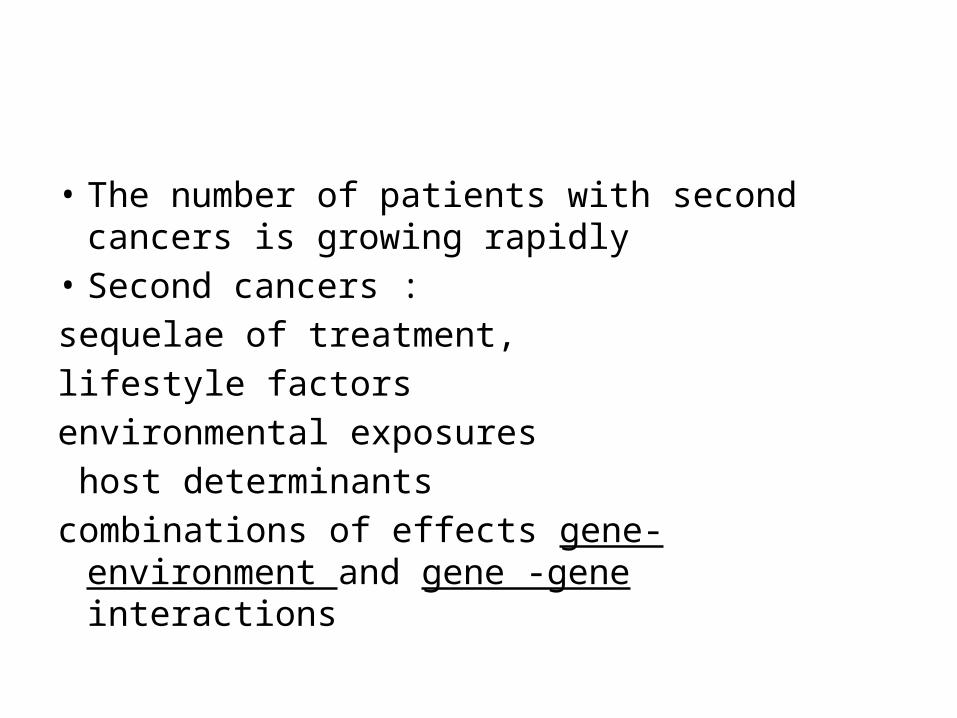
• The number of patients with second cancers is growing rapidly
• Second cancers :sequelae of treatment, lifestyle factorsenvironmental exposures host determinantscombinations of effects gene- environment and
gene -gene interactions
• Methods to Assess Second Cancer RiskCohort StudiesCase-Control Studies
• Carcinogenicity of Individual Treatment Modalities
Radiation TherapyChemotherapy

Radiation Therapy
Elevated risks of second cancer have been documented among patients treated with radiation therapy (RT) for
HL,testicular cancer,cervix cancer, breast cancer, childhood cancer. prostate cancer cancer of the nasopharynx

low-dose radiation
• Japanese atomic bomb survivors,• occupational radiation exposure,• sizable amounts of diagnostic radiation• RT for nonmalignant diseases. Consequently, the relationship between
radiation dose and cancer risk is better understood for these low-dose exposures.

• For most solid tumors, there is an approximately linear increase in risk with increasing radiation dose up to about 5 Gy.
• leukemia :a linear increase in risk with dose up to 1.5 to 2 Gy
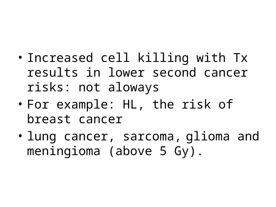
• Increased cell killing with Tx results in lower second cancer risks: not aloways
• For example: HL, the risk of breast cancer • lung cancer, sarcoma, glioma and meningioma
(above 5 Gy).

• risk of radiation-induced solid cancer increases with increasing dose well into the RT dose range prescribed to treat many cancers.
• It is also apparent that there is variation in the sensitivity of different tissues with regard to cancer induction by radiation, with sites such as the thyroid, female breast, and bone marrow being most radiosensitive
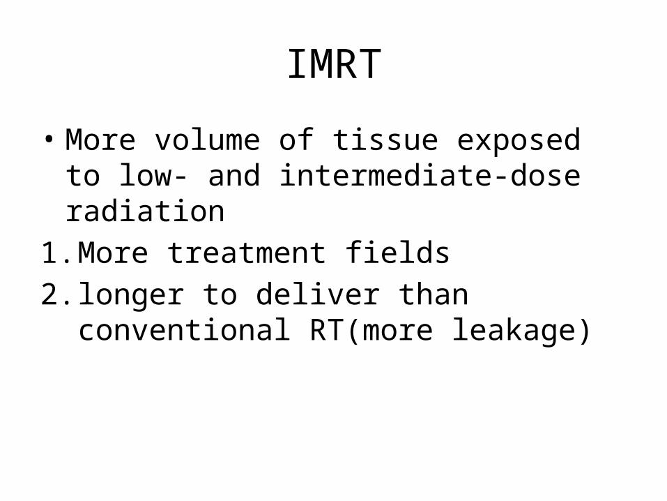
IMRT
• More volume of tissue exposed to low- and intermediate-dose radiation
1. More treatment fields 2. longer to deliver than conventional RT(more
leakage)

IMRT
• The additional low-dose exposure may increase the risk of second cancers especially in
high energy beamsyoung ages

RT+
• Hormonal factors• other carcinogenic agents• known genetic factors
Chemotherapy
• Leukemia (model disease)• In contrast, the induction of solid tumors after
chemotherapy is less well understood

Two major types of treatment-related leukemia
1. Alkylating agent-induced AML 2. Topoisomerase inhibitor-induced AML.
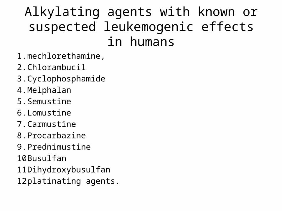
Alkylating agents with known or suspected leukemogenic effects in humans
1. mechlorethamine,2. Chlorambucil3. Cyclophosphamide 4. Melphalan 5. Semustine 6. Lomustine 7. Carmustine 8. Procarbazine9. Prednimustine 10. Busulfan 11. Dihydroxybusulfan12. platinating agents.

• The risk of alkylating agent-related AML has been shown to increase with increasing cumulative dose, duration of therapy, or dose intensity

Topoisomerase II inhibitors
• etoposide and teniposide• As compared with alkylating agent-induced
AML, epipodophyllotoxin-related AML has a shorter induction period (median, 2 to 3 years) and generally presents without preceding MDS, and its risk appears not to be dose dependent.

susceptibility to chemotherapy-induced cancers.
1. host phenotype (e.g., age, gender)2. host genotype (e.g., polymorphisms)3. exposure-related variables (e.g.,
chemotherapy dose, regimen, co-treatment with radiotherapy),

• Risk of Second Malignancy in Patients with Selected Primary Cancers
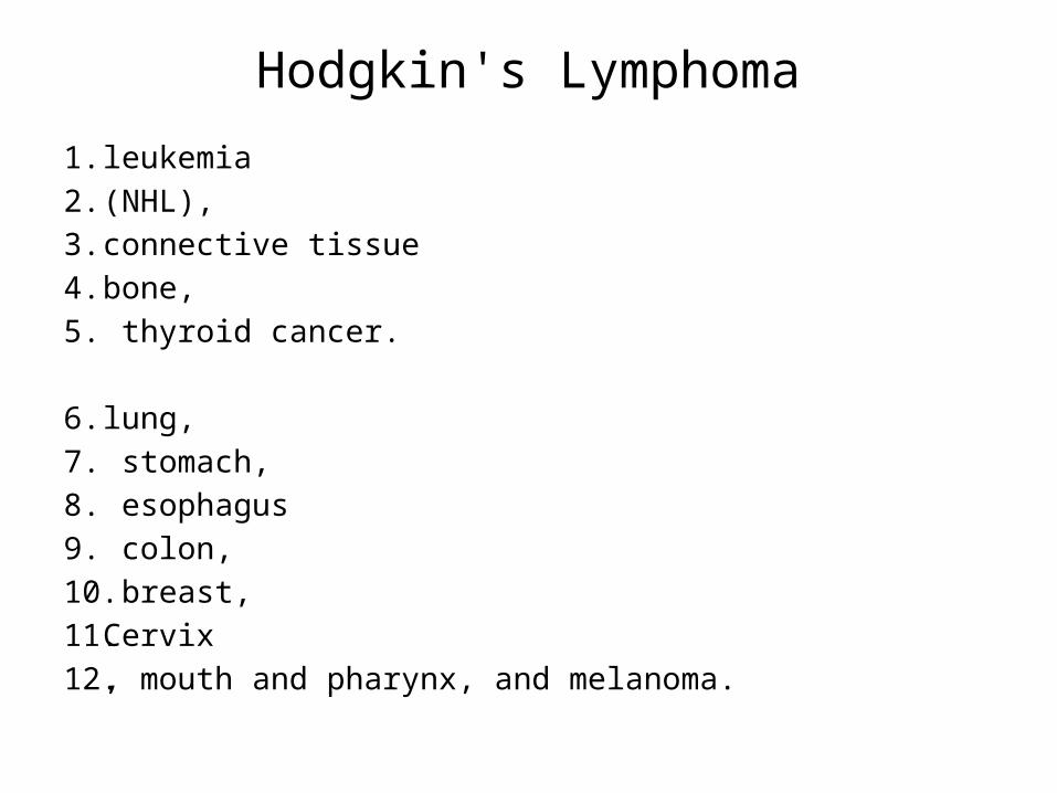
Hodgkin's Lymphoma
1. leukemia 2. (NHL), 3. connective tissue4. bone, 5. thyroid cancer.
6. lung,7. stomach,8. esophagus9. colon,10. breast, 11. Cervix12. , mouth and pharynx, and melanoma.

Time
• increased leukemia risk has its peak occurrence 3 to 9 years after chemotherapy,
• The RR of solid tumors is minimally elevated in the 1- to 4-year follow-up period, and increases steadily with increasing follow-up time from 5 years until at least 20 years since first treatment.
• For several tumor sites (breast, thyroid, esophagus), the excess risk does not become apparent until after 10 or even 15 years of observation.

• Risk Factors for Leukemia and NHL after H Lکموتراپی رژیم
استخوان مغز پیوند
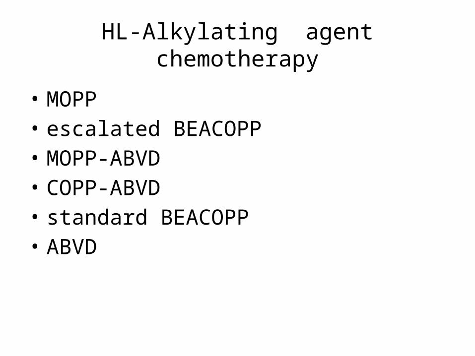
HL-Alkylating agent chemotherapy
• MOPP• escalated BEACOPP• MOPP-ABVD• COPP-ABVD • standard BEACOPP• ABVD
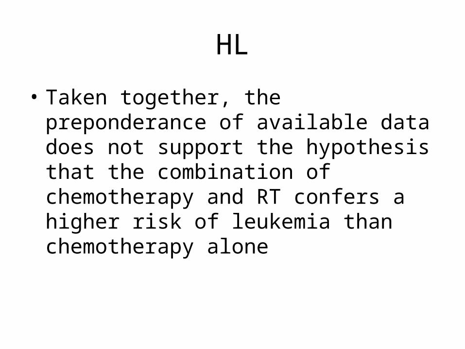
HL
• Taken together, the preponderance of available data does not support the hypothesis that the combination of chemotherapy and RT confers a higher risk of leukemia than chemotherapy alone

HL
• In some series, relatively high actuarial risks (4% to 15% at 5 years) of AML/MDS have been observed after autologous stem cell transplantation for HL.
• The prognosis of AML/MDS after HL treatment is extremely poor, with only 15% of patients surviving more than 1 year and no apparent survival benefit from allogenic stem cell transplantation.

• The causes of the excess risk of NHL after HL treatment are not well understood.
• Risk Factors for Solid Malignancies after Treatment for Hodgkin's Lymphoma
Role of ChemotherapyEffect of Age at Treatment
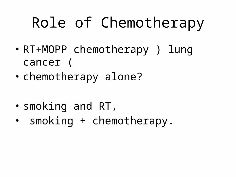
Role of Chemotherapy
• RT+MOPP chemotherapy ) lung cancer (• chemotherapy alone?
• smoking and RT, • smoking + chemotherapy.
• The inconsistent results with regard to the influence of chemotherapy on solid tumor risk may be due to the fact that most studies considered all solid tumors combined, whereas chemotherapy may differentially affect the risk of tumors at disparate sites.

Effect of Age at Treatment
• Several studies have shown that the RR of solid tumors increases strongly with younger age at first treatment .The effect is most notable for breast cancer
Conclusion-HL
• the occurrence of treatment-related second cancers is a major problem in survivors of HL.
• The substantial increase in solid tumor risk with greater follow-up time necessitates careful, lifelong medical surveillance of all patients.
• dissuade HL patients from smoking even before treatment starts.
• screening for lung cancer ?
Women with HL
• regular breast examinations should be emphasized
• From 8 years after irradiation (but not before age 25), the follow-up program of these survivors should include :
yearly breast palpation and breast imaging(MRI)
GI cancer -HL
• in patients who have received para-aortic and pelvic radiation fields and thoroughly examine any gastrointestinal complaints.

Modern chemotherapy
• Important questions to be answered in future research are whether modern chemotherapy regimens with lower doses of procarbazine and mechlorethamine also contribute to the risk of nonbreast solid tumors, and, if so, which cytotoxic drugs are responsible for the excess risk
Non-Hodgkin's Lymphoma• second malignancies following NHL(etiologic factors?, possibly immune
related?)
malignant melanoma Kaposi's sarcoma AMLbuccal cavity salivary gland Mouthlung Vaginaurinary bladder kidney parenchyma Bone

MDS/AML
• Additional work needs to be undertaken in order to more fully delineate the roles of the various types of prior therapy for lymphoma, the preparative regimen for transplantation, and other factors in the subsequent development of MDS/AML.
summary
• survivors of NHL are at increased risk for a number of second malignancies, although to a considerably smaller degree than are HL patients.
• The excess risks of AML and bladder cancer have been shown to be treatment related.
• The persistent increase in the risk of all second cancers for more than 20 years after NHL treatment alerts clinicians to the importance of long-term surveillance.
• smoking cessation. • Health care providers should also be aware of the large risk of
bladder cancer among patients who had been treated with the high-dose cyclophosphamide regimens of the past

Testicular Cancer
• Solid Tumor standard infradiaphragmatic radiotherapy fields ssupradiaphragmatic radiotherapy fields
Testicular Cancer
• chemotherapy alone is associated with significantly increased risks of solid cancers,
• Platinum is retained in the human body for prolonged durations after treatment and causes both leukemia and solid tumors in preclinical studies.(TC or ovarian cancer)
Leukemias IN TC survivors
• studies have described the role of both radiotherapy and chemotherapy
Ovarian Cancer
• Most :acute leukemia• breast, colon, rectum, small intestine, bladder,
renal pelvis, eye (ocular melanoma), and intrahepatic bile ducts.
• Reproductive and genetic factors predisposing to ovarian cancer may have contributed to the elevated risk of breast, colorectal, and other neoplasms.

Breast Cancer
1. contralateral breast2. Ovary3. Lung4. Esophagus5. Colon6. Connective tissue7. Melanoma8. Leukemia

• For :contralateral breast, ovary, and uterus, and possibly melanoma, the excesses may be fully or partly explained by a common etiology (e.g., genetic predisposition or hormonal risk factors).
• Other excess risks may be treatment related or reflect the interaction of several factors. Adjuvant CT, hormonal treatment, and RT, and combinations of these modalities, are being administered to a growing proportion of breast cancer patients.

• It is unfortunate, that it has not yet been examined whether RT as applied from the 1980s onward, especially postlumpectomy radiation, affects the risk of contralateral breast cancer
contralateral breast cancer
• How long :protective effect of tamoxifen • adjuvant chemotherapy may reduce the risk of
contralateral breast cancer,
• AML :use of dose-intensification strategies in
chemotherapy protocols for breast cancer?granulocyte colony stimulating factor?
• Conclusive evidence has emerged that tamoxifen is associated with a moderately increased risk of endometrial cancer
( بدتر پروگنوز با های ( پاتولوژیmalignant mixed mesodermal tumors uterine sarcoma
Conclusion(Breast)• only part of the elevated risk of second malignancies
following breast cancer is due to treatment.• a contralateral tumor is unlikely to be meaningfully
affected by current RT for the initial breast cancer, whereas tamoxifen reduces the risk of contralateral disease.
• Conventional anthracycline-based chemotherapy is associated with a low excess risk of leukemia.
• However, leukemia risk appears to increase with larger cumulative doses of anthracyclines and cyclophosphamide, and RT adds to this risk.
• The effectiveness of screening for endometrial cancer has not been demonstrated. Consequently, outside of research settings, there is no basis for regular gynecologic examinations in asymptomatic patients taking tamoxifen.
stop smoking
• The risk of lung cancer is about a twofold increase after postmastectomy RT, but not after postlumpectomy radiation. There is ample reason to advise breast cancer patients to stop smoking, especially if they receive radiation treatment

Pediatric Malignancies
• second primary bone tumors • soft tissue sarcoma• leukemia,• cancers of the brain, thyroid, and breast • breast ,skin,197 and other carcinomas• head and neck• adult-type carcinomas overall showed a four-fold
excess at an early age (median age at diagnosis: 27 years).
• Only a portion of the excess second cancer risk in survivors of childhood cancer is related to treatment
• Over the past decade, prophylactic cranial RT has been largely replaced by intrathecal methotrexate, but the number of these injections has not been related to the subsequent risk of brain tumors
conclusion• The magnitude of new malignancies depends on treatment and the
type of initial malignancy, si• • It is therefore imperative that survivors of childhood cancer be
carefully monitored to assess the long-term risks of various types of second cancers.
• Bone sarcoma is highest. • The leukemogenic potential of epipodophyllotoxin-containing
regimens that vary in cumulative dose and schedule of administration should continue to be rigorously assessed, as holds true for other new chemotherapy regimens
• RT is the main risk factor although chemotherapy has recently been shown to add substantially to the increased risk from radiation.