Embed Size (px)
Citation preview

foods
Review
Searching for the Antioxidant, Anti-Inflammatory, andNeuroprotective Potential of Natural Food and NutritionalSupplements for Ocular Health in the Mediterranean Population
Mar Valero-Vello 1,† , Cristina Peris-Martínez 2,3,4,† , José J. García-Medina 1,4,5,6 ,Silvia M. Sanz-González 1,4,7 , Ana I. Ramírez 4,8 , José A. Fernández-Albarral 8 , David Galarreta-Mira 4,9,Vicente Zanón-Moreno 1,4,10,* , Ricardo P. Casaroli-Marano 4,11,‡ and María D. Pinazo-Duran 1,4,7,‡
�����������������
Citation: Valero-Vello, M.;
Peris-Martínez, C.; García-Medina, J.J.;
Sanz-González, S.M.; Ramírez, A.I.;
Fernández-Albarral, J.A.;
Galarreta-Mira, D.; Zanón-Moreno, V.;
Casaroli-Marano, R.P.;
Pinazo-Duran, M.D. Searching for the
Antioxidant, Anti-Inflammatory, and
Neuroprotective Potential of Natural
Food and Nutritional Supplements
for Ocular Health in the
Mediterranean Population. Foods
2021, 10, 1231. https://doi.org/
10.3390/foods10061231
Academic Editor: Amit K. Jaiswal
Received: 18 April 2021
Accepted: 25 May 2021
Published: 28 May 2021
Publisher’s Note: MDPI stays neutral
with regard to jurisdictional claims in
published maps and institutional affil-
iations.
Copyright: © 2021 by the authors.
Licensee MDPI, Basel, Switzerland.
This article is an open access article
distributed under the terms and
conditions of the Creative Commons
Attribution (CC BY) license (https://
creativecommons.org/licenses/by/
4.0/).
1 Ophthalmic Research Unit “Santiago Grisolía” Foundation for the Promotion of Health and BiomedicalResearch of Valencia FISABIO, 46017 Valencia, Spain; [email protected] (M.V.-V.);[email protected] (J.J.G.-M.); [email protected] (S.M.S.-G.); [email protected] (M.D.P.-D.)
2 Ophthalmic Medical Center (FOM), Foundation for the Promotion of Health and Biomedical Research ofValencia (FISABIO), 46015 Valencia, Spain; [email protected]
3 Department of Surgery, University of Valencia, 46019 Valencia, Spain4 Spanish Net of Ophthalmic Research “OFTARED” RD16/0008/0022, Institute of Health Carlos III,
28029 Madrid, Spain; [email protected] (A.I.R.); [email protected] (D.G.-M.);[email protected] (R.P.C.-M.)
5 Department of Ophthalmology, General University Hospital “Morales Meseguer”, 30007 Murcia, Spain6 Department of Ophthalmology and Optometry, University of Murcia, 30120 Murcia, Spain7 Cellular and Molecular Ophthalmobiology Group, Department of Surgery, Faculty of Medicine and
Odontology, University of Valencia, 46010 Valencia, Spain8 Department of Immunology, Ophthalmology and Otorrinolaringology, Institute of Ophthalmic Research
“Ramón Castroviejo”, Complutense University of Madrid, 28040 Madrid, Spain; [email protected] Department of Ophthalmology. University Clinic Hospital of Valladolid, 47003 Valladolid, Spain10 Faculty of Health Sciences, International University of Valencia, 46002 Valencia, Spain11 Departament of Surgery, School of Medicine and Health Sciences, Clinic Hospital of Barcelona,
Universitat de Barcelona, 08036 Barcelona, Spain* Correspondence: [email protected]† Both (These) authors contributed equally to this work.‡ Group leaders contributing equally to this work.
Abstract: Adherence to a healthy diet offers a valuable intervention to compete against the increas-ing cases of ocular diseases worldwide, such as dry eye disorders, myopia progression, cataracts,glaucoma, diabetic retinopathy, or age macular degeneration. Certain amounts of micronutrientsmust be daily provided for proper functioning of the visual system, such as vitamins, carotenoids,trace metals and omega-3 fatty acids. Among natural foods, the following have to be considered forboosting eye/vision health: fish, meat, eggs, nuts, legumes, citrus fruits, nuts, leafy green vegetables,orange-colored fruits/vegetables, olives-olive oil, and dairy products. Nutritional supplements havereceived much attention as potential tools for managing chronic-degenerative ocular diseases. Asystematic search of PubMed, Web of Science, hand-searched publications and historical archiveswere performed by the professionals involved in this study, to include peer-reviewed articles in whichnatural food, nutrient content, and its potential relationship with ocular health. Five ophthalmologistsand two researchers collected the characteristics, quality and suitability of the above studies. Finally,177 publications from 1983 to 2021 were enclosed, mainly related to natural food, Mediterranean diet(MedDiet) and nutraceutic supplementation. For the first time, original studies with broccoli andtigernut (chufa de Valencia) regarding the ocular surface dysfunction, macular degeneration, diabeticretinopathy and glaucoma were enclosed. These can add value to the diet, counteract nutritionaldefects, and help in the early stages, as well as in the course of ophthalmic pathologies. The mainpurpose of this review, enclosed in the Special Issue “Health Benefits and Nutritional Quality ofFruits, Nuts and Vegetables,” is to identify directions for further research on the role of diet andnutrition in the eyes and vision, and the potential antioxidant, anti-inflammatory and neuroprotec-tive effects of natural food (broccoli, saffron, tigernuts and walnuts), the Mediterranean Diet, andnutraceutic supplements that may supply a promising and highly affordable scenario for patients
Foods 2021, 10, 1231. https://doi.org/10.3390/foods10061231 https://www.mdpi.com/journal/foods
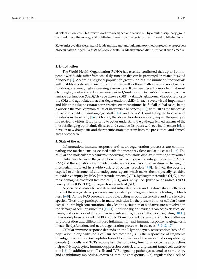
Foods 2021, 10, 1231 2 of 27
at risk of vision loss. This review work was designed and carried out by a multidisciplinary groupinvolved in ophthalmology and ophthalmic research and especially in nutritional ophthalmology.
Keywords: eye diseases; natural food; antioxidant/anti-inflammatory/neuroprotective properties;broccoli; saffron; tigernuts-chufa de Valencia; walnuts; Mediterranean diet; nutritional supplements
1. Introduction
The World Health Organization (WHO) has recently confirmed that up to 1 billionpeople worldwide suffer from visual dysfunction that can be prevented or treated to avoidblindness [1]. According to global population growth indices, the number of individualswith mild-to-moderate visual impairment as well as those with severe vision loss andblindness, are worryingly increasing everywhere. It has been recently reported that mostchallenging ocular disorders are uncorrected/under-corrected refractive errors, ocularsurface dysfunction (OSD)/dry eye disease (DED), cataracts, glaucoma, diabetic retinopa-thy (DR) and age-related macular degeneration (AMD). In fact, severe visual impairmentand blindness due to cataract or refractive error constitutes half of all global cases, beingglaucoma the most common cause of irreversible blindness [1–3], with DR as the first causeof visual disability in working-age adults [1–4] and the AMD constituting the first cause ofblindness in the elderly [1–5]. Overall, the above disorders seriously impair the quality oflife related to vision. It is a priority to better understand the pathogenic mechanisms of themost challenging ophthalmic diseases and systemic disorders with eye involvement [6], todevelop new diagnostic and therapeutic strategies from both the pre-clinical and clinicalareas of concern.
2. State of the Art
Inflammation/immune response and neurodegeneration processes are commonpathogenic mechanisms associated with the most prevalent ocular diseases [1–6] Thecellular and molecular mechanisms underlying these shifts display interesting similarities.
Disbalance between the generation of reactive oxygen and nitrogen species (ROS andRNS) and the activation of antioxidant defenses is known as oxidative stress, a challengingmechanism involved in a wide variety of ocular disorders [7,8]. In fact, the eyes areexposed to environmental and endogenous agents which makes them especially sensitiveto oxidative injury by ROS [superoxide anions (·O¯), hydrogen peroxides (H2O2), themost damaging hydroxyl free radical (·OH)] and/or by RNS [nitric oxide radical (NO·),peroxynitrite (ONOO−), nitrogen dioxide radical (NO2·)
Associated diseases to oxidative and nitrosative stress and its downstream effectors,much of these age-related processes, are prevalent pathologies potentially leading to blind-ness [6–9]. Active ROS present a dual role, acting as both destructive and constructivespecies. Thus, they participate in many activities for the preservation of cellular home-ostasis, but in high concentrations, they lead to a situation of oxidative stress involved inthe damage of cellular structures [10,11]. Additionally, antioxidants can act as classical de-fenses, and as sensors of intracellular oxidants and regulators of the redox signaling [10,11].It has widely been reported that ROS and RNS are involved in signal transduction pathwaysof proliferation and differentiation, inflammation and immune response, angiogenesis,metabolic dysfunction, and neurodegeneration processes, in the eyes [7–9,12–17].
Cellular immune response depends on the T lymphocytes, representing 70% of allpopulation, along with the T-cell surface receptor (TCR) the responsible of fragmentsof antigen recognition (as peptides bound to molecules of the major histocompatibiligycomplex). T-cells and TCRs accomplish the following functions: cytokine production,helper-T-lymphocytes, immunosuppression control, and unpleasant target cell destruc-tion [18]. In addition to the T-cells and TCRs signaling pathways, several co-stimulatoryand co-inhibitory molecules, known as immune checkpoints (ICs), regulate the T-cell ac-

Foods 2021, 10, 1231 3 of 27
tivities. Under normal conditions, these molecules are essential in maintaining immunehomeostasis and preventing autoimmunity by controlling the type, extent, and time spanof the immune responses [19]. There are numerous different T cell subtypes having theirown specific identifiable surface markers and displaying different roles. Two major Tlymphocyte types can be identified according to the presence of the CD4 and CD8 cellsurface molecules, and subsequently classified into the Th1 cells and Th2 cells, also pro-ducing the correspondent cytokines [9]. A wide variety of pro-inflammatory cytokineshave been reported in humans and animal models, with an increasing interest in relationto ophthalmic diseases, over recent years [20–24]. The microglia of the central nervoussystem protect the organs and tissues under normal conditions, by responding swiftly tothe injury signals. However, these activities are reverted in chronic neurodegenerativediseases, where the activated microglia are shifted on a pro-inflammatory phenotype that,in turn, release pro-inflammatory cytokines and different neurotoxic substances such asROS and RNS, proteolytic enzymes, specific neurotransmitters, and others [12,15–17,22,24].
Neurodegeneration refers to the progressive damage and loss of both the structureand function of nerve cells and the vascular system, occurring in specific sites of thecentral and peripheral nervous system and the neurosensory organs [15]. Retinal andoptic nerve neurodegenerative diseases, the majority of them incurable, are increasingworldwide, representing a serious health problem and a big financial burden for thecountries [6,12,15–17,24–27], among them, the multifactorial sight-threatening retinal andoptic nerve diseases: AMD, RD, glaucoma, retinitis pigmentosa, macular dystrophies,uveoretinitis, pathologic myopia, retinal vascular occlusion, as well as the optic neu-ropathies [6,12,15–17,24–32].
Nutritional intervention has been evoked as one important tool for protecting theeyes and vision. Natural food and its derivatives play essential roles in health and well-being, and it has been characteristically associated with defense mechanisms againstmicrobial agents, xenobiotics and/or physical factors, also demonstrating an immenserange of health-promoting perspectives for achieving a pleasant lifespan [33,34]. Largepopulation studies on the role of nutrition in eye health have reported controversial resultsover decades, mainly regarding cataracts, AMD, RD, OSD/DED, and glaucoma, and alsoslowing progression of the above sight-threatening diseases, by dietary interventions hasalso been discussed [6,8,35–54].
It has recently shown a reduced global risk of DR in a Mediterranean population withtype 2 diabetes mellitus (T2DM), according to clinical, biochemical and lifestyle biomarkersincluding the adherence to the Mediterranean Diet (MedDiet) [40,55], and also a reductionin the enlargement of drusen, the most relevant manifestation of AMD, by the MedDietand lifestyle incorporating fruits, vegetables, legumes and fish [56]. In this concern, therandomized clinical trial “Prevención con Dieta Mediterránea” (PREDIMED) demonstrateda reduced risk of DR in middle-aged/older individuals with type 2 diabetes, with theintake of at least 500 mg/d of dietary long-chain omega 3 polyunsaturated fatty acids (ω3PUFAs), by means of two weekly servings of oily fish [57]. Additionally, the PREDIMEDgroup showed that intake of skimmed yogurt was associated with lower risk of cataractsin the elderly Mediterranean population with high cardiovascular risk [58].
Nutrition clearly makes a difference to eye health and vision care [54]. In fact, coordi-nated, multidisciplinary interventions are essential to deal with the role of natural food,and nutritional supplements to achieve better knowledge of the diet and ocular diseases,as in the present work. In fact, our review article included four subsections regardingthe benefits of natural food for vision health. Broccoli, nuts, saffron and tigernuts areawesome single foods that can help prevent/manage ocular diseases and also can helpfight against certain risk factors related to visual impairment. Practically all of these fooddisplay anti-inflammatory, detoxicating, anti-angiogenic, anti-apoptotic, photo-protective,antioxidant and neuroprotective effects to some extent. Because of this, here we soughtto address the role of diet and nutrition in the eyes and vision, focusing on the potentialbenefit of natural food (broccoli, saffron, tiger nuts and walnuts), as well as the benefits

Foods 2021, 10, 1231 4 of 27
of the MedDiet and the positive effects of appropriated nutraceutical supplements on eyehealth in order to prevent vision loss.
3. Material and MethodsDesign
An extensive systematic search of PubMed, Web of Science, Scopus, Google Scholar,hand-searched publications and historical archives were performed by the profession-als involved in this review, according to the standardized search of publications withthe following keywords: eye, health, disease, vision loss, dry eyes, glaucoma, cataracts,retinopathies, macular degeneration, blindness, natural food, risk factors, pathogenic mech-anisms, antioxidant, anti-inflammatory, neuroprotective, broccoli, saffron, tigernuts-chufade Valencia, walnuts, Mediterranean diet (MedDiet), and nutritional supplements. More-over, we utilized several combinations of the above terms. The main objective was toinclude English language peer-reviewed reports in which natural food, nutrient content,and its potential relationship with ocular health and disease were treated. Additionally,other important documents were revised in its native language. Five ophthalmologists andtwo researchers collected the characteristics, quality and suitability of the above studies,to ensure as much as possible the scientific interest and quality as well as to minimizethe risk of bias. The MeaSurement Tool to Assess systematic Reviews (AMSTAR-2), animportant tool for critically appraising systematic reviews of randomized controlled clinicaltrials) [59], the risk-of-bias (ROB) assessment of systematic reviews [60] and the Appraisalof Guidelines for Research and Evaluation (AGREE II), the most widely used guidelinesappraisal tool [61] were utilized. Finally, 177 papers, from a period between 1983 to 2021were selected for this study, mainly related to natural food, MedDiet, nutraceutic supple-mentation and methodology in the context of ocular health and the most prevalent eyediseases. Original studies from our group done with broccoli, and tigernut (chufa de Valen-cia) regarding the ocular surface dysfunction, macular degeneration, diabetic retinopathyand glaucoma were enclosed.
The specific methodology that was followed on the not previously published studiesdealing with the effects of daily intake of broccoli and tigernuts, were precisely describedin the corresponding subsections.
4. Natural Food and Ocular Health4.1. Broccoli
The Brassica Oleracea, belonging to the vegetable Brassicaceae family, is popularlyknown as broccoli (Italian variety), brecol (spanish) and ka-i-lan or kale (Chinese variety)with the new food baby broccoli or tenderstem being a mix between the traditional broccoliand the kale. This family of natural foods constitutes a group of vegetables including thefollowing: cabbage, broccoli, cauliflower, red cabbage, Brussels sprouts, radish, turnip,and others, all of them important sources of micronutrients and fiber [62]. The greenbroccoli (Calabrese) is the most common variety of this plant, sized 10–20 cm, weighing500 g, with dark green and light green stems and buds from chlorophyll pigment. Thebroccoli contains high levels of water, carotenes (beta-carotene, lutein), vitamins (A, B, C, E),isothiocyanates, fatty acids (linoleic acid, palmitic acid) and diverse minerals (calcium, iron,magnesium, potassium, phosphorus, sodium). Additionally, amino acids (tyrosine, asparticacid, glutamic acid, proline, valine) were found in larger concentrations. Even more,broccoli sprouts, have been implicated in many biological activities such as antiapoptotic,anti-inflammatory, antioxidant, antimicrobial, as well as neuroprotectant properties, asrecently reviewed [63–65]. The ethyl acetate fraction of broccoli florets was reported toexert potent antioxidant and anti-inflammatory effects, by inhibiting nitric oxide release,counteracting the ROS and nuclear factor-κB activation in a dose-dependent manner,concluding that broccoli can be utilized as a dietary supplement to improve nutrition aswell as for the adjunctive intervention in chronic inflammation [66]. The pro-apoptoticfunction of broccoli has been previously demonstrated in different cancers [67–69]. In this
Foods 2021, 10, 1231 5 of 27
concern, the effects of the bio-accessible fraction of broccoli, kale, mustard, and radish,were evaluated on colon cancer cells, showing its usefulness to reduce this disease incombination with a balanced diet. However, a review about the targets and mechanismon breast cancer reported the contradictory roles of sulforaphane derivatives in breastcancer therapy [70]. The effects of the broccoli isothiocyanates, amino acid compoundsthat are detoxified by conjugation with glutathione, have also been reviewed in bothin vitro and in vivo models of acute and chronic neurodegenerative diseases [71]. Similarly,the sulphoraphane (glucoraphanin), a phytocompound belonging to the isothiocyanatefamily with a role in preventing vascular complications in diabetes [72], also demonstratedimportant benefits for neurodegenerative disorders [73].
Nowadays, AMD in the dry and wet clinical types is the first cause of blindnessamong the elder population [2,5,6,42,56]. It has been estimated that early AMD caseswill augment to approximately 17.8 million in 2050 [74]. Increasing the consumption ofspecific nutrients may be an effective intervention to vision care in AMD as well as inother sight-threatening ocular diseases. Among these nutrients, the xanthophyll pigmentslutein and zeaxanthin and its metabolic by-products, were identified in the macula in1985, at the highest concentrations of the whole human body, suggesting pivotal rolesfor these carotenoids in the retina [75]. In fact, lutein and zeaxantin have been proved toanatomically and functionally protecting the macula against photo-oxidative attack [76].The Age-Related Eye Disease Study (AREDS) concluded that the daily intake of 10 mg oflutein and 2 mg of zeaxantin alone or in combination with docosahexaenoic acid (DHA)(350 mg/day) and eicosapentaenoic acid (EPA) (650 mg/day) to the original AREDSsupplement formula composed by vitamin C, vitamin E, beta carotene, zinc oxide andcupric oxide [77], except the beta carotene, demonstrated efficacy in the prevention ofAMD progression to advanced forms in right risk eyes [78]. The question arose as towhether it is possible to identify individuals at risk of AMD based on the findings of theircentral macular pigment optical density (MPOD) levels. In this concern, Berstein et al. [79]concluded that a central MPOD below 0.2 d.u. should be taken as low levels, 0.2–0.5. d.u.as mild levels and more than 0.5 as high levels.
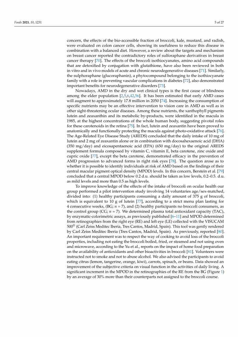
To improve knowledge of the effects of the intake of broccoli on ocular health ourgroup performed a pilot intervention study involving 14 voluntaries age/sex-matched,divided into: (1) healthy participants consuming a daily amount of 375 g of broccoli,which is equivalent to 10 g of lutein [77], according to a strict menu plan lasting for4 consecutive weeks, (BG; n = 7), and (2) healthy participants no broccoli consumers, asthe control group (CG; n = 7). We determined plasma total antioxidant capacity (TAC),by enzymatic-colorimetric assays, as previously published [6–11] and MPOD determinedfrom retinographies from the right eye (RE) and left eye (LE) collected with the VISUCAM500® (Carl Zeiss Meditec Iberia, Tres Cantos, Madrid, Spain). This tool was gently renderedby Carl Zeiss Meditec Iberia (Tres Cantos, Madrid, Spain). As previously, reported [80].An important requirement was to respect the way of cooking to avoid loss of the broccoliproperties, including not eating the broccoli boiled, fried, or steamed and not using ovenand microwave, according to the Yu et al., reports on the impact of home food preparationon the availability of antioxidants and other bioactivities in broccoli [81]. Volunteers wereinstructed not to smoke and not to abuse alcohol. We also advised the participants to avoideating citrus (lemon, tangerine, orange, kiwi), carrots, spinach, or beans. Data showed animprovement of the subjective criteria on visual function in the activities of daily living. Asignificant increment in the MPOD in the retinographies of the RE from the BG (Figure 1)by an average of 30% more than their counterparts not assigned to the broccoli course.
Foods 2021, 10, 1231 6 of 27
Figure 1. Comparative evaluation of the MPOD measured with the Visucam 500®. (A) Retinographyof the central retina of the right eye at baseline, (B) Distribution of the macular pigment and thepeak at the foveal level of the right eye at baseline. (C) Retinography of the central retina of theright eye of the same participant at end-of-study. (D) Distribution of the macular pigment andthe peak at the foveal levels of the righ eye of the same participant at end-of-study (E) Schematicrepresentation of the retinal area where the pigment is placed (a). (F) Illustrative drawing of theVisucam 500 parameters: total pigment volume (b), maximum of the pigment density (c), mean ofthe pigment density (d) [81].
Moreover, it was also found a significant increase in plasma TAC in the BG (baseline:1.231 ± 0.120 mM; end-of-study: 1.858 ± 0.393 mM; p = 0.002). This study mainly suggeststhat the broccoli intake improved the antioxidant load and the MPOD associated withlutein dietary supplementation, thus helping in protect the macula against oxidative injury.
In summary, the biochemical and physicochemical characteristics of broccoli make thisfood optimal to fight against age-related chronic inflammatory and/or neurodegenerativedisorders, to better eye and vision care, as widely suggested [62–66,72,73,80–85].
4.2. Saffron
The Crocus Sativus is a plant that provides a spice, saffron, which has been classicallyused in food preparation, being the most expensive spice in the world [86]. Etymolog-
Foods 2021, 10, 1231 7 of 27
ically saffron comes from the Arabic term za’faran (yellow) as well as from the Persianza’ferân (golden stigmas), the Latin word safranum and the Spanish azafrán. Since ancienttimes, medicinal properties have been attributed to this species because it has more than100 metabolites in the composition of its stigmas, including crocin isomers, zeaxanthin,lycopene and vitamin B12, among others [87].
Major saffron components are crocin and crocetin (that gives the yellow color to thestigmas), picrocrocin (that contributes to the bittersweet flavor), kaempferol (from thecrocus sativus petals) and safranal (which lends the fragrance to the spice, also contributingto the flavor) [86–88]. However, the main therapeutic activities of saffron are due to itsmain bioactive components, the carotenoids crocetin and crocin [88]. Crocin is hydrolyzedto crocetin when absorption occurs in the intestine [89], and once in the blood it can betransported to different tissues and can even cross the blood-brain barrier reaching tissuesof the central nervous system [90]. This spice has been used in traditional medicine asanti-ischemic, hypolipidemic, anti-hypertensive, anxiolytic, antidiabetic, antidepressant,anticancer, and cardioprotective [91,92]. It has been possible due to the various propertiesattributed to crocetin as an anti-inflammatory, anti-apoptotic and antioxidant [92]. An-tioxidant activities are due to their ability to scavenge free radicals [93], their capacity todecrease telomerase activity and to increase proapoptotic effects in cancer cells. In addition,the anti-inflammatory effect is due to the regulation of genes that control the release ofproinflammatory cytokines, adhesion molecules and proinflammatory enzymes by glialcells, as well as modulation of inflammatory pathways (e.g., nuclear factor-κB) [94].
The beneficial effects of saffron have been demonstrated in neurodegenerative dis-eases such as Alzheimer’s and Parkinson’s, where it has been shown to exert a certainneuroprotective effect [95,96]. In addition, in neurodegenerative diseases of the eye, saffronmay also have these beneficial effects [94].
In AMD, it has been shown that in the early stages of the disease, saffron can improvevisual function by reversing the damage to photoreceptors and bipolar cells caused byoxidative stress [97]. In addition, daily supplementation with saffron improved retinalchanges observed with optical coherence tomography and electroretinogram in patientswith both dry and wet AMD [98].
In DR, saffron can reduce insulin resistance in pre-diabetic patients [99]. In vitromodels of diabetes have shown that saffron can control microglia activation. In addition,crocin supplementation decreases macular thickness improving visual acuity in patientswith diabetic macular edema probably by its anti-inflammatory effects [100].
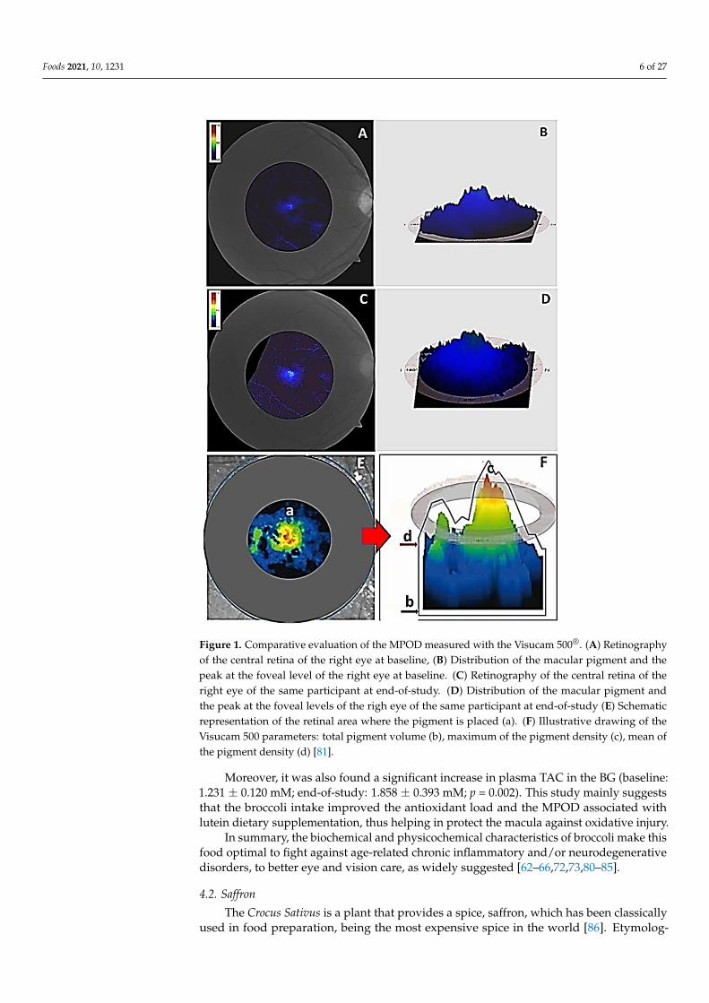
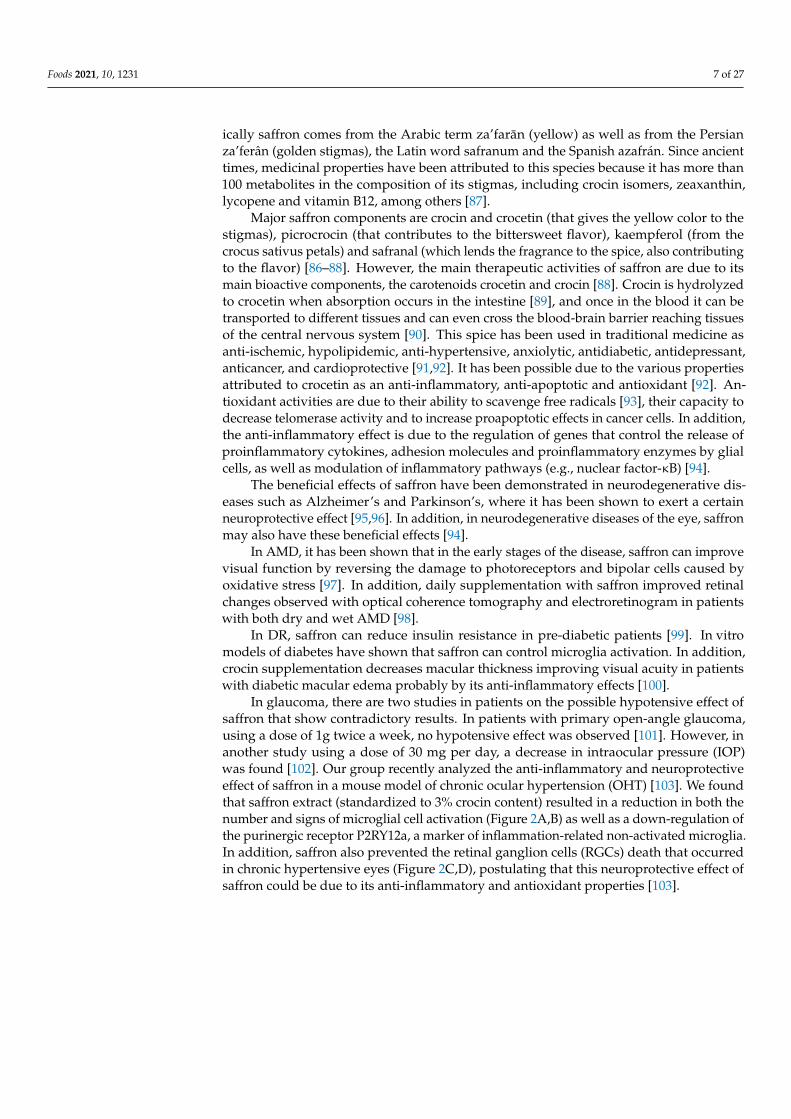
In glaucoma, there are two studies in patients on the possible hypotensive effect ofsaffron that show contradictory results. In patients with primary open-angle glaucoma,using a dose of 1g twice a week, no hypotensive effect was observed [101]. However, inanother study using a dose of 30 mg per day, a decrease in intraocular pressure (IOP)was found [102]. Our group recently analyzed the anti-inflammatory and neuroprotectiveeffect of saffron in a mouse model of chronic ocular hypertension (OHT) [103]. We foundthat saffron extract (standardized to 3% crocin content) resulted in a reduction in both thenumber and signs of microglial cell activation (Figure 2A,B) as well as a down-regulation ofthe purinergic receptor P2RY12a, a marker of inflammation-related non-activated microglia.In addition, saffron also prevented the retinal ganglion cells (RGCs) death that occurredin chronic hypertensive eyes (Figure 2C,D), postulating that this neuroprotective effect ofsaffron could be due to its anti-inflammatory and antioxidant properties [103].
Foods 2021, 10, 1231 8 of 27
Figure 2. Immunohistochemistry of retinal whole-mount micrographs of mice eyes. The A,B retinas were labeled withanti-Iba-1 (microglia), showing the comparison of Iba-1 + microglia in the OHT untreated (A) and treated with saffronextract (B) eyes in the outer plexiform layer of the retina. It has been noted that in the OHT mice eyes treated with saffronextracts, less activation and fewer microglial cells were observed than in the untreated eyes. The C,D retinas were stainedwith anti Brn3a (RGCs). The C,D comparative micrographs showed the Brn3a + RGCs in the untreated (C) and treated withsaffron extract (D) mice retinas. It was also detected a higher RGCs density in the treated versus the untreated rat retinas.RGC: retinal ganglion cells.
4.3. Tigernut-Chufa de Valencia
The Cyperus Esculentus is a herbaceous, perennial, fasciculate-rooted plant foundacross the world, but distributed mostly in Egypt, Nigeria and Spain. Additionally knownas “Juncia Avellanada” and tigernut (TN), it has a highly developed rhizomatic systemwith the tubers produced at the apical ends of the rhizomes [104]. It is known that onlyone specimen can produce hundreds or thousands of tubers through a growing season.According to the macroscopic characteristics three main tuber types have been described:brown, black and yellow [105]. Over the last decades, substantial research has evidencedthat TN is a good source of oil, and its by-products are rich in various nutrients andbioactive compounds [105,106]. The available data reveals that tubers are rich in essentialdietary constituents such as proteins (3.28–8.45%), fats (22.14–44.92%), fibers (8.26–15.47%)and ashes (1.60–2.60%). The lipid profiling interestingly revealed that TN oil has a similarfatty acid composition to olive oil [107].
The TN finds the Mediterranean climate of Valencia particularly favorable for itscultivation and development. In this area, it is known as chufa de Valencia. It was introducedin the Valencian region in the 8th century CE, a multi-cultural period lasting 711–1492 inwhich Christians, Jews and Muslims created a high degree of civilization in Spain andEurope. The curative properties of the chufa de Valencia date from 1297 [108]. Arnau deVilanova (1232–1311), a famous physician and theologian of this time, prescribed eatingchufa de Valencia to alleviate different disorders [109]. The Valencian botanist Cavanilles(1745–1804) reflected in his works from 1795, the cultivation of the TN in the town of Albo-raya (Valencia, Spain) [110]. The chufa de Valencia is dark brown, sized 0.9-1.6 cm long and

Foods 2021, 10, 1231 9 of 27
0.7–1.1 cm wide. According to the shape, two types are distinguished: the elongated (chufallargueta) and the rounded (chufa armela). The chufa de Valencia is a historical gastronomicand cultural wholesome brown tuber crop, with outstanding nutritional properties. Thisfresh functional food consists in carbohydrates 18%, fats 17% (includingω6/ω9 fatty acids),proteins 8%, fiber 13%, oligoelements (calcium, copper, iron, magnesium, phosphorus,potassium, zinc) and vitamins (C, E), providing 460 kcal/100 g [111]. It is also extensivelyused to prepare a cold beverage, known as “horchata de chufa” typical to Valencia [112].
Several epidemiologic and experimental studies pointed out the wide variety oftherapeutic benefits of TN, such as cardioprotector, antioxidant, anti-inflammatory andneuroprotectant [113,114]. The TN also contributes to lowering total cholesterol andtriglycerides, stabilizes glycemic profile, provides amino acids, vitamins, minerals, andfiber. Its salt content is low, and it does not contain lactose or fructose. Thus, the TN and itsderivatives (horchata, flours, oils, spices, etc.) constitute a very complete food, as they offerlarge proportions of vitamins and minerals (such as vitamins C and E), lipids and oleicacid which are useful for the control of cholesterol and triglycerides [115,116]. As for thepresence of vitamin E, it is important to highlight its importance as it is an essential vitamin,not synthesized by the body, but necessary for its proper functioning, and therefore mustbe included in the diet [117]. In addition to being one of the major antioxidants, vitamin Ehas the ability to scavenge free radicals, which reduces the risk of cancer and prevents theprogression of pre-cancerous lesions [118,119].
Among the most prevalent eye diseases are ocular surface disorders. DED definesthe pathology of the ocular surface that induces tear-film deficiency and dry eyes [120].The term includes complex diseases that affect the eyelids, lacrimal glands, conjunctiva,cornea and the tear film, with very high global prevalence, affecting both genders andpeople aged 60 years and above. The signs and symptoms range from mild redness, foreignbody sensation, photophobia, and/or blurred vision, to intense and diffuse hyperemia,epiphora, continuous sensations of itchiness, stinging and burning, as well as visual impair-ment. First-line therapy includes eye drops of artificial tears, lubricant gels and ointments.The imbalance between prooxidant and antioxidant sources damages the ocular surfacestructures [121,122]. It has also been assumed that chronic inflammation is involved inOSD/DED, as demonstrated by the release of pro-inflammatory mediators and the positiveresponse to the oral supplementation with antioxidants and essential fatty acids [123,124].In this concern, anti-inflammatory eye drops can also be prescribed [125,126]. In spiteof this, further research is needed to improve the eyes and the quality of life of patientsaffected by DED.
In this concern, our group conducted one study in the past years (2016–2018) aboutthe role of the daily intake of chufa de Valencia in eye health, with the main purpose ofevaluating its effects on DED by integrating clinical and biochemical data. A pilot studyon 20 women aged 45–70 years, office employees of the administration services of theUniversity of Valencia, with the common characteristic of working with computers duringthe workday were included in the study and classified as: (1) women working withcomputers assigned to a daily ration of 30 g of fresh chufa de Valencia, kindly given by theRegulatory Council of the Designation of Origin Chufa de Valencia (Alboraya, Valencia,Spain) during 3 consecutive months (n = 10; ChG) and (2) women working with computerswithout consuming the tuber (n = 10; CG). A personal interview including the ocularsurface disorder questionnaire (OSDI; Allergan Inc., Irvine, CA, USA) to discriminatebetween normal-mild-moderate-severe DED, and ocular examination (best corrected visualacuity, the spontaneous number of closing eyelids in 1 min: blinking frequency that innormal conditions 9–12/min; quantitative Schirmer test, to evaluate the amount of wettingthe strip located on the inferior inner eyelid during 5 min, that in normal conditions is morethan 10 mm; qualitative break up time test (BUT), the time interval between last blink andthe appearance of first dry spot over the cornea, that in normal conditions is more than5 s), were carried out for all women participants. One important point of this study wasto ensure compliance with the supplement food by the participants, which is essential to

Foods 2021, 10, 1231 10 of 27
optimize the effectiveness of the nutritional intervention. Average duration of computeruses during the workday was 5.8 ± 2 h with similar type of screen and computer for thetwo groups of participants. Tear samples from the inferior eyelid lacrimal meniscus werecollected with capillaire microtubes, labeled and stored at −80 ◦C until processing.
Mean age of participants was 55.4 ± 6.2 years. The OSDI questionnaire revealedthat 68.4% of the ChG had moderate dry eyes at baseline and the same participants hadmild-to-moderate dry eyes at the end of study (61.6%). All volunteers displayed signsand symptoms of DED, ranging from redness, grittiness, itchiness, foreign body sensation,burning, stinging and blurred vision. Eye discomfort, visual impairment, and reductionof the quality of life related to the eyes and vision, were referred by the volunteers at theonset of this study. However, a noticeable reduction of the signs, symptoms and subjectivesensations was recorded at the end of the food supplementation. The blinking frequencywas significantly and positively reduced in the ChG after the oral intake period as comparedto the non-supplemented employers (p = 0.042). The BUT test was significantly higherin the ChG at the end of study (RE: 7.4 ± 0.7 s, vs. 9.8 ± 0.4 s; p = 0.011; LE: 7.5 ± 0.7 svs. 9.7 ± 0.4 s, p = 0.016). Additionally, the ChG showed higher Schirmer test marks atend-of-study, as compared to baseline (RE: 7.1 ± 0.7 mm vs. 10.5 ± 0.9 mm, p = 0.002;LE: 7.0 ± 0.6 mm vs. 12.9 ± 1.8 mm, p = 0.001), as reflected in Figure 3. In addition, noadverse effects were reported in relation to the supplementation in the assigned participantsto this regime. In contrast, all participants declared to be satisfied with the organolepticproperties of the intake of chufa de Valencia.
Figure 3. Clinical probes to qualitative and quantitative evaluating the tear film from baseline to theend of study in the participants randomly assigned to a daily intake of 30 g. of the fresh TN Chufa deValencia. (A) Data from the FTBUT in both eyes. (B) Schirmer test determination in both eyes. F-BUTtest: Fluorescein break up time test; RE: right eye LE: left eye. F p < 0.05 statistically significant.
Foods 2021, 10, 1231 11 of 27
In conclusion, the daily intake of 30 g. of chufa de Valencia improved the amount andstability of the tear film, decreasing the signs, symptoms, and subjective sensations of theDED patients.
As reflected in the anterior subsection (3.1 Broccoli) of this review, AMD refers to thechronic, progressive degeneration of the macula, a common eye disorder affecting peopleaged 60 years and more [1–3,6,36–38,77–79]. Up to 200 million people worldwide currentlyhave AMD which is caused by complex interactions between aging comorbidities andgenetics with the environmental factors, and other unknown situations [1–3,6]. The clinicalAMD forms, dry and wet, can be clinically distinguished, with the dry AMD accountingfor 90% of all cases. AMD progressively leads to central vision loss and reduced quality oflife in the affected individuals.
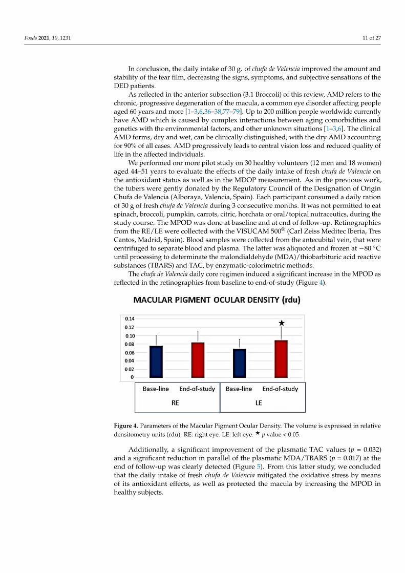
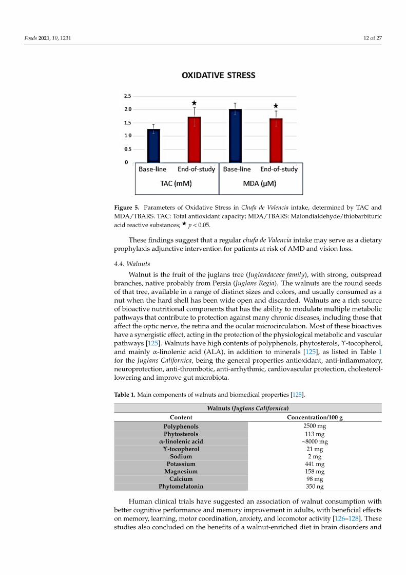
We performed onr more pilot study on 30 healthy volunteers (12 men and 18 women)aged 44–51 years to evaluate the effects of the daily intake of fresh chufa de Valencia onthe antioxidant status as well as in the MDOP measurement. As in the previous work,the tubers were gently donated by the Regulatory Council of the Designation of OriginChufa de Valencia (Alboraya, Valencia, Spain). Each participant consumed a daily rationof 30 g of fresh chufa de Valencia during 3 consecutive months. It was not permitted to eatspinach, broccoli, pumpkin, carrots, citric, horchata or oral/topical nutraceutics, during thestudy course. The MPOD was done at baseline and at end of follow-up. Retinographiesfrom the RE/LE were collected with the VISUCAM 500® (Carl Zeiss Meditec Iberia, TresCantos, Madrid, Spain). Blood samples were collected from the antecubital vein, that werecentrifuged to separate blood and plasma. The latter was aliquoted and frozen at −80 ◦Cuntil processing to determinate the malondialdehyde (MDA)/thiobarbituric acid reactivesubstances (TBARS) and TAC, by enzymatic-colorimetric methods.
The chufa de Valencia daily core regimen induced a significant increase in the MPOD asreflected in the retinographies from baseline to end-of-study (Figure 4).
Figure 4. Parameters of the Macular Pigment Ocular Density. The volume is expressed in relativedensitometry units (rdu). RE: right eye. LE: left eye. F p value < 0.05.
Additionally, a significant improvement of the plasmatic TAC values (p = 0.032)and a significant reduction in parallel of the plasmatic MDA/TBARS (p = 0.017) at theend of follow-up was clearly detected (Figure 5). From this latter study, we concludedthat the daily intake of fresh chufa de Valencia mitigated the oxidative stress by meansof its antioxidant effects, as well as protected the macula by increasing the MPOD inhealthy subjects.
Foods 2021, 10, 1231 12 of 27
Figure 5. Parameters of Oxidative Stress in Chufa de Valencia intake, determined by TAC andMDA/TBARS. TAC: Total antioxidant capacity; MDA/TBARS: Malondialdehyde/thiobarbituricacid reactive substances; F p < 0.05.
These findings suggest that a regular chufa de Valencia intake may serve as a dietaryprophylaxis adjunctive intervention for patients at risk of AMD and vision loss.
4.4. Walnuts
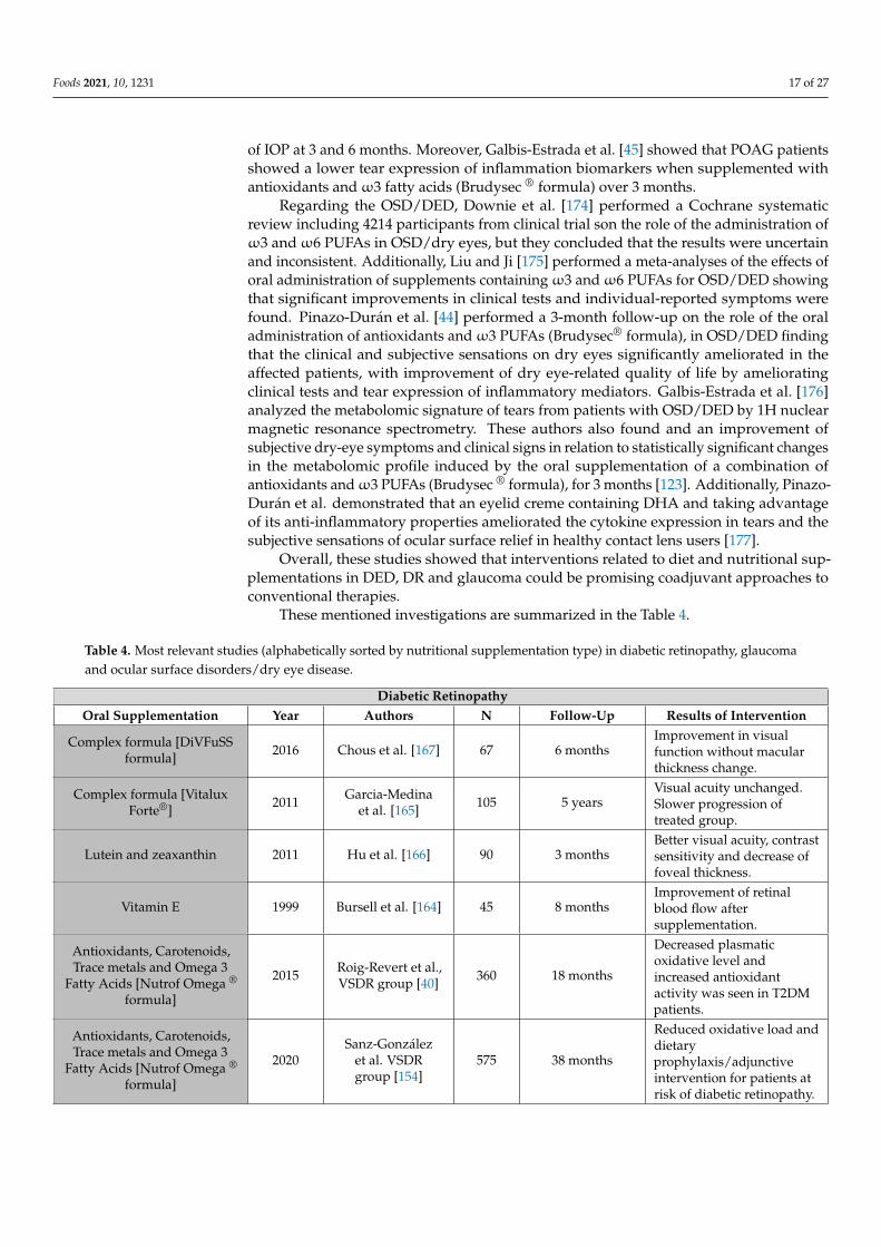
Walnut is the fruit of the juglans tree (Juglandaceae family), with strong, outspreadbranches, native probably from Persia (Juglans Regia). The walnuts are the round seedsof that tree, available in a range of distinct sizes and colors, and usually consumed as anut when the hard shell has been wide open and discarded. Walnuts are a rich sourceof bioactive nutritional components that has the ability to modulate multiple metabolicpathways that contribute to protection against many chronic diseases, including those thataffect the optic nerve, the retina and the ocular microcirculation. Most of these bioactiveshave a synergistic effect, acting in the protection of the physiological metabolic and vascularpathways [125]. Walnuts have high contents of polyphenols, phytosterols, Υ-tocopherol,and mainly α-linolenic acid (ALA), in addition to minerals [125], as listed in Table 1for the Juglans Californica, being the general properties antioxidant, anti-inflammatory,neuroprotection, anti-thrombotic, anti-arrhythmic, cardiovascular protection, cholesterol-lowering and improve gut microbiota.
Table 1. Main components of walnuts and biomedical properties [125].
Walnuts (Juglans Californica)Content Concentration/100 g
Polyphenols 2500 mgPhytosterols 113 mg
α-linolenic acid ~8000 mgΥ-tocopherol 21 mg
Sodium 2 mgPotassium 441 mg
Magnesium 158 mgCalcium 98 mg
Phytomelatonin 350 ng
Human clinical trials have suggested an association of walnut consumption withbetter cognitive performance and memory improvement in adults, with beneficial effectson memory, learning, motor coordination, anxiety, and locomotor activity [126–128]. Thesestudies also concluded on the benefits of a walnut-enriched diet in brain disorders and

Foods 2021, 10, 1231 13 of 27
other chronic diseases [126–129]. The additive effect of the essential components of wal-nuts is proven, with protective action against the events related to oxidative stress andinflammation present in different chronic diseases [125,130]. All these positive healtheffects can be obtained in different eye diseases, such as glaucoma, DR and age-AMD [131],chronic pathologies with a degenerative character for the ocular structures, which sharecommon pathophysiological mechanisms, characterized by the presence of events relatedto oxidative stress and inflammation. Likewise, in recent decades, with the increase in lifeexpectancy and the progressive growth of the population with its consequent aging, wehave noticed a significant increase in the incidence of chronic neurodegenerative disorders,as in the case of AMD. Its socio-economic consequences are evident, both in terms of thedecrease in the quality of life of those affected and in terms of a considerable pressing inthe health care system and increased financial burden.
Recent experimental evidence suggests that the main polyphenols of walnuts, ellagi-tannins and their metabolites (urolithins), have beneficial properties against the oxidationprocesses of cellular components and in the inflammation pathways, in addition to posi-tively influencing the intestinal microbiome [125,132]. Phytosterols have proven antioxidantproperties and are partly responsible for their cholesterol-lowering effect. They are pow-erful free radical scavengers, acting to reduce pro-inflammatory eicosanoids, and thenmitigating the inflammatory response [130,131].
The metabolism of ALA—the vegetableω3 fatty acid—gives rise to vasodilator andanti-inflammatory oxylipins, which can be the basis for a protective action on the functionof capillary endothelial cells. Its neuroprotective capacity has also been described onbrain function, inducing vasodilation of the cerebral arteries with improved irrigationand contributing to phenomena related to neuroplasticity. These effects could also beobserved in the retina and the optic nerve [133–138]. Interestingly, in addition to its alreadyknown anti-arrhythmic potential, ALA can exert other beneficial effects on cardiovascularfunction, through an anti-thrombotic, anti-inflammatory, and cholesterol-lowering action.The latter are considered protective factors against atherosclerosis [137]. On the oculartissues, its vasculoprotective action could contribute to an improved endothelial functionin the microcirculation of the retina, the cribriform plate and the choriocapillaries [137,138].
Finally, walnuts are also rich in Υ-tocopherol—a form of vitamin E—as we have ex-plained before, a powerful antioxidant with anti-inflammatory properties, with protectiveand preventive action in macular diseases, such as AMD [139]. Non-sodium minerals suchas potassium, calcium and magnesium, shared by all nuts, and especially in walnuts, alsohave a protective effect on cardio-metabolic risk, as confirmed by recent evidence [138,139].
Primary prevention in many of these neurodegenerative diseases is crucial from thepoint of view of public health and could be achieved early in life by introducing a healthydiet, rich in antioxidant and anti-inflammatory phytochemicals, as is the case with dietarysupplementation with walnuts, as for its nutritional value for ocular chronic diseases.
5. Mediterranean Diet—Current Knowledge on Eye Diseases
Dr. Keys, a physiologist from Minnesota (USA), analyzed in the second half of thetwentieth century the nutritional habits and lifestyle of people settled in the EuropeanMediterranean countries. He conducted various larger population studies to find a notice-able reduction in the incidence of cardiovascular and other chronic disorders, altogetherwith a higher lifespan of this area. In his renowned publication of “the seven countriesstudy,” [140] he hypothesized basically that cholesterol fat and the intake of food contain-ing cholesterol are important hallmarks of morbimortality. Among the milestones of theMedDiet are the following: (1) the elevated consumption of bread, cereals, olive oil, fruits,legumes, and vegetables altogether with the low consumption of saturated fat; (2) themoderate-to-high intake of chicken and fish; (3) the moderate intake of cheese, yogurt, andwine; and (4) the low consumption of red meat. In addition, a healthy lifestyle should befollowed, doing physical activity regularly. Its beneficial effects on health motivated itsappointment of Intangible Cultural Heritage of Humanity in November 2010. Therefore,

Foods 2021, 10, 1231 14 of 27
the United Nations Educational, Scientific and Cultural Organization (UNESCO) raisedthe MedDiet as a lifestyle and cultural heritage for humanity.
Numerous studies have been conducted on its protective effects against chroniccardiovascular-inflammatory-metabolic- neurodegenerative diseases, such as cardiovas-cular disorders [140–142], diabetes mellitus [143,144], obesity [145,146], cognitive de-cline/dementia [147,148], and Alzheimer disease [149]. Additionally, there are studiesshowing the possible protective effect of MedDiet in age-related ocular pathologies.
In this sense, Raimundo et al. [150] carried out a nested case-control study withinthe Coimbra Eye Study in patients with AMD. They used a food consumption frequencyquestionnaire and a scale initially developed for the Greek population (mediSCORE). Theauthors concluded that high adherence to a MedDiet protects against AMD due, mainly,to the high intake of antioxidants. Keenan and colleagues [145] found similar results in aretrospective cohort-based study of the AREDS and AREDS2. In this case, the authors usedthe Alternative Mediterranean diet Index (aMedi) based on food frequency questionnairesand observed a lower risk of AMD progression in those patients with high adherenceto MedDiet.
There is also evidence of the protective effect of the MedDiet on the onset of DR.Díaz-López et al. [151] conducted a nutritional intervention study in patients with T2DMwho did not have microvascular complications at the beginning of the study. Threedietary interventions were analyzed: MedDiet supplemented with extra virgin olive oil,MedDiet supplemented with walnuts and a low-fat control diet. After a 6-year follow-up,it was found that MedDiet enriched with extra virgin olive oil had a protective effecton the development of DR. Other research carried out within the Prevention with theMediterranean Diet (PREDIMED) study concludes that 500 mg/day ofω3 fatty acids, anamount easily achievable with good adherence to MedDiet, significantly reduces the riskof developing DR [57] and mortality was analyzed under dietary α-Linolenic Acid andmarineω3 fatty acids [152].
Regarding glaucoma, Abreu-Reyes et al. [153] conducted an interesting observationalstudy in the Spanish Canary Islands on the adherence to MedDiet in 100 patients withprimary open-angle glaucoma (POAG). The authors reported moderate adherence toMedDiet in 71% of the subjects. It would be advisable to carry out more interventionstudies to estimate the potential benefit of MedDiet in the glaucoma risk and progression.
In the Valencia study of diabetic retinopathy (VSDR) the influence of MedDiet innutritional outcomes of type 2 diabetics with or without DR were analyzed [40,154]. Ac-cording to the PREDIMED study [57] a 14-item questionnaire to assess the adherence tothe MedDiet of T2DM patients was applied, to be compared with those from the healthycontrols (CG). Scores indicating compliance to MedDiet distinguished between those par-ticipants with a high intake of bread, cereals, fish, fruits, legumes, olive oil, vegetables, andred wine that were positives (1), while those with a lower intake of the above foods werenegatives (0). Data from this questionnaire showed average values significantly higher inthe CG (9.8 ± 2.1) versus the T2DM patients (6.4 ± 1.1) (p < 0.05). The comparison of theprooxidant and antioxidant markers analyzed in the present study in the T2DMG withpoor and good adherence to the MedDiet is reflected in Table 2.
Table 2. Comparative data on plasmatic redox biomarkers according to the adherence to MedDiet.
Type 2 Diabetics with DR Type 2 Diabetics without DR p ValuePoor Adherence
MedDietGood Adherence
MedDietPoor Adherence
MedDietGood Adherence
MedDiet
MDA/TBARS (mm/L) 4 ± 1 3 ± 1 2 ± 1 2 ± 1 p < 0.001
TAC(mM) 2 ± 1 2 ± 2 3 ± 1 3 ± 2 p < 0.051
DR: Diabetic Retinopathy; MDA/TBARS: Malondialdehyde/Thiobarbituric Acid Reactive Substances; TAC: Total Antioxidant Capacity;MedDiet: Mediterranean Diet. Data are shown as mean ± standard deviation. p value obtained from ANOVA analysis [40].

Foods 2021, 10, 1231 15 of 27
Controversial results have been reported on the effects of the MedDiet regarding DED,cataracts, and glaucoma. Molina-Leiva and the PREDIMED PLUS study [155] concludedthat implementing the MedDiet and lifestyle pattern to benefit dry eye patients.
A prospective case-control study was done by our group in 2016–2017, in collaborationwith the Department of Ophthalmology of the Careggi Hospital (University of Florence,Italy), including 112 consecutive individuals of both sexes aged 25–80 years, to addressthe adherence to the MedDiet in an ophthalmologic population of Valencia (Spain) [156].Patients were included on the basis of the diagnosis of DR, AMD, POAG, or cataracts. Bymeans of the slit lamp (ImageNet, Topcon, Barcelona, Spain) the ocular fundus examinationwas done to confirm DR diagnosis, and biomicrosopy of the anterior eye segment andmedia was done to confirm cataracts diagnosis. The POAG group was addressed by theIOP elevation (>21 mm Hg) according to the central corneal thickness values, as well as bymorphologic/structural and functional probes (optic disc examination, optic coherencetomography (OCT) and visual field performance), as suggested by Castejon-Cerveró et al.,over the European Glaucoma Society Guidelines for Spain [157]. The AMD was diagnosedon the basis of the ocular fundus and OCT examination as suggested by the Spanish Societyof Retina and Vitreous (SERV) guidelines [158]. Personal interviews and the 14-itemsvalidated MedDiet questionnaire were done. Additionally, socio-demographic data, riskfactors for ocular diseases and ocular examination were performed. The questionnairescores indicating compliance to the MedDiet is able to distinguish between participantswith a high intake of cereals, legumes, fruits, vegetables, olive oil, fish, nuts, industrialpastry, wine and meat (red or white meat, cold meat) and those with a low intake of thesefoods. Good adherence to the MedDiet is scored with a total of 9 points of 14. Mean age ofthe suitable participants was 55.8 ± 14.1 years (58,7 % females and 41.3% males). Averagescore of the questionnaire was 8.4 ± 2.2 suggesting suboptimal adherence for almost halfof the selected participants without differences across the cases and the controls (Table 3).
Table 3. Adherence to MedDiet in eye patients and healthy controls [155].
CG (n = 58) Ocular Pathologies (n = 52)
Cataracts(n = 16)
Glaucoma(n = 13)
DR(n = 23)
Adherence level 8.4 ± 1.8 8.8 ± 2.3 8.9 ± 2.2 8.3 ± 2.1
p value Ref 0.519 0.371 0.775CG: control group; DR: diabetic retinopathy.
No statistical differences were found in adherence to Mediterranean diet by age(p = 0.536) or sex (p = 0.104) in the participants suffering the above ocular diseases. More-over, no individual item differed significantly between the analyzed ocular disease patientsexcept for a trend towards a larger consumption of fish.
This work concluded that adherence to MedDiet was suboptimal in half of patients,regardless of their ocular disease status, including diagnoses of cataract, glaucoma and DR.Data suggested that educational interventions to improve the dietary habits of ophthalmicoutpatients could be investigated in further prospective studies.
In summary, MedDiet may be considered a protective factor against the onset of theleading causes of blindness. Further investigations are essential to deep on the knowledgeof benefits of this diet to appropriately recommend it to the population, and to assess thepossibility of using nutritional supplementation in cases of low-medium adherence to aMedDiet, to uncover methods for prevention of vision loss and quality of life.
6. Nutritional Supplements
The most prevalent sight-threatening ophthalmic diseases are DR and glaucoma,leading causes of vision loss in the global population. Dietary habits can contribute toDR initiation and progression. Lower dietary fiber is associated with increased risk ofdeveloping DR [159]. Plus, once these complications appear, intensive glycemic control

Foods 2021, 10, 1231 16 of 27
has been demonstrated to reduce the rate of diabetic DR [158]. MedDiet, characterizedby being reach in fruits and vegetables and unrefined carbohydrates, has been related toreduced risk of DR incidence [151,160].
Concerning glaucoma, two epidemiological studies included in the Study of Osteo-porotic Fractures found a decrease in risk of developing glaucoma when high consumptionrates of green leaves and vegetables, fruits and fruit juice were consumed [161,162]. Recentsystematic reviews found that selenium and iron may increase the risk of glaucoma but therelationships of dietary intake of other substances did not present a strong association withthe risk of glaucoma [50] and a beneficial association among dietary intake of vitamin Aand C with open angle glaucoma [163].
There is an increased interest in nutritional supplementation as coadjuvant therapyin DR and glaucoma patients. Different types of methodologies and supplements, mainlyvitamins, have been studied. Outcomes have been considered in terms of clinical parame-ters (visual acuity, IOP, global indices of visual field examination, degree of retinopathy orthicknesses at different ocular locations assessed by OCT. The most relevant investigationsin this sense are mentioned below.
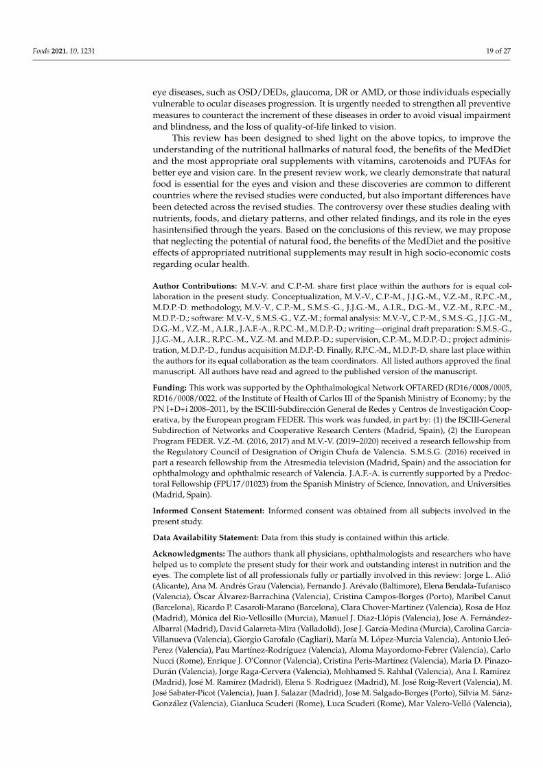
In relation to DR, Bursell et al. [164] investigated the supplementation with high-doseof α-tocopherol in a group of type-1 diabetes mellitus patients and found an increaseof retinal blood flow that improved after 8-month supplementation. Combination ofvitamins and other microelements have also been considered. A study by our group withfive-year follow-up dealt with the effects of a complex composed of lutein, α-tocopherol,niacin, beta-carotene, zinc and selenium in 105 type-2 diabetic patients suffering from non-proliferative diabetic retinopathy (NPDR). We found a decrease in the clinical funduscopicprogression but visual acuity did not change [165]. In contrast, Hu et al. [166] found animprovement of visual acuity, and also of contrast sensitivity and foveal thickness in type-1and type-2 diabetic patients with NPDR after a 3-month supplementation with lutein adzeaxanthin. Similarly, Chous et al. [167] demonstrated an increase of visual acuity with noalteration of retinal thickness with the administration during 6 months to in type-1 andtype-2 diabetic patients with no retinopathy or NPDR of DiVFuSS complex containingvitamins C, D3 and E, zinc oxide, eicosapentaenoic acid, docosahexaenoic acid, α-lipoicacid (racemic mixture), coenzyme Q10, mixed tocotrienols/tocopherols, zeaxanthin, lutein,benfotiamine, N-acetyl cysteine, grape seed extract, resveratrol, turmeric root extract, greentea leaf, and pycnogenol. The VSDR group performed a prospective case-control studyin 575 participants during 38-month follow-up on the effects of the daily intake of a pillcontaining antioxidants, trace metals and ω3 fatty acids (Nutrof Omega ® formula), inT2DM patients, with and without DR and healthy controls, concluding that this coursechanges reduced the oxidative load in patients at risk of DR [40,153].
In relation to glaucoma, supplementation with black currant anthocyanins extractfor two years was related to a better visual field performance in comparison with non-supplemented group in glaucoma [168]. Plus, this extract was proved to be related to adecrease of IOP in healthy and glaucoma patients [169]. In addition, supplementation withginkgo biloba extract during a mean period of over 12 years showed a slower progressionin perimetric global indices in normotensive glaucoma [170]. Alternatively, nutritionalsupplementation by our group with two complexes of vitamins and other substances(one with and one without ω3 fatty acid supplementation) on IOP-controlled primaryopen-angle glaucoma patients did not overcome control group in terms of visual field,peripapillary nerve fiber layer thickness or macular ganglion cell complex thickness in atwo-year follow-up. Thus, we concluded that this kind of supplementation did not seemto be useful [171]. In contrast, Mutolo et al. [172] administered another complex (contain-ing different vitamins such as B1, B2, and B6) and they found a decrease of IOP and anamelioration in eletroretinogram results in a 12-month follow-up in open-angle glaucomapatients. More recently, Romeo Villadoniga et al. [173] found that supplementation to pseu-doexfoliative glaucoma patients with a complex containing DHA (and other componentssuch as EPA, vitamins B, C, E, lutein, zeaxanthin, and minerals) was related to a decrease
Foods 2021, 10, 1231 17 of 27
of IOP at 3 and 6 months. Moreover, Galbis-Estrada et al. [45] showed that POAG patientsshowed a lower tear expression of inflammation biomarkers when supplemented withantioxidants andω3 fatty acids (Brudysec ® formula) over 3 months.
Regarding the OSD/DED, Downie et al. [174] performed a Cochrane systematicreview including 4214 participants from clinical trial son the role of the administration ofω3 andω6 PUFAs in OSD/dry eyes, but they concluded that the results were uncertainand inconsistent. Additionally, Liu and Ji [175] performed a meta-analyses of the effects oforal administration of supplements containingω3 andω6 PUFAs for OSD/DED showingthat significant improvements in clinical tests and individual-reported symptoms werefound. Pinazo-Durán et al. [44] performed a 3-month follow-up on the role of the oraladministration of antioxidants andω3 PUFAs (Brudysec® formula), in OSD/DED findingthat the clinical and subjective sensations on dry eyes significantly ameliorated in theaffected patients, with improvement of dry eye-related quality of life by amelioratingclinical tests and tear expression of inflammatory mediators. Galbis-Estrada et al. [176]analyzed the metabolomic signature of tears from patients with OSD/DED by 1H nuclearmagnetic resonance spectrometry. These authors also found and an improvement ofsubjective dry-eye symptoms and clinical signs in relation to statistically significant changesin the metabolomic profile induced by the oral supplementation of a combination ofantioxidants andω3 PUFAs (Brudysec ® formula), for 3 months [123]. Additionally, Pinazo-Durán et al. demonstrated that an eyelid creme containing DHA and taking advantageof its anti-inflammatory properties ameliorated the cytokine expression in tears and thesubjective sensations of ocular surface relief in healthy contact lens users [177].
Overall, these studies showed that interventions related to diet and nutritional sup-plementations in DED, DR and glaucoma could be promising coadjuvant approaches toconventional therapies.
These mentioned investigations are summarized in the Table 4.
Table 4. Most relevant studies (alphabetically sorted by nutritional supplementation type) in diabetic retinopathy, glaucomaand ocular surface disorders/dry eye disease.
Diabetic RetinopathyOral Supplementation Year Authors N Follow-Up Results of Intervention
Complex formula [DiVFuSSformula] 2016 Chous et al. [167] 67 6 months
Improvement in visualfunction without macularthickness change.
Complex formula [VitaluxForte®] 2011 Garcia-Medina
et al. [165] 105 5 yearsVisual acuity unchanged.Slower progression oftreated group.
Lutein and zeaxanthin 2011 Hu et al. [166] 90 3 monthsBetter visual acuity, contrastsensitivity and decrease offoveal thickness.
Vitamin E 1999 Bursell et al. [164] 45 8 monthsImprovement of retinalblood flow aftersupplementation.
Antioxidants, Carotenoids,Trace metals and Omega 3
Fatty Acids [Nutrof Omega ®
formula]
2015 Roig-Revert et al.,VSDR group [40] 360 18 months
Decreased plasmaticoxidative level andincreased antioxidantactivity was seen in T2DMpatients.
Antioxidants, Carotenoids,Trace metals and Omega 3
Fatty Acids [Nutrof Omega ®
formula]
2020Sanz-González
et al. VSDRgroup [154]
575 38 months
Reduced oxidative load anddietaryprophylaxis/adjunctiveintervention for patients atrisk of diabetic retinopathy.

Foods 2021, 10, 1231 18 of 27
Table 4. Cont.
GlaucomaOral Supplementation Year Authors N Follow-Up Results of Intervention
AREDS-based formulas 2015 Garcia-Medinaet al. [171] 117 2 years
No differences in visualfield indexes, RGClcomplex, peripapillaryretinal nerve fiber layer.
Black currant anthocyanins 2012 Ohguro et al. [168] 38 24 monthsBetter mean deviationchange (visual field index)in supplemented group.
Black currant anthocyanins 2013 Ohguro et al. [169] 21 4 weeksIOP decrease at 2 and 4weeks in treated group butno change in placebo group.
Docosahexaenoic acid 2018 Romeo Villadonigaet al. [173] 47 6 months IOP decrease at 3 and 6
months in treated eyes.Formula containing forskolin,homotaurine, carnosine, and
folic acid2016 Mutolo et al. [172] 44 1 year
IOP lowering, ERGimprovement and fovealsensitivity.
Antioxidants and Omega 3fatty acids [Brudysec ®
formula]2013 Galbis-Estrada
et al. [45] 97 3 monthsReduced inflammationbiomarkers in glaucomatoustears.
Ginkgo biloba extract 2013 Lee et al. [170] 42 12 yearsSlower progression of visualfield damage in treatedpatients.
Ocular Surface Disorders/Dry Eyes
Omega 3 and Omega 6polyunsaturated fatty acids 2019 Downie et al. [174] 4214
Cochrane systematic reviewof clinical trials evidenceuncertain/inconsistent forDEDs.
Omega 3 and Omega 6polyunsaturated fatty acids 2014 Liu and Ji, [175] 790
Meta-analyses ofrandomized,placebo-controlled studies.Improvement of clinicaltests, individual-reportedsymptoms in DEDs.
Antioxidants and Omega 3fatty acids [Brudysec ®
formula]2013 Pinazo-Duran
et al. [44] 66
Benefit DED patients.Improvement of dryeye-related quality of life.Ameliorating clinical testsand tear expression ofinflammatory mediators.
Antioxidants and Omega 3fatty acids [Brudysec ®
formula]2015 Galbis-Estrada
et al. [123] 90
Improvement of subjectivedry-eye symptoms bychanging the tearmetabolomic profile
Omega 3 fatty acids [BrudyDerm Dry Eye Gel ®] 2021 Pinazo-Durán et al.
[177] 72
Ameliorating ocular surfacerelief and decreasingcytokine expression in tearsfrom contact lenses users.
7. Concluding Remarks
Knowledge on nutrition interventions for the prevention of chronic eye diseases isfar from complete, as reflected in this review article. In fact, the extended lifespan and theincreased prevalence of sight-threatening diseases have a huge socio-economic impact onhealthcare systems, individuals, and their families worldwide.
Ophthalmologists, researchers, and policymakers, have to pay special attention to thediet and lifestyle patterns of the population, to identify people at high risk of developing
Foods 2021, 10, 1231 19 of 27
eye diseases, such as OSD/DEDs, glaucoma, DR or AMD, or those individuals especiallyvulnerable to ocular diseases progression. It is urgently needed to strengthen all preventivemeasures to counteract the increment of these diseases in order to avoid visual impairmentand blindness, and the loss of quality-of-life linked to vision.
This review has been designed to shed light on the above topics, to improve theunderstanding of the nutritional hallmarks of natural food, the benefits of the MedDietand the most appropriate oral supplements with vitamins, carotenoids and PUFAs forbetter eye and vision care. In the present review work, we clearly demonstrate that naturalfood is essential for the eyes and vision and these discoveries are common to differentcountries where the revised studies were conducted, but also important differences havebeen detected across the revised studies. The controversy over these studies dealing withnutrients, foods, and dietary patterns, and other related findings, and its role in the eyeshasintensified through the years. Based on the conclusions of this review, we may proposethat neglecting the potential of natural food, the benefits of the MedDiet and the positiveeffects of appropriated nutritional supplements may result in high socio-economic costsregarding ocular health.
Author Contributions: M.V.-V. and C.P.-M. share first place within the authors for is equal col-laboration in the present study. Conceptualization, M.V.-V., C.P.-M., J.J.G.-M., V.Z.-M., R.P.C.-M.,M.D.P.-D. methodology, M.V.-V., C.P.-M., S.M.S.-G., J.J.G.-M., A.I.R., D.G.-M., V.Z.-M., R.P.C.-M.,M.D.P.-D.; software: M.V.-V., S.M.S.-G., V.Z.-M.; formal analysis: M.V.-V., C.P.-M., S.M.S.-G., J.J.G.-M.,D.G.-M., V.Z.-M., A.I.R., J.A.F.-A., R.P.C.-M., M.D.P.-D.; writing—original draft preparation: S.M.S.-G.,J.J.G.-M., A.I.R., R.P.C.-M., V.Z.-M. and M.D.P.-D.; supervision, C.P.-M., M.D.P.-D.; project adminis-tration, M.D.P.-D., fundus acquisition M.D.P.-D. Finally, R.P.C.-M., M.D.P.-D. share last place withinthe authors for its equal collaboration as the team coordinators. All listed authors approved the finalmanuscript. All authors have read and agreed to the published version of the manuscript.
Funding: This work was supported by the Ophthalmological Network OFTARED (RD16/0008/0005,RD16/0008/0022‚ of the Institute of Health of Carlos III of the Spanish Ministry of Economy; by thePN I+D+i 2008–2011, by the ISCIII-Subdirección General de Redes y Centros de Investigación Coop-erativa, by the European program FEDER. This work was funded, in part by: (1) the ISCIII-GeneralSubdirection of Networks and Cooperative Research Centers (Madrid, Spain), (2) the EuropeanProgram FEDER. V.Z.-M. (2016, 2017) and M.V.-V. (2019–2020) received a research fellowship fromthe Regulatory Council of Designation of Origin Chufa de Valencia. S.M.S.G. (2016) received inpart a research fellowship from the Atresmedia television (Madrid, Spain) and the association forophthalmology and ophthalmic research of Valencia. J.A.F.-A. is currently supported by a Predoc-toral Fellowship (FPU17/01023) from the Spanish Ministry of Science, Innovation, and Universities(Madrid, Spain).
Informed Consent Statement: Informed consent was obtained from all subjects involved in thepresent study.
Data Availability Statement: Data from this study is contained within this article.
Acknowledgments: The authors thank all physicians, ophthalmologists and researchers who havehelped us to complete the present study for their work and outstanding interest in nutrition and theeyes. The complete list of all professionals fully or partially involved in this review: Jorge L. Alió(Alicante), Ana M. Andrés Grau (Valencia), Fernando J. Arévalo (Baltimore), Elena Bendala-Tufanisco(Valencia), Óscar Álvarez-Barrachina (Valencia), Cristina Campos-Borges (Porto), Maribel Canut(Barcelona), Ricardo P. Casaroli-Marano (Barcelona), Clara Chover-Martínez (Valencia), Rosa de Hoz(Madrid), Mónica del Rio-Vellosillo (Murcia), Manuel J. Diaz-Llópis (Valencia), Jose A. Fernández-Albarral (Madrid), David Galarreta-Mira (Valladolid), Jose J. García-Medina (Murcia), Carolina García-Villanueva (Valencia), Giorgio Garofalo (Cagliari), María M. López-Murcia Valencia), Antonio Lleó-Perez (Valencia), Pau Martínez-Rodríguez (Valencia), Aloma Mayordomo-Febrer (Valencia), CarloNucci (Rome), Enrique J. O’Connor (Valencia), Cristina Peris-Martínez (Valencia), Maria D. Pinazo-Durán (Valencia), Jorge Raga-Cervera (Valencia), Mohhamed S. Rahhal (Valencia), Ana I. Ramírez(Madrid), José M. Ramírez (Madrid), Elena S. Rodriguez (Madrid), M. José Roig-Revert (Valencia), M.José Sabater-Picot (Valencia), Juan J. Salazar (Madrid), Jose M. Salgado-Borges (Porto), Silvia M. Sánz-González (Valencia), Gianluca Scuderi (Rome), Luca Scuderi (Rome), Mar Valero-Velló (Valencia),

Foods 2021, 10, 1231 20 of 27
Carlos Vergés (Barcelona), Gianni Virgili (Florence), Vicente Zanón-Moreno (Valencia). We especiallywish to thank D. Corella (Valencia, Spain), G. Virgili (Florence, Italy), and J. F. Arévalo (Baltimore,MD, USA) for peer interactive communication, and for the thoughtful comments and critical readingof the manuscript. The authors and the involved professionals in the present study want to thankLaboratorios Thea, S.A., (Barcelona, Spain) for gently providing the Nutrof Omega® formulation thatwas given to the participants during the described study course on diabetic retinopathy. Also, wewould like to thank Brudylab S. L. (Barcelona, Spain) for the donation of the Brudysec ® formula thatwas used in the DED studies described herein. In addition, the authors want to thank the RegulatoryCouncil of Designation of Origin Chufa de Valencia (Alboraya, Valencia, Spain) for kindly given thetubers that were consumed by the volunteers during the corresponding studies.
Conflicts of Interest: All authors of this work have disclosed that they have no significant financialrelationships or financial interests in the commercial companies that are related to this study or paper.
References1. WHO. World Report on Vision; World Health Organization: Geneva, Switzerland, 2019; Available online: https://www.who.int/
publications-detail-redirect/world-report-on-vision (accessed on 10 May 2021).2. GBD 2019 Blindness and Vision Impairment Collaborators; Vision Loss Expert Group of the Global Burden of Disease Study.
Causes of blindness and vision impairment in 2020 and trends over 30 years, and prevalence of avoidable blindness in relation toVISION 2020: The Right to Sight: An analysis for the Global Burden of Disease Study. Lancet Glob. Health 2021, 9, e144–e160.
3. Zhang, J.; Tuo, J.; Wang, Z.; Zhu, A.; Machalinska, A.; Long, Q. Pathogenesis of Common Ocular Diseases. J. Ophthalmol. 2015,2015, 734527. [CrossRef]
4. Lanzetta, P.; Sarao, V.; Scanlon, P.H.; Barratt, J.; Porta, M.; Bandello, F.; Loewenstein, A. Fundamental principles of an effectivediabetic retinopathy screening program. Acta Diabetol. 2020, 57, 785–798. [CrossRef]
5. Pondorfer, S.G.; Terheyden, J.H.; Heinemann, M.; Wintergerst, M.W.M.; Holz, F.G.; Finger, R.P. Association of Vision-relatedQuality of Life with Visual Function in Age-Related Macular Degeneration. Sci. Rep. 2019, 9, 15326. [CrossRef]
6. Pinazo-Durán, M.D.; Zanón-Moreno, V.; García-Medina, J.J.; Arévalo, J.F.; Gallego-Pinazo, R.; Nucci, C. Eclectic Ocular Comor-bidities and Systemic Diseases with Eye Involvement: A Review. BioMed Res. Int. 2016, 2016, 6215745. [CrossRef] [PubMed]
7. Pinazo-Duran, M.D.; Zanon-Moreno, V.; Garcıa-Medina, J.J.; Gallego-Pinazo, R. Evaluation of presumptive biomarkers of oxidativestress, immune response and apoptosis in primary open-angle glaucoma. Curr. Opin. Pharmacol. 2013, 13, 98–107. [CrossRef]
8. Pinazo-Durán, M.; Gallego-Pinazo, R.; García-Medina, J.J.; Zanón-Moreno, V.; Nucci, C.; Dolz-Marco, R.; Martínez-Castillo, S.;Galbis-Estrada, C.; Marco-Ramírez, C.; López-Gálvez, M.I.; et al. Oxidative stress in aging eyes. Clin. Interv. Aging 2014, 9,637–652. [CrossRef] [PubMed]
9. Nita, M.; Grzybowski, A. The Role of the Reactive Oxygen Species and Oxidative Stress in the Pathomechanism of the Age-RelatedOcular Diseases and Other Pathologies of the Anterior and Posterior Eye Segments in Adults. Oxidative Med. Cell. Longev. 2016,2016, 3164734. [CrossRef]
10. Finkel, T. Signal transduction by reactive oxygen species. J. Cell Biol. 2011, 194, 7–15. [CrossRef]11. Schieber, M.; Chandel, N.S. ROS function in redox signaling and oxidative stress. Curr. Biol. 2014, 24, R453–R462. [CrossRef]12. Pinazo-Durán, M.D.; Muñoz-Negrete, F.J.; Sanz-González, S.M.; Benítez-Del-Castillo, J.; Giménez-Gómez, R.; Valero-Velló, M.;
Zanón-Moreno, V.; García-Medina, J.J. The role of neuroinflammation in the pathogenesis of glaucoma neurodegeneration. Prog.Brain Res. 2020, 256, 99–124.
13. Kim, Y.-M.; Byzova, T.V. Oxidative stress in angiogenesis and vascular disease. Blood 2014, 5, 625–631. [CrossRef]14. Dong, A.; Xie, B.; Shen, J.; Yoshida, T.; Yokoi, K.; Hackett, S.F.; Campochiaro, P.A. Oxidative stress promotes ocular neovascular-
ization. J. Cell Physiol. 2009, 219, 544–552. [CrossRef] [PubMed]15. Przedborski, S.; Vila, M.; Jackson-Lewis, V. Neurodegeneration: What is it and where are we? J. Clin. Investig. 2003, 111, 3–10.
[CrossRef] [PubMed]16. Maresca, A.; la Morgia, C.; Caporali, L.; Valentino, M.L.; Carelli, V. The optic nerve: A “mito-window” on mitochondrial
neurodegeneration. Mol. Cell Neurosci. 2013, 55, 62–76. [CrossRef]17. Pinazo-Duran, M.D.; Zanon-Moreno, V.; Gallego-Pinazo, R.; Garcıa-Medina, J.J. Oxidative stress and mitochondrial failure in the
pathogenesis of glaucoma neurodegeneration. Prog. Brain Res. 2015, 220, 127–153. [PubMed]18. Garcia, K.C.; Adams, E.J. How the T cell receptor sees antigen—A structural view. Cell 2005, 122, 333–336. [CrossRef] [PubMed]19. Lemke, H. Immune Response Regulation by Antigen Receptors’ Clone-Specific Nonself Parts. Front. Immunol. 2018, 9, 1471.
[CrossRef] [PubMed]20. Kumar, B.V.; Connors, T.J.; Farber, D.L. Human T Cell Development, Localization, and Function throughout Life. Immunity 2018,
48, 202–2013. [CrossRef]21. Benitez-Del-Castillo, J.; Cantu-Dibildox, J.; Sanz-González, S.M.; Zanón-Moreno, V.; Pinazo-Duran, M.D. Cytokine expression
in tears of patients with glaucoma or dry eye disease: A prospective, observational cohort study. Eur. J. Ophthalmol. 2019, 29,437–443. [CrossRef]
Foods 2021, 10, 1231 21 of 27
22. Murakami, Y.; Ishikawa, K.; Nakao, S.; Sonoda, K.H. Innate immune response in retinal homeostasis and inflammatory disorders.Prog. Retin. Eye Res. 2020, 74, 100778. [CrossRef]
23. Wooff, Y.; Man, S.M.; Aggio-Bruce, R.; Natoli, R.; Fernando, N. IL-1 family members mediate cell death, inflammation andangiogenesis in retinal degenerative diseases. Front. Immunol. 2019, 10, 1618. [CrossRef]
24. Zindler, E.; Zipp, F. Neuronal injury in chronic CNS inflammation. Best Pract. Res. Clin. Anaesthesiol. 2010, 24, 551–562. [CrossRef]25. Osborne, N.N. Pathogenesis of ganglion “cell death” in glaucoma and neuroprotection: Focus on ganglion cell axonal mitochon-
dria. Prog. Brain Res. 2008, 173, 339–352.26. Carelli, V.; La Morgia, C.; Ross-Cisneros, F.N.; Sadun, A.A. Optic neuropathies: The tip of the neurodegeneration iceberg. Hum.
Mol. Genet. 2017, 26, R139–R150. [CrossRef] [PubMed]27. Chitranshi, N.; Dheer, Y.; Abbasi, M.; You, Y.; Graham, S.L.; Gupta, V. Glaucoma pathogenesis and neurotrophins: Focus on the
molecular and genetic basis for therapeutic prospects. Curr. Neuropharmacol. 2018, 16, 1018–1035. [CrossRef]28. Stein, J.D.; Newman-Casey, P.A.; Mrinalini, T.; Lee, P.P.; Hutton, D.W. Cost-effectiveness of bevacizumab and ranibizumab for
newly diagnosed neovascular macular degeneration (an American Ophthalmological Society thesis). Trans. Am. Ophthalmol. Soc.2013, 111, 56–69. [PubMed]
29. Niketeghad, S.; Pouratian, N. Brain Machine Interfaces for Vision Restoration: The Current State of Cortical Visual Prosthetics.Neurotherapeutics. J. Am. Soc. Exp. Neuro Ther. 2019, 16, 134–143. [CrossRef] [PubMed]
30. Micera, A.; Balzamino, B.O.; Di Zazzo, A.; Dinice, L.; Bonini, S.; Coassin, M. Biomarkers of Neurodegeneration and PrecisionTherapy in Retinal Disease. Front. Pharmacol. 2021, 11, 601647. [CrossRef]
31. Galvin, O.; Chi, G.; Brady, L.; Hippert, C.; Del Valle Rubido, M.; Daly, A.; Michaelides, M. The Impact of Inherited RetinalDiseases in the Republic of Ireland (ROI) and the United Kingdom (UK) from a Cost-of-Illness Perspective. Clin. Ophthalmol.2020, 14, 707–719. [CrossRef]
32. Singh, R.K.; Nasonkin, I.O. Limitations and Promise of Retinal Tissue from Human Pluripotent Stem Cells for DevelopingTherapies of Blindness. Front. Cell. Neurosci. 2020, 14, 179. [CrossRef]
33. Ebi, K.L.; Harris, F.; Sioen, G.B.; Wannous, C.; Anyamba, A.; Bi, P.; Boeckmann, M.; Bowen, K.; Cissé, G.; Dasgupta, P.; et al.Transdisciplinary Research Priorities for Human and Planetary Health in the Context of the 2030 Agenda for SustainableDevelopment. Int. J. Environ. Res. Public Health 2020, 17, 8890. [CrossRef]
34. Singh, A.; Banerjee, P.; Anas, M.; Singh, N.; Qamar, I. Traditional Nutritional and Health Practices Targeting Lifestyle BehavioralChanges in Humans. J. Lifestyle Med. 2020, 10, 67–73. [CrossRef]
35. Bae, J.H.; Shin, D.S.; Lee, S.C.; Hwang, I.C. Sodium Intake and Socioeconomic Status as Risk Factors for Development ofAge-Related Cataracts: The Korea National Health and Nutrition Examination Survey. PLoS ONE 2015, 10, e0136218. [CrossRef]
36. Age-Related Eye Disease Study Research Group. A randomized, placebo-controlled, clinical trial of high-dose supplementationwith vitamins C and E, beta carotene, and zinc for age-related macular degeneration and vision loss: AREDS report no. 8. Arch.Ophthalmol. 2001, 119, 1417–1436. [CrossRef] [PubMed]
37. Age-Related Eye Disease Study 2 Research Group. Lutein + zeaxanthin and omega-3 fatty acids for age-related macular degeneration:The Age-Related Eye Disease Study 2 (AREDS2) randomized clinical trial. JAMA 2013, 309, 2005–2015. [CrossRef] [PubMed]
38. Agrón, E.; Mares, J.; Clemons, T.E.; Swaroop, A.; Chew, E.Y.; Keenan, T.D.L.; AREDS and AREDS2 Research Groups. DietaryNutrient Intake and Progression to Late Age-Related Macular Degeneration in the Age-Related Eye Disease Studies 1 and 2.Ophthalmology 2021, 128, 425–442. [CrossRef] [PubMed]
39. Snodderly, D.M.; Mares, J.A.; Wooten, B.R.; Oxton, L.; Gruber, M.; Ficek, T.; CAREDS Macular Pigment Study Group. Macularpigment measurement by heterochromatic flicker photometry in older subjects: The carotenoids and age-related eye diseasestudy. Investig. Ophthalmol. Vis. Sci. 2004, 45, 531–538. [CrossRef]
40. Roig-Revert, M.J.; Lleó-Pérez, A.; Zanón-Moreno, V.; Vivar-Llopis, B.; Marín-Montiel, J.; Dolz-Marco, R.; Alonso-Muñoz, L.;Albert-Fort, M.; López-Gálvez, M.I.; Galarreta-Mira, D.; et al. Valencia Study On Diabetic Retinopathy (VSDR). EnhancedOxidative Stress and Other Potential Biomarkers for Retinopathy in Type 2 Diabetics: Beneficial Effects of the NutraceuticSupplements. BioMed Res. Int. 2015, 2015, 408180. [CrossRef]
41. Moeller, S.M.; Voland, R.; Tinker, L.; Blodi, B.A.; Klein, M.L.; Gehrs, K.M.; Johnson, E.J.; Snodderly, D.M.; Wallace, R.B.;Chappell, R.J.; et al. Associations between age-related nuclear cataract and lutein and zeaxanthin in the diet and serum in theCarotenoids in the Age-Related Eye Disease Study, an Ancillary Study of the Women’s Health Initiative. Arch. Ophthalmol. 2008, 126,354–364. [CrossRef]
42. Ferrero, A.; Alassane, S.; Binquet, C.; Bretillon, L.; Acar, N.; Arnould, L.; Muselier-Mathieu, A.; Delcourt, C.; Bron, A.M.;Creuzot-Garcher, C. Dry eye disease in the elderly in a French population-based study (the Montrachet study: Maculopathy, OpticNerve, nuTRition, neurovAsCular and HEarT diseases): Prevalence and associated factors. Ocul. Surf. 2018, 16, 112–119. [CrossRef]
43. Rand, A.L.; Asbell, P.A. Nutritional supplements for dry eye syndrome. Curr. Opin. Pharmacol. 2011, 22, 279–282. [CrossRef]44. Pinazo-Durán, M.D.; Galbis-Estrada, C.; Pons-Vázquez, S.; Cantú-Dibildox, J.; Marco-Ramírez, C.; Benítez-del-Castillo, J. Effects of a
nutraceutical formulation based on the combination of antioxidants andω-3 essential fatty acids in the expression of inflammationand immune response mediators in tears from patients with dry eye disorders. Clin. Interv. Aging 2013, 8, 139–148. [CrossRef]
45. Galbis-Estrada, C.; Pinazo-Durán, M.D.; Cantú-Dibildox, J.; Marco-Ramírez, C.; Díaz-Llópis, M.; Benítez-del-Castillo, J. Patientsundergoing long-term treatment with antihypertensive eye drops responded positively with respect to their ocular surfacedisorder to oral supplementation with antioxidants and essential fatty acids. Clin. Interv. Aging 2013, 8, 711–719.

Foods 2021, 10, 1231 22 of 27
46. Barabino, S.; Horwath-Winter, J.; Messmer, E.M.; Rolando, M.; Aragona, P.; Kinoshita, S. The role of systemic and topical fattyacids for dry eye treatment. Prog. Retin. Eye Res. 2017, 61, 23–34. [CrossRef] [PubMed]
47. Signes-Soler, I.; Javaloy Estañ, J. Nutrition and dry eye: A systematic review. Exp. Rev. Ophthalmol. 2019, 14, 1–18. [CrossRef]48. Francisco, S.G.; Smith, K.M.; Aragonès, G.; Whitcomb, E.A.; Weinberg, J.; Wang, X.; Bejarano, E.; Taylor, A.; Rowan, S. Dietary
Patterns, Carbohydrates, and Age-Related Eye Diseases. Nutrients 2020, 12, 2862. [CrossRef] [PubMed]49. Morrone, L.A.; Rombolà, L.; Corasaniti, M.T.; Bagetta, G.; Nucci, C.; Russo, R. Natural compounds and retinal ganglion cell
neuroprotection. Prog. Brain Res. 2015, 220, 257–281. [PubMed]50. Ramdas, W.D. The relation between dietary intake and glaucoma: A systematic review. Acta Ophthalmol. 2018, 96, 550–556.
[CrossRef] [PubMed]51. Al Owaifeer, A.M.; Al Taisan, A.A. The Role of Diet in Glaucoma: A Review of the Current Evidence. Ophthalmol. Ther. 2018, 7,
19–31. [CrossRef]52. Pinazo-Duran, M.D.; Shoaie-Nia, K.; Zanon-Moreno, V.; Sanz-Gonzalez, S.M.; Del Castillo, J.B.; Garcia-Medina, J.J. Strategies to
Reduce Oxidative Stress in Glaucoma Patients. Curr. Neuropharmacol. 2018, 16, 903–918. [CrossRef]53. Saccà, S.C.; Corazza, P.; Gandolfi, S.; Ferrari, D.; Sukkar, S.; Iorio, E.L.; Traverso, C.E. Substances of Interest That Support
Glaucoma Therapy. Nutrients 2019, 11, 239. [CrossRef] [PubMed]54. Pinazo-Duran, M.D.; Bendala Tufanisco, E. Guidelines for Nutrition in Retinal Diseases; Domenech Pujades: Barcelona, Spain, 2018;
ISBN 978-84-697-4909-8.55. Salas-Salvadó, J.; Guasch-Ferré, M.; Lee, C.H.; Estruch, R.; Clish, C.B.; Ros, E. Protective Effects of the Mediterranean Diet on
Type 2 Diabetes and Metabolic Syndrome. J. Nutr. 2015, 146, 920S–927S. [CrossRef] [PubMed]56. Merle, B.M.J.; Rosner, B.; Seddon, J.M. Genetic Susceptibility, Diet Quality, and Two-Step Progression in Drusen Size. Investig.
Ophthalmol. Vis. Sci. 2020, 61, 17. [CrossRef] [PubMed]57. Sala-Vila, A.; Díaz-López, A.; Valls-Pedret, C.; Cofán, M.; García-Layana, A.; Lamuela-Raventós, R.M.; Castañer, O.;
Zanon-Moreno, V.; Martinez-Gonzalez, M.A.; Toledo, E.; et al. Dietary Marineω-3 Fatty Acids and Incident Sight-ThreateningRetinopathy in Middle-Aged and Older Individuals With Type 2 Diabetes: Prospective Investigation From the PREDIMED Trial.JAMA Ophthalmol. 2016, 134, 1142–1149. [CrossRef] [PubMed]
58. Camacho-Barcia, L.; Bulló, M.; García-Gavilán, J.F.; Martínez-González, M.A.; Corella, D.; Estruch, R.; Fitó, M.; Gómez-Gracia, E.;Arós, F.; Fiol, M.; et al. Dairy products intake and the risk of incident cataracts surgery in an elderly Mediterranean population:Results from the PREDIMED study. Eur. J. Nutr. 2019, 58, 619–627. [CrossRef]
59. Shea, B.J.; Reeves, B.C.; Wells, G.; Thuku, M.; Hamel, C.; Moran, J.; Moher, D.; Tugwell, P.; Welch, V.; Kristjansson, E.; et al.AMSTAR 2: A critical appraisal tool for systematic reviews that include randomised or non-randomised studies of healthcareinterventions, or both. BMJ (Clin. Res. Ed.) 2017, 358, j4008. [CrossRef] [PubMed]
60. Viswanathan, M.; Patnode, C.; Berkman, N.D.; Bass, E.B.; Chang, S.; Hartling, L.; Murad, H.M.; Treadwell, J.R.; Kane, R.L.Assessing the Risk of Bias in Systematic Reviews of Health Care Interventions. Methods Guide for Comparative Effectiveness Reviews.(Prepared by the Scientific Resource Center under Contract No. 290-2012-0004-C); AHRQ Publication No. 17(18)-EHC036-EF; Agencyfor Healthcare Research and Quality: Rockville, MD, USA, 2017; pp. 1–29.
61. Hoffmann-Eßer, W.; Siering, U.; Neugebauer, E.A.; Brockhaus, A.C.; Lampert, U.; Eikermann, M. Guideline appraisal withAGREE II: Systematic review of the current evidence on how users handle the 2 overall assessments. PLoS ONE 2017, 12,e0174831. [CrossRef]
62. Favela-González, K.M.; Hernández-Almanza, A.Y.; De la Fuente-Salcido, N.M. The value of bioactive compounds of cruciferousvegetables (Brassica) as antimicrobials and antioxidants: A review. J. Food Biochem. 2020, 44, e13414. [CrossRef] [PubMed]
63. Campas-Baypoli, O.N.; Sánchez-Machado, D.I.; Bueno-Solano, C.; Núñez-Gastélum, J.A.; Reyes-Moreno, C.; López-Cervantes, J.Biochemical composition and physicochemical properties of broccoli flours. Int. J. Food Sci. Nutr. 2009, 60 (Suppl. 4), 163–173. [CrossRef]
64. Le, T.N.; Luong, H.Q.; Li, H.P.; Chiu, C.H.; Hsieh, P.C. Broccoli (Brassica oleracea L. var. italica) Sprouts as the Potential FoodSource for Bioactive Properties: A Comprehensive Study on In Vitro Disease Models. Foods 2019, 8, 532. [CrossRef]
65. Le, T.N.; Chiu, C.H.; Hsieh, P.C. Bioactive Compounds and Bioactivities of Brassica oleracea L. var. Italica Sprouts and Microgreens:An Updated Overview from a Nutraceutical Perspective. Plants 2020, 9, 946. [CrossRef]
66. Hwang, J.-H.; Lim, S.B. Antioxidant and Anti-inflammatory Activities of Broccoli Florets in LPS-stimulated RAW 264.7 Cells.Prev. Nutr. Food Sci. 2014, 19, 89–97. [CrossRef]
67. Kalpana Deepa Priya, D.; Gayathri, R.; Gunassekaran, G.R.; Murugan, S.; Sakthisekaran, D. Apoptotic role of natural isothiocyanatefrom broccoli (Brassica oleracea italica) in experimental chemical lung carcinogenesis. Pharm. Biol. 2013, 51, 621–628. [CrossRef]
68. Elkashty, O.A.; Tran, S.D. Broccoli extract increases drug-mediated cytotoxicity towards cancer stem cells of head and necksquamous cell carcinoma. Br. J. Cancer 2020, 123, 1395–1403. [CrossRef]
69. Fuente, B.; López-García, G.; Máñez, V.; Alegría, A.; Barberá, R.; Cilla, A. Antiproliferative Effect of Bioaccessible Fractions ofFour Brassicaceae Microgreens on Human Colon Cancer Cells Linked to Their Phytochemical Composition. Antioxidants 2020, 9,368. [CrossRef] [PubMed]
70. Jabbarzadeh Kaboli, P.; Afzalipour Khoshkbejari, M.; Mohammadi, M.; Abiri, A.; Mokhtarian, R.; Vazifemand, R.; Amanollahi, S.;Yazdi Sani, S.; Li, M.; Zhao, Y.; et al. Targets and mechanisms of sulforaphane derivatives obtained from cruciferous plants with specialfocus on breast cancer—Contradictory effects and future perspectives. BioMed Pharmacother. 2020, 121, 109635. [CrossRef] [PubMed]

Foods 2021, 10, 1231 23 of 27
71. Giacoppo, S.; Galuppo, M.; Montaut, S.; Iori, R.; Rollin, P.; Bramanti, P.; Mazzon, E. An overview on neuroprotective effects ofisothiocyanates for the treatment of neurodegenerative diseases. Fitoterapia 2015, 106, 12–21. [CrossRef]
72. Schepici, G.; Bramanti, P.; Mazzon, E. Efficacy of Sulforaphane in Neurodegenerative Diseases. Int. J. Mol. Sci. 2020, 21, 8637.[CrossRef] [PubMed]
73. Yamagishi, S.; Matsui, T. Protective role of sulphoraphane against vascular complications in diabetes. Pharm. Biol. 2016, 54,2329–2339. [CrossRef]
74. Rein, D.B.; Wittenborn, J.S.; Zhang, X.; Honeycutt, A.A.; Lesesne, S.B.; Saaddine, J. Vision Health Cost-Effectiveness Study Group.Forecasting age-related macular degeneration through the year 2050: The potential impact of new treatments. Arch. Ophthalmol.2009, 127, 533–540. [CrossRef]
75. Bone, R.A.; Landrum, J.T.; Tarsis, S.L. Preliminary identification of the human macular pigment. Vis. Res. 1985, 25, 1531–1535. [CrossRef]76. Arunkumar, R.; Calvo, C.M.; Conrady, C.D.; Bernstein, P.S. What do we know about the macular pigment in AMD: The past, the
present, and the future. Eye 2018, 2, 992–1004. [CrossRef]77. Age-Related Eye Disease Study Research Group; SanGiovanni, J.P.; Chew, E.Y.; Clemons, T.E.; Ferris, F.L., 3rd; Gensler, G.;
Lindblad, A.S.; Milton, R.C.; Seddon, J.M.; Sperduto, R.D. The relationship of dietary carotenoid and vitamin A, E, and C intakewith age-related macular degeneration in a case-control study: AREDS Report No. 22. Arch. Ophthalmol. 2007, 125, 1225–1232.
78. Age-Related Eye Disease Study 2 (AREDS2) Research Group; Chew, E.Y.; Clemons, T.E.; Sangiovanni, J.P.; Danis, R.P.; Ferris, F.L.,3rd; Elman, M.J.; Antoszyk, A.N.; Ruby, A.J.; Orth, D.; et al. Secondary analyses of the effects of lutein/zeaxanthin on age-relatedmacular degeneration progression: AREDS2 report No. 3. JAMA Ophthalmol. 2014, 132, 142–149. [CrossRef]
79. Bernstein, P.S.; Delori, F.C.; Richer, S.; van Kuijk, F.J.; Wenzel, A.J. The value of measurement of macular carotenoid pigment opticaldensities and distributions in age-related macular degeneration and other retinal disorders. Vis. Res. 2010, 50, 716–728. [CrossRef]
80. Dolz-Marco, R.; Gallego-Pinazo, R.; Pinazo-Durán, M.D.; Diaz-Llópis, M. Macular pigment optical density. In Thea Monographiesof Information and Research in Ophthalmology; 2013; Volume 68, pp. 1–20. ISSN 84-1887-4096. Available online: https://www.laboratoriosthea.com/medias/thea_informacion_68.pdf (accessed on 10 May 2021).
81. Yu, L.; Gao, B.; Li, Y.; Wang, T.T.Y.; Luo, Y.; Wang, J.; Yu, L.L. Home food preparation techniques impacted the availability ofnatural antioxidants and bioactivities in kale and broccoli. Food Funct. 2018, 9, 585–593. [CrossRef]
82. Hwang, J.H.; Lim, S.B. Antioxidant and anticancer activities of broccoli by-products from different cultivars and maturity stagesat harvest. Prev Nutr Food Sci. 2015, 20, 8–14. [CrossRef] [PubMed]
83. Matsui, T.; Nakamura, N.; Ojima, A.; Nishino, Y.; Yamagishi, S.I. Sulforaphane reduces advanced glycation end products(AGEs)-induced inflammation in endothelial cells and rat aorta. Nutr Metab Cardiovasc Dis. 2016, 26, 797–807. [CrossRef]
84. Klomparens, E.A.; Ding, Y. The neuroprotective mechanisms and effects of sulforaphane. Brain Circ. 2019, 5, 74–83.85. Vanduchova, A.; Anzenbacher, P.; Anzenbacherova, E. Isothiocyanate from Broccoli, Sulforaphane, and Its Properties. J. Med.
Food 2019, 22, 121–126. [CrossRef]86. Basker, D.; Negbi, M. Uses of saffron. Econ. Bot. 1983, 37, 228–236. [CrossRef]87. Karimi, E.; Oskoueian, E.; Hendra, R.; Jaafar, H.Z.E. Evaluation of Crocus sativus L. Stigma Phenolic and Flavonoid Compounds
and Its Antioxidant Activity. Molecules 2010, 15, 6244–6256. [PubMed]88. Hashemi, M.; Hosseinzadeh, H. A comprehensive review on biological activities and toxicology of crocetin. Food Chem. Toxicol.
2019, 130, 44–60. [CrossRef]89. Xi, L.; Qian, Z.; Du, P.; Fu, J. Pharmacokinetic properties of crocin (crocetin digentiobiose ester) following oral administration in
rats. Phytomedicine 2007, 14, 633–636. [CrossRef]90. Hosseini, A.; Razavi, B.M.; Hosseinzadeh, H. Pharmacokinetic Properties of Saffron and its Active Components. Eur. J. Drug
Metab. Pharmacokinet. 2018, 43, 383–390. [CrossRef] [PubMed]91. Ghaffari, S.; Roshanravan, N. Saffron; An updated review on biological properties with special focus on cardiovascular effects.
Biomed. Pharmacother. 2019, 109, 21–27. [CrossRef] [PubMed]92. Fernández-Albarral, J.A.; De Hoz, R.; Ramírez, A.I.; López-Cuenca, I.; Salobrar-García, E.; Pinazo-Durán, M.D.; Ramírez, J.M.;
Salazar, J.J. Beneficial effects of saffron (Crocus sativus L.) in ocular pathologies, particularly neurodegenerative retinal diseases.Neural Regen. Res. 2020, 15, 1408–1416. [CrossRef] [PubMed]
93. Assimopoulou, A.N.; Sinakos, Z.; Papageorgiou, V.P. Radical scavenging activity of Crocus sativus L. extract and its bioactiveconstituents. Phyther. Res. 2005, 19, 997–1000. [CrossRef] [PubMed]
94. Zeinali, M.; Zirak, M.R.; Rezaee, S.A.; Karimi, G.; Hosseinzadeh, H. Immunoregulatory and anti-inflammatory properties ofCrocus sativus (Saffron) and its main active constituents: A review. Iran. J. Basic Med. Sci. 2019, 22, 334–344. [PubMed]
95. Khazdair, M.R.; Boskabady, M.H.; Hosseini, M.; Rezaee, R.; Tsatsakis, A.M. The effects of Crocus sativus (saffron) and itsconstituents on nervous system: A review. Avicenna J. Phytomed. 2015, 5, 376–391. [PubMed]
96. Akhondzadeh, S.; Sabet, M.S.; Harirchian, M.H.; Togha, M.; Cheraghmakani, H.; Razeghi, S.; Hejazi, S.S.; Yousefi, M.H.;Alimardani, R.; Jamshidi, A.; et al. Saffron in the treatment of patients with mild to moderate Alzheimer’s disease: A 16-week,randomized and placebo-controlled trial. J. Clin. Pharm. Ther. 2010, 35, 581–588. [CrossRef] [PubMed]
97. Piccardi, M.; Marangoni, D.; Minnella, A.M.; Savastano, M.C.; Valentini, P.; Ambrosio, L.; Capoluongo, E.; Maccarone, R.; Bisti, S.;Falsini, B. A longitudinal follow-up study of saffron supplementation in early age-related macular degeneration: Sustainedbenefits to central retinal function. Evid. Based Complement. Altern. Med. 2012, 2012, 429124. [CrossRef] [PubMed]

Foods 2021, 10, 1231 24 of 27
98. Lashay, A.; Sadough, G.; Ashrafi, E.; Lashay, M.; Movassat, M.; Akhondzadeh, S. Short-term Outcomes of Saffron Supplementationin Patients with Age-related Macular Degeneration: A Double-blind, Placebo-controlled, Randomized Trial. Med. HypothesisDiscov. Innov. Ophthalmol. J. 2016, 5, 32–38.
99. Yaribeygi, H.; Atkin, S.L.; Sahebkar, A. A review of the molecular mechanisms of hyperglycemia-induced free radical generationleading to oxidative stress. J. Cell. Physiol. 2019, 234, 1300–1312. [CrossRef] [PubMed]
100. Sepahi, S.; Mohajeri, S.A.; Hosseini, S.M.; Khodaverdi, E.; Shoeibi, N.; Namdari, M.; Tabassi, S.A.S. Effects of Crocin on DiabeticMaculopathy: A Placebo-Controlled Randomized Clinical Trial. Am. J. Ophthalmol. 2018, 190, 89–98. [CrossRef]
101. Hecht, I.; Achiron, A.; Bartov, E.; Maharshak, I.; Mendel, L.; Pe’er, L.; Bar, A.; Burgansky-Eliash, Z. Effects of dietary and lifestylerecommendations on patients with glaucoma: A randomized controlled pilot trial. Eur. J. Integr. Med. 2019, 25, 60–66. [CrossRef]
102. Jabbarpoor Bonyadi, M.H.; Yazdani, S.; Saadat, S. The ocular hypotensive effect of saffron extract in primary open angle glaucoma:A pilot study. BMC Complement. Altern. Med. 2014, 14, 399. [CrossRef]
103. Fernández-Albarral, J.A.; Ramírez, A.I.; de Hoz, R.; López-Villarín, N.; Salobrar-García, E.; López-Cuenca, I.; Licastro, E.;Inarejos-García, A.M.; Almodóvar, P.; Pinazo-Durán, M.D.; et al. Neuroprotective and anti-inflammatory effects of a hydrophilicsaffron extract in a model of glaucoma. Int. J. Mol. Sci. 2019, 20, 4110. [CrossRef]
104. Bixquert Jiménez, M. Horchata y Salud. Propiedades saludables y de prevención de enfermedades digestivas. In Jornada Chufa yHorchata, Tradición y Salud. Generalitat Valenciana; Conselleria de Agricultura, Pesca y Alimentación: Valencia, Spain, 2003; pp. 71–85.
105. Codina-Torrella, I.; Guamis, B.; Trujillo, A.J. Characterization and comparison of tiger nuts (Cyperus esculentus L.) from differentgeographical origin. Ind. Crops Prod. 2015, 65, 406–414. [CrossRef]
106. Granato, D.; Nunes, D.S.; Barba, F.J. An integrated strategy between food chemistry, biology, nutrition, pharmacology, andstatistics in the development of functional foods: A proposal. Trends Food Sci. Technol. 2017, 62, 13–22. [CrossRef]
107. Adel, A.A.M.; Awad, A.M.; Mohamed, H.H.; Iryna, S. Chemical composition, physicochemical properties and fatty acid profile oftiger nut (Cyperus esculentus L) seed oil as affected by different preparation methods. Int. Food Res. J. 2015, 22, 1931–1938.
108. Roselló-Soto, E.; Poojary, M.M.; Barba, F.J.; Lorenzo, J.M.; Mañes, J.; Moltó, J.C. Tiger nut and its by-products valorization: Fromextraction of oil and valuable compounds to development of new healthy products. Innov. Food Sci. Emerg. Technol. 2017, 45,306–312. [CrossRef]
109. King Jaime II of Aragon. Letter to Ermengol Blasi about the Chufa Properties. (6 December 1297). Available online: http://www.chufadevalencia.org/ver/244/Historia.html (accessed on 10 May 2021).
110. Vilanova, A. Valencia Cathedral Archive. Manuscrito de la Biblioteca Capitular de Valencia, nº 123, 1307.111. Cavanilles, A.J. Observaciones Sobre la Historia Natural, Geografía, Agricultura, Población y Frutos del Reino de Valencia; Imprenta Real:
Madrid, Spain, 1975; Available online: https://bivaldi.gva.es/es/consulta/registro.cmd?id=284 (accessed on 10 May 2021).112. Denominación de Origen Chufa de Valencia. Available online: http://www.chufadevalencia.org/ (accessed on 10 May 2021).113. Udefa, A.L.; Amama, E.A.; Archibong, E.A.; Nwangwa, J.N.; Adama, S.; Inyang, V.U.; Inyaka, G.U.; Aju, G.J.; Okpa, S.; Inah, I.O.
Antioxidant, anti-inflammatory and anti-apoptotic effects of hydro-ethanolic extract of Cyperus esculentus L. (tigernut) on leadacetate-induced testicular dysfunction in Wistar rats. Biomed. Pharmacother. 2020, 129, 110491. [CrossRef]
114. Oguwike, F.N.; Nwosu, P.N.; Nwafor, C.; Onumonu, C.; Eluke, B.C.; Eze, R.I.; Asika, C.M. The effects of Cyperus esculentus (Tigernut) on Haematological and Biochemical Profile of Male Hypercholesteremic Subjects in Uli, Anambra State Nigeria. Greener J.Med. Sci. 2017, 7, 36–41. [CrossRef]
115. Zommara, M.; Imaizumi, K. Antiatherogenic effect of Tiger nut tubers (Cyperus esculentus L.) supplemented diet in apolipoproteinE knockout mice. J. Sustain. Agric. Sci. 2018, 43, 197–2014. [CrossRef]
116. Albahrani, A.A.; Greaves, R.F. Fat-Soluble Vitamins: Clinical Indications and Current Challenges for Chromatographic Measure-ment. Clin. Biochem. Rev. 2016, 37, 27–47.
117. Böhm, V. Vitamin E. Antioxidants 2018, 7, 44. [CrossRef]118. Pinheiro-Sant’Ana, H.; Guinazi, M.; Oliveira, D.; Della Lucia, C.; Reis, B.; Brandão, S. Method for simultaneous analysis of eight
vitamin E isomers in various foods by high performance liquid chromatography and fluorescence detection. J. Chromatogr. 2011,1218, 8496–8502. [CrossRef] [PubMed]
119. Tavares, F.P.; Fernandes, R.S.; Bernardes, T.F.; Bonfioli, A.A.; Soares, E.J. Dry eye disease. Semin. Ophthalmol. 2010, 25, 84–93.[CrossRef] [PubMed]
120. Tsubota, K.; Kawashima, M.; Inaba, T.; Dogru, M.; Matsumoto, Y.; Ishida, R.; Kaido, M.; Kojima, T.; Uchino, M.; Uchino, Y.; et al.The antiaging approach for the treatment of dry eye. Cornea 2012, 31 (Suppl. 1), S3–S8. [CrossRef]
121. Nakamura, S.; Shibuya, M.; Nakashima, H.; Hisamura, R.; Masuda, N.; Imagawa, T.; Uehara, M.; Tsubota, K. Involvement of oxidativestress on corneal epithelial alterations in a blink-suppressed dry eye. Investig. Ophthalmol. Vis. Sci. 2007, 48, 1552–1558. [CrossRef]
122. Enríquez-de-Salamanca, A.; Castellanos, E.; Stern, M.E.; Fernández, I.; Carreño, E.; García-Vázquez, C.; Herreras, J.M.; Calonge,M. Tear cytokine and chemokine analysis and clinical correlations in evaporative-type dry eye disease. Mol. Vis. 2010, 16, 862–873.
123. Galbis-Estrada, C.; Pinazo-Durán, M.D.; Martínez-Castillo, S.; Morales, J.M.; Monleón, D.; Zanon-Moreno, V. A metabolomicapproach to dry eye disorders. The role of oral supplements with antioxidants and omega 3 fatty acids. Mol Vis. 2015, 21, 555–567.
124. Saccà, S.C.; Cutolo, C.A.; Ferrari, D.; Corazza, P.; Traverso, C.E. The Eye, Oxidative Damage and Polyunsaturated Fatty Acids.Nutrients 2018, 10, 668. [CrossRef]
125. Ros, E.; Izquierdo-Pulido, M.; Sala-Vila, A. Beneficial Effects of Walnut Consumption on Human Health: Role of Micronutrients.Curr. Opin. Clin. Nutr. Metab. Care 2018, 21, 498–504. [CrossRef] [PubMed]
Foods 2021, 10, 1231 25 of 27
126. Poulose, S.M.; Miller, M.G.; Shukitt-Hale, B. Role of Walnuts in Maintaining Brain Health with Age. J. Nutr. 2014, 144, 561S–566S.[CrossRef] [PubMed]
127. Chauhan, A.; Chauhan, V. Beneficial Effects of Walnuts on Cognition and Brain Health. Nutrients 2020, 12, 550. [CrossRef] [PubMed]128. Sala-Vila, A.; Valls-Pedret, C.; Rajaram, S.; Coll-Padrós, N.; Cofán, M.; Serra-Mir, M.; Pérez-Heras, A.M.; Roth, I.; Freitas-Simoes, T.M.;
Doménech, M.; et al. Effect of a 2-Year Diet Intervention with Walnuts on Cognitive Decline. The Walnuts and Healthy Aging(WAHA) Study: A Randomized Controlled Trial. Am. J. Clin. Nutr. 2020, 111, 590–600. [CrossRef] [PubMed]
129. Rajaram, S.; Valls-Pedret, C.; Cofán, M.; Sabaté, J.; Serra-Mir, M.; Pérez-Heras, A.M.; Arechiga, A.; Casaroli-Marano, R.P.;Alforja, S.; Sala-Vila, A.; et al. The Walnuts and Healthy Aging Study (WAHA): Protocol for a Nutritional Intervention Trial withWalnuts on Brain Aging. Front. Aging Neurosci. 2017, 8, 333. [CrossRef] [PubMed]
130. Nunes, S.; Alves, D.; Barreto, P.; Raimundo, M.; da Luz Cachulo, M.; Farinha, C.; Laíns, I.; Rodrigues, J.; Almeida, C.;Ribeiro, L.; et al. Adherence to a Mediterranean Diet and Its Association with Age-Related Macular Degeneration. The CoimbraEye Study–Report 4. Nutrition 2018, 51–52, 6–12. [CrossRef] [PubMed]
131. Braakhuis, A.; Raman, R.; Vaghefi, E. The Association between Dietary Intake of Antioxidants and Ocular Disease. Diseases 2017,5, 3. [CrossRef] [PubMed]
132. Rinninella, E.; Mele, M.; Merendino, N.; Cintoni, M.; Anselmi, G.; Caporossi, A.; Gasbarrini, A.; Minnella, A. The Role of Diet,Micronutrients and the Gut Microbiota in Age-Related Macular Degeneration: New Perspectives from the Gut–Retina Axis.Nutrients 2018, 10, 1677. [CrossRef] [PubMed]
133. Tan, J.S.L. Dietary Fatty Acids and the 10-Year Incidence of Age-Related Macular Degeneration: The Blue Mountains Eye Study.Arch. Ophthalmol. 2009, 127, 656. [CrossRef]
134. Schleicher, M.; Weikel, K.; Garber, C.; Taylor, A. Diminishing Risk for Age-Related Macular Degeneration with Nutrition: ACurrent View. Nutrients 2013, 5, 2405–2456. [CrossRef]
135. Walchuk, C.; Suh, M. Nutrition and the aging retina: A comprehensive review of the relationship between nutrients and their rolein age-related macular degeneration and retina disease prevention. Adv. Food Nutr. Res. 2020, 93, 293–332.
136. Garcia-Medina, J.J.; Rubio-Velazquez, E.; Foulquie-Moreno, E.; Casaroli-Marano, R.P.; Pinazo-Duran, M.D.; Zanon-Moreno, V.;del-Rio-Vellosillo, M. Update on the Effects of Antioxidants on Diabetic Retinopathy: In Vitro Experiments, Animal Studies andClinical Trials. Antioxidants 2020, 9, 561. [CrossRef]
137. Berryman, C.E.; Grieger, J.A.; West, S.G.; Chen, C.-Y.O.; Blumberg, J.B.; Rothblat, G.H.; Sankaranarayanan, S.; Kris-Etherton,P.M. Acute Consumption of Walnuts and Walnut Components Differentially Affect Postprandial Lipemia, Endothelial Function,Oxidative Stress, and Cholesterol Efflux in Humans with Mild Hypercholesterolemia. J. Nutr. 2013, 143, 788–794. [CrossRef]
138. Sun, Y.; Smith, L.E.H. Retinal Vasculature in Development and Diseases. Annu. Rev. Vis. Sci. 2018, 4, 101–122. [CrossRef]139. Evans, J.R.; Lawrenson, J.G. Antioxidant Vitamin and Mineral Supplements for Slowing the Progression of Age-Related Macular
Degeneration. Cochrane Database Syst. Rev. 2017, 11, CD000254. [CrossRef]140. Keys, A.; Mienotti, A.; Karvonen, M.J.; Aravanis, C.; Blackburn, H.; Buzina, R.; Djordjevic, B.S.; Dontas, A.S.; Fidanza, F.;
Keys, M.H.; et al. The diet and 15-year death rate in the seven countries study. Am. J. Epidemiol. 1986, 124, 903. [CrossRef]141. Martín-Peláez, S.; Fito, M.; Castaner, O. Mediterranean Diet Effects on Type 2 Diabetes Prevention, Disease Progression, and
Related Mechanisms. A Review. Nutrients 2020, 12, 2236. [CrossRef]142. Esposito, K.; Maiorino, M.I.; Bellastella, G.; Chiodini, P.; Panagiotakos, D.; Giugliano, D. A journey into a Mediterranean diet and
type 2 diabetes: A systematic review with meta-analyses. BMJ Open 2015, 5, e008222. [CrossRef] [PubMed]143. D’Innocenzo, S.; Biagi, C.; Lanari, M. Obesity and the Mediterranean Diet: A Review of Evidence of the Role and Sustainability of
the Mediterranean Diet. Nutrients 2019, 11, 1306. [CrossRef]144. Martin-Calvo, N.; Chavarro, J.E.; Falbe, J.; Hu, F.B.; Field, A.E. Adherence to the Mediterranean dietary pattern and BMI change
among US adolescents. Int. J. Obes. 2016, 40, 1103–1108. [CrossRef]145. Keenan, T.D.; Agrón, E.; Mares, J.; Clemons, T.E.; van Asten, F.; Swaroop, A.; Chew, E.Y.; Age-Related Eye Disease Studies (AREDS)
1 and 2 Research Groups. Adherence to the Mediterranean Diet and Progression to Late Age-Related Macular Degeneration inthe Age-Related Eye Disease Studies 1 and 2. Ophthalmology 2020, 127, 1515–1528. [CrossRef] [PubMed]
146. Casas, R.; Sacanella, E.; Estruch, R. The immune protective effect of the Mediterranean diet against chronic low-grade inflamma-tory diseases. Endocr. Metab. Immune Disord. Drug Targets 2014, 14, 245–254. [CrossRef] [PubMed]
147. Karstens, A.J.; Tussing-Humphreys, L.; Zhan, L.; Rajendran, N.; Cohen, J.; Dion, C.; Zhou, X.J.; Lamar, M. Associations of theMediterranean diet with cognitive and neuroimaging phenotypes of dementia in healthy older adults. Am. J. Clin. Nutr. 2019,109, 361–368. [CrossRef] [PubMed]
148. Dominguez, L.J.; Barbagallo, M.; Muñoz-Garcia, M.; Godos, J.; Martinez-Gonzalez, M.A. Dietary Patterns and Cognitive Decline:Key features for prevention. Curr. Pharm. Des. 2019, 25, 2428–2442. [CrossRef] [PubMed]
149. Grewal, R.; Reutzel, M.; Dilberger, B.; Hein, H.; Zotzel, J.; Marx, S.; Tretzel, J.; Sarafeddinov, A.; Fuchs, C.; Eckert, G.P. Purifiedoleocanthal and ligstroside protect against mitochondrial dysfunction in models of early Alzheimer’s disease and brain ageing.Exp. Neurol. 2020, 328, 113248. [CrossRef]
150. Raimundo, M.; Mira, F.; Cachulo, M.D.L.; Barreto, P.; Ribeiro, L.; Farinha, C.; Laíns, I.; Nunes, S.; Alves, D.; Figueira, J.; et al.Adherence to a Mediterranean diet, lifestyle and age-related macular degeneration: The Coimbra Eye Study—Report 3. ActaOphthalmol. 2018, 96, e926–e932. [CrossRef]

Foods 2021, 10, 1231 26 of 27
151. Díaz-López, A.; Babio, N.; Martínez-González, M.A.; Corella, D.; Amor, A.J.; Fitó, M.; Estruch, R.; Arós, F.; Gómez-Gracia, E.;Fiol, M.; et al. Mediterranean Diet, Retinopathy, Nephropathy, and Microvascular Diabetes Complications: A Post Hoc Analysisof a Randomized Trial. Diabetes Care 2015, 38, 2134–2141. [CrossRef] [PubMed]
152. Sala-Vila, A.; Guasch-Ferré, M.; Hu, F.B.; Sánchez-Tainta, A.; Bulló, M.; Serra-Mir, M.; López-Sabater, C.; Sorlí, J.V.; Arós, F.; Fiol, M.; et al.Dietary α-Linolenic Acid, Marineω-3 Fatty Acids, and Mortality in a Population with High Fish Consumption: Findings From thePREvención con DIeta MEDiterránea (PREDIMED) Study. J. Am. Heart Assoc. 2016, 5, e002077. [CrossRef] [PubMed]
153. Abreu-Reyes, J.A.; Álvarez-Luis, D.; Arteaga-Hernández, V.; Sánchez-Mendez, M.; Abreu-González, R. Mediterranean dietadherence by patients with primary open angle glaucoma. Arch. Soc. Esp. Oftalmol. 2017, 92, 353–358. [CrossRef] [PubMed]
154. Sanz-González, S.M.; García-Medina, J.J.; Zanón-Moreno, V.; López-Gálvez, M.I.; Galarreta-Mira, D.; Duarte, L.; Valero-Velló, M.;Ramírez, A.I.; Arévalo, J.F.; Pinazo-Durán, M.D. On Behalf Of The Valencia Study Group On Diabetic Retinopathy VSDR. ReportNumber 5. Clinical and Molecular-Genetic Insights into the Role of Oxidative Stress in Diabetic Retinopathy: AntioxidantStrategies and Future Avenues. Antioxidants 2020, 9, 1101. [CrossRef] [PubMed]
155. Molina-Leyva, I.; Molina-Leyva, A.; Riquelme-Gallego, B.; Cano-Ibáñez, N.; García-Molina, L.; Bueno-Cavanillas, A. Effectivenessof Mediterranean Diet Implementation in Dry Eye Parameters: A Study of PREDIMED-PLUS Trial. Nutrients 2020, 12, 1289.[CrossRef] [PubMed]
156. Garofalo, G. Ocular Pathology and Adherence to the Mediterranean Diet: Scuola di Scienze della Salute Umana; Corso di Laurea/Laurea;Specialistica/Laurea Magistrale in Medicina e Chirurgia: Florence, Italy, 2016; pp. 1–28.
157. Castejón-Cervero, M.A.; Jiménez-Parras, R.; Fernandez-Arias, I.; Teus-Guezala, M.A.; IMCA Study Group. Evaluation ofcompliance with the EGS guidelines in Spain, using Achievable Benchmarks of Care (ABC®) methodology: The IMCA Study.Eur. J. Ophthalmol. 2011, 21, 149–155. [CrossRef]
158. Pareja-Ríos, A.; Serrano-García, M.A.; Marrero-Saavedra, M.D.; Abraldes-López, V.M.; Reyes-Rodríguez, M.A.; Cabrera-López, F.;López-Gálvez, M.; Cardona-Guerra, P.; Abreu-Reyes, P.; Quijada-Fumero, E.; et al. Guías de práctica clínica de la SERV: Manejode las complicaciones oculares de la diabetes. Retinopatía diabética y edema macular [Guidelines of clinical practice of theSERV (Spanish Retina and Vitreous Society): Management of ocular complications of diabetes. Diabetic retinopathy and macularoedema]. Arch. Soc. Esp. Oftalmol. 2009, 84, 429–450.
159. Diabetes Control and Complications Trial Research Group; Nathan, D.M.; Genuth, S.; Lachin, J.; Cleary, P.; Crofford, O.;Davis, M.; Rand, L.; Siebert, C. The Effect of Intensive Treatment of Diabetes on the Development and Progression of Long-TermComplications in Insulin-Dependent Diabetes Mellitus. N. Engl. J. Med. 1993, 329, 977–986.
160. Salas-Salvadó, J.; Bulló, M.; Babio, N.; Martínez-González, M.Á.; Ibarrola-Jurado, N.; Basora, J.; Estruch, R.; Covas, M.I.;Corella, D.; Arós, F.; et al. Erratum. Reduction in the incidence of type 2 diabetes with the Mediterranean diet: Results of thePREDIMED-Reus nutrition intervention randomized trial. Diabetes Care 2011, 34, 14–19. [CrossRef]
161. Coleman, A.L.; Stone, K.L.; Kodjebacheva, G.; Yu, F.; Pedula, K.L.; Ensrud, K.E.; Cauley, J.A.; Hochberg, M.C.; Topouzis, F.;Badala, F.; et al. Glaucoma Risk and the Consumption of Fruits and Vegetables among Older Women in the Study of OsteoporoticFractures. Am. J. Ophthalmol. 2008, 145, 1081–1089. [CrossRef]
162. Giaconi, J.A.; Yu, F.; Stone, K.L.; Pedula, K.L.; Ensrud, K.E.; Cauley, J.A.; Hochberg, M.C.; Coleman, A.L.; Study of OsteoporoticFractures Research Group. The Association of Consumption of Fruits/Vegetables with Decreased Risk of Glaucoma among OlderAfrican-American Women in the Study of Osteoporotic Fractures. Am. J. Ophthalmol. 2012, 154, 635–644. [CrossRef] [PubMed]
163. Ramdas, W.D.; Schouten, J.S.A.G.; Webers, C.A.B. The Effect of Vitamins on Glaucoma: A Systematic Review and Meta-Analysis.Nutrients. 2018, 10, 359. [CrossRef] [PubMed]
164. Bursell, S.E.; Clermont, A.C.; Aiello, L.P.; Aiello, L.M.; Schlossman, D.K.; Feener, E.P.; Laffel, L.; King, G.L. High-dose vitamin Esupplementation normalizes retinal blood flow and creatinine clearance in patients with type 1 diabetes. Diabetes Care 1999, 22,1245–1251. [CrossRef] [PubMed]
165. Garcia-Medina, J.J.; Pinazo-Duran, M.D.; Garcia-Medina, M.; Zanon-Moreno, V.; Pons-Vazquez, S. A 5-year follow-up ofantioxidant supplementation in type 2 diabetic retinopathy. Eur. J. Ophthalmol. 2011, 21, 637–643. [CrossRef]
166. Hu, B.J.; Hu, Y.N.; Lin, S.; Ma, W.J.; Li, X.R. Application of Lutein and Zeaxanthin in nonproliferative diabetic retinopathy. Int. J.Ophthalmol. 2011, 4, 303–306. [PubMed]
167. Chous, A.P.; Richer, S.P.; Gerson, J.D.; Kowluru, R.A. The Diabetes Visual Function Supplement Study (DiVFuSS). Br. J. Ophthalmol.2016, 100, 227–234. [CrossRef] [PubMed]
168. Ohguro, H.; Ohguro, I.; Katai, M.; Tanaka, S. Two-Year Randomized, Placebo-Controlled Study of Black Currant Anthocyanins onVisual Field in Glaucoma. Ophthalmologica 2012, 228, 26–35. [CrossRef]
169. Ohguro, H.; Ohguro, I.; Yagi, S. Effects of Black Currant Anthocyanins on Intraocular Pressure in Healthy Volunteers and Patientswith Glaucoma. J. Ocul. Pharmacol. Ther. 2013, 29, 61–67. [CrossRef]
170. Lee, J.; Sohn, S.W.; Kee, C. Effect of Ginkgo Biloba Extract on Visual Field Progression in Normal Tension Glaucoma. J. Glaucoma2013, 22, 780–784. [CrossRef]
171. Garcia-Medina, J.J.; Garcia-Medina, M.; Garrido-Fernandez, P.; Galvan-Espinosa, J.; Garcia-Maturana, C.; Zanon-Moreno, V.;Pinazo-Duran, M.D. A Two-Year Follow-up of Oral Antioxidant Supplementation in Primary Open-Angle Glaucoma: AnOpen-Label, Randomized, Controlled Trial. Acta Ophthalmol. 2015, 93, 546–554. [CrossRef] [PubMed]
Foods 2021, 10, 1231 27 of 27
172. Mutolo, M.G.; Albanese, G.; Rusciano, D.; Pescosolido, N. Oral administration of forskolin, homotaurine, carnosine, and folicacid in patients with primary open angle glaucoma: Changes in intraocular pressure, pattern electroretinogram amplitude, andfoveal sensitivity. J. Ocul. Pharmacol. Ther. 2016, 32, 178–183. [CrossRef] [PubMed]
173. Romeo Villadóniga, S.; Rodríguez García, E.; Sagastagoia Epelde, O.; Álvarez Díaz, M.D.; Domingo Pedrol, J.C. Effects of OralSupplementation with Docosahexaenoic Acid (DHA) plus Antioxidants in Pseudoexfoliative Glaucoma: A 6-Month Open-LabelRandomized Trial. J. Ophthalmol. 2018, 2018, 8259371. [CrossRef] [PubMed]
174. Downie, L.E.; Ng, S.M.; Lindsley, K.B.; Akpek, E.K. Omega-3 and omega-6 polyunsaturated fatty acids for dry eye disease.Cochrane Database Syst. Rev. 2019, 12, CD011016. [CrossRef]
175. Liu, A.; Ji, J. Omega-3 essential fatty acids therapy for dry eye syndrome: A meta-analysis of randomized controlled studies. Med.Sci. Monit. 2014, 20, 1583–1589. [PubMed]
176. Galbis-Estrada, C.; Martinez-Castillo, S.; Morales, J.M.; Vivar-Llopis, B.; Monleón, D.; Díaz-Llopis, M.; Pinazo-Durán, M.D.Differential effects of dry eye disorders on metabolomic profile by 1H nuclear magnetic resonance spectroscopy. Biomed. Res. Int.2014, 2014, 542549. [CrossRef] [PubMed]
177. Pinazo-Durán, M.D.; Raga-Cervera, J.; Sanz-González, S.M.; Salgado-Borges, J.; Benítez-Del-Castillo, J.; Ramírez, A.I.;Zanón-Moreno, V. Efficacy and safety study of an eyelid gel after repeated nocturnal application in healthy contact lens usersand non-users. J. Optom. 2021, 14, 28–36. [CrossRef]