Embed Size (px)
Citation preview

Functional Foods in Health and Disease 2013; 3(11):447-461 Page 447 of 461
Research Article Open Access
Antioxidant and anti-inflammatory activities of loquat
(Eriobotrya japonica) tea
Phyu Phyu Khine Zar1, Kozue Sakao
2, Fumio Hashimoto
1,3, Akiko Morishita
2, Makoto
Fujii4, Koji Wada
1,5 and De-Xing Hou
1, 2*
1Course of Biochemical Science and Technology, United Graduate School of Agricultural
Sciences; 2Department of Biochemical Science and Technology;
3Department of Horticultural
Science, Faculty of Agriculture, Kagoshima University, Kagoshima 890-0065, Japan; 4Totsukawa Noujou Ltd Co, Nejime, Kagoshima 893-2503, Japan;
5Faculty of Agriculture,
University of the Ryukyus, Senbaru, Nishihara 1, Okinawa 903-0213, Japan
Corresponding author: Prof. De-Xing Hou, Faculty of Agriculture, Kagoshima University,
Kagoshima 890-0065, Japan
Submission date: October 14, 2013; Acceptance date: November 24, 2013; Publication date:
November 28, 2013
ABSTRACT
Background: Fresh loquat leaves contain several kinds of flavonoids and have been reported to
have preventive effects against some human diseases such as diabetes, coughs and ulcers,.
Recently, fresh loquat leaves in Japan were processed to a beverage, called loquat tea, after the
fresh leaves are roasted at 350C for 30 minutes. However, the scientific evidence supporting the
functions of these processed leaves is still minimal.
Objective: The aim of this study is to investigate the antioxidant and anti-inflammatory
activities of roasted loquat tea extract (LTE) in vitro and in culture cells.
Methods: Bioactive fractions of LTE were separated by column chromatograph. Antioxidant
activities were determined by DPPH and ROS assay. Pro-inflammatory mediators
cyclooxygenase-2 (COX-2) and prostaglandin E2 (PGE2) were determined by Western blot and
ELISA assay, respectively. Chemical quantification and characterization were analyzed by
HPLC, FR-IR, and NMR. Phenolic content was measured by Folin-Ciocalteu assay.
Results: The results showed that loquat tea extract (LTE) possessed stronger DPPH scavenging
activity than fresh. Cellular data revealed that LTE inhibited the production of reactive oxygen
species (ROS), and further suppressed the production of COX-2 and PGE2 in lipopolysaccharide
(LPS)-activated RAW 264.7 cells. Chemical quantification and characterization data indicated
that LTE contained new bioactive phenolic components that were produced from the roasting
processes of fresh loquat leaves.

Functional Foods in Health and Disease 2013; 3(11):447-461 Page 448 of 461
Conclusions: Loquat tea made from roasted loquat leaves contained new bioactive phenolic
compounds that contribute to its antioxidant and anti-inflammatory activities.
Keywords: Loquat tea, Antioxidant activity, Anti-inflammatory activity, Chemical
characterization
INTRODUCTION:
Loquat (Eriobotrya japonica) belongs to the Rosaceae family. All parts of loquat, such as fruits,
leaves, and peels have been reported to have health benefits. In particular, the leaves have a
higher flavonoid content than the peel or fruits, with stronger radical scavenging activity [1] and
have been reported to have preventive effects against skin diseases, diabetes, chronic bronchitis,
coughs, phlegm, ulcers, allergies, and cancer [2, 3]. Recently, fresh loquat leaves were processed
into a beverage, called loquat tea, after the fresh leaves are roasted at 350C for 30 minutes.
However, the scientific evidence supporting the functions is still minimal.
Dietary antioxidants can scavenge reactive oxygen species (ROS), which are implicated in a
wide range of human diseases such as atherosclerosis and certain cancers [4]. On the other hand,
inflammation is the first physiological defense system and is present in two forms: short term
inflammation and long term inflammation. Long term inflammation occurs in many kinds of
inflammatory diseases and stimulated macrophages to produce excess amounts of inflammatory
mediators, such as prostaglandins (PGE2) [5]. COX-2 is one of the most pivotal enzymes, and is
induced by proinflammatory stimuli and growth factors (LPS), and it is responsible for the
production of PGE2 at the inflammatory sites [6, 7]. Inhibition of the overproduction of PGE2 in
macrophages by inhibiting COX-2 expression may have therapeutic potential in inflammatory
diseases.
The aim of the present study was to investigate the antioxidant and anti-inflammatory
activities of roasted loquat tea. Therefore, fresh and different fractions of roasted loquat tea were
extracted by boiling water according to folk customs. The antioxidant activities from the
different fractions of loquat tea extracts (LTE) were compared with fresh loquat leaves by 1-
diphenyl-2-picrylhydrazyl assay (DPPH) assay in vitro and by dichlorofluorescein-diacetate
(DCFH-DA) assay in mouse RAW264.7 cells. Since RAW264.7 cells can be used to mimic a
state of oxidative stress and inflammation [5, 8], the inhibitory effects on the production of
cyclooxygenase-2 (COX-2) and prostagladin E2 (PGE2) were further examined in
lipopolysaccharide (LPS)-activated RAW 264.7 cells. Finally, the HPLC profiles of LTE and its
fractions were compared with the extract of fresh loquat leaves to clarify the bioactive
compounds contributing to the antioxidant and anti-inflammatory activities in LTE.
METHODS:
Fresh and Loquat tea extraction
Loquat leaves were washed and dried, then roasted at 350C for 30 minutes in a ceramic vessel.
Then, for both the fresh and roasted samples, the leaves were boiled at 100ºC for 15 minutes, and
supernatants were collected after centrifugation at 12000 rpm for 5 minutes. The supernatant
fluid of roasted leaves (M fraction) was then separated by MCI gel column, and A, B, C, and D

Functional Foods in Health and Disease 2013; 3(11):447-461 Page 449 of 461
fractions were obtained by eluting with water, 30% EtOH, 50% EtOH, and 100% acetone,
respectively. According to the antioxidant activity-based purification, the C fraction was further
separated with ODS gel column, and C1-C9 fractions were finally obtained by elution with 10 to
90 % MeOH (Figure 1A). All of extracts and fractions were evaporated and stored at -20 C until
use.
DPPH assay
The radical scavenging activity of LTE and its fractions were measured by the DPPH (1-
diphenyl-2-picrylhydrazyl) method [9]. Briefly, ten microliters of each extraction fraction
(1mg/ml) was mixed with 190 l of 100 M DPPH in 96-well plates and a final concentration of
50 μg/ml. The plate was covered with aluminum foil and left for 30 minutes at room temperature,
with the samples being mixed every 10 minutes. The absorbance was then measured at 492 nm
with a microplate reader (Thermo scientific Multiscan FC, version 1.00.79). 6-Hydroxy -2,5,7,8-
tetramethyl chroman-2-carboxylic acid (Trolox), which has high antioxidant capacity, was used
as a standard. The percentage activity of DPPH scavenging was calculated with the formula (A0-
A1/ A0) ×100 where A0 was the absorbance of the control, and A1 was the absorbance of LTE
and its fractions [10].
Cell culture
Murine macrophage-like RAW 264.7 cells were purchased from RIKEN Bioresource Center
Cell Bank of Japan (RCB0535), and cultured at 37ºC in a 5% CO2 atmosphere in Dulbecco's
Modified Eagle Medium (DMEM) containing 10% FBS, 1% of penicillin and streptomycin, and
2% glutamin. Fetal bovine serum (FBS) was purchased from Equitech-Bio (Kerrville, TX, USA),
and LPS (Escherichia coli Serotype 055:B5) was purchased from Sigma (St. Louis, MO, USA).
Cell viability assay
The cell survival rate was measured by a MTT assay [11]. Briefly, RAW 264.7 cells (2×10⁴
cells/100 µl) were seeded into each well of 96-well plates. After an incubation period of 24 hours,
the cells were treated with different concentrations of LTE or its fractions for 12 hours. Then, 10
µl of MTT solution (5 mg/ml) was added to each well. After incubating the cells for another 4
hours, the resulting MTT-formazan product was dissolved by adding 100 µl of 0.04 N HCl-
isopropanol solution. The amount of formazan was determined by measuring the absorbance at
595 nm in a microplate reader (Thermo scientific Multiscan FC, version 1.00.79). The results
were expressed as the optical density ratio of the treatment to control.
Measurement of ROS production
Intracellular ROS were determined using the oxidation-sensitive dichlorofluorescein-diacetate
(DCFH-DA) fluorescent dye. RAW 264.7 cells were seeded into 96-well plates at a starting
density of 2×10⁴ cell/well. After pre-incubation for 24 hours, the culturing medium was replaced
with a fresh one. The cells were treated with or without LTE and its fractions for 30 minutes
before exposure to LPS (1µg/ml) for 12 hours, and DCFH-DA with a final concentration of 20
µM was then added for an additional 2 hours. Fluorescence was measured at 485 nm excitation
and 530 nm emission using a fluorescent Multilable Counter (Perkin-Elmer). The relative
Functional Foods in Health and Disease 2013; 3(11):447-461 Page 450 of 461
amount of intracellular ROS production was expressed as the fluorescence ratio of the treatment
to control.
Measurement of PGE2 production
PGE2 in the culture medium was measured with a PGE2 enzyme immunoassay kit (Cayman Co.,
St. Luris, MO, USA) according to manufacturer’s manual [12]. In brief, RAW 264.7 cells (5×10⁵
cells) were seeded into each well of 6-well plates. After an incubation period of 24 hours, the
cells were starved by being cultured without serum for another 2.5 hours to eliminate the
influence of FBS. The cells were then treated with or without LTE and its fractions for 30
minutes before exposure to LPS (40 ng/ml) for 12 hours. The amount of PGE2 released into the
medium was determined by measuring absorbance at 405 nm with a microplate reader.
Western blot analysis
Western blotting was performed as described previously [13]. RAW 264.7 (1 x 10⁶) cells were
pre-cultured in 6-cm dish for 21 hours and then starved by being cultured serum-free another 2.5
hours to eliminate the influence of FBS. The cells were treated with LTE and its fractions for 30
minutes and then exposed to LPS (40 ng/ml) for 12 hours. Equal amounts of lysated protein were
separated on SDS-polyacrylamide gel and transferred onto the PVDF membrane. Afterwards, the
membrane was blotted at room temperature for 2 hours in blocking buffer and incubated with
specific primary antibody overnight at 4 ºC, following a three-time wash with TBS-Tween
solution. The membrane was further incubated for 1 hour with HRP-conjugated secondary
antibodies and washed three times again. Band intensities bound with antibodies were detected
by ECL system in a luminivision PRO machine (TAITEC Co., Japan). Antibodies against COX-
2 and α-tubulin were from Santa Cruz Biotechnology (CA, USA).
Chemical quantification and characterization
Ten microliters of each extract were analyzed using a HPLC unit and a 250 × 4.6 mm i.d., Crest
Pak C18 T-5 column. The solvent system was a mixture of 0.05 μM H3PO4 in CH3CN (A) and
0.05 μM H3PO4 in water (B), with a flow rate of 0.8 ml/min, and the gradient was as follows: 39
minutes - 4% A ; 96% B and 6 minutes -75% A ; 25% B. Spectroscopic data from all peaks were
accumulated in the range of 200-700 nm, and chromatograms were recorded at 280 nm. C2
fraction separated from roasted loquat leaf extract was detected using a JASCO FT-IR/IRT-3000
ATR-30-Z (Tokyo, Japan) equipped with an ATR attachment. The FT-IR frequencies were
detected between 400 and 4000 cm-1
. Moreover, the MALDI-TOF-MS was obtained in a 2,5-
dihydroxybenzoic acid (DHB) matrix in positive ion mode on a Bruker Autoflex
Speed/TOF/TOF(Bruker Daltonics, USA/CA) and the 1H-NMR spectra were determined using a
JEOL JNM-ECA600 (Tokyo, Japan). DMSO-d6 was used as the solvent, and chemical shifts
were expressed ppm with reference to tetramethylsilane.
Measurement of phenolic contents
The concentration of the total phenolic substances was measured according to the previous
method with some modification [9]. Briefly, 10 l of LTE or its fractions was mixed with 200 l
of 2% Na2CO3 in 96-well plates. After 3 minutes, 10 l of 50% diluted Folin-Ciocalteu reagent

Functional Foods in Health and Disease 2013; 3(11):447-461 Page 451 of 461
was added to each well. The mixture was allowed to stand for 30 minutes at room temperature
with mixing every 10 minutes, and then the absorbance was measured at 595 nm with a
microplate reader (Thermo scientific Multiscan FC, version 1.00.79). The gallic acid was used as
standard, and the total phenolic content was expressed as a gallic acid equivalent (GAE) in
milligrams per gram of LTE or its fractions.
Statistical Analysis
All data were statistically analyzed by a student’s t-test. Differences were considered significant
for p < 0.05 and p < 0.01.
RESULTS:
In vitro antioxidant activities of fresh and LTE
Antioxidant activities of fresh leaves and fractions of LTE were examined using a DPPH assay.
As shown in Figure 1B, DPPH scavenging activities of fresh (Fresh) and LTE (M) at the
concentration of 50 μg/ml were 18.34 and 44.81, respectively. Thus, LTE possessed higher
levels of antioxidant activity than fresh loquat leaves. Furthermore, the DPPH value of fraction A,
B, C, or D fractionated from LTE (M) were 44.24, 54.00, 69.31, and 25.24 % at the
concentration of 50μg/ml, respectively. According to antioxidant activity-guided purification, C
fraction was further separated into C1~C9 fractions by ODS gel column. Their DPPH
scavenging activities at the concentration of 50 µg/ml were 69.02, 75.00, 68.52, 67.61, 65.28,
38.54, 21.44, 27.25, and 49.67%, respectively (Figure 1C), suggesting that C1~C5 fractions
contain higher antioxidant activity than C6~C9. Thus, we chose C2 fraction with highest
antioxidant activity as a sample to further studies.
Antioxidant activities of LTE in culture cells
To investigate whether the LTE also showed antioxidant activity in cellular level, we measured
the change of ROS level in LPS-activated RAW 264.7 cells with or without treatment of LTE,
using DCFH-DA fluorescent dye. As shown in Figure 2A, LPS induced ROS production (lane 2),
and C fraction showed the highest inhibitory effect on LPS-induced ROS among M, A, B, C, and
D fractions at a concentration of 25 μg/ml. We further investigated the inhibitory effects of C and
its C2 fractions on LPS-induced ROS, and found a dose-dependent inhibition of the ROS at the
concentration of 0-50 µg/ml (Figure 2B). These data revealed that LTE and its fractions also had
antioxidant activity at the cellular level.
Innhibition of LTE on PGE2 production in LPS-stimulated RAW 264.7 cells
Since the antioxidant activity of phytochemicals has been considered to link to anti-inflammation
[8], we next investigated the anti-inflammatory activities of LTE in mouse macrophage-like cell
RAW 264.7, which is a cell model to investigate inflammation mechanisms. As shown in Figure
3A, LPS-induced PGE2 productions were significantly attenuated by treatment with LTE (M, A,
B, C, and D). Moreover, C and C2 fraction strongly inhibited PGE2 production at the
concentration range of 100-200 µg/ml (Figure 3B). In addition, there is no significant difference
in the cell viability between the treatments and controls (Figure 3A and 3B). Thus, the inhibitory
effects by LTE and its fractions were not caused by their cytotoxicity.

Functional Foods in Health and Disease 2013; 3(11):447-461 Page 452 of 461
Fig. 1. (A) LTE fractionation. Fresh loquat leaves were roasted at 350C for 30 minutes. Both
fresh and roasted leaves were boiled at 100 ºC for 15 minutes. The aqueous layer of roasted leaves was
chromatographed by MCI gel column with water, 30% MeOH, 50% EtOH and acetone. The fraction
which contained high antioxidant activity was further chromatographed by ODS gel column with 10-90%
MeOH. DPPH scavenging activities of fresh loquat leaves and LTE (B), and C1-9 fractions and (C). The
values were expressed as the percentage of the control value. M, crude extract; A, water elution fraction;
B, 30% EtOH fraction; C, 50% EtOH fraction; D, 100% acetone fraction. C1~C9, fractions 1-9 obtained
from C fraction eluted by 10~90% MeOH, respectively. The data represent the mean ± SD of three
separated experiments.

Functional Foods in Health and Disease 2013; 3(11):447-461 Page 453 of 461
Fig. 2. Iinhibition of LTE (A), C and C2 fractions (B) on ROS production in LPS-activated
RAW 264.7 cells. The cells were seeded into 96-well plate (2×10⁴
cells/well) and pre-cultured for 24
hours. The cells were treated with or without LTE fractions at different concentrations for 30 minutes
before exposure to LPS (1 μg/ml) for 12 hours. DCFH-DA was then added to the medium with a final
concentration of 20 μM for an additional 2 hours. The fluorescence intensity was then measured at an
excitation (485 nm) and emission (530 nm) wavelength using a fluorescent Mutilabel Counter (Perkin-
Elmer), and was expressed as the percentage of control in the absence of LPS. Data are the mean ± SD of
three separated experiments. Asterisk shows significant inhibition to LPS only (P< 0.05) and (P< 0.01).
Inhibition of LTE on COX-2 expression in LPS-stimulated RAW 264.7 cells
PGE2 is usually synthesized at inflammatory site by the enzyme, cyclooxygenase-2 (COX-2).
Thus, we further investigated the effect of LTE and its fractions on the LPS-induced COX-2
expression. As shown in Figure 4A, LPS-induced COX-2 production was markedly inhibited by
LTE at the concentration (200 µg/ml), and C and C2 fractions showed a dose-dependent
inhibition on LPS-induced COX-2 expression (Figure 4B and 4C). As a control, α-tubulin
expression was not changed. These results suggested that LTE, especially C fraction, inhibited
PGE2 production by suppressing COX-2 expression.

Functional Foods in Health and Disease 2013; 3(11):447-461 Page 454 of 461
Fig. 3. Inhibition of PGE2 production and influence on cell viability by LTE (A), C fraction and C2
fraction (B) in LPS-induced RAW 264.7 cells. RAW 264.7 (5 × 10⁵ cells) were pretreated with the
indicated concentrations of LTE, C and C2 fractions for 30 minutes and then incubated with LPS (40
ng/ml) for 12 hours. The level of PGE2 production in culture media was determined using enzyme
immunoassays (ELISA) kit, and expressed as pg/ml (left vertical axis). Asterisk shows significant
inhibition to LPS only (P< 0.01). The cell viability was simultaneously estimated by MTT assay, and
expressed as viability percentage to control cell (right vertical axis). The data represent the mean ± SD of
three separated experiments.
Fig. 4. Inhibition of LTE (A), C fraction (B) and C2 fraction (C) on COX-2 expression in LPS-activated
RAW 264.7 cells. The cells were treated by different fractions of Loquat tea with indicated concentrations
for 30 minutes, and stimulated with 40 ng/ml LPS for 12 hours. COX-2 and α-tubulin were detected by
Western Blotting and analysis with their antibodies, respectively. The values show the densitometry fold
of COX-2 protein normalized to α-tubulin. The data represent the mean of three separated experiments.
Functional Foods in Health and Disease 2013; 3(11):447-461 Page 455 of 461
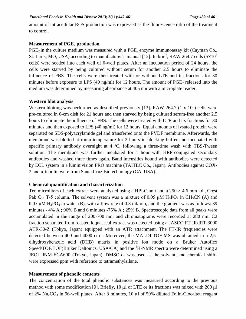
Chemical quantification and characterization of LTE
To analyze the bioactive components in LTE, we first compared the HPLC profiling of LTE to
fresh loquat leaves with known compounds as standards. As shown in Figure 5A and Table 1,
larger amounts of 5-caffeonylquinic acid and 3-caffeonylquinic acid, and smaller amounts of (-)-
epicatechin and procyanidin B2 were detected in fresh loquat leaves, but smaller amounts of
everything were in LTE crude (b) and none were detected in the C and C2 fraction (c and d) of
LTE. Some new peaks were detected (c and d). To characterize these chemicals, C2 fraction was
further investigated by FT-IR spectra. The strong and broad band of O-H stretching was
observed at 3252 cm-1
. The existence of one or more aromatic rings in a structure is normally
readily determined from the C–H, C=C and C–C ring. Medium strong and finger peaks at 1593,
1543, and 1515 assigned to C=C stretching modes. The generated C-C aromatic stretch was
observed at 1387.53, with strong absorptions of FT-IR. The peaks at 7193 and 6733 cm−1
could
be assigned as mono substitute benzene. These data suggest that some new phenolic compounds
might be produced during the roasting process of fresh loquat leaves. Moreover, the C2 fraction
showed primary three peaks at m/z 170 - m/z 330 on MALDI-TOF MS spectra, and the signals
for aromatic or olefinic protons at 6.256-8.308 ppm as well as for the methylene or alicyclic
protons at 0.823-2.995 ppm were detected in the 1H-NMR spectra. Thus, we gather from these
data that these compounds in LTE might be several kinds of phenolic compounds.
Since phenolic compounds generally have antioxidant capacity, we thus quantified the
phenolic contents of fresh and its fractions of LTE, using gallic acid as reference standard. As
shown in Figure 5B, phenolic contents of LTE fractions (M) are higher than fresh loquat leaves.
Moreover, the fraction C and C2 contained higher phenolic amount among these fractions.
Table1 HPLC data of fresh loquat leaves and roasted loquat tea
Retension time Peak area Amount
(min) (μV.S) (mg/kg)
Fresh leaves 5-caffeonylquinic acid 21.07 1673131 6.59
3-caffeonylquinic acid 28.08 4225609 16.64
Procyanidin B2 31.27 295307 1.16
Epicatechin 36.26 1174750 4.62
LTE (M) 5-caffeonylquinic acid 21.07 27036 0.26
3-caffeonylquinic acid 28.08 106130 1.03
Procyanidin B2 n.d - -
Epicatechin n.d - -
LTE (C or C2) 5-caffeonylquinic acid n.d - -
3-caffeonylquinic acid n.d - -
Procyanidin B2 n.d - -
Epicatechin n.d - -
Chemical compoundFractions

Functional Foods in Health and Disease 2013; 3(11):447-461 Page 456 of 461
Fig. 5. HPLC profiles (A) of fresh Loquat leave (a), LTE M (b), C fraction (c) and C2 fraction (d). (-)-
Epicathechin, 3-caffeonylquinic acid, 5-caffeonylquinic acid and procyanidin B2 were used as standards.
The CresPak C18T-5 column (4.6 mm i.d. × 250 mm) was set in 40 ºC. Ten microliters of standards or
LTE solution was injected to the column after filtered with a millipore filter (0.45 μM) and flowed with
0.05 M of H3PO4 in CH3CN from 4% to 30% for 39 minutes and then changed 30% to 75% for 6 minutes
under a flow rate of 0.8 ml/min. Spectroscopic data from all peaks were accumulated in the range of 200-
700 nm, and chromatograms were recorded at 280 nm. Phenolic contents (B) of fresh loquat leaf, LTE,
LTE and C2 fractions. Phenolic contents were determined by Folin-Ciocalteu method. The amounts were
presented as gallic acid equivalents (GAE mg/ml). M, crude extract; A, water elution fraction; B, 30%
EtOH fraction; C, 50% EtOH fraction; D, 100% acetone fraction, C2 fraction purified from C fraction,
respectively. The data represent the mean ± SD of three separated experiments.

Functional Foods in Health and Disease 2013; 3(11):447-461 Page 457 of 461
DISCUSSION:
Loquat tea is made from loquat leaves roasted at 350C for 30 minutes, and usually used as
beverage according to the folk customs. Although fresh loquat leaves have been reported to have
biological activities such as antioxidant and anti-inflammatory activities [14-16], there is no
report on the biological activities of roasted loquat leaves (loquat tea). In the present study, we
used antioxidant activity-guided fractionation to investigate the biological activities of loquat tea.
Our data showed that loquat tea had stronger antioxidant activities than fresh loquat leaves.
Loquat tea revealed stronger antioxidant activities not only in vitro, but also in mouse
macrophage-like cell RAW264.7, which is a cell model to investigate antioxidant and anti-
inflammation mechanisms. COX-2 is only induced during inflammation by pro-inflammatory
stimuli including bacterial LPS, growth factor and cytokines. COX-2 is the rate-limiting enzyme
in the conversion of arachidonic acid to PGE2 which is a pro-inflammatory mediator present in
the inflammatory site [17,18]. Persistence inflammation and continuous production of COX-2
has been linked to development of cancer and autoimmune disorders [19,20]. In this study, we
found that loquat tea could attenuate the production of PGE2 and COX-2 induced by LPS.
Therefore, our data indicate that loquat tea has antioxidant and anti-inflammatory properties.
We were then interested in the bioactive compounds contributing to the antioxidant and
anti-inflammatory activities in loquat tea extract (LTE). Thus, we compared the HPLC profiles
of LTE fractions with the extract of fresh loquat leaves because some bioactive flavonoids such
as (-)-epicatechin, 3-caffeonylquinic acid, 5-caffeonylquinic acid, and procyanidin B2 have been
clarified in fresh loquat leaves, and have been suggested to contribute the biological activities
[21-23].
We confirmed these compounds in fresh loquat leaves using their standard samples (Figure
5A-a). These results are in agreement with the previous report [1, 24]. However, their
compounds were much lower in LTE (Figure 5A-b), and finally disappeared in the C and C2
fractions. In place of them, some new compounds were detected in the C fractions (Figure 5A-d).
Although we could not determine the chemical structure at this moment, the data from FT-IR
spectra showed the existence of aromatic rings and broad band of O-H stretching. Moreover, the
C2 fraction showed primary three peaks at m/z 170 - m/z 330 on the MALDI-TOF MS spectra,
and the signals for aromatic or olefinic protons at 6.256-8.308 ppm as well as for the methylene
or alicyclic protons at 0.823-2.995 ppm were detected in the 1H-NMR spectra. Thus, we gather
that the bioactive compounds in LTE might be, at least partly, several kinds of phenolic
compounds, which are produced from the release and/or degradation of bound phenolic
compounds in fresh loquat leaves during roasting process.
Some similar findings have also been reported that (-)-epicatechin and procyanidin
significantly decreased after roasting cocoa beans and coca ingredients due to epimerization [25,
26]. Since phenolic compounds have been reported to have antioxidant activities, we next
measured the phenolic amount of the C and C2 fractions, comparing with LTE using gallic acid
as standard. As shown in Figure 5B, phenol content in C and C2 fraction of LTE were 258 ±
22mg/g and 267 ± 21 mg/g. In fresh loquat leaves and LTE (M), the total phenolic contents were
26 ± 4.2 mg/g and 77.4 ± 2.2 mg/g, respectively. These data indicated that phenolic contents
were increased when loquat leaves were roasted at 350C for 30 min. It has been reported that
high temperature treatment changed phenolic content of samples caused by the release of bound

Functional Foods in Health and Disease 2013; 3(11):447-461 Page 458 of 461
phenolic compound, release of phenolic acid derivatives, and thermal degradation of the phenolic
compounds [27].
CONCLUSION:
Loquat tea, made from roasted loquat leaves, revealed stronger antioxidant activity than its fresh
leaves by scavenging DPPH and suppressing cellular ROS, and also showed anti-inflammatory
activity by suppressing the production of pro-inflammatory mediators such as COX-2 and PGE2.
The bioactive components are speculated to be phenolic compounds that were produced from
fresh loquat leaves during the roasting processes.
Abbreviations:
LTE, loquat tea extract; COX-2, cyclooxygenase-2; DCFH-DA, dichlorofluorescein-diacetate;
DMEM, Dulbecco's Modified Eagle Medium; DPPH, 1-diphenyl-2-picrylhydrazyl; HPLC, High
performance liquid chromatography; LPS, lipopolysaccharide; PGE2, prostaglandin E2; ROS,
reactive oxygen species; Trolox, 6-Hydroxy -2.5.7.8-tetramethyl chroman-2-carboxylic acid.
Competing interest:
The authors have no financial interests or other conflicts of interest.
Authors’ Contribution:
Ms. Phyu Phyu Khine Zar is the primary investigator in this study. Dr. Kozue Sakao participated
in chemical characterization. Dr. Fumio Hashimoto and Dr. Koji Wada participated in the
extraction and purification. Ms. Akiko Morishita helped the culture cell experiments. Dr. Makato
Fujii helped the preparation of roasted loquat leaves. Dr. De-Xing Hou designed this study and
wrote the manuscript as corresponding author.
Acknowledgments:
This work was supported by the Fund of Scholar Research of Kagoshima University in Japan (to
D.-X., Hou).
REFERENCES:
1. Ferreres F, Gomes D, Valentao P, Goncalves R, Pio R, Chagas EA, Seabra RM at al.
Improved loquat (Eriobotrya japonica Lindl.) cultivars: Variation of phenolics and
antioxidative potential. Food Chem 2009; 114: 1019-1027.
2. De Tommasi N, Aquino R, De Simona F, Pizza C. Plant metabolites. New sesquiterpene
and ionona glycosides from Eriobotrya japonica. J Nat Prod 1992; 55: 1025-1032.
3. Ito H, Kobayashi E, Takamatsu Y, Li SH, Hatano T, Sakagami H, Kusama K, et al.
Polyphenols from Eriobotrya japonica and their cytotoxicity against human oral tumor
cell lines. Chem Pharm Bull 2000; 48: 687-693.
4. Wootton-Beard PC, Ryan L. Improving public health?: The role of antioxidant-rich fruit
and vegetable beverages. Food Res Int 2011; 44: 3135-3148.
5. Laskin DL, Laskin JD. Role of macrophages and inflammatory mediators in chemically
Functional Foods in Health and Disease 2013; 3(11):447-461 Page 459 of 461
induced toxicology. J Toxicol 2001; 160: 111-118.
6. Laskin DL, Pendino KJ. Macrophages and inflammatory mediators in tissue injury. Annu
Rev Pharmacol Toxicol 1995; 35: 655-677
7. Akira S, Takeda K. Toll-like receptor signaling. Nature Reviews Immunology 2004; 4:
499-511.
8. Su YW, Chiou WF, Chao SH, Lee MH, Chen CC, Tsai YC. Ligustilide prevents LPS-
induced iNOS expression in RAW 264.7 macrophages by preventing ROS production
and down-regulating the MAPK, NF-κB and AP-1 signaling pathways. Int
Immunopharmacol 2011; 11: 1166-1172.
9. Rabah IO, Hou D-X, Komine S, Shono M, Fuji M. Increase in antioxidant and
cytotoxicity through apoptosis-induction of HL-60 of sweet potato (Ipomoea Batatas
Lam. Cv. Koganesengan) by sub-critical water treatment. Food Sci Technol Res 2005;
11: 122-126.
10. Xie Z, Huang J, Xu X, Jin Z. Antioxidant activity of peptides isolated from alfalfa leaf
protein hydrolysate. Food Chem 2008; 111: 370-376.
11. Hou D-X, Fukuda M, Jonson JA, Miyamori K, Ushikai M, Fujii M. Fisetin induces
transcription of NADPH: Quinone oxidoreductase gene through an antioxidant
responsive element-involved activation. Int J Oncol 2001; 18: 1175-1179.
12. Hou D-X, Luo D, Tanigawa S, Hashimoto F, Uto T, Masuzaki S, Fujii M et al.
Prodelphinidin B-4 3 -O-gallate, a tea polyphenol, is involoved in the inhibition of COX-2
and iNOS via the downregulation of TAK1-NF-κB pathway. Biochem Pharmacol 2007;
74: 742-751.
13. Hou D-X, Yanagita T, Uto T, Masuzaki S, Fujii M. Anthocyanidins inhibit
cyclooxygenase-2 expression in LPS-evoked macrophages: Strucutre-activity relationship
and molecular mechanisms involved. Biochem Pharmacol 2005; 70: 417-425.
14. Banno N, Akihisa T, Tokuda H, Yasukawa K, Taguchi Y, Akazawa H, Ukiya M,
et al. Anti-inflammatory and antitumor-promoting effects of the triterpene acid from the
leaves of Eriobotrya japonica. Biol Pharm Bull 2005; 28: 1995-1999.
15. Hong Y, Lin S, Jiang Y, Ashraf M. Variations and contents of total phenolic and
flavonoids and antioxidant activities in the leaves of 11 Eriobotrya species. Plant Foods
Hum Nutr 2008; 63: 200-204.
16. Kim SH, Shin TY. Anti-inflammatory effect of leaves of Eriobotrya japonica correlating
with attenuation of p38 MAPK, ERK, and NF-κB activation in mast cells. J Toxicol In
Vitro 2009; 23: 1215-1219.
17. O’Sullivan MG, Huggins Jr. EM, Meade EA, DeWitt DL, McFall CE.
Lipopolysaccharide induces prostaglandin H synthase-2 in alveolar macrophages.
Biochim Biophys Res Commun1992; 187: 1123-1127.
18. Adelizzi RA. COX-1 and COX-2 in health and disease. J American Osteropathic
Association, 1999; 99: S7-12.
19. Fitzpatrick FA. Inflammation, carcinogenesis and cancer. Int Immunopharmacol 2001; 1:
1651-1667.
20. Kubatka P, Ahler I, Ahlersova E, Adamekova E, Luk P, Bojkova B, Markova M.
Chemoprevention of mammary carcinogenesis in female rats by rofecoxib. Cancer Lett

Functional Foods in Health and Disease 2013; 3(11):447-461 Page 460 of 461
2003; 202: 131-136.
21. Jung HA, Park JC, Chung HY, Kim J, Choi JS. Antioxidant flavonoids and chlorogenic
acid from the leaves Eriobotrya japonica. Arch Pharm Res 1999; 22: 213-218.
22. Ito H, Kobayashi E, Li SH, Hatano T, Sugita D, Kubo N, Shimura S et al. Antitumor
activity of compounds isolated from leaves of Eriobotrya japonica. J Agric Food Chem
2002; 50: 2400-2403.
23. Bae YI, Jeong CH, Shim KH. Antioxidative and Antimicrobial activities of Epicatechin
isolated from leaves of loquat (Eriobotrya japonica). Int Journal Food Science 2005; 10:
118
24. Tanaka K, Tamaru S, Nishizono S, Miyata Y, Tamaya K, Matsui T, Tanaka T, et al.
Hypotriacylglycerolemic and antiobesity properties of a new fermented tea product
obtained by tea-rolling processing of third-crop green tea (Camellia sinensis) leaves and
loquat (Eriobotrya japonica) leaves. Biosci Biotechnol Biochem 2010; 74; 1606-1612.
25. Hurst WJ, Krake SH, Bergmeier SC, Payne MJ, Miller KB, Stuart DA. Impact of
fermentation, drying, and roasting dutch procession on flavon-3-ol stereo chemistry in
coca beans and coca ingredients. Chem Cent J 2001; 53: 1-5.
26. Kothe L, Zimmermann BF, Galensa R.Temperature influences epimerization and
composition of flavanol monomers, dimmers, and trimers during cocoa bean roasting. J
Food Chem 2013; 141: 3656-3653.
27. Ross CF, Hoye CJ, Fernandez-Plotka VC. Influence of heating on the polyphenolic
content and antioxidant activity of grape seed flour. J Food Sci 2011; 76: C884-90.