Embed Size (px)
Citation preview

Searching for interesting channels: pairing selection and molecular
evolution methods to study ion channel structure and function
Daniel L. Minor, Jr*ab
Received 26th January 2009, Accepted 11th May 2009
First published as an Advance Article on the web 19th June 2009
DOI: 10.1039/b901708a
The pairing of selection and screening methods with randomly mutated libraries can be an
exceptionally powerful means for probing the functions of biological molecules and for
developing novel regents from random libraries of peptides and oligonucleotides. The use of such
approaches is beginning to permeate the ion channel field where they are being deployed to
uncover fundamental aspects about ion channel structure and gating, small molecule–channel
interactions, and the development of novel agents to control channel activity.
Introduction
Brains, hearts, senses, and muscles all run on bioelectrical
signals that race along cell membranes on the millisecond
timescale. To make these exceptionally rapid signals, cells rely
on the activity of a large, diverse set of transmembrane
macromolecular complexes known as ion channels.
The hydrophobic nature of the cell membrane presents a
significant barrier to the passage of charged particles such as
ions. Cells expend a great deal of their ATP resources to drive a
variety of pumps that establish asymmetric ion gradients across
their cell membranes. When ion channel proteins open, energy
stored in these ionic gradients is released as the ions flow down
their electrochemical gradients and across cell membranes.1
This rapid transport of ions, catalyzed by ion channel proteins,
is the fundamental process that creates the electrical signals that
underlie the normal functioning of our cardiovascular and
nervous systems. Without such activity, there would be no
thoughts, no racing heart at the sight of a loved one, no feeling
of pain, and no warm embraces. Further, ion channel
misfunction is linked to an ever-growing range of human
diseases including arrhythmias, migraine, diabetes, and
movement disorders.2,3 Consequently, there is a great interest
both in understanding the molecular basis for how channels
work and in the development of new reagents that can control
their functions.
Because ion channels are membrane proteins, the use of
high-resolution biophysical techniques to elaborate the
molecular architectures that underlie channel function remains
very challenging.4 Thus, there has been a great deal of effort
focused on other types of approaches that can enlighten the
connections between ion channel molecules and their activities.
In this regard, genetic methods constitute an exceedingly
powerful means for querying biological systems and for
establishing insights into how macromolecules function. One
of the biggest strengths of genetic approaches is that they offer
an assumption-free method in which a system can be probed
to identify functional alterations that are rooted in mutational
changes in specific macromolecules. Classical genetic studies in
which functional defects in both multicellular and unicellular
organisms were traced to ion channel gene mutations have
played a large role in ion channel studies. These investigations
have determined the identities of founding members of many
important ion channel families, such as voltage-gated
potassium channels,5–7 sensory transduction TRP channels,8–10
and centrally important proteins involved in channel
regulation.11,12 Over the past ten years or so, a different sort
of channel-focused genetics has been emerging, one that starts
a Cardiovascular Research Institute, Departments of Biochemistry andBiophysics, and Cellular and Molecular Pharmacology, CaliforniaInstitute for Quantitative Biosciences, University of California,San Francisco, CA 94158-2330, USA.E-mail: [email protected]
b Physical Biosciences Division, Lawrence Berkeley NationalLaboratory, Berkeley, CA 94720, USA
Daniel L. Minor
Daniel L. Minor, Jr., is anAssociate Professor at theUniversity of California, SanFrancisco, in the Cardio-vascular Research Institute,Departments of Biochemistryand Biophysics, and Cellularand Molecular Pharmacology,and California Institute forQuantitative Biosciences. Healso holds a position asFaculty Scientist at theLawrence Berkeley NationalLaboratory. Minor receivedhis BA in Biophysics and Bio-chemistry magna cum laude
from the University of Pennsylvania. He earned his PhD inchemistry at the Massachusetts Institute of Technology forstudies on protein structure and design. He began his studiesof ion channels during postdoctoral training at the MRCLaboratory of Molecular Biology with Nigel Unwin, and UCSFDepartment of Physiology with Lily Jan. He was named aBeckman Young Investigator, McKnight Scholar, Rita AllenScholar, Searle Scholar, and Sloan Fellow, and is currently anAmerican Heart Association Established Investigator. Hislaboratory applies multidisciplinary approaches includingselection methods, electrophysiology, and X-ray crystallographyto dissect ion channel structure and function.
802 | Mol. BioSyst., 2009, 5, 802–810 This journal is �c The Royal Society of Chemistry 2009
REVIEW www.rsc.org/molecularbiosystems | Molecular BioSystems

not with an investigation of a physiological process but with a
molecule. This approach is typically termed ‘reverse genetics’.
Rather than look for mutant genes in an organism to identify a
specific channel that is key to some process, researchers have
established a number of heterologous expression systems in
which large numbers of mutant channels can be assayed
directly for new or altered properties. The experimental
advantage of such gene-based methods is that none require
purification of the protein of interest. Thus, all of the power of
molecular biology and molecular evolution methods can be
brought to bear on discovery-oriented selections and screens
that when paired with electrophysiological analysis lead to
deep molecular insight into the mechanisms of ion channel
function.
Basic considerations
Genetic systems that use unicellular organisms, such as
bacteria (e.g., Escherichia coli) or yeast (e.g., Saccharomyces
cerevisiae), have been one of the mainstays of biological
investigation and provide a potent means to assay large
numbers of variants, up to B1 million, in a parallel manner
in a short period of time, typically within a week or so. The
challenge of using genetic methods to study ion channels in
unicellular systems is that one needs to establish a robust
phenotype that can be the focus of either a selection or simple
assay that can constitute a screen.w Systems in which expression
of an ion channel gene overcomes a specific functional deficit
that allows the cell to survive some external challenge are the
strongest in this regard. Alternatively, fluorescence-based
methods that monitor calcium signals resulting from the
activity of ion channels constitute a second productive approach.
Once a microorganism-based genetic system is established,
one can readily examine the properties of libraries of large
numbers of mutant channels.
Libraries of mutant channels can be generated using a
variety of approaches: chemical mutagenesis,13,14 error-prone
PCR,15–22 passage of the target gene through a bacterial
mutator strain,23–25 designed mutant libraries made from
synthetic oligonucleotides encoding whole gene or targeted
to key gene portions,26–28 and DNA shuffling approaches.29,30
The method of library generation is less important than the
coverage and amount of sequence diversity that it contains.
Given a good library and a robust selection or screen, one can
readily find a host of interesting mutants that merit character-
ization by other methods.
Ion channel subunits are generally medium to large sized
proteins. Pore-forming subunits from members of the voltage-
gated channel family are predominantly in the range of
300–500 amino acids and some are as large as 2500–3000
amino acids.31 Considering these subunit sizes and the limits
imposed on the level of diversity by host organism transfor-
mation efficiencies, typically 105–106 individual clones, none of
the current mutagenesis schemes can yield libraries that
contain enough mutants to sample all possible variant
sequences for a given subunit. For example, a 300-residue
subunit has 20300 possible sequences, a number that surpasses
all estimates of the total numbers of atoms in the universe.
Given the paltry amount of sequence space that can be
explored for a given subunit, one might imagine that the
chance of discovering a mutation that changes function or
that identifies a key functional residue by a completely blind
mutagenesis approach might have little chance of success.
In spite of these seemingly insurmountable odds, there is
ample evidence that the situation is not as dire as might be
initially predicted. There are many experimental strategies for
making the most of number limits that are inherent to the
selection/screening process. An initial broad sweep in which an
entire channel gene is targeted for mutation so that each
position is likely to be changed at least once can lead to the
identification of a particular region or set of residues that can
be more intensely explored by subsequent focused libraries
that more extensively test the amino acid restrictions of
particular positions. For example, there are 6000 possible
variants for a 300 amino acid subunit if each position is
substituted with all 20 amino acids. In a well-made random
library made by error-prone PCR there is a good chance that
most positions would be changed to at least a few amino acids
of very different chemical character and allow the investigator
to uncover a few key regions that might affect function from
the first pass selection or screen. Alternatively, if one has an
interest in a region with known functional importance, one can
directly employ focused libraries that target a particular
channel element. To date, a combination of molecular
evolution–selection approaches has been applied to four
classes of channels: potassium channels, TRP channels,
mechanosensitive bacterial channels from the MscL family,
and voltage-gated calcium channels. These efforts have yielded
a multitude of interesting channel mutants that have brought
genuinely new insight into channel function.
Rescue of ion transport deficient microorganisms
The biggest challenge in establishing a genetic system to study
a particular ion channel is to devise a situation in which
activity of the channel of interest is intimately tied to cell
survival or to a robust secondary assay. One powerful approach
has been the use of systems in which expression of the channel
of interest affects ion homeostasis. To this end, systems that
rely on potassium uptake assays have been particularly
fruitful.
All cells need potassium to survive. Bacteria and yeast have
special uptake systems that harvest potassium from the
environment.32,33 Deletion or inactivation of the genes
responsible for potassium uptake (E. coli TK242034,35 and
S. cerevisiae Dtrk1Dtrk236,37) yields strains that survive when
bathed in high concentrations of potassium (B100 mM) but
not when subjected to low external potassium concentrations
(0.5–2 mM). The activity of the plasma membrane proton-
ATPase sets the membrane potentials of both microorganisms
w Definitions: classical genetics (forward genetics), a procedure thatconnects a phenotype to a particular genotype; reverse genetics,identification of phenotypes that result from specific mutations in agene of interest; selection, a protocol in which functional molecules(in this case channels) are required for cell survival or yield a toxicphenotype; screen, application of an assay to a pool of mutantchannels. In a screen every mutant must be examined; GOF, gain offunction; LOF, loss of function.
This journal is �c The Royal Society of Chemistry 2009 Mol. BioSyst., 2009, 5, 802–810 | 803

in a very negative range (B�300 mV).38 Because this range is
below the equilibrium potential for potassium ions under low
external potassium (�133 mV for 1 mM [K+]out/150 mM
[K+]in at 37 1C), expression of a functional potassium channel
can provide a route for potassium uptake under low external
potassium conditions and rescue the growth of the potassium-
starved strain. Initially, potassium-uptake deficient yeast
strains were used to clone and study the properties of plant
potassium channel genes (see for example ref. 37, 39–41). The
demonstration of functional expression of a mammalian
inward rectifier potassium channel in potassium transport
deficient yeast42 ushered in a series of studies on inward
rectifiers and voltage-dependent potassium channels in which
large libraries of channel mutants were used to study basic
channel architectural constraints,15,26 ion selectivity,30,43 and
gating properties.16,29 In these studies, a central element was
the pairing of the results of the yeast selections with the
biophysical characterization of channel function using electro-
physiological approaches. Such correlations are essential. The
power of genetic screens is that one can find interesting
mutants with relative ease. The potential drawback is that
one never knows all of the parameters that are actually being
tested by the system. Thus, it is crucial to follow the selections
or screens with extensive characterization to understand how
the isolated mutants might be working. That caution aside, the
combination of mutant libraries with channel selections or
screens has led to the isolation of a remarkable number of new
channel mutants with very interesting properties.
Selections for new activities
Many channels move between a closed state that does not
conduct ions, and an open state that passes ions as the result of
some type of stimulus such as ligand binding or voltage
change. Such channels are excellent candidates for
gain-of-function (GOF) selections and screens in which one
hunts for mutations that endow the target channel with a new
functional property such as activation in the absence of the
normal gating stimulus. This type of GOF investigation has
proven instrumental for the identification of residues that have
important roles in maintaining the integrity of the closed
state or in stabilizing the open state and that therefore may
constitute key moving parts of the channel.
A common type of GOF selection is one in which the
investigator tries to bring a non-functional channel that is
closed due to the absence of activating signal to life (Fig. 1A).
In these life and death selections, only those bacteria or yeasts
fortunate enough to receive a gene encoding a functional
channel survive when the selective pressure is applied. Such
selections are straightforward and require little sophisticated
equipment. The library of channel genes is transformed into the
host strain and plated under conditions where the channel is not
required for survival. Subsequently, the colonies are transferred
by replica plating onto plates that have selective conditions,
such as low potassium, where only microbes carrying a plasmid
that encodes for a functional channel survive. From these
remaining colonies one can recover the plasmid, sequence the
gene, retest the plasmid to verify the phenotype, and scrutinize
the mutant by biophysical characterization.
GOF selection experiments have a distinct advantage as a
means to find mutants that confer a new activity to the channel
of interest. There are many ways a mutation can kill a channel
but far fewer in which a mutational change can impart a new
functionality. The types of mutations that constitute the bulk
of a randomly mutated library (neutral mutations and
mutations that result in channels that are non-functional due
to premature stop codons, missense mutations in the active
site, misfolding or faulty membrane insertion) result in cell
death under selective conditions and as a consequence are
readily culled from the library. After application of selective
pressure, one is left with a small number of colonies, which
should bear channels with increased activity. Thus, rare
mutations that have strong effects on function can be found.
A good example of the power of this type of selection
approach was a pair of studies that aimed to isolate mutants
of G-protein activated inward rectifier channels (Kir3.1 and
Kir3.2) that had enhanced activity in the absence of their
normal ligand, the Gbg complex.16,29 Independent work in the
Jan and Reuveny groups identified a number of Kir3.1 and
Kir3.2 mutants that suggested a crucial role for the movement
and bending of the inner pore-lining transmembrane segment
Fig. 1 Schematic for selection of functional channels from mutant
libraries. Top, depicts a collection of mutants of a two-transmembrane
domain channel subunit encoded by plasmids. Path A shows the
procedure when functional channels confer rescue under selective
conditions. The library is first plated onto non-selective conditions.
Under these conditions, channel function is not required for survival.
After colonies are established they are replica-plated onto plates that
have selective conditions in which channel activity is required for
survival. All non-functional clones are lost at this step and only
functional mutants remain. Path B shows the outcome when functional
channels confer a lethal phenotype. In this case, all colonies survive the
non-selective conditions, but only those colonies expressing active
channels die on the selective plates (indicated by the red dashed circles).
In both cases, recovery of the plasmid, retesting to verify phenotype,
DNA sequencing to identify the mutations, and subsequent character-
ization by electrophysiological studies are essential.
804 | Mol. BioSyst., 2009, 5, 802–810 This journal is �c The Royal Society of Chemistry 2009

in gating. These ideas were derived prior to the advancement
of similar potassium channel gating hypotheses based on
structural studies.44 Similar GOF studies with the bacterial
channel KcsA have identified mutants that increase channel
opening28 and a set of activatory mutations that cluster in the
portion of the structure that is essential for maintaining the
closed state.22
A different type of GOF selection searches for mutant
channels that kill the host (Fig. 1B). This type of GOF
selections is more technically challenging than GOF rescue
selections as the colonies that do not survive transfer to
selective conditions will be far fewer than those that do.
Nevertheless, selections that look for activated channels that
are lethal have proved to be an effective way to find interesting
gating mutants. In one of the first reported lethal GOF
screens, Loukin and colleagues uncovered gating mutants in
likely pore-lining segments of the yeast potassium channel
TOK1 (YKC1) that have an important role in closed state
stabilization.13 A related study of the E. coli potassium
channel Kch identified GOF mutations resident in a key
cytoplasmic element that is thought to control channel
gating.21 More recently, Myers et al. have used a lethal GOF
screen to isolate mutants of the heat and capsaicin sensitive
TRP channel, TRPV1, that increase basal activity and
discovered that the pore-helix segment likely plays an
important role in channel activation.17
Instead of a selection, GOF experiments can also be
conducted as a screen in which channel function is coupled
to a secondary assay. One productive approach has been to
use fluorescence-based detection to monitor intracellular
calcium changes that are linked to the activity of the channel
of interest. The Kung and Saimi groups have developed such
an assay for yeast and employed it to identify mutants that
increase the activity of the native yeast TRP channel
TRPY1,24 an intracellular ion channel that resides in the yeast
vacuole. Expression of a calcium-sensor protein, apoaqueorin,
allowed the measurement of a calcium-dependent lumines-
cence signal resulting from the release of calcium from the
vacuole into the cytoplasm through TRPY1 following osmotic
shock. By screening B4000 TRPY1 mutants in a multiwell
format, the investigators identified a set of mutants with
increased calcium signals. Follow-up studies by patch clamp
electrophysiology showed that the mutants have increased
activity and identify key elements of the pore domain that
are excellent candidates for part of the channel gate.23,24
Genetic selections have also been important for isolating
interesting mutants of a bacterial mechanosensitive ion
channel MscL.45 Two different approaches have been reported:
one looked for GOFmutants that impair host growth,14,25 and
the other used dyes that allow one to asses cell viability
following an osmotic challenge20,46 and that could distinguish
GOF and loss of function (LOF) mutants by exploiting
differences in vital dyes, propidium iodide and SYTO 9, that
report on cell viability. One important difference highlighted
by these MscL studies is that screens, while powerful, are still
more labor intensive and limited in scope than selections. The
vital dye screen examinedB400 mutants whereas the functional
selections were able to sort through two orders of magnitude
more mutants (B50 000) and correctly identify a residue that
is centrally important for maintaining the integrity of the
closed state.14,45
High throughput functional screens
A different way to deploy the power of reverse genetics is to
establish a screen in which particular functional properties of a
large number of mutants can be measured in cultured
mammalian cells. By coupling calcium imaging in transfected
mammalian cell lines with studies of channels that are both
temperature and ligand gated, TRPM818 and TRPV3,19 the
Patapoutian lab was able to examine pools of B14 000
mutants of each channel and isolate mutants that displayed
selective alterations in either ligand or thermal sensitivity.
Together with the TRPV1 study,17 the results of screening of
TRPM8 and TRPV3 channel mutants indicate that at least
some of the activation modes of TRP channels can be uncoupled
(ligand vs. temperature, protons vs. ligand). Even though the
exact molecular mechanisms remain to be resolved, these
studies provide further examples of the power of unbiased
screens as in all three cases the investigators identified key
regions of the channels that are likely to be important for
gating responses.
Taken together, the results from these varied investigations
of different channel types demonstrate that genetic screens of a
modest number of mutants (in the thousands to 10 000s) can
be an exceptionally productive means to uncover mutants that
identify key elements that are likely to be core elements of the
gating mechanism. This linkage is further reinforced by the
cases where activating mutants reside in essential gating elements
that are characterized by high-resolution structural
studies.16,21,28,29 Overall, the relatively high frequency with
which GOF mutants occur in ion channels bodes well for
future screens for other channels as it appears that one does
not have to rely on sifting through massive numbers of
mutants to find interesting and informative ones.
Feeling around in the dark—random mutagenesis
and selection as a means for defining architectural
elements and testing protein–protein interactions
Many membrane proteins are assembled from helical bundles
that are largely perpendicular to the membrane. This situation
facilitates the use of extensive mutagenesis as a means to derive
constraints that inform ideas about membrane protein
architecture. Patterns of the physicochemical properties of
allowed amino acid substitutions at specific positions can be
used to identify secondary structures, lipid-facing residues,
and protein–protein interaction surfaces. Such indirect
structural analysis of membrane proteins has a long history
best exemplified by the work done on the transmembrane
dimer glycophorin.47–49
Access to a genetically tractable system enables another
powerful type of experiment that can yield structural constraints,
the search for intragenic suppressors that rescue a defect that
destroys channel function (Fig. 2A). The strength of the
approach is that in the complete absence of structural data
one can identify residue pairs that are functionally coupled.
The search for suppressors can also be enlightening when set in
This journal is �c The Royal Society of Chemistry 2009 Mol. BioSyst., 2009, 5, 802–810 | 805
the context of testing a structural model as one can focus the
mutagenesis efforts towards candidate segments that might
interact. Work on structurally well-characterized systems
indicates that, in general, suppressor pairs are the most
effective at restoring protein function when the suppressor lies
in close proximity to the original perturbation.50 While it is
not strictly necessary for suppressor and defect mutations to
interact directly to restore function, second site suppressor
experiments can provide constraints that can vet models for
how transmembrane segments might be arranged.
Based on a genetic selection that characterized large
numbers of mutant channels, analysis of transmembrane
amino acid substitution patterns together with second site
suppressor experiments was used to probe the transmembrane
architecture of the mammalian inward rectifier Kir2.1. This
study established the overall helix packing arrangement of
mammalian inward rectifiers,26 which was subsequently
validated by crystallographic studies,51 and uncovered an
intrasubunit intramembrane hydrogen bond that is essential
for channel assembly. Suppressor studies have also given
important insight into regarding residues that may interact
in the open state of MscL.25 The most extensive use of
suppressors to extract functional constraints comes from a
combined modeling–selection study on the hyperpolarization
activated channel KAT1 that helped to establish the likely
range of voltage sensor motion.15,52 These studies complement
the ongoing crystallographic work in the field and provide
new ideas for testing key questions about what sorts of
conformational transitions underlie channel gating.
Selections and libraries meet channel
blockers—second site suppressor to map sites and
mechanisms of action
There are many diverse classes of ion channels that are now
known from extensive gene characterization efforts. Unfortu-
nately, the ability to identify ion channel genes has far
surpassed the ability to define novel pharmacological agents
for particular channels. Consequently, many ion channels
have poor to no pharmacology. This situation limits the ability
of investigators to make the connections between a particular
ion channel gene and its exact biological function. Thus, one
of the key challenges for ion channel research is to develop
means to identify new agents that can control channel activity.
Genetic selections offer a novel, unbiased way to identify
channel-modifying compounds. A number of studies have
used the Dtrk yeast system to screen for and map the sites of
action of ion channel blockers. Studies by Zaks-Makhina and
colleagues identified a novel potassium channel blocker using
a yeast genetic screen based on Kir2.1 rescue. Surprisingly, the
compound turned out to be a better inhibitor of the voltage-
gated potassium channel Kv2.1,53 a result that may be related
to the high degree of structural conservation present in
potassium channel pore domains.54
Identification of a new channel modulator is only a first
step. One of the immediate questions that a researcher faces
once a new channel blocker or activator is identified is: ‘How
does the compound act?’ The use of genetic selections to find
suppressors of channel blockers is a potent approach for
addressing this question as one demands two stringent criteria:
the channel must become insensitive to the blocker as a result
of the mutational change but still function as an ion channel
(Fig. 2B and C). My laboratory used a combination of blocker
screening and the selection of blocker resistant mutants from
pore-domain libraries to examine whether the selection system
would be a fruitful way not only to find blockers but also to
map their sites of action.27 By focusing on the well-known
potassium channel blocker barium and selecting for barium
resistant Kir2.1 channels, we uncovered an unusual mutation
located very near the barium binding site that could make the
channels resistant to the blocker without perturbing other
functional properties. The mutation placed a positively
charged residue in close proximity to the ion conduction
pathway at a position that should effectively cancel any effects
from a putative helix macrodipole that was thought to be
important for ion conduction.55 Extensive biophysical
characterization and computational studies established that
the barium resistance was electrostatic in origin and showed
that the helix macrodipole could not be an important factor
for ion conduction.27
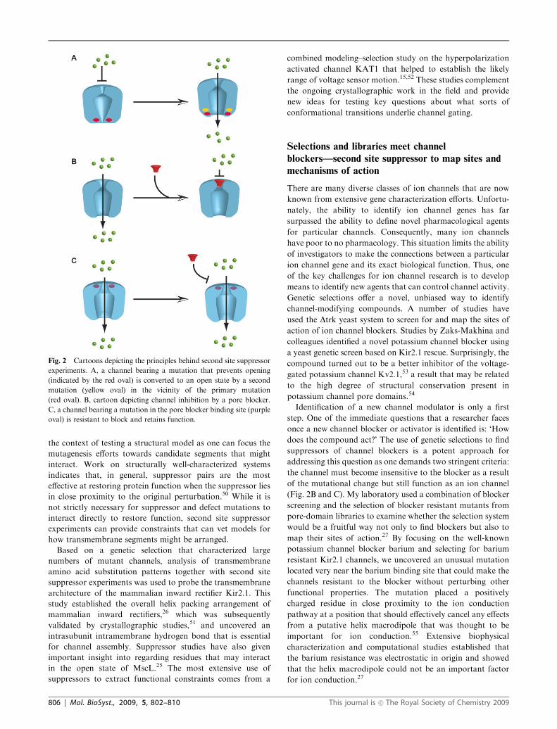
Fig. 2 Cartoons depicting the principles behind second site suppressor
experiments. A, a channel bearing a mutation that prevents opening
(indicated by the red oval) is converted to an open state by a second
mutation (yellow oval) in the vicinity of the primary mutation
(red oval). B, cartoon depicting channel inhibition by a pore blocker.
C, a channel bearing a mutation in the pore blocker binding site (purple
oval) is resistant to block and retains function.
806 | Mol. BioSyst., 2009, 5, 802–810 This journal is �c The Royal Society of Chemistry 2009
Recently, an elegant set of studies reported the identification
and characterization of a new voltage-gated calcium channel
blocker through the use of a genetic selection based on the
roundworm Caenorhabditis elegans.56,57 Roy and colleagues
initially searched a B14 000 compound library for new
small molecules that could be used to explore the biology of
C. elegans. One of the B300 hits yielded a novel compound,
nemadipine-A, that caused a variety of growth and egg-laying
defects in the worms. Nemadipine-A is related to the class
of drugs known as 1,4-dihydropyridines (DHPs) that affect
voltage-gated calcium channel function and are used to treat
hypertension. Subsequent studies for suppressors of
nemadipine-A activity identified the target as the sole
C. elegans voltage-gated calcium channel a1-subunit,Egl-19.56 This channel is homologous to the human L-type
CaV1 family. CaV1 subunits are large (B2500 amino acids)
and might seem to be an unlikely candidate for a productive
unbiased screen. Nevertheless, a follow-up study in which
chemically mutagenized worms were used to look for suppressors
of nemadipine-A identified a number of mutants in the worm
CaV1 channel.57 Remarkably, the mutants identified eleven
residues that had been previously shown to be critical for DHP
binding in mammalian CaV1 channels and a new set of eight
mutants at previously uncharacterized positions. When tested
in the context of the electrophysiologically well-characterized
rat CaV1.2 channel, six of the novel mutants altered DHP
sensitivity and convincingly demonstrated the potential for
using this system as a means for finding new important
elements of drug sensitivity and channel gating. The set of
studies by Roy and colleagues is a fantastic demonstration of
the power of organism based genetic screens to identify novel
small molecules and to gain important and unexpected insights
into the mechanism of action. Together, the yeast and worm-
based channel blocker identification and suppressor studies
establish important proof-of-concept examples that will hope-
fully inspire further development of channel selection systems
that can further enrich channel pharmacology and extend our
understanding of drug–channel interactions.
In vitro evolution methods and channels, breaking
over the horizon
The evolution of new traits that arise from the combination of
individual variation in a population and application of
selective pressure is the fundamental principle that underpins
all of modern biology. This principle is not limited to living
biological systems but can also be harnessed to shape molecules.
In vitro evolution experiments have been among the most
powerful ones deployed by biochemists for finding molecules
with novel properties and have been a robust area of
biochemical research with a more than 40 year history.58 These
experiments use Darwinian selection to cull polynucleotides or
polypeptides having novel properties from large libraries of
variants through multiple rounds of competition, selection,
and amplification (Fig. 3). One major advantage of in vitro
evolution methods is that one can access exceptionally large
libraries that contain up to 1013–1015 unique molecules.
The main in vitro evolution technologies focus on the two
types of biopolymers that have well-known sequence-dependent
folding and self-assembly properties: oligonucleotides, both
DNA and RNA, and peptides and proteins. Nucleic acid poly-
mers have the advantage that the molecule contains both the
information for folding and the information for direct amplifica-
tion (using enzymes). Peptide and protein display methods
require a means to link the functional molecule (the polypeptide)
with the information required for directing its synthesis (a piece
of DNA). A wide variety of in vitro evolution systems that link
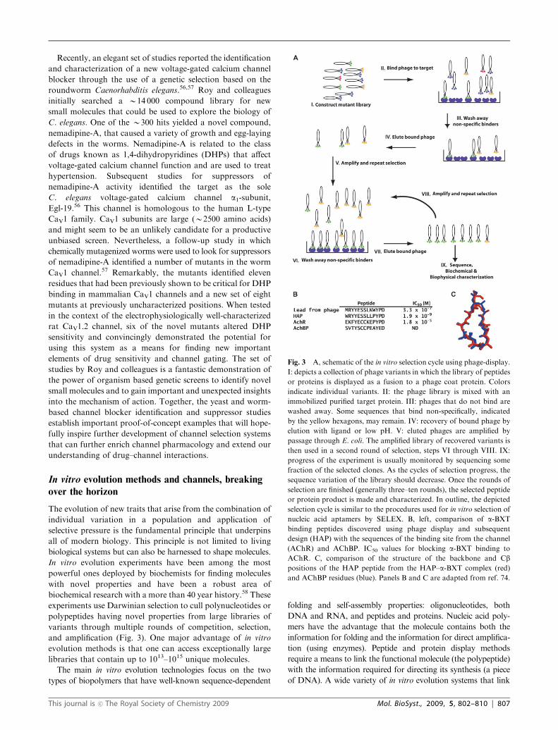
Fig. 3 A, schematic of the in vitro selection cycle using phage-display.
I: depicts a collection of phage variants in which the library of peptides
or proteins is displayed as a fusion to a phage coat protein. Colors
indicate individual variants. II: the phage library is mixed with an
immobilized purified target protein. III: phages that do not bind are
washed away. Some sequences that bind non-specifically, indicated
by the yellow hexagons, may remain. IV: recovery of bound phage by
elution with ligand or low pH. V: eluted phages are amplified by
passage through E. coli. The amplified library of recovered variants is
then used in a second round of selection, steps VI through VIII. IX:
progress of the experiment is usually monitored by sequencing some
fraction of the selected clones. As the cycles of selection progress, the
sequence variation of the library should decrease. Once the rounds of
selection are finished (generally three–ten rounds), the selected peptide
or protein product is made and characterized. In outline, the depicted
selection cycle is similar to the procedures used for in vitro selection of
nucleic acid aptamers by SELEX. B, left, comparison of a-BXT
binding peptides discovered using phage display and subsequent
design (HAP) with the sequences of the binding site from the channel
(AChR) and AChBP. IC50 values for blocking a-BXT binding to
AChR. C, comparison of the structure of the backbone and Cbpositions of the HAP peptide from the HAP–a-BXT complex (red)
and AChBP residues (blue). Panels B and C are adapted from ref. 74.
This journal is �c The Royal Society of Chemistry 2009 Mol. BioSyst., 2009, 5, 802–810 | 807
these two together using bacteriophage,59–61 ribosome
display,60–62 and mRNA display61–63 are now widely used. All
of these methods work best when they are directed against a
purified target. As the expression, purification, and biochemical
isolation of ion channels is still not routine these technologies
have not yet been fully harnessed in the service of studying ion
channels. Nevertheless, it has been demonstrated that one can
run selections using membranes or cells that bear the target
receptor to isolate target-specific polymers. Thus, the ability to
isolate a purified target is not absolutely essential.
Aptamers are nucleic acid polymers that act as high-affinity
binders for a particular target,64 such as a protein or small
molecule, and are evolved by an in vitro selection method
SELEX (systematic evolution of ligands by exponential
enrichment).65 The concept is straightforward. One starts with
a large library of randomized nucleic acid sequences flanked
by fixed sequences that can be used for enzymatic amplifica-
tion. Typically, aptamer libraries are made from DNA or
RNA polymers of 20–100 nucleotides and can contain up to a
trillion unique members. The library is then subjected to a
selection procedure that involves incubation with the target,
some procedure to separate the bound from unbound
molecules, and capture of the few molecules that bind.
Following recovery, binders are amplified, for example by
PCR, and the process is repeated multiple times in order to
isolate sequences that have a high-affinity interaction with the
target. One of the biggest challenges with such approaches is
coming up with a good strategy to squelch the background
binding. Successful approaches include elution by competition
with a known ligand of the target or counterselections against
decoy targets to eliminate background binders.
A number of groups have succeeded at evolving channel-
directed nucleic acid aptamers by employing approaches that
target a channel that is not a purified protein, but that is
presented in a cell membrane environment. The Hess group
has used the fact that the Torpedo electric organ is an
exceptionally enriched source of nicotinic acetylcholine
receptors (nAChRs) and conducted SELEX experiments using
a combination of gel-shifts and high-affinity binder displace-
ment experiments to isolate aptamer sequences that bind to
nAChRs and inhibit AChR activity in isolated muscle
cells.66,67 A similar approach using picrotoxin displacement
of aptamers from rat forebrain preparations has led to the
isolation of RNA aptamers that bind GABAA receptors with
nanomolar affinity and inhibitory activity against heterolo-
gously expressed channels.68 The apparent success at isolating
aptamers that are specific for a target displayed in a very
heterogeneous environment indicates that there may be a great
potential for using similar approaches for other ion channels.
To date, few of the ever-growing numbers of channels and
channel domains that have been purified and expressed for
crystallographic studies have been exploited as selection targets.
This situation is starting to change. Two recent reports make
use of the soluble, ligand-binding extracellular domain of the
glutamate receptor subtype GluR2 in SELEX experiments that
are no doubt a harbinger of the near future of this exciting area of
research. The Niu group has recently reported the isolation
of an RNA aptamer having nanomolar affinity for GluR2 by
using SELEX on HEK cells that expressed glutamate receptors
following transient transfection.69 The authors show that the
RNA aptamer can inhibit channel function and also characterize
its binding properties against the soluble version of the GluR2
extracellular domain. In an approach that exploited binding to
the structurally well-characterized S1/S2 soluble domain,70 the
Jayaraman group was also able to isolate an RNA aptamer that
is a competitive antagonist of GluR2 and that displays subtype
specificity as it is inactive against the related glutamate receptor
GluR6.71 Together, these reports highlight the exciting
possibilities for developing novel molecules that may prove
useful for studies of ion channel function.
Phage display libraries offer a useful platform for the
isolation and evolution of peptides and proteins with unique
properties (Fig. 3A). In this format, randomized sequences are
displayed in the form of fusion proteins that are linked to
particular phage coat proteins. Such formats have been extremely
useful for the evolution of antibodies59 and antibody-like
molecules.72 Selection involves binding, washing, and elution
steps having the same possible pitfalls of non-specific binding
as the SELEX experiments. Library construction and phage
amplification and propagation are done through steps that
require E. coli and as a result the library sizes are a good deal
smaller than what one can work with in SELEX (the best
being 109). Nevertheless, recent work shows that this is not a
serious limitation as specific molecules have been evolved that
can bind a variety of targets.
a-Bungarotoxin (a-BXT) is a peptide toxin found in
elapid snake venom and is a potent inhibitor of nAChRs
(EC50 E 10�11 M). Using phage display of random fifteen
residue peptides, Fuchs and colleagues identified a peptide that
bound to a-BXT with micromolar affinity, could prevent toxin
binding to the receptor, and that had a sequence that resembled
the sequence found in the agonist binding site73 (Fig. 3B). By
incorporating a few amino acid changes, the investigators were
able to turn this lead peptide into one having almost two
orders of magnitude higher affinity for a-BXT. Comparison of
the structure of a designed higher affinity version of the a-BXT
inhibitory peptide complexed with a-BXT and the conforma-
tion of the agonist binding loop of a soluble homolog of the
extracellular domain of nAChR revealed a remarkable structural
similarity74 (Fig. 3C). This work provides an elegant example
of the power of phage display to discover new reagents
and new biological insights. Peptides such as these that are
discovered by phage display may prove to be particularly
useful new reagents for controlling channel function.
The types of protein and peptide molecules that can be
displayed on phage are enormous. Peptide toxins from the
venoms of snakes, insects, and marine snails have been indis-
pensable for ion channel research and have even led to new
therapeutics.75–77 It may be possible to display libraries of
these types of molecules on a phage and evolve new toxins
with altered specificities or that interact with ion channels that
presently lack such modulators.
Conclusions and perspectives
The use of genetic selections in cellular and in vitro systems is
becoming an important strategy for dissecting the ion channel
functional mechanisms and holds great promise for the
808 | Mol. BioSyst., 2009, 5, 802–810 This journal is �c The Royal Society of Chemistry 2009

discovery of new biopolymers and small molecules that affect
channel function. The initial reports using in vitro evolution
experiments to develop channel-directed reagents offer a
promising view of the types of applications that are well within
reach for a variety of targets. As more and more channels and
channel domains are produced for structural studies, one
natural byproduct is likely to be the use of phage or RNA
display methods to create new agents. Such applications offer
an exciting new avenue for the intersection of channels and
molecular evolution methods.
Finally, one wonders how far such laboratory-based
evolution experiments can be pushed. One intriguing question
is how did nature invent the various folds that became the ion
channels we now know. The microorganism-based channel
selection methods have thus far only been used to explore
questions about the structure gating properties of existing
channels. The application of molecular evolution approaches
has yielded exciting new prospects for evolving soluble
proteins with new functions.78 One can anticipate that similar
exciting discoveries await those who can develop a system for
it allows the directed evolution of ion channels with
completely new functions or the evolution of an ion channel
from scratch. Such research directions would greatly enhance
our ability to turn channels into novel devices and to address
fundamental questions regarding ion channel evolution.
Acknowledgements
I thank S. Bagriantsev, K. Brejc, B. Myers, A. Moroni, E.
Reuveny, and G. Thiel for comments on the manuscript. This
work was supported by grants to DLM from NIH-NINDS and
American Heart Association. DLM is an AHA Established
Investigator.
References
1 B. Hille, Ion Channels of Excitable Membranes, Sinauer Associates,Inc., Sunderland, MA, 3rd edn, 2001.
2 F. M. Ashcroft, Ion Channels and Disease, Academic Press,San Diego, CA. 2000.
3 F. M. Ashcroft, From molecule to malady, Nature, 2006, 440,440–447.
4 D. L. Minor Jr., The neurobiologist’s guide to structural biology: aprimer on why macromolecular structure matters and how toevaluate structural data, Neuron, 2007, 54, 511–533.
5 B. L. Tempel, D. M. Papazian, T. L. Schwarz, Y. N. Jan andL. Y. Jan, Sequence of a probable potassium channel componentencoded at Shaker locus of Drosophila, Science, 1987, 237,770–775.
6 D. M. Papazian, T. L. Schwarz, B. L. Tempel, Y. N. Jan andL. Y. Jan, Cloning of genomic and complementary DNA fromShaker, a putative potassium channel gene from Drosophila,Science, 1987, 237, 749–753.
7 L. Y. Jan and Y. N. Jan, Cloned potassium channels fromeukaryotes and prokaryotes, Annu. Rev. Neurosci., 1997, 20,91–123.
8 R. C. Hardie and B. Minke, The trp gene is essential for a light-activated Ca2+ channel in Drosophila photoreceptors, Neuron,1992, 8, 643–651.
9 C. Montell and G. M. Rubin, Molecular characterization of theDrosophila trp locus: a putative integral membrane proteinrequired for phototransduction, Neuron, 1989, 2, 1313–1323.
10 K. Venkatachalam and C. Montell, TRP channels, Annu. Rev.Biochem., 2007, 76, 387–417.
11 Y. Saimi and C. Kung, Calmodulin as an ion channel subunit,Annu. Rev. Physiol., 2002, 64, 289–311.
12 J. A. Kink, M. E. Maley, R. R. Preston, K. Y. Ling, M. A. Wallen-Friedman, Y. Saimi and C. Kung, Mutations in parameciumcalmodulin indicate functional differences between the C-terminaland N-terminal lobes in vivo, Cell, 1990, 62, 165–174.
13 S. H. Loukin, B. Vaillant, X. L. Zhou, E. P. Spalding, C. Kung andY. Saimi, Random mutagenesis reveals a region important forgating of the yeast K+ channel Ykc1, EMBO J., 1997, 16,4817–4825.
14 X. Ou, P. Blount, R. J. Hoffman and C. Kung, One face of atransmembrane helix is crucial in mechanosensitive channel gating,Proc. Natl. Acad. Sci. U. S. A., 1998, 95, 11471–11475.
15 H. C. Lai, M. Grabe, Y. N. Jan and L. Y. Jan, The S4 voltagesensor packs against the pore domain in the KAT1 voltage-gatedpotassium channel, Neuron, 2005, 47, 395–406.
16 R. Sadja, K. Smadja, N. Alagem and E. Reuveny, CouplingGbetagamma-dependent activation to channel opening via poreelements in inwardly rectifying potassium channels, Neuron, 2001,29, 669–680.
17 B. R. Myers, C. J. Bohlen and D. Julius, A yeast genetic screenreveals a critical role for the pore helix domain in TRP channelgating, Neuron, 2008, 58, 362–373.
18 M. Bandell, A. E. Dubin, M. J. Petrus, A. Orth, J. Mathur,S. W. Hwang and A. Patapoutian, High-throughput randommutagenesis screen reveals TRPM8 residues specifically requiredfor activation by menthol, Nat. Neurosci., 2006, 9, 493–500.
19 J. Grandl, H. Hu, M. Bandell, B. Bursulaya, M. Schmidt,M. Petrus and A. Patapoutian, Pore region of TRPV3 ion channelis specifically required for heat activation, Nat. Neurosci, 2008, 11,1007–1013.
20 J. A. Maurer and D. A. Dougherty, Generation and evaluation of alarge mutational library from the Escherichia coli mechanosensitivechannel of large conductance, MscL: implications for channelgating and evolutionary design, J. Biol. Chem., 2003, 278,21076–21082.
21 M. M. Kuo, Y. Saimi and C. Kung, Gain-of-function mutationsindicate that Escherichia coli Kch forms a functional K+ conduitin vivo, EMBO J., 2003, 22, 4049–4058.
22 J. J. Paynter, P. Sarkies, I. Andres-Enguix and S. J. Tucker,Genetic selection of activatory mutations in KcsA, Channels(Austin), 2008, 2, 413–418.
23 Z. Su, X. Zhou, W. J. Haynes, S. H. Loukin, A. Anishkin, Y. Saimiand C. Kung, Yeast gain-of-function mutations reveal structure-function relationships conserved among different subfamilies oftransient receptor potential channels, Proc. Natl. Acad. Sci. U. S. A.,2007, 104, 19607–19612.
24 X. Zhou, Z. Su, A. Anishkin, W. J. Haynes, E. M. Friske,S. H. Loukin, C. Kung and Y. Saimi, Yeast screens show aromaticresidues at the end of the sixth helix anchor transient receptorpotential channel gate, Proc. Natl. Acad. Sci. U. S. A., 2007, 104,15555–15559.
25 Y. Li, R. Wray and P. Blount, Intragenic suppression of gain-of-function mutations in the Escherichia coli mechanosensitivechannel, MscL, Mol. Microbiol., 2004, 53, 485–495.
26 D. L. Minor Jr., S. J. Masseling, Y. N. Jan and L. Y. Jan,Transmembrane structure of an inwardly rectifying potassiumchannel, Cell, 1999, 96, 879–891.
27 F. C. Chatelain, N. Alagem, Q. Xu, R. Pancaroglu, E. Reuvenyand D. L. Minor Jr., The pore helix dipole has a minor role ininward rectifier channel function, Neuron, 2005, 47, 833–843.
28 S. N. Irizarry, E. Kutluay, G. Drews, S. J. Hart andL. Heginbotham, Opening the KcsA K+ channel: tryptophanscanning and complementation analysis lead to mutants withaltered gating, Biochemistry, 2002, 41, 13653–13662.
29 B. A. Yi, Y. F. Lin, Y. N. Jan and L. Y. Jan, Yeast screen forconstitutively active mutant G protein-activated potassiumchannels, Neuron, 2001, 29, 657–667.
30 D. Bichet, Y. F. Lin, C. A. Ibarra, C. S. Huang, B. A. Yi, Y. N. Janand L. Y. Jan, Evolving potassium channels by means of yeastselection reveals structural elements important for selectivity,Proc. Natl. Acad. Sci. U. S. A., 2004, 101, 4441–4446.
31 F. H. Yu, V. Yarov-Yarovoy, G. A. Gutman and W. A. Catterall,Overview of molecular relationships in the voltage-gated ionchannel superfamily, Pharmacol. Rev., 2005, 57, 387–395.
This journal is �c The Royal Society of Chemistry 2009 Mol. BioSyst., 2009, 5, 802–810 | 809
32 W. Epstein, The roles and regulation of potassium in bacteria,Prog. Nucleic Acid Res. Mol. Biol., 2003, 75, 293–320.
33 C. H. Ko and R. F. Gaber, TRK1 and TRK2 encode structurallyrelated K+ transporters in Saccharomyces cerevisiae, Mol. Cell.Biol., 1991, 11, 4266–4273.
34 E. T. Buurman, D. McLaggan, J. Naprstek and W. Epstein,Multiple paths for nonphysiological transport of K+ inEscherichia coli, J. Bacteriol., 2004, 186, 4238–4245.
35 W. Epstein, E. Buurman, D. McLaggan and J. Naprstek, Multiplemechanisms, roles and controls of K+ transport in Escherichia coli,Biochem. Soc. Trans., 1993, 21, 1006–1010.
36 H. Sentenac, N. Bonneaud, M. Minet, F. Lacroute, J. M. Salmon,F. Gaymard and C. Grignon, Cloning and expression in yeast of aplant potassium ion transport system, Science, 1992, 256, 663–665.
37 J. A. Anderson, R. L. Nakamura and R. F. Gaber, Heterologousexpression of K+ channels in Saccharomyces cerevisiae: strategiesfor molecular analysis of structure and function, Symp. Soc. Exp.Biol., 1994, 48, 85–97.
38 A. Rodriguez-Navarro, Potassium transport in fungi and plants,Biochim. Biophys. Acta, 2000, 1469, 1–30.
39 F. Rubio, W. Gassmann and J. I. Schroeder, Sodium-drivenpotassium uptake by the plant potassium transporter HKT1 andmutations conferring salt tolerance, Science, 1995, 270, 1660–1663.
40 N. Uozumi, W. Gassmann, Y. Cao and J. I. Schroeder, Identifica-tion of strong modifications in cation selectivity in an Arabidopsisinward rectifying potassium channel by mutant selection in yeast,J. Biol. Chem., 1995, 270, 24276–24281.
41 R. L. Nakamura and R. F. Gaber, Studying ion channels usingyeast genetics, Methods Enzymol., 1998, 293, 89–104.
42 W. Tang, A. Ruknudin, W. Yang, S. Shaw, A. Knickerbocker andS. Kurtz, Functional expression of a vertebrate inwardly rectifyingK+ channel in yeast, Mol. Biol. Cell, 1995, 6, 1231–1240.
43 R. L. Nakamura, J. A. Anderson and R. F. Gaber, Determinationof key structural requirements of a K+ channel pore, J. Biol.Chem., 1997, 272, 1011–1018.
44 Y. Jiang, A. Lee, J. Chen, M. Cadene, B. T. Chait andR. MacKinnon, Crystal structure and mechanism of a calcium-gated potassium channel, Nature, 2002, 417, 515–522.
45 E. Perozo, Gating prokaryotic mechanosensitive channels,Nat. Rev. Mol. Cell Biol., 2006, 7, 109–119.
46 J. A. Maurer and D. A. Dougherty, A high-throughput screen forMscL channel activity and mutational phenotyping, Biochim.Biophys. Acta, Biomembr., 2001, 1514, 165.
47 M. A. Lemmon, J. M. Flanagan, H. R. Treutlein, J. Zhang andD. M. Engelman, Sequence specificity in the dimerization of trans-membrane alpha-helices, Biochemistry, 1992, 31, 12719–12725.
48 M. A. Lemmon, H. R. Treutlein, P. D. Adams, A. T. Brunger andD. M. Engelman, A dimerization motif for transmembrane alpha-helices, Nat. Struct. Biol., 1994, 1, 157–163.
49 M. A. Lemmon and D.M. Engelman, Specificity and promiscuity inmembrane helix interactions, Q. Rev. Biophys., 1994, 27, 157–218.
50 G. Schreiber and A. R. Fersht, Energetics of protein–proteininteractions: analysis of the barnase–barstar interface by singlemutations and double mutant cycles, J. Mol. Biol., 1995, 248,478–486.
51 A. Kuo, J. M. Gulbis, J. F. Antcliff, T. Rahman, E. D. Lowe,J. Zimmer, J. Cuthbertson, F. M. Ashcroft, T. Ezaki andD. A. Doyle, Crystal structure of the potassium channel KirBac1.1in the closed state, Science, 2003, 300, 1922–1926.
52 M. Grabe, H. C. Lai, M. Jain, Y. N. Jan and L. Y. Jan, Structureprediction for the down state of a potassium channel voltagesensor, Nature, 2007, 445, 550–553.
53 E. Zaks-Makhina, Y. Kim, E. Aizenman and E. S. Levitan, Novelneuroprotective K+ channel inhibitor identified by high-throughputscreening in yeast, Mol. Pharmacol., 2004, 65, 214–219.
54 D. A. Doyle, J. Morais Cabral, R. A. Pfuetzner, A. Kuo,J. M. Gulbis, S. L. Cohen, B. T. Chait and R. MacKinnon,The structure of the potassium channel: molecular basis of K+
conduction and selectivity, Science, 1998, 280, 69–77.55 B. Roux and R. MacKinnon, The cavity and pore helices in the
KcsA K+ channel: electrostatic stabilization of monovalentcations, Science, 1999, 285, 100–102.
56 T. C. Kwok, N. Ricker, R. Fraser, A. W. Chan, A. Burns,E. F. Stanley, P. McCourt, S. R. Cutler and P. J. Roy, Asmall-molecule screen in C. elegans yields a new calcium channelantagonist, Nature, 2006, 441, 91–95.
57 T. C. Kwok, K. Hui, W. Kostelecki, N. Ricker, G. Selman,Z. P. Feng and P. J. Roy, A genetic screen for dihydropyridine(DHP)-resistant worms reveals new residues required forDHP-blockage of mammalian calcium channels, PLoS Genet.,2008, 4, e1000067.
58 G. F. Joyce, Forty years of in vitro evolution, Angew. Chem., Int.Ed., 2007, 46, 6420–6436.
59 S. S. Sidhu and S. Koide, Phage display for engineering andanalyzing protein interaction interfaces, Curr. Opin. Struct. Biol.,2007, 17, 481–487.
60 P. Dufner, L. Jermutus and R. R. Minter, Harnessing phage andribosome display for antibody optimisation, Trends Biotechnol.,2006, 24, 523–529.
61 A. M. Levin and G. A. Weiss, Optimizing the affinity andspecificity of proteins with molecular display, Mol. BioSyst.,2006, 2, 49.
62 D. Lipovsek and A. Pluckthun, In-vitro protein evolution byribosome display and mRNA display, J. Immunol. Methods,2004, 290, 51–67.
63 L. Gold, mRNA display: diversity matters during in vitro selection,Proc. Natl. Acad. Sci. U. S. A., 2001, 98, 4825–4826.
64 R. R. Breaker, Natural and engineered nucleic acids as tools toexplore biology, Nature, 2004, 432, 838–845.
65 S. M. Shamah, J. M. Healy and S. T. Cload, Complex targetSELEX, Acc. Chem. Res., 2008, 41, 130–138.
66 Y. Cui, H. Ulrich and G. P. Hess, Selection of 20-fluoro-modifiedRNA aptamers for alleviation of cocaine and MK-801 inhibitionof the nicotinic acetylcholine receptor, J. Membr. Biol., 2004, 202,137–149.
67 H. Ulrich, J. E. Ippolito, O. R. Pagan, V. A. Eterovic, R. M. Hann,H. Shi, J. T. Lis, M. E. Eldefrawi and G. P. Hess, In vitro selection ofRNA molecules that displace cocaine from the membrane-boundnicotinic acetylcholine receptor, Proc. Natl. Acad. Sci. U. S. A.,1998, 95, 14051–14056.
68 Y. Cui, P. Rajasethupathy and G. P. Hess, Selection of stableRNA molecules that can regulate the channel-opening equilibriumof the membrane-bound gamma-aminobutyric acid receptor,Biochemistry, 2004, 43, 16442–16449.
69 Z. Huang, W. Pei, S. Jayaseelan, H. Shi and L. Niu, RNAaptamers selected against the GluR2 glutamate receptor channel,Biochemistry, 2007, 46, 12648–12655.
70 M. L. Mayer, Glutamate receptor ion channels, Curr. Opin.Neurobiol., 2005, 15, 282–288.
71 M. Du, H. Ulrich, X. Zhao, J. Aronowski and V. Jayaraman,Water soluble RNA based antagonist of AMPA receptors,Neuropharmacology, 2007, 53, 242–251.
72 G. Sennhauser and M. G. Grutter, Chaperone-assisted crystallo-graphy with DARPins, Structure, 2008, 16, 1443–1453.
73 M. Balass, E. Katchalski-Katzir and S. Fuchs, The alpha-bungarotoxin binding site on the nicotinic acetylcholine receptor:analysis using a phage-epitope library, Proc. Natl. Acad. Sci. U. S. A.,1997, 94, 6054–6058.
74 M. Harel, R. Kasher, A. Nicolas, J. M. Guss, M. Balass,M. Fridkin, A. B. Smit, K. Brejc, T. K. Sixma, E. Katchalski-Katzir, J. L. Sussman and S. Fuchs, The binding site of acetyl-choline receptor as visualized in the X-Ray structure of a complexbetween alpha-bungarotoxin and a mimotope peptide, Neuron,2001, 32, 265–275.
75 H. Terlau and B. M. Olivera, Conus venoms: a rich source of novelion channel-targeted peptides, Physiol. Rev., 2004, 84, 41–68.
76 K. J. Swartz, Tarantula toxins interacting with voltage sensors inpotassium channels, Toxicon, 2007, 49, 213–230.
77 W. A. Catterall, S. Cestele, V. Yarov-Yarovoy, F. H. Yu,K. Konoki and T. Scheuer, Voltage-gated ion channels and gatingmodifier toxins, Toxicon, 2007, 49, 124–141.
78 S. Bershtein and D. S. Tawfik, Advances in laboratory evolution ofenzymes, Curr. Opin. Chem. Biol., 2008, 12, 151–158.
810 | Mol. BioSyst., 2009, 5, 802–810 This journal is �c The Royal Society of Chemistry 2009