Embed Size (px)
Citation preview
RESEARCH ARTICLE Open Access
Screening of antibiofilm and anti-quorumsensing activty of Actinomycetes isolatesextracts against aquaculture pathogenicbacteriaGloria Raissa1, Diana Elizabeth Waturangi1* and Dinamella Wahjuningrum2
Abstract
Background: Indonesia is the third largest producer of fish and other aquaculture products in the world, makingthis industry a major contributor in the economy of Indonesia. However, this industry continually overcomechallenges, one of them are bacterial outbreaks. In addition, the emergence of these bacterial outbreaks wereworsen due to the biofilm produced by many significant pathogenic bacteria and the impact of increasedantibiotic resistance. These issues have become a global concern, because antibiotics are currently one of the maintreatments available to overcome this problems. Therefore, studies aimed at finding and characterizing bioactivecompounds to combat these issues. In this study actinomycetes isolates were screened and characterized for theirbioactive compounds produced which have inhibitory and destructive activity and also QS inhibitors against biofilmstructure of aquatic pathogenic bacteria, such as Vibrio harveyi, A. hydrophila, and S. agalactiae.
Result: Extracts (20 mg/mL) produced by sixteen Actinomycetes isolates showed anti-quorum sensing activitytowards reporter stain Chromobacterium violaceum wild-type. Most of these extracts showed better inhibitoryactivity on all of the pathogenic bacteria biofilm structure tested than the destructive activity on the preformed ofthose biofilm structure. Subsequently, we also performed characterization of bioactive compound and found that inthis study, polysaccharide is the most common antibiofilm agents, which were responsible to their antibiofilmactivity. Finally, we found that the value of LC50 of all extracts tested were more than 1 mg/mL, thereby all ofextracts tested did not show cyto-toxic effect against Artemia salina.
Conclusion: All of the extracts of Actinomycetes isolates showed promising inhibitory activity towards biofilmstructure of pathogenic bacteria tested. So far, all of the extracts are potential to be QS inhibitors and antibiofilmagents of all pathogenic bacteria tested.
Keywords: Actinomyces, Antibiofilm, Aquatic pathogen, Biofilm, Quorum sensing
© The Author(s). 2020 Open Access This article is licensed under a Creative Commons Attribution 4.0 International License,which permits use, sharing, adaptation, distribution and reproduction in any medium or format, as long as you giveappropriate credit to the original author(s) and the source, provide a link to the Creative Commons licence, and indicate ifchanges were made. The images or other third party material in this article are included in the article's Creative Commonslicence, unless indicated otherwise in a credit line to the material. If material is not included in the article's Creative Commonslicence and your intended use is not permitted by statutory regulation or exceeds the permitted use, you will need to obtainpermission directly from the copyright holder. To view a copy of this licence, visit http://creativecommons.org/licenses/by/4.0/.The Creative Commons Public Domain Dedication waiver (http://creativecommons.org/publicdomain/zero/1.0/) applies to thedata made available in this article, unless otherwise stated in a credit line to the data.
* Correspondence: [email protected] of Biotechnology Program, Faculty of Biotechnology, Atma JayaCatholic University of Indonesia, Jalan Jenderal Sudirman, Jakarta 12930,IndonesiaFull list of author information is available at the end of the article
Raissa et al. BMC Microbiology (2020) 20:343 https://doi.org/10.1186/s12866-020-02022-z
BackgroundThe aquaculture industry is one of the main producers inthe food sector globally by providing high-protein foodsource for world population. In fact, capita food fish con-sumption increased by 1.5% per year from 9.0 kg in 1961 to20.5 kg in 2018. Indonesia is the third largest producer offish aquaculture products in the world, which it has beenestimated that Indonesia had produced 5.4 million tons offishes, 3.5 million tons of aquaculture products includingaquatic plants, and 0.9 million tons of finfish until 2018. Inaddition to that, it is known that Indonesia is the secondlargest producer of crustaceans producing 0.9 million tonsof them [1]. However this industry is continually overcom-ing the same challenges, one of them are bacterial out-breaks, causing crop failures in the aquaculture industryand income loss. This emergence of these bacteria out-breaks also cause problems especially due to the biofilmproduced by several pathogenic bacteria and its impact ofincreased antibiotic resistance [2].Biofilms are bacterial multispecies communities, which
attach to a surface and are embed by extracellular poly-meric substances (EPS). These communities are formed asa bacterial response in the face of hostile environments,such as nutritional deficiencies, desiccation, and high fre-quency antibiotic and disinfectant exposure. The forma-tion of biofilm structure is known to have implications forthe increase in bacterial resistance to immune system ofthe host and antimicrobial agents, one of which is antibi-otics, up to 1000 times the normal dose [3]. This is due tothe nature of the sessile cells (cells which live within thebiofilm structure) and biofilm structure. For example, thelow nutrient state in the biofilm structure could result incell dormancy, where the rate of cell metabolic is very low,causing cells to become insensitive to particular typeof antibiotics, such as β-lactam [4]. In addition, closecell contact within biofilm structure is known to in-crease the efficiency of horizontal gene transfer, result-ing in increased the spread of genes associated withresistance [5].In the aquaculture system, biofilm structures were also
found. It is known that there are many fish pathogenicbacteria which also have the ability to form these struc-tures, such as Vibrio harveyi and Aeromonas hydrophila.It has become a concern, because biofilm structures canact as reservoirs of those bacteria populations and thisstructure makes the bacteria become more resistant to an-tibiotics [6]. This also has become major concern, consid-ering antibiotics are major front liner treatment in theface bacterial outbreak. In addition, the prolonged use ofantibiotics in aquaculture could increase the spread of re-sistance genes even to humans. Due to the nature of anti-biotics being relatively stable and non-biodegradable,thereby the residual of these compounds could remain inthe aquaculture product for human consumption [7].
The process of biofilm formation is regulated by a cellto cell communication, called quorum sensing (QS) sys-tem. This system is also involved in various regulations ofmany gene expressions, such as bioluminescence, secre-tion of virulence factors, and formation of biofilm struc-tures [8], making this system a promising target in dealingwith infections and bacteria pathogenicity. Therefore,studies aimed at finding and characterizing bioactive com-pounds with antibiofilm activity is necessary, as an alter-native step in overcoming this problem.Natural products have become the source of novel ther-
apeutics discovery, hence microbes as one of them havealso become the primary source of drug discovery [9].Actinomycetes are Gram-positive bacteria which are foundin nature. These bacteria are known to produce manybioactive compounds [10]. Based on our previous studies,we found extracts of actinomycetes isolates which showedboth inhibitory and destructive antibiofilm towards biofilmstructure of Gram-positive and Gram-negative associ-ated bacteria, such as Pseudomonas aeruginosa, Sal-monella typhimurium, Vibrio cholera, Bacillus cereus,and Staphylococcus aureus. In addition, these extractsalso produced quorum quenching compounds [11].Therefore, in this study we screen the antibiofilm andanti-quorum sensing activity of these extracts on sev-eral aquaculture pathogenic bacteria.
ResultBacterial cultivationSixteen isolates of Actinomycetes recovered from marineenvironments from our previous study were cultivatedin yeast malt extract agar (YMEA) + 1% calcium carbon-ate. All of these isolates were attached on agar mediaand their colonies showed calcification due to theaddition of calcium carbonate.
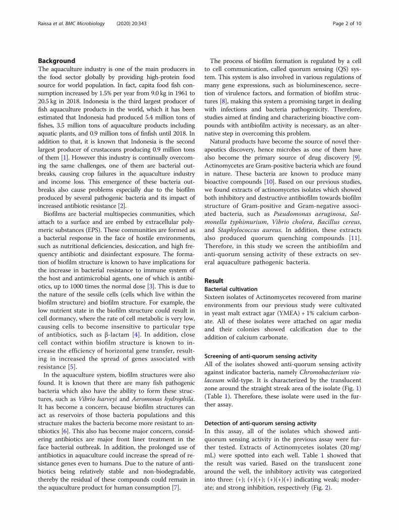
Screening of anti-quorum sensing activityAll of the isolates showed anti-quorum sensing activityagainst indicator bacteria, namely Chromobacterium vio-laceum wild-type. It is characterized by the translucentzone around the straight streak area of the isolate (Fig. 1)(Table 1). Therefore, these isolate were used in the fur-ther assay.
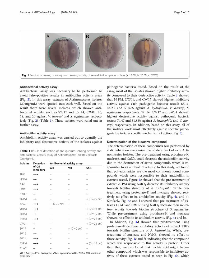
Detection of anti-quorum sensing activityIn this assay, all of the isolates which showed anti-quorum sensing activity in the previous assay were fur-ther tested. Extracts of Actinomycetes isolates (20 mg/mL) were spotted into each well. Table 1 showed thatthe result was varied. Based on the translucent zonearound the well, the inhibitory activity was categorizedinto three: (+); (+)(+); (+)(+)(+) indicating weak; moder-ate; and strong inhibition, respectively (Fig. 2).
Raissa et al. BMC Microbiology (2020) 20:343 Page 2 of 10
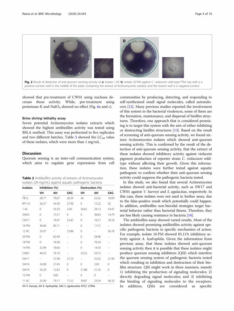
Antibacterial activity assayAntibacterial assay was necessary to be performed toavoid false-positive results in antibiofilm activity assay(Fig. 3). In this assay, extracts of Actinomycetes isolates(20 mg/mL) were spotted into each well. Based on theresult there were several isolates, which showed anti-bacterial activity, such as SW17 and 15, 14, CW01, 16,18, and 20 against V. harveyi and S. agalactiae, respect-ively (Fig. 2) (Table 1). These isolates were ruled out infurther assay.
Antibiofilm activity assayAntibiofilm activity assay was carried out to quantify theinhibitory and destructive activity of the isolates against
pathogenic bacteria tested. Based on the result of theassay, most of the isolates showed higher inhibitory activ-ity compared to their destructive activity. Table 2 showedthat 16 PM, CW01, and CW17 showed highest inhibitoryactivity against each pathogenic bacteria tested: 85.11,44.23, and 53.42% against A. hydrophila, V. harveyi, S.agalactiae respectively. While, CW17 and SW14 showedhighest destructive activity against pathogenic bacteriatested: 74.47 and 51.88% against A. hydrophila and V. har-veyi, respectively. In addition, based on this assay, all ofthe isolates work most effectively against specific patho-genic bacteria in specific mechanism of action (Fig. 3).
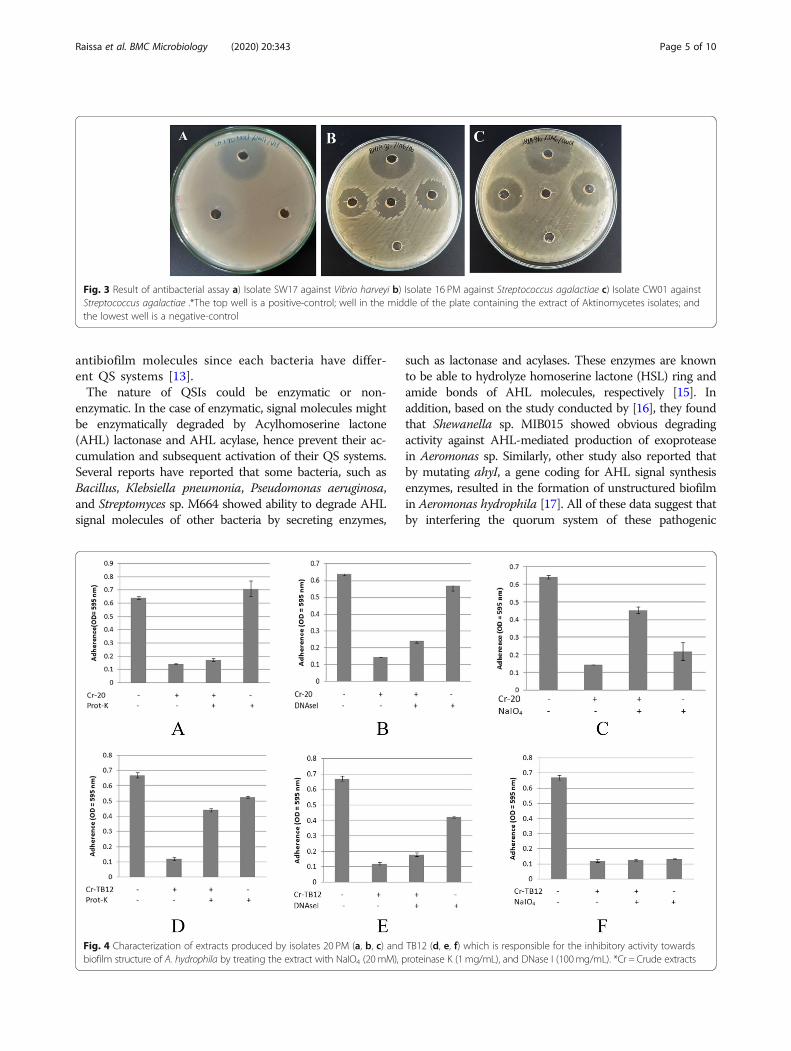
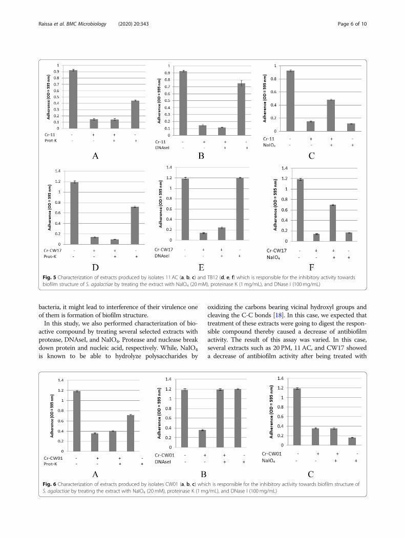
Determination of the bioactive compoundThe determination of these compounds was performed bystatic inhibition assay using the crude extract of each Acti-nomycetes isolates. The pre-treatment using proteinase-K,nuclease, and NaIO4 could decrease the antibiofilm activitydue to the destruction of active compounds, which is re-sponsible to its antibiofilm activity. In this study, we foundthat polysaccharides are the most commonly found com-pounds which were responsible to their antibiofilm inextracts tested. Figure 4c showed that the pre-treatment ofextract 20 PM using NaIO4 decrease its inhibitory activitytowards biofilm structure of A. hydrophila. While pre-treatment using proteinase-K and nuclease showed rela-tively no effect to its antibiofilm activity (Fig. 4a and b).Similarly, Fig. 5c and f showed that pre-treatment of ex-tracts 11 AC and CW17 using NaIO4 decrease their inhibi-tory activity towards biofilm structure of S. agalactiae.While pre-treatment using proteinase-K and nucleaseshowed no effect to its antibiofilm activity (Fig. 4a and b).In addition, Fig. 4d showed that pre-treatment using
proteinase-K decrease inhibitory activity of extract TB12towards biofilm structure of A. hydrophila. While, pre-treatment of nuclease and NaIO4 showed no effect tothose activity (Fig. 4e and f), indicating that the compoundwhich was responsible to this activity is protein. Otherthan that, we also found that nucleic acid might be an-other compound which was responsible to inhibitory ac-tivity of these extracts tested as seen in Fig. 6b, which
Fig. 1 Result of screening of anti-quorum sensing activity of several Actinomycetes isolates (a: 18 PM; b: 20 PM; c: SW03)
Table 1 Result of detection of anti-quorum sensing activity andanti-bacterial activity assay of Actinomycetes isolates extracts(20 mg/mL)
Isolates Detectionof QSinhibitors
Antibacterial activity assay
AH VH SAG
TB12 +++ – – –
KP110 ++ – – –
1 AC +++ – – –
SW03 +++ – – –
CW17 + – – –
16 PM ++ – – + (D = 2.2 cm)
12 AC +++ + (D = 2 cm) – –
20 PM +++ – – + (D = 1.5 cm)
18 PM ++ – – + (D = 1.2 cm)
14 PM +++ – – + (D = 2.1 cm)
CW01 + – – + (D = 2.5 cm)
SW17 + – + (D = 2 cm) –
SW16 ++ – – –
SW14 +++ – – –
15 PM +++ – – + (D = 1.1 cm)
11 AC + – – –
VH V. harveyi, AH A. hydrophila, SAG S. agalacatiae ATCC 27956, D Diameter ofclear zone
Raissa et al. BMC Microbiology (2020) 20:343 Page 3 of 10
showed that pre-treatment of CW01 using nuclease de-crease those activity. While, pre-treatment usingproteinase-K and NaIO4 showed no effect (Fig. 6a and c).
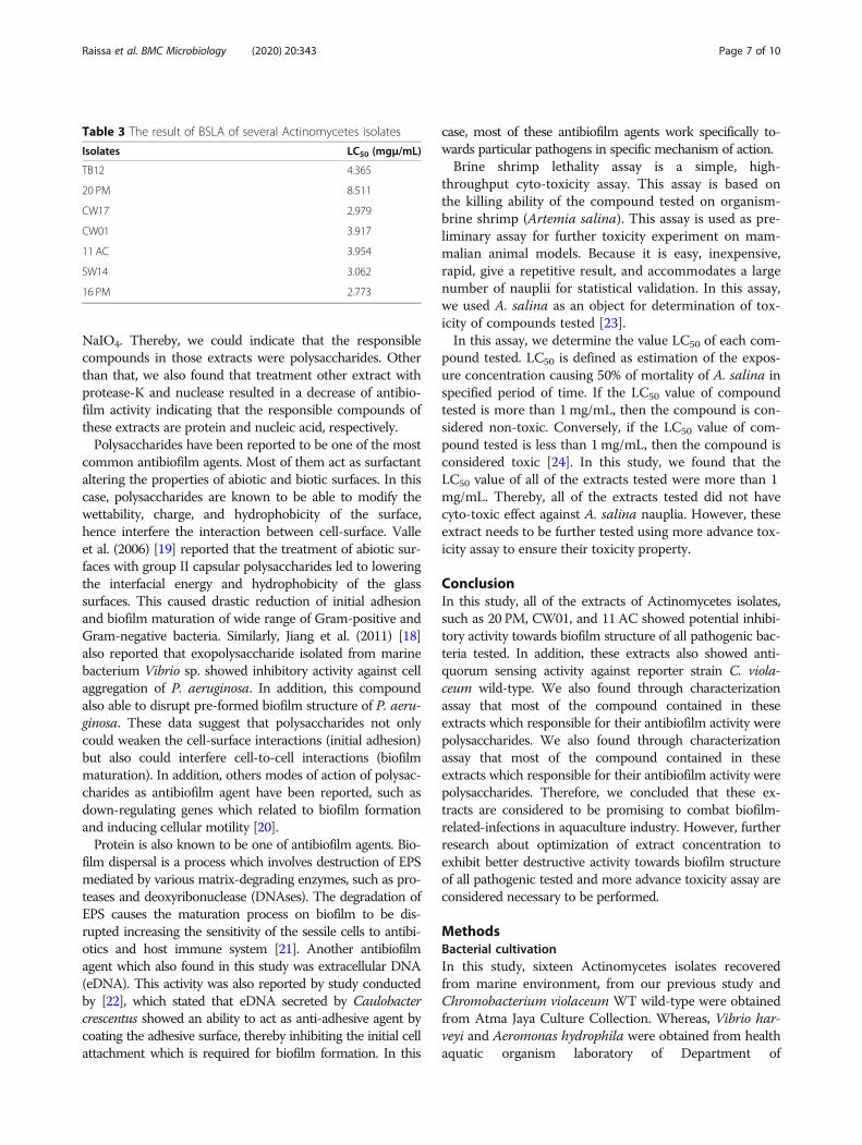
Brine shrimp lethality assaySeven potential Actinomycetes isolates extracts whichshowed the highest antibiofilm activity was tested usingBSLA method. This assay was performed in five replicatesand two different batches. Table 3 showed the LC50 valueof these isolates, which were more than 1mg/mL.
DiscussionQuorum sensing is an inter-cell communication system,which aims to regulate gene expressions from cell
communities by producing, detecting, and responding toself-synthesized small signal molecules, called autoindu-cers [12]. Many previous studies reported the involvementof this system in the bacterial virulences, some of them arethe formation, maintenance, and dispersal of biofilm struc-tures. Therefore, one approach that is considered promis-ing is to target this system with the aim of either inhibitingor destructing biofilm structures [13]. Based on the resultof screening of anti-quorum sensing activity, we found six-teen Actinomycetes isolates which showed anti-quorumsensing activity. This is confirmed by the result of the de-tection of anti-quorum sensing activity, that the extract ofthese isolates showed inhibitory activity against violaceinpigment production of reporter strain C. violaceum wild-type without affecting their growth. Given this informa-tion, these isolates were further tested against aquaticpathogenic to confirm whether their anti-quorum sensingactivity could suppress the pathogenic bacteria tested.In this study, we also found that several Actinomycetes
isolates showed anti-bacterial activity, such as SW17 andCW01 against V. harveyi and S. agalactiae, respectively. Inthis case, these isolates were not used in further assay, dueto the false-positive result which potentially could happen.In addition, antibiofilm non-biocidal strategies target bac-terial behavior rather than bacterial fitness. Therefore, theyare less likely causing resistance in bacteria [14].The antibiofilm assay showed varied results. Most of the
isolates showed promising antibiofilm activity against spe-cific pathogenic bacteria in specific mechanism of action.For example, isolate 16 PM showed 85.11% inhibitory ac-tivity against A. hydrophila. Given the information fromprevious assay, that these isolates showed anti-quorumsensing activity then it is possible that these isolates mightproduce quorum sensing inhibitors (QSI) which interferethe quorum sensing system of pathogenic bacteria testedwhich resulting in inhibition and destruction of their bio-film structure. QSI might work in three manners, namely1) inhibiting the production of signaling molecules; 2)directly degrading signal molecules; and 3) inhibitingthe binding of signaling molecules to the receptors.In addition, QSIs are considered as specific
Fig. 2 Result of detection of anti-quorum sensing activity of a) Isolate 1 AC b) Isolate 20 PM against C. violaceum wild-type.*The top well is apositive-control; well in the middle of the plate containing the extract of Actinomycetes isolates; and the lowest well is a negative-control
Table 2 Antibiofilm activity of extracts of Actinomycetesisolates (20 mg/mL) against aquatic pathogenic bacteria
Isolates Inhibition (%) Destruction (%)
VH AH SAG VH AH SAG
TB12 28.77 78.67 26.34 36 22.62 18.09
KP110 26.37 34.49 27.90 0 15.22 42
1 AC 0 33.33 3.30 26.65 29.13 33.47
SW03 0 75.57 0 0 38.83 14.79
CW17 0 74.47 53.42 0 16.57 18.16
16 PM 36.66 85.11 – 0 17.31 –
12 AC 33.07 – 23.86 0 – 36.78
20 PM 0 82.44 – 0 28.46 –
18 PM 0 76.90 – 0 76.54 –
14 PM 32.94 78.60 – 0 14.54 –
CW01 44.23 76.10 – 23.52 59.75 –
SW17 – 37.99 27.23 – 25.02 27.99
SW16 24.08 27.45 0 0 3.83 0
SW14 35.29 12.62 0 51.88 21.35 0
15 PM 0 9.85 – 0 0 –
11 AC 35.99 79.17 71.52 39.87 23.54 36.33
VH V. harveyi, AH A. hydrophila, SAG S. agalacatiae ATCC 27956
Raissa et al. BMC Microbiology (2020) 20:343 Page 4 of 10
antibiofilm molecules since each bacteria have differ-ent QS systems [13].The nature of QSIs could be enzymatic or non-
enzymatic. In the case of enzymatic, signal molecules mightbe enzymatically degraded by Acylhomoserine lactone(AHL) lactonase and AHL acylase, hence prevent their ac-cumulation and subsequent activation of their QS systems.Several reports have reported that some bacteria, such asBacillus, Klebsiella pneumonia, Pseudomonas aeruginosa,and Streptomyces sp. M664 showed ability to degrade AHLsignal molecules of other bacteria by secreting enzymes,
such as lactonase and acylases. These enzymes are knownto be able to hydrolyze homoserine lactone (HSL) ring andamide bonds of AHL molecules, respectively [15]. Inaddition, based on the study conducted by [16], they foundthat Shewanella sp. MIB015 showed obvious degradingactivity against AHL-mediated production of exoproteasein Aeromonas sp. Similarly, other study also reported thatby mutating ahyI, a gene coding for AHL signal synthesisenzymes, resulted in the formation of unstructured biofilmin Aeromonas hydrophila [17]. All of these data suggest thatby interfering the quorum system of these pathogenic
Fig. 3 Result of antibacterial assay a) Isolate SW17 against Vibrio harveyi b) Isolate 16 PM against Streptococcus agalactiae c) Isolate CW01 againstStreptococcus agalactiae .*The top well is a positive-control; well in the middle of the plate containing the extract of Aktinomycetes isolates; andthe lowest well is a negative-control
Fig. 4 Characterization of extracts produced by isolates 20 PM (a, b, c) and TB12 (d, e, f) which is responsible for the inhibitory activity towardsbiofilm structure of A. hydrophila by treating the extract with NaIO4 (20mM), proteinase K (1mg/mL), and DNase I (100mg/mL). *Cr = Crude extracts
Raissa et al. BMC Microbiology (2020) 20:343 Page 5 of 10
bacteria, it might lead to interference of their virulence oneof them is formation of biofilm structure.In this study, we also performed characterization of bio-
active compound by treating several selected extracts withprotease, DNAseI, and NaIO4. Protease and nuclease breakdown protein and nucleic acid, respectively. While, NaIO4
is known to be able to hydrolyze polysaccharides by
oxidizing the carbons bearing vicinal hydroxyl groups andcleaving the C-C bonds [18]. In this case, we expected thattreatment of these extracts were going to digest the respon-sible compound thereby caused a decrease of antibiofilmactivity. The result of this assay was varied. In this case,several extracts such as 20 PM, 11 AC, and CW17 showeda decrease of antibiofilm activity after being treated with
Fig. 5 Characterization of extracts produced by isolates 11 AC (a, b, c) and TB12 (d, e, f) which is responsible for the inhibitory activity towardsbiofilm structure of S. agalactiae by treating the extract with NaIO4 (20 mM), proteinase K (1 mg/mL), and DNase I (100mg/mL)
Fig. 6 Characterization of extracts produced by isolates CW01 (a, b, c) which is responsible for the inhibitory activity towards biofilm structure ofS. agalactiae by treating the extract with NaIO4 (20 mM), proteinase K (1 mg/mL), and DNase I (100 mg/mL)
Raissa et al. BMC Microbiology (2020) 20:343 Page 6 of 10
NaIO4. Thereby, we could indicate that the responsiblecompounds in those extracts were polysaccharides. Otherthan that, we also found that treatment other extract withprotease-K and nuclease resulted in a decrease of antibio-film activity indicating that the responsible compounds ofthese extracts are protein and nucleic acid, respectively.Polysaccharides have been reported to be one of the most
common antibiofilm agents. Most of them act as surfactantaltering the properties of abiotic and biotic surfaces. In thiscase, polysaccharides are known to be able to modify thewettability, charge, and hydrophobicity of the surface,hence interfere the interaction between cell-surface. Valleet al. (2006) [19] reported that the treatment of abiotic sur-faces with group II capsular polysaccharides led to loweringthe interfacial energy and hydrophobicity of the glasssurfaces. This caused drastic reduction of initial adhesionand biofilm maturation of wide range of Gram-positive andGram-negative bacteria. Similarly, Jiang et al. (2011) [18]also reported that exopolysaccharide isolated from marinebacterium Vibrio sp. showed inhibitory activity against cellaggregation of P. aeruginosa. In addition, this compoundalso able to disrupt pre-formed biofilm structure of P. aeru-ginosa. These data suggest that polysaccharides not onlycould weaken the cell-surface interactions (initial adhesion)but also could interfere cell-to-cell interactions (biofilmmaturation). In addition, others modes of action of polysac-charides as antibiofilm agent have been reported, such asdown-regulating genes which related to biofilm formationand inducing cellular motility [20].Protein is also known to be one of antibiofilm agents. Bio-
film dispersal is a process which involves destruction of EPSmediated by various matrix-degrading enzymes, such as pro-teases and deoxyribonuclease (DNAses). The degradation ofEPS causes the maturation process on biofilm to be dis-rupted increasing the sensitivity of the sessile cells to antibi-otics and host immune system [21]. Another antibiofilmagent which also found in this study was extracellular DNA(eDNA). This activity was also reported by study conductedby [22], which stated that eDNA secreted by Caulobactercrescentus showed an ability to act as anti-adhesive agent bycoating the adhesive surface, thereby inhibiting the initial cellattachment which is required for biofilm formation. In this
case, most of these antibiofilm agents work specifically to-wards particular pathogens in specific mechanism of action.Brine shrimp lethality assay is a simple, high-
throughput cyto-toxicity assay. This assay is based onthe killing ability of the compound tested on organism-brine shrimp (Artemia salina). This assay is used as pre-liminary assay for further toxicity experiment on mam-malian animal models. Because it is easy, inexpensive,rapid, give a repetitive result, and accommodates a largenumber of nauplii for statistical validation. In this assay,we used A. salina as an object for determination of tox-icity of compounds tested [23].In this assay, we determine the value LC50 of each com-
pound tested. LC50 is defined as estimation of the expos-ure concentration causing 50% of mortality of A. salina inspecified period of time. If the LC50 value of compoundtested is more than 1mg/mL, then the compound is con-sidered non-toxic. Conversely, if the LC50 value of com-pound tested is less than 1mg/mL, then the compound isconsidered toxic [24]. In this study, we found that theLC50 value of all of the extracts tested were more than 1mg/mL. Thereby, all of the extracts tested did not havecyto-toxic effect against A. salina nauplia. However, theseextract needs to be further tested using more advance tox-icity assay to ensure their toxicity property.
ConclusionIn this study, all of the extracts of Actinomycetes isolates,such as 20 PM, CW01, and 11AC showed potential inhibi-tory activity towards biofilm structure of all pathogenic bac-teria tested. In addition, these extracts also showed anti-quorum sensing activity against reporter strain C. viola-ceum wild-type. We also found through characterizationassay that most of the compound contained in theseextracts which responsible for their antibiofilm activity werepolysaccharides. We also found through characterizationassay that most of the compound contained in theseextracts which responsible for their antibiofilm activity werepolysaccharides. Therefore, we concluded that these ex-tracts are considered to be promising to combat biofilm-related-infections in aquaculture industry. However, furtherresearch about optimization of extract concentration toexhibit better destructive activity towards biofilm structureof all pathogenic tested and more advance toxicity assay areconsidered necessary to be performed.
MethodsBacterial cultivationIn this study, sixteen Actinomycetes isolates recoveredfrom marine environment, from our previous study andChromobacterium violaceum WT wild-type were obtainedfrom Atma Jaya Culture Collection. Whereas, Vibrio har-veyi and Aeromonas hydrophila were obtained from healthaquatic organism laboratory of Department of
Table 3 The result of BSLA of several Actinomycetes isolates
Isolates LC50 (mgμ/mL)
TB12 4.365
20 PM 8.511
CW17 2.979
CW01 3.917
11 AC 3.954
SW14 3.062
16 PM 2.773
Raissa et al. BMC Microbiology (2020) 20:343 Page 7 of 10
Aquaculture, Faculty of Fisheries and Marine Sciences,Bogor Agricultural Univeristy. Streptococcus agalactiaewhich were used in this study had been characterized asATCC 27956.Cryopreservation isolates of Actinomycetes were streaked
onto Tryptic Soya Agar (TSA) (Oxoid) and incubated at28 °C for 7 days. Afterwards, single colony was picked,streaked onto yeast malt extract agar (10 g Malt extract; 4 gYeast extract; 4 g Glucose; 2 g CaCO3; 1 L distilled water)(YMEA), and incubated at 28 °C for 7 days. Whereas,pathogenic bacteria were streaked onto luria agar (5 g NaCl;5 g Yeast extract; 10 g Tripton; 20 g Agar Bacto; 1 L distilledwater) (LA) and incubated at 28 °C for overnight.
Primary screening of anti-quorum sensing activityThis primary screening was performed based on Abu-doleh and Mahasneh (2017) [25] method with somemodifications. Firstly, Actinomycetes isolates werestraight-streak onto LA and incubated at 28 °C for 3days. Subsequently, C. violaceum WT wild-type as re-porter strain was grown in luria broth (LB) and incu-bated at 28 °C for overnight. This culture was dilutedin sterile LB until the absorbance value reaches 0.132at 600 nm (McFarland 0.5). Then, 100 μL of theculture was put into 3 mL semisolid LA (0.75% w/vagarbacto). After incubation time of Actinomycetesisolates, the culture was poured onto the LA as anoverlay. These plates were incubated at 28 °C for an-other 4 days. The result was observed. In this case,positive result was indicated by the inhibition of vio-lacein pigment around the Actinomycetes isolates.Conversely, negative result was indicated by the pro-duction of violacein pigment around Actinomycetesisolates.
Fermentation conditions and extracts preparationFermentation and extraction was performed using Bala-subramanian et al. (2017) [26] with some modifications.Isolates of Actinomycetes were inoculated into Trypticsoy broth (TSB) + 1% glucose and incubated at 28 °C for7 days at 150 rpm using rotary shaker. Afterwards, theculture was centrigufed at 6900 x g for 15 min. Super-natant was harvested and an equal volume of ethyl acet-ate was added. The supernatant was incubated at 28 °Cfor overnight at 150 rpm using rotary shaker. Then, solv-ent layer was harvested and evaporated using rotaryevaporator to generate extracts. These extracts were dis-solved in 1% (v/v) dimethyl sulfoxide (DMSO) to gener-ate final concentration (20 mg/mL). The extracts wereused for further assays.
Detection of anti-quorum sensing activityThis step was performed based on the method published byRajivgandhi et al. (2018) [27] with some modifications.
Chromobacterium violaceum wild-type was streaked con-tinuously in three directions onto LA using sterile cottonbuds. Then, sterile cork borer was used to remove plugs ofthe agar. This plug was replaced by extract of Actinomycetesisolates. In this assay, DMSO was used as control negative,while Streptomycin (10mg/mL) was used as positive con-trol. Plates were incubated at 28 °C for overnight. Finally,translucent zone against violacein pigmentation of C. viola-ceum wild-type was observed.
Anti-bacterial assayAntibacterial assay was performed by using diffusionmethod according to Bauer et al. (1966) [28] with somemodifications. Pathogenic bacteria were inoculated intoLB and incubated at 28 °C for overnight at 150 rpmusing rotary shaker. Afterwards, these cultures were di-luted into sterile LB until the absorbance value reaches0.132 at 600 nm (McFarland 0.5).Pathogenic bacteria were streaked in three directions
onto Mueller Hinton Agar (MHA) using sterile cottonbuds. Afterwards, a sterile cork borer was used to re-move the agar plugs. Then, these plugs were replaced bythe extract of Actinomycetes isolates. In this assay,DMSO was used as control negative, while Streptomycin(10 mg/mL) was used as positive control. Plates were in-cubated at 28 °C for overnight. Then, clearing zone wasobserved. The isolate which showed anti-bacterial activ-ity was ruled out in antibiofilm activity assay.
Antibiofilm assayAntibiofilm assay was performed in two categories basedon their mechanisms, namely inhibitory and destructiveactivity assay. Preparation of this assay was performed,by inoculating pathogenic bacteria into LB medium.Then, cultures were incubated at 28 °C for overnight anddiluted into sterile LB until the absorbance valuesreaches 0.132 at 600 nm (McFarland 0.5).Antibiofilm assay was performed according to
O’Toole and Kolter (1998) [29] with some modifica-tions. For inhibitory activity assay, 200 μL of patho-genic bacteria suspension and Actinomycetes isolatesextracts was loaded simultaneously into 96-well poly-styrene plates. Then, plates were incubated at 28 °Cfor overnight, then staining process was performed.Conversely for destructive activity assay, firstly 200 μLof pathogenic bacteria suspension was loaded into 96-well polystyrene plates. Plates were incubated at 28 °Cfor overnight and Actinomycetes isolates extractswere loaded, then staining process was performed.For both assay, pathogenic bacteria suspensions wereused as positive control, while sterile LB was used asnegative control.The process of staining was performed un-aseptically.
Planktonic cells and media were discarded. Then,
Raissa et al. BMC Microbiology (2020) 20:343 Page 8 of 10
adherent biofilm cells were rinsed with water twice andair-dried for 30 min. These cells were stained by using200 μL of 0.4% (b/v) crystal violet for 30 min. Then, crys-tal violet was discarded. These stained cells were rinsedwith water for five times to remove any remaining crys-tal violet and air-dried for 30 min. Then, 100 μL of abso-lute ethanol was added and re-suspended to dissolve thestained cells. Subsequently, this ethanol was transferredinto new 96-well polystyrene plates. The absorbance ofeach well was measured with microplate reader. Finally,the percentage of antibiofilm activity of Actinomycetesisolates extract was calculated using the formula below[30]:
%Activity ¼ OD Positive control −OD sampleOD positive control
x 100%ð Þ
Determination of the bioactive compoundsThis method was based on this research (Jiang et al.2011) [18] with some modifications. Some selected ex-tracts of Actinomycetes isolates were treated with pro-teinase K (1 mg/mL), DNase I (100 μg/mL), and NaIO4
20mM at 37 °C for 12 h. Then, the post-treated extractswere used for biofilm inhibition and destruction activityassay. Finally, the activity of pre and post-treated ex-tracts were measured and compared.
Sample preparation for brine shrimp lethality assay (BSLA)Samples were prepared by dissolving 1 mL extract of Ac-tinomycetes isolates in 100mL artificial sea water inorder to get a stock solution (1000 ppm). Then, stock so-lution was diluted into final concentration (10, 100, 500,and 1000 ppm).
Hatching the shrimpThis method was based on this research (Krishnarajuet al. 2006) [31] with some modifications. Three mg ofbrine shrimp eggs was added into 3
4 volume of 1 L min-eral water bottle filled with artificial sea water. Moreoverto optimize the hatching process, the growing media wasconstantly aerated with air pump and illuminated withlamp. After 24 h, the nauplia were collected by pipetteand put into tubes.
BioassayBioassay was performed according to the method pub-lished by Ramachandran et al. (2011) [32] with somemodifications. Ten nauplia were transferred into tubesfilled with 4.5 mL artificial sea water. Then, 500 μL of ex-tracts was added into each tube. Tubes were maintainedunder illumination for 24 h at room temperature. Finally,
the death percentage of nauplii was determined usingthis formula below:
%Death ¼ Death naupliiTotal nauplii
x 100%ð Þ
In case the negative control does not give 0% of mor-tality, the formula was corrected using Abotts’s formulabelow:
%Death ¼ Death nauplii in tested vial − death nauplii in control vialDeath nauplii in control vial
x 100%ð Þ
In this assay, 500 μL of K2Cr2O7 was be used as posi-tive control, while 500 μL of artificial water was used asnegative control. The lethal concentration (LC50) wascollected.
AbbreviationsEPS: Extracellular polymeric substances; YMEA: Yeast Malt Extract Agar;QSI: Quorum Sensing Inhibitor; AHL: Acylhomoserine lactone;HSL: Homoserine lactone; LA: Luria Agar; LB: Luria Broth; TSB: Tryptic SoyBroth; DMSO: Dimethyl Sulfoxide; MHA: Mueller Hinton Agar; QS: QuorumSensing; BSLA: Brine Shrimp Lethality Assay
AcknowledgementsThe authors are grateful toward all the supports and would like to thankeveryone who contributed in this study. We would like to thank Ministry ofResearch and Technology of Indonesia which had funded this research. Wealso thank faculty of fisheries and marine science of Institute of PertanianBogor University which had provided several of the pathogenic bacteriacultures.
LimitationThis study only screened for bioactive compounds which have anti-quorumsensing and antibiofilm activity for several aquaculture pathogenic bacteria,thereby more researches need to be conducted to screen and characterizeanother compounds which have those activities against another aquaculturepathogenic bacteria. In addition, in this study we only performed preliminaryassay of toxicity assay, thereby more advance toxicity assay need to be per-formed to assure the toxicity property of these extracts.
Authors’ contributionsDEW involved in research design and advisory. GR conducted the research,collected the data, analyzed and processed the data, and prepared themanuscript. DW provided several of pathogen culture and involved inadvisory. All authors read and approved the final manuscript.
FundingThis study was funded by Ministry of Research and Technology of Indonesia(Kementrian Riset dan Teknologi Indonesia). The funder has no contributionin this study.
Availability of data and materialsAll data generated or analysed during this study are included in thispublished article.
Ethics approval and consent to participateNot applicable.
Consent for publicationNot applicable.
Competing interestsThe authors declare that they have no competing interests.
Raissa et al. BMC Microbiology (2020) 20:343 Page 9 of 10
Author details1Master of Biotechnology Program, Faculty of Biotechnology, Atma JayaCatholic University of Indonesia, Jalan Jenderal Sudirman, Jakarta 12930,Indonesia. 2Department of Aquaculture, Faculty of Fisheries and MarineScience, Bogor Agricultural University, Jalan Raya Dramaga, Bogor 16680,Indonesia.
Received: 10 July 2020 Accepted: 27 October 2020
References1. FAO. 2020. The state of world fisheries and aquaculture.2. MMAF. Marine affairs and fisheries in figures 2013. Jakarta: MMAF; 2014.3. Flemming HC, Wingender J. The biofilm matrix. Nat Rev Microbiol. 2010;8:
623–33. https://doi.org/10.1038/nrmicro2415.4. Anderl JN, Franklin MJ, Stewart PS. Role of antibiofilm penetration limitation
in Klebsiella pneumoniae biofilm resistance to ampicillin and ciprofloxacin.Antimicrob Agents Chemother. 2000;44:1818–24. https://doi.org/10.1128/AAC.44.7.1818-1824.2000.
5. Ma H, Bryers JD. Non-invasive determination of conjugative transfer ofplasmids bearing antibiotic-resistance genes in biofilm-bound bacteria:effects of substrate loading and antibiotic selection. Appl MicrobiolBiotechnol. 2013;97(1):317–28. https://doi.org/10.1007/s00253-012-4179-9.
6. King RK, Flick GJ, Pierson D, Smith SA, Boardman GD, Coale CW.Identification of bacterial pathogens in biofilms of recirculating aquaculturesystems. J Aquat Food Product Technol. 2004;13(1):125–31. https://doi.org/10.1300/J030v13n01_11.
7. Gauthier DT. Bacterial zoonoses of fishes: a review and appraisal of evidencefor linkages between fish and human infections. Vet J. 2015;203:27–35.
8. Rutherford ST, Bassler BL. Bacterial quorum sensing: its role in virulence andpossibilities for its control. Cold Spring Harb Perspect Med. 2012;2:a012427.https://doi.org/10.1101/cshperspect.a012427.
9. Courtois S, Cappellano CM, Ball M. Recombinant environmental librariesprovide access to microbial diversity for drug discovery from naturalproducts. Appl Environ Microbiol. 2003;69:49–55. https://doi.org/10.1128/AEM.69.1.49-55.2003.
10. Pandey B, Ghimire P, Agrawal VP. Studies on the bacterial activity of theActinomycetes isolated from the Khumbu region of Nepal. Appl Microbiol.2008;5:235–61.
11. Wijaya M, Delicia D, Waturangi DE. Screening and quantification of antiquorum-sensing activity of Actinobacteria isolates against gram-positive and gram-negative biofilm associated bacteria. Durham: Research Square; 2020.
12. Van Bodman SB, Willey JM, Diggle SP. Cell-cell communication in bacteria: unitedwe stand. J Bacteriol. 2008;190:4377–91. https://doi.org/10.1128/JB.00486-08.
13. Brackman G, Coenye T. Quorum sensing inhibitors as antibiofilm agents.Curr Pharm Des. 2015;21(1):5–11. https://doi.org/10.2174/1381612820666140905114627.
14. Rosenthal CB, Mootz JM, Horswill AR. Staphylococcus aureus biofilmformation and inhibition. In: Rumbaugh KP, Ahmad I, editors. Antibiofilmagents: from diagnosis to treatment and prevention. London: Springer;2015. p. 233–56.
15. Uroz S, Dessaux Y, Oger P. Quorum sensing and quorum quenching: the yinand yang of bacterial communication. Chem Biol Chem. 2009;10:205–16.https://doi.org/10.1002/cbic.200800521.
16. Morohoshi T, Ebata A, Nakazawa S, Kato N, Ikeda T. N-acyl homoserinelactone-producing or-degrading bacteria isolated from intestinal microbialflora of Ayu fish (Plecoglossus altivelis). Microbes Environ. 2005;20(4):264–8.
17. Lynch MJ, Swift S, Kirke DF, Keevil CW, Dodd CER, Williams P. The regulationof biofilm development by quorum sensing in Aeromonas hydrophila.Environ Microbiol. 2002;4(1):18–28.
18. Jiang P, Li J, Han F, Duan G, Lu X, Gu Y, Yu W. Antibiofilm activity of anexopolysaccharide from marine bacterium Vibrio sp. QY101. PLoS One. 2011;6(4):1–11.
19. Valle J, Re SD, Henry N, Fontaine T, Balestrino D, Lambert PL, Ghigo JM.Broad-spectrum biofilm inhibition by a secreted bacterial polysaccharide.PNAS. 2006;103(33):12558–63. https://doi.org/10.1073/pnas.0605399103.
20. Rendueles O, Kaplan JB, Ghigo JM. Antibiofilm polysaccharides. EnvironMicrobiol. 2013;15(2):334–46. https://doi.org/10.1111/j.1462-2920.2012.02810.x.
21. Roy R, Tiwari M, Donelli G, Tiwari V. Strategies for combathing bacterialbiofilms: a focus on antibiofilm agents and their mechanisms of action.Virulence. 2018;9(1):522–54. https://doi.org/10.1080/21505594.2017.1313372.
22. Berne C, Kysela DT, Brun YV. A bacterial extracellular DNA inhibits settling ofmotile progeny cells within biofilm. Mol Microbiol. 2010;77(4):815–29.https://doi.org/10.1111/j.1365-2958.2010.07267.x.
23. Rajabi S, Ramazani A, Hamidi M, Naji T. Artemia salina as a model organismin toxicity assessment of nanoparticles. DARU J Pharm Sci. 2015;23:20–6.
24. Wu C. An important player in brine shrimp lethality bioassay: the solvent. JAdv Pharm Technol Res. 2014;5(1):57–8.
25. Abudoleh SM, Mahasneh AM. Anti-quorum sensing activity of substancesisolated from wild berry associated bacteria. Avicenna J Med Biotechnol.2017;9(1):23–30.
26. Balasubramanian S, Othman EM, Kampik D, Stopper H, Hentschel U, ZiebuhrW, Oelschlaeger TA, Abdelmohsen UR. Marine sponge-derived Streptomycessp. SBT343 extract inhibits Stpahylococcal biofilm formation. FrontMicrobiol. 2017;8:236. https://doi.org/10.3389/fmicb.2017.00236.
27. Rajivgandhi G, Vijayan R, Maruthupandy M, Vaseeharan B, Manoharan N.Antibiofilm effect of Nocardiopsis sp. GRG1 (KT235640) compound againstbiofilm forming gram negative bacteria on UTIs. Microb Pathog. 2018;118:190–8. https://doi.org/10.1016/j.micpath.2018.03.011.
28. Bauer AW, Kirby WMM, Sherris JC, Turck M. Antibiotic susceptibility testingby a standardized single disk method. Am J Clin Pathol. 1966;45:493–6.
29. O’Toole GA, Kolter R. Flagellar and twitching motility are necessary forPseudomonas aeruginosa biofilm development. Mol Microbiol. 1998;30:295–304.
30. Djordjevic D, Wiedmann M, McLandsborough LA. Microtiter plate assay forassessment of Listeria monocytogenes biofilm formation. Appl EnvironMicrobiol. 2002;68(6):2950–8. https://doi.org/10.1128/aem.68.6.2958.2002.
31. Krishnaraju AV, Rao TVN, Sundararaju D, Vanisree M, Tsay HS, Subbaraju GV.Biological screening of medicinal plants collected from eastern Ghats of Indiausing Artemia salina (brine shrimp test). Int J Appl Sci Eng. 2006;4(2):115–25.
32. Ramachandran S, Vamsikrishna M, Gowthami KV, Heera B, DhanarajuMD. Assessment of cytotoxic activity of Agave cantula using brineshrimp (Artemia salina) lethality bioassay. Asian J Sci Res. 2011;4(1):90–4.https://doi.org/10.3923/ajsr.2011.90.94.
Publisher’s NoteSpringer Nature remains neutral with regard to jurisdictional claims inpublished maps and institutional affiliations.
Raissa et al. BMC Microbiology (2020) 20:343 Page 10 of 10