Embed Size (px)
Citation preview
Scoliosis
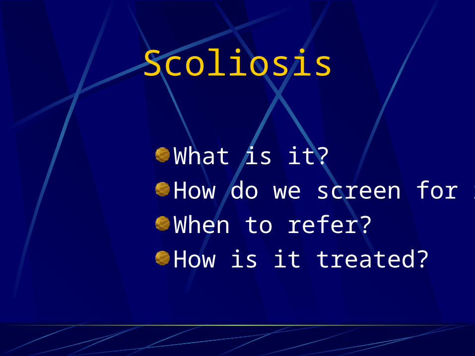
Scoliosis
What is it?How do we screen for it?When to refer?How is it treated?
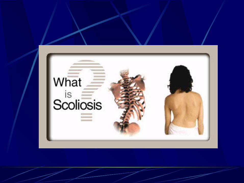
What is scoliosis?
Lateral curvature of the spine >10º accompanied by vertebral rotationIdiopathic scoliosis - Multigene dominant condition with variable phenotypic expression & no clear causeMultiple causes exist for secondary scoliosis
Secondary causes for scoliosis:Inherited connective tissue disorders
- Ehler’s Danlos syndrome
- Marfan syndrome- Homocystinuria
Secondary causes for scoliosis:Neurologic disorders
Tethered cord syndromeSyringomyeliaSpinal tumorNeurofibromatosisMuscular dystrophy
Cerebral palsyPolioFriedeich’s ataxiaFamilial dysautonomiaWerdnig-Hoffman disease
Secondary causes for scoliosis:Musculoskeletal disorders
Leg length discrepancyDevelopmental hip dysplasia
Osteogenesis imperfectaKlippel-Feil syndrome
Characteristics of idiopathic scoliosis:
Present in 2 - 4% of kids aged 10 – 16 yearsRatio of girls to boys with small curves (<10º) is equal, but for curves >30º the ratio is 10:1Scoliosis tends to progress more often in girls (so girls with scoliosis are more likely to require treatment)
Natural history of scoliosis
Of adolescents diagnosed with scoliosis, only 10% have curve progression requiring medical interventionThree main determinants of curve progression are:(1) Patient gender(2) Future growth potential(3) Curve magnitude at time of diagnosis
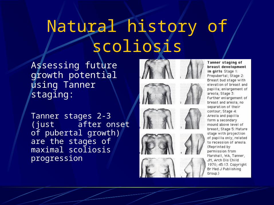
Natural history of scoliosis
Assessing future growth potential using Tanner staging:
Tanner stages 2-3 (just after onset of pubertal growth) are the stages of maximal scoliosis progression
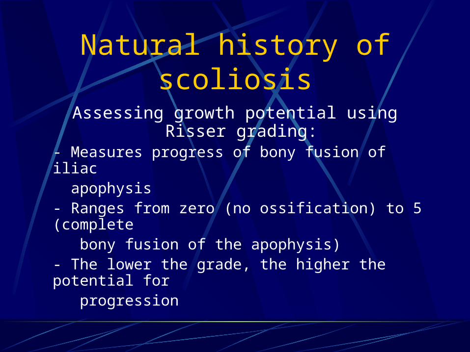
Natural history of scoliosis
Assessing growth potential using Risser grading:
- Measures progress of bony fusion of iliac apophysis- Ranges from zero (no ossification) to 5 (complete
bony fusion of the apophysis)- The lower the grade, the higher the potential for progression
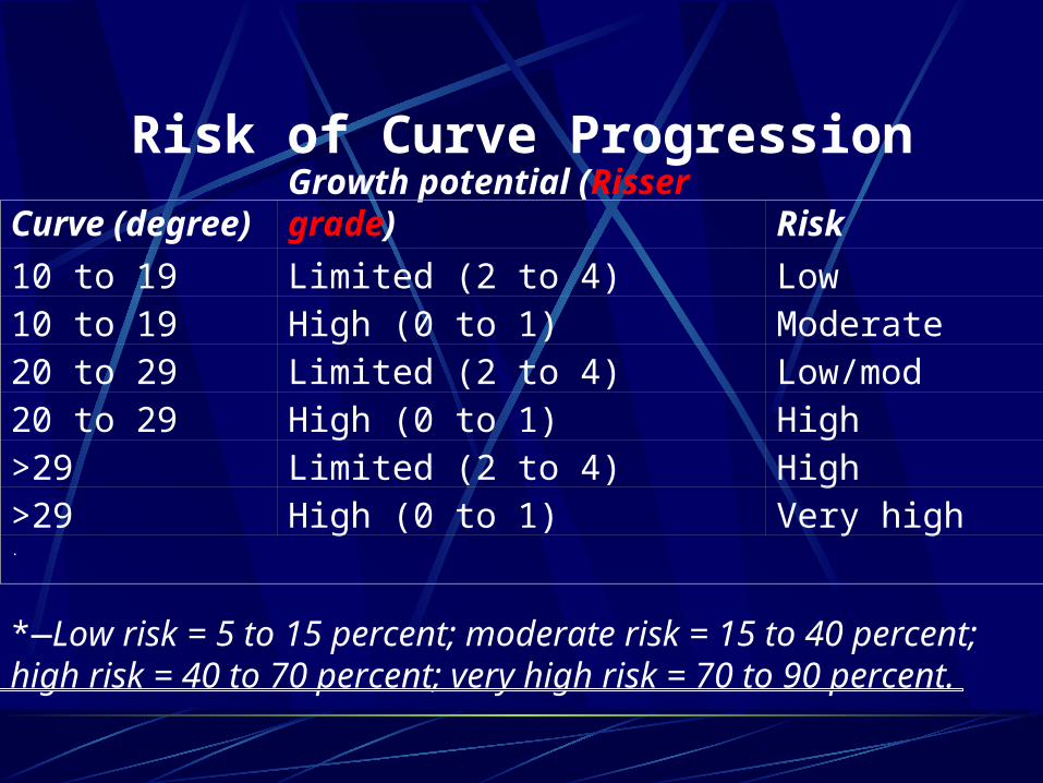
Risk of Curve Progression
Curve (degree) Growth potential (Risser grade) Risk
10 to 19 Limited (2 to 4) Low10 to 19 High (0 to 1) Moderate20 to 29 Limited (2 to 4) Low/mod20 to 29 High (0 to 1) High>29 Limited (2 to 4) High>29 High (0 to 1) Very high.
*—Low risk = 5 to 15 percent; moderate risk = 15 to 40 percent; high risk = 40 to 70 percent; very high risk = 70 to 90 percent.
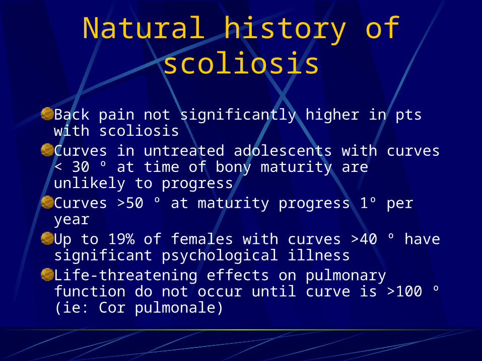
Natural history of scoliosis
Back pain not significantly higher in pts with scoliosisCurves in untreated adolescents with curves < 30 º at time of bony maturity are unlikely to progressCurves >50 º at maturity progress 1º per yearUp to 19% of females with curves >40 º have significant psychological illnessLife-threatening effects on pulmonary function do not occur until curve is >100 º (ie: Cor pulmonale)
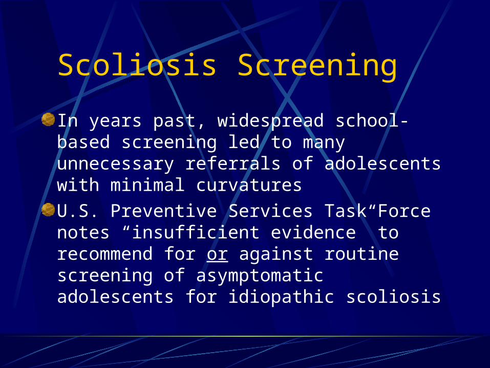
Scoliosis Screening
In years past, widespread school-based screening led to many unnecessary referrals of adolescents with minimal curvaturesU.S. Preventive Services Task Force notes “insufficient evidence” to recommend for or against routine screening of asymptomatic adolescents for idiopathic scoliosis
Scoliosis Screening Recommendations
American Academy of Orthopedic Surgeons - Screen girls at ages 11 and 13- Screen boys once at age 13 or 14American Academy of Pediatrics- Screen at 10, 12, 14 and 16 years
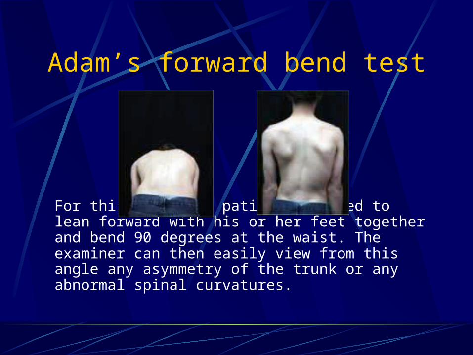
Adam’s forward bend test
For this test, the patient is asked to lean forward with his or her feet together and bend 90 degrees at the waist. The examiner can then easily view from this angle any asymmetry of the trunk or any abnormal spinal curvatures.
Screening hints:
Shoulders are different heights – one shoulder blade is more prominent than the other Head is not centered directly above the pelvis Appearance of a raised, prominent hip Rib cages are at different heights Uneven waist Changes in look or texture of skin overlying the spine (dimples, hairy patches, color changes) Leaning of entire body to one side
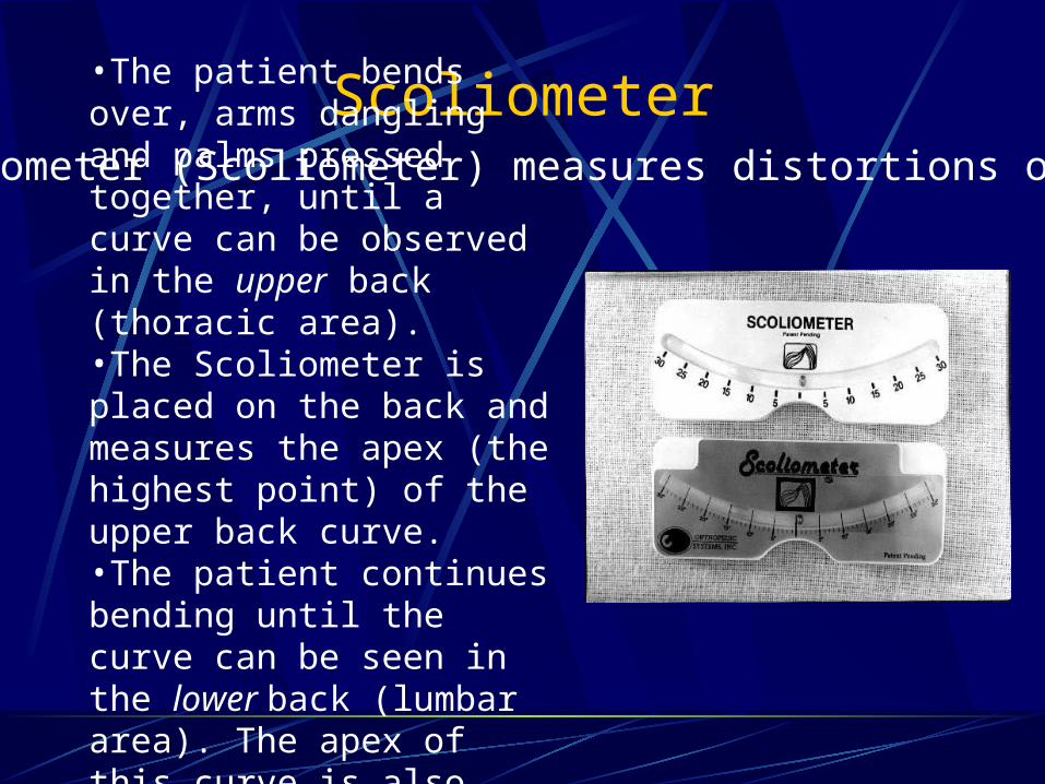
Scoliometer•The patient bends over, arms dangling and palms pressed together, until a curve can be observed in the upper back (thoracic area). •The Scoliometer is placed on the back and measures the apex (the highest point) of the upper back curve. •The patient continues bending until the curve can be seen in the lower back (lumbar area). The apex of this curve is also measured.
An inclinometer (Scoliometer) measures distortions of the torso.
Red flags on PE:
Left-sided thoracic curvature PainSignificant stiffnessAbnormal neurologic findingsStigmata of other clinical syndromes associated with curvature
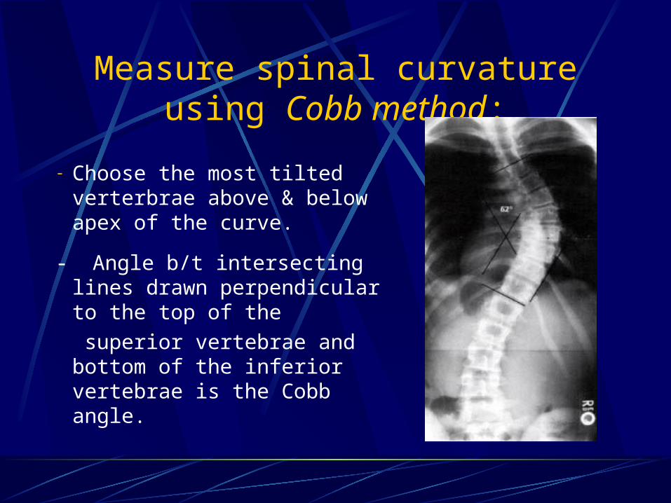
Measure spinal curvature using Cobb method:
- Choose the most tilted verterbrae above & below apex of the curve.
- Angle b/t intersecting lines drawn perpendicular to the top of the superior vertebrae and bottom of the inferior vertebrae is the Cobb angle.
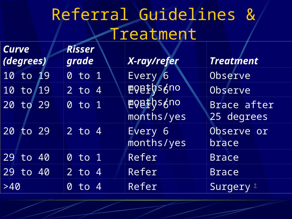
Referral Guidelines & Treatment
Curve (degrees) Risser grade X-ray/refer Treatment
10 to 19 0 to 1 Every 6 months/no Observe
10 to 19 2 to 4 Every 6 months/no Observe
20 to 29 0 to 1 Every 6 months/yes
Brace after 25 degrees
20 to 29 2 to 4 Every 6 months/yes
Observe or brace
29 to 40 0 to 1 Refer Brace
29 to 40 2 to 4 Refer Brace
>40 0 to 4 Refer Surgery †
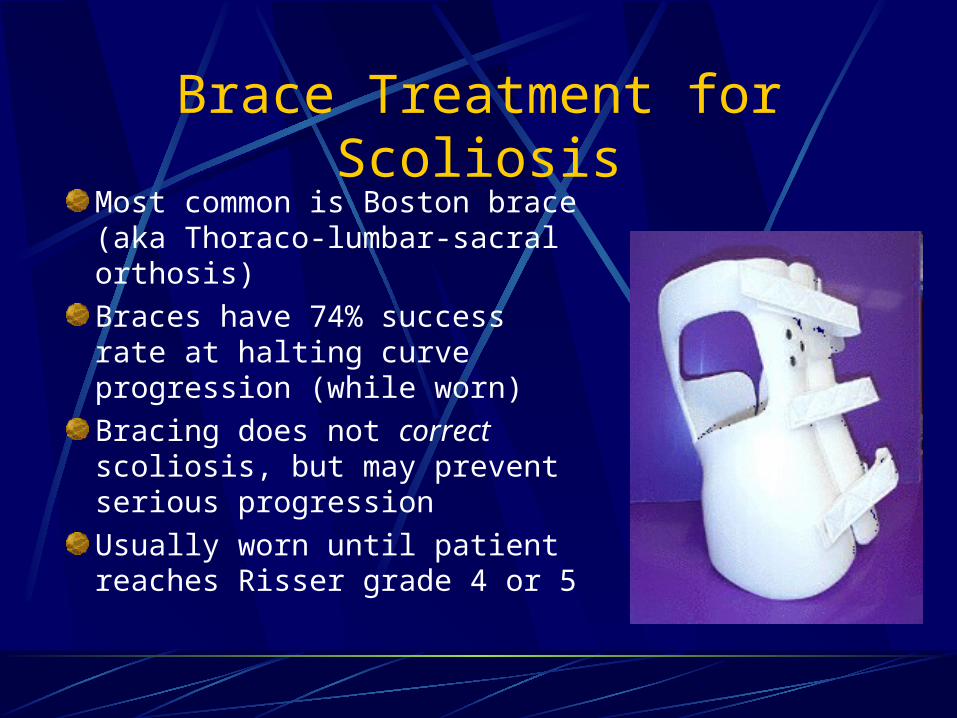
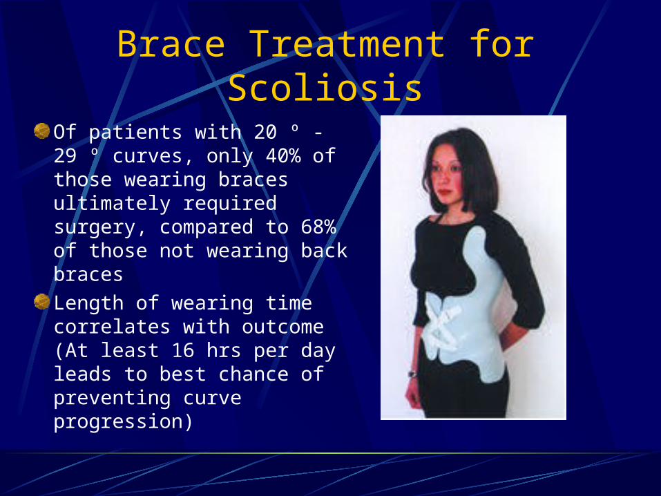
Brace Treatment for ScoliosisMost common is Boston brace (aka Thoraco-lumbar-sacral orthosis)Braces have 74% success rate at halting curve progression (while worn)Bracing does not correct scoliosis, but may prevent serious progressionUsually worn until patient reaches Risser grade 4 or 5
Brace Treatment for Scoliosis
Of patients with 20 º - 29 º curves, only 40% of those wearing braces ultimately required surgery, compared to 68% of those not wearing back bracesLength of wearing time correlates with outcome (At least 16 hrs per day leads to best chance of preventing curve progression)
Surgical Treatment for Scoliosis
Curves in growing children greater than 40 º require a spinal fusion (Risser grade 0 to 1 in girls and Risser 2 or 3 in boys) Skeletally mature patients can be observed until their curves reach 50 º Posterior spinal fusion is best choice for thoracic curvesAnterior spinal fusion is best treatment for thoracolumbar and lumbar curves
Surgical Treatment for Scoliosis
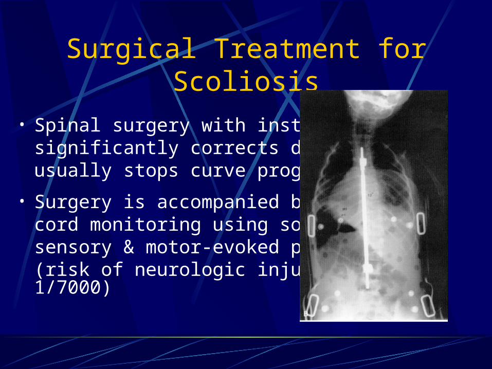
• Spinal surgery with instrumentationsignificantly corrects deformity &usually stops curve progression
• Surgery is accompanied by spinalcord monitoring using somato-sensory & motor-evoked potentials (risk of neurologic injury is 1/7000)
Post-Op Treatment & Long Term Consequences of Spinal Fusion
If segmental instrumentation used, no post-op cast or brace requiredPost-fusion back pain does occur and is more common in distal spinal fusionsUsually out of hospital in 4-5 days & back at school in 2 wksOK to participate in athletics after 9 – 12 months (should avoid contact sports)
Case #1
MP is a 16-year-old male who presents to your office for his annual health assessment and sports physical. During the course of his examination, you note a mild convexity in the thoracic region of his spine with forward flexion at the hips. Based on your clinical examination, you estimate a lateral spinal curvature of about 5 degrees. You note these findings to the patient and then to his mother.
Question 1
C.
Order a radiograph of the back to quantify the curvature (e.g., Cobb angle).
C.
Order a radiograph of the back to quantify the curvature (e.g., Cobb angle).
1. Which one of the following procedures should be implemented next?
A. Recommend back-strengthening exercises.B. Refuse to permit participation in contact sports.C.Order a radiograph of the back to quantify the
curvature (e.g., Cobb angle).D. Monitor the patient's condition.E.Refer for orthopedic consultation.
Answer 1
The answer is D: monitor the patient's condition.
Question 2
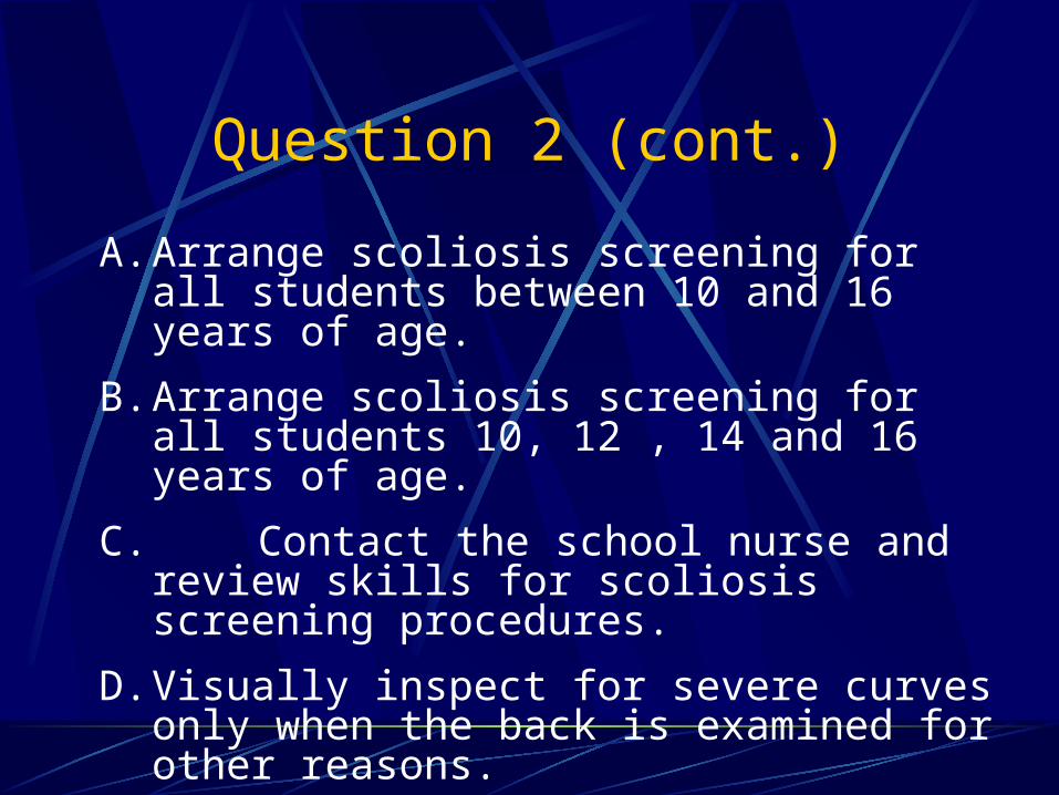
Because you have recently agreed to serve as school physician in the district where your office is located, you wonder what scoliosis screening programs are in place and who has been examining these school children for scoliosis. Which one of the following procedures should you implement?
Question 2 (cont.)
A. Arrange scoliosis screening for all students between 10 and 16 years of age.
B. Arrange scoliosis screening for all students 10, 12 , 14 and 16 years of age.
C. Contact the school nurse and review skills for scoliosis screening procedures.
D. Visually inspect for severe curves only when the back is examined for other reasons.
E. Screen girls for scoliosis at 11 and 13 years of age and boys at 13 and 15 years of age.
Answer 2
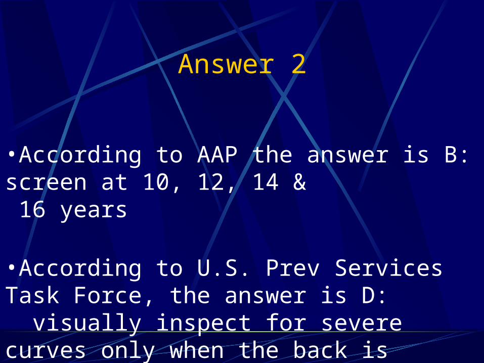
•According to AAP the answer is B: screen at 10, 12, 14 & 16 years
•According to U.S. Prev Services Task Force, the answer is D: visually inspect for severe curves only when the back is examined for other reasons.
Question 3
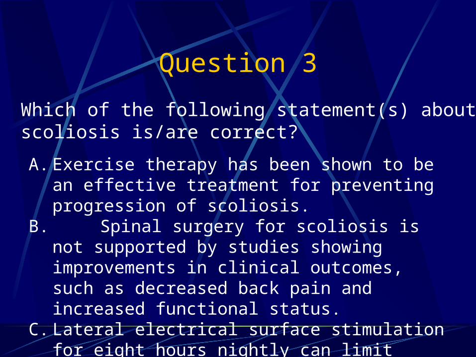
Which of the following statement(s) about treatment for adolescent scoliosis is/are correct?
A. Exercise therapy has been shown to be an effective treatment for preventing progression of scoliosis.
B. Spinal surgery for scoliosis is not supported by studies showing improvements in clinical outcomes, such as decreased back pain and increased functional status.
C. Lateral electrical surface stimulation for eight hours nightly can limit progression of spinal curvature
D. Back bracing (e.g., orthoses) reduces symptoms of low back pain.
Answer 3
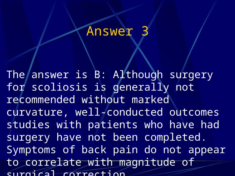
The answer is B: Although surgery for scoliosis is generally not recommended without marked curvature, well-conducted outcomes studies with patients who have had surgery have not been completed. Symptoms of back pain do not appear to correlate with magnitude of surgical correction.
ConclusionsScreening for scoliosis remains controversial & has led to many unnecessary referralsAdolescent scoliosis can be followed by family docs if the curve has a low risk of progression & underlying causes have been excludedCurves demonstrating significant progression with continued growth remaining or those at high risk of progression should be referred for orthopedic evaluationAlways refer when red flags are present on PE or X-ray
Conclusions
90% of kids with scoliosis will not require medical interventionGirls are much more likely than boys to need intervention for scoliosisBracing can slow progression of many curves and significantly decrease need for surgerySpinal fusion surgery is recommended for curves greater than 45 – 50 degrees
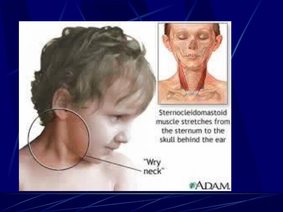
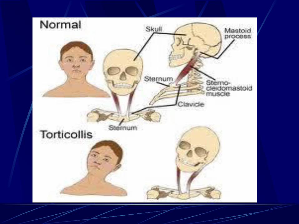
Torticollis
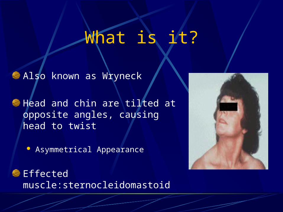
What is it?
Also known as Wryneck
Head and chin are tilted at opposite angles, causing head to twist
Asymmetrical Appearance
Effected muscle:sternocleidomastoid
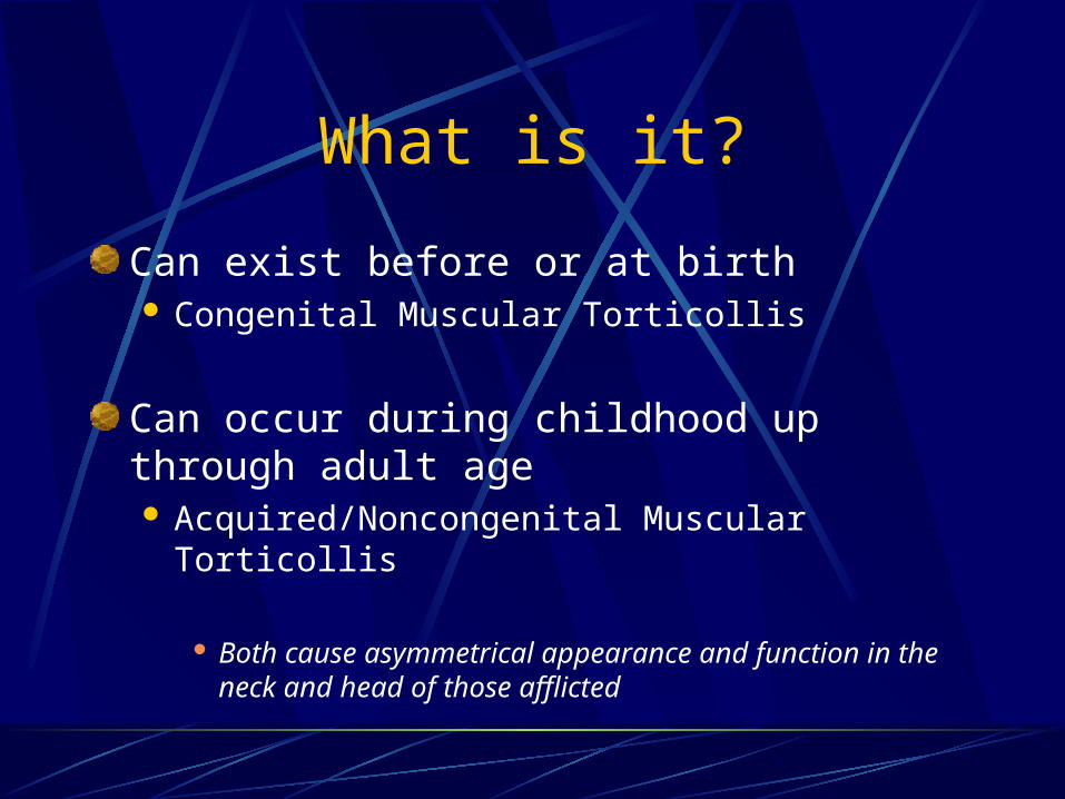
What is it?
Can exist before or at birth Congenital Muscular Torticollis
Can occur during childhood up through adult age Acquired/Noncongenital Muscular Torticollis
Both cause asymmetrical appearance and function in the neck and head of those afflicted
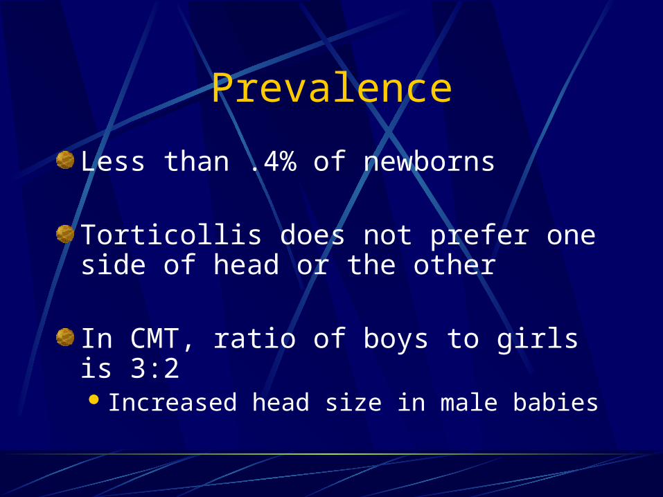
Prevalence
Less than .4% of newborns
Torticollis does not prefer one side of head or the other
In CMT, ratio of boys to girls is 3:2 Increased head size in male babies
Prevalence
In adults, noncongenital muscular torticollis has an average onset of 40 years old Females twice as likely afflicted than males
Usually equal distribution between right and left side of body afflicted
Slightly more right torticollis in older female populations
Causes?
Not well understood
Almost 80 entities have been reported to cause torticollis
Common causes: Developmental disorders affecting
sternocleidomastoid muscle Imbalance in function of cervical muscles Other abnormalities in skull/cervical area
Other Causes
Genetic defectInfants position during pregnancy or deliveryTumors in head or neckArthritis of neck Pseudotumors in infants
Certain medications Genes More likely to be afflicted if family member had
torticollis or similar disorder
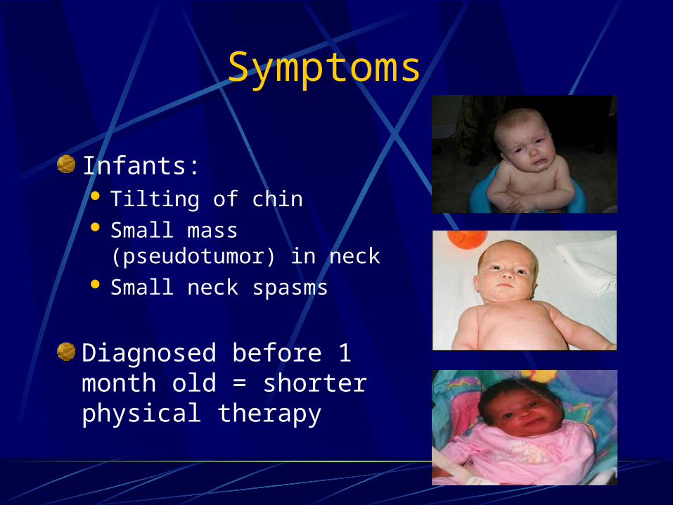
Symptoms
Adults and Children: Abnormal contraction of the neck Limited range of motion Stiff neck muscles Possible swelling and pain
Can often be mistaken for more serious condition
• See medical professional immediately
Symptoms
Infants: Tilting of chin Small mass (pseudotumor)
in neck Small neck spasms
Diagnosed before 1 month old = shorter physical therapy
Prognosis
Most helpful diagnosis is made early
Not life threatening
May self correct itself
May be chronic and reoccurring
Any complications may result from compressed nerve roots
Treatments
Stretching and lengthening affected neck muscles
Applying heat, massage, analgesicsCan be combined with TENS
Transcutaneous Electrical Nerve Stimulation
Medical treatment—Bacolfen or Botox Injection every three months
Treatments
Surgery in severe casesPatients whose pathology does not resolve
after 12 months of physical therapy or who develops facial asymmetry
Risk of injury to spinal nerves

Preventive Measures
Nearly impossible to prevent
Become familiar with symptoms
Seek medical attention Other serious conditions may be confused
for Torticollis and are not treated correctly
Any Questions?