Embed Size (px)
Citation preview
SCIENTIFIC REPORT
Automated identification of diabetic retinal exudates indigital colour imagesA Osareh, M Mirmehdi, B Thomas, R Markham. . . . . . . . . . . . . . . . . . . . . . . . . . . . . . . . . . . . . . . . . . . . . . . . . . . . . . . . . . . . . . . . . . . . . . . . . . . . . . . . . . . . . . . . . . . . . . . . . . . . . . . . . . . . . . . . . . . . . . . . . . . . . . .
Br J Ophthalmol 2003;87:1220–1223
Aim: To identify retinal exudates automatically from colourretinal images.Methods: The colour retinal images were segmented usingfuzzy C-means clustering following some key preprocessingsteps. To classify the segmented regions into exudates andnon-exudates, an artificial neural network classifier wasinvestigated.Results: The proposed system can achieve a diagnosticaccuracy with 95.0% sensitivity and 88.9% specificity forthe identification of images containing any evidence ofretinopathy, where the trade off between sensitivity andspecificity was appropriately balanced for this particularproblem. Furthermore, it demonstrates 93.0% sensitivity and94.1% specificity in terms of exudate based classification.Conclusions: This study indicates that automated evaluationof digital retinal images could be used to screen for exudativediabetic retinopathy.
Intraretinal fatty (hard) exudates are a visible sign ofdiabetic retinopathy and a marker for the presence of co-existent retinal oedema. If present in the macular area,
they are a major cause of treatable visual loss in the non-proliferative forms of diabetic retinopathy. It would be usefulto have an automated method of detecting exudates in digitalretinal images produced from diabetic retinopathy screeningprogrammes.
Sinthanayothin1 identified exudates in grey level imagesbased on a recursive region growing technique. The sensitiv-ity and specificity reported were 88.5% and 99.7%; however,these measurements were based on 10610 windows whereeach window was considered as an exudate or a non-exudateregion. The reported sensitivity and specificity only representan approximate accuracy of exudate recognition, because anyparticular 10610 window may be only partially affected byexudates. Gardner et al2 used a neural network (NN) toidentify the exudates in grey level images. The authorsreported a sensitivity of 93.1%. Again this was the result ofclassifying whole 20620 regions rather than a pixel levelclassification. One novelty of our proposed method here isthat we locate exudates at pixel resolution rather thanestimate for regions. We evaluate the performance of oursystem applying both lesion based and image based criteria incolour retinal images.
MATERIALS AND METHODSWe used 142 colour retinal images obtained from a CanonCR6-45 non-mydriatic retinal camera with a 45˚field of viewas our initial image dataset. This consisted of 75 images fortraining and testing our NN classifier in the exudate basedclassification stage. The remaining 67 colour images wereemployed to investigate the diagnostic accuracy of our systemfor identification of images containing any evidence of
retinopathy. The image resolution was 7606570 at 24 bitRGB.
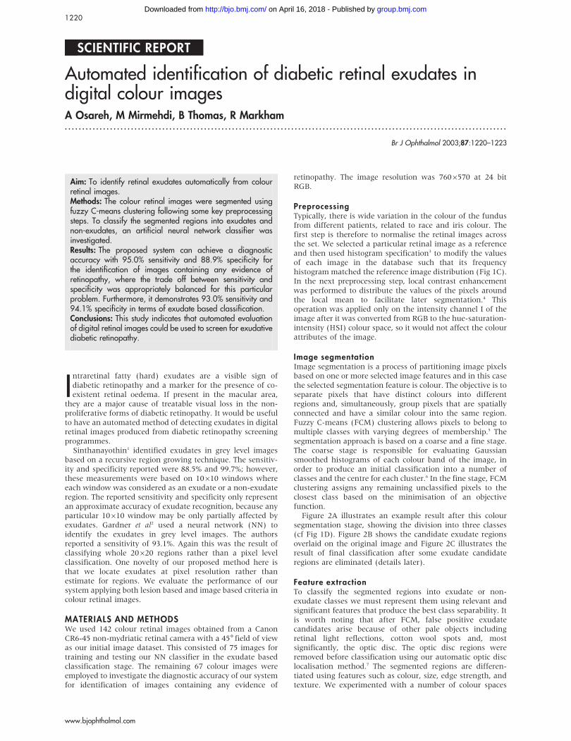
PreprocessingTypically, there is wide variation in the colour of the fundusfrom different patients, related to race and iris colour. Thefirst step is therefore to normalise the retinal images acrossthe set. We selected a particular retinal image as a referenceand then used histogram specification3 to modify the valuesof each image in the database such that its frequencyhistogram matched the reference image distribution (Fig 1C).In the next preprocessing step, local contrast enhancementwas performed to distribute the values of the pixels aroundthe local mean to facilitate later segmentation.4 Thisoperation was applied only on the intensity channel I of theimage after it was converted from RGB to the hue-saturation-intensity (HSI) colour space, so it would not affect the colourattributes of the image.
Image segmentationImage segmentation is a process of partitioning image pixelsbased on one or more selected image features and in this casethe selected segmentation feature is colour. The objective is toseparate pixels that have distinct colours into differentregions and, simultaneously, group pixels that are spatiallyconnected and have a similar colour into the same region.Fuzzy C-means (FCM) clustering allows pixels to belong tomultiple classes with varying degrees of membership.5 Thesegmentation approach is based on a coarse and a fine stage.The coarse stage is responsible for evaluating Gaussiansmoothed histograms of each colour band of the image, inorder to produce an initial classification into a number ofclasses and the centre for each cluster.6 In the fine stage, FCMclustering assigns any remaining unclassified pixels to theclosest class based on the minimisation of an objectivefunction.
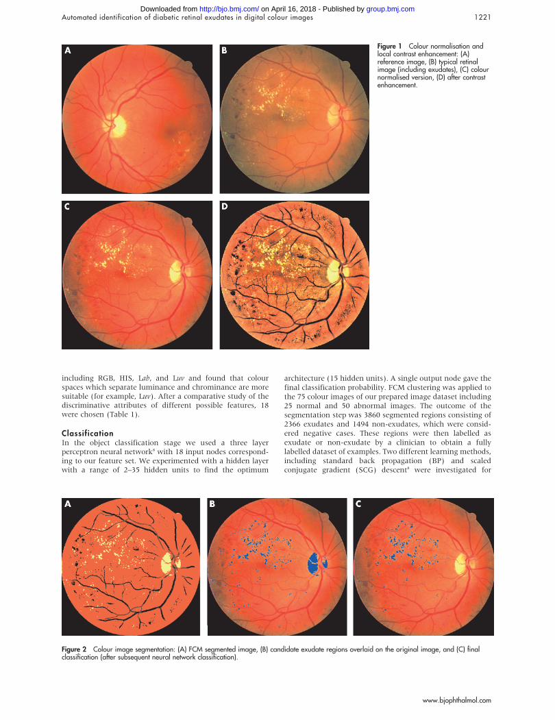
Figure 2A illustrates an example result after this coloursegmentation stage, showing the division into three classes(cf Fig 1D). Figure 2B shows the candidate exudate regionsoverlaid on the original image and Figure 2C illustrates theresult of final classification after some exudate candidateregions are eliminated (details later).
Feature extractionTo classify the segmented regions into exudate or non-exudate classes we must represent them using relevant andsignificant features that produce the best class separability. Itis worth noting that after FCM, false positive exudatecandidates arise because of other pale objects includingretinal light reflections, cotton wool spots and, mostsignificantly, the optic disc. The optic disc regions wereremoved before classification using our automatic optic disclocalisation method.7 The segmented regions are differen-tiated using features such as colour, size, edge strength, andtexture. We experimented with a number of colour spaces
1220
www.bjophthalmol.com
group.bmj.com on April 16, 2018 - Published by http://bjo.bmj.com/Downloaded from
including RGB, HIS, Lab, and Luv and found that colourspaces which separate luminance and chrominance are moresuitable (for example, Luv). After a comparative study of thediscriminative attributes of different possible features, 18were chosen (Table 1).
ClassificationIn the object classification stage we used a three layerperceptron neural network8 with 18 input nodes correspond-ing to our feature set. We experimented with a hidden layerwith a range of 2–35 hidden units to find the optimum
architecture (15 hidden units). A single output node gave thefinal classification probability. FCM clustering was applied tothe 75 colour images of our prepared image dataset including25 normal and 50 abnormal images. The outcome of thesegmentation step was 3860 segmented regions consisting of2366 exudates and 1494 non-exudates, which were consid-ered negative cases. These regions were then labelled asexudate or non-exudate by a clinician to obtain a fullylabelled dataset of examples. Two different learning methods,including standard back propagation (BP) and scaledconjugate gradient (SCG) descent8 were investigated for
Figure 1 Colour normalisation andlocal contrast enhancement: (A)reference image, (B) typical retinalimage (including exudates), (C) colournormalised version, (D) after contrastenhancement.
Figure 2 Colour image segmentation: (A) FCM segmented image, (B) candidate exudate regions overlaid on the original image, and (C) finalclassification (after subsequent neural network classification).
Automated identification of diabetic retinal exudates in digital colour images 1221
www.bjophthalmol.com
group.bmj.com on April 16, 2018 - Published by http://bjo.bmj.com/Downloaded from

training the NNs. Table 2 summarises the optimum resultsobtained on the test set for our NN configurations. For eachNN the output threshold (T) value giving the balancebetween sensitivity and specificity is also shown. The BP-NN with 15 hidden units represented better balance betweensensitivity and specificity. On the other hand, SCG couldrepresent a higher level of sensitivity accuracy.
The optimum threshold value depends on the diagnosticrequirements and can be defined based on the requirementsset by medical experts. In order to demonstrate howchanging this value can affect the performance of a NNclassifier and the balance between sensitivity and specificitycriteria, a receiver operating characteristic (ROC) curve9 wasproduced. The bigger the area under the ROC curve (Az), thehigher is the probability of making a correct decision.Figure 3A compares the behaviour of the NN classifiers forthe full range of output threshold values. The BP-NNachieved a very good performance with Az = 0.966. Figure 2Cillustrates the final classification result for a typical retinalimage using this BP network classifier. Similarly, the SCGnetwork with 15 hidden units, also demonstrated a highgeneralisation performance with Az = 0.962. Figure 3B illus-trates the behaviour of the BP classifier and provides a guidefor selecting the most appropriate output threshold based onthe problem requirements and the related diagnostic accu-racy including sensitivity and specificity.
So far we have discussed pixel by pixel based lesionclassification. We can use this to evaluate the effectiveness ofour proposed approach by assessing the image based accuracyof the system. A population of 67 different retinal images,from our initial dataset, was considered (27 normal 40abnormal). Each retinal image was evaluated using the BPneural network classifier and a final decision was made todetermine whether the image had some evidence of diabeticretinopathy. The BP classifier could identify affected retinas(including exudates) with 95.0% sensitivity while it correctlyclassified 88.9% of normal images—that is, the specificity.
RESULTS AND DISCUSSIONWe presented a study in detecting retinal exudates usingFCM segmentation and a NN based on different learningmethods. The best diagnostic accuracy was 93.0% sensitivityand 94.1% specificity in terms of lesion based classification,and 95.0% sensitivity and 88.9% specificity for the identifica-tion of patients with evidence of retinopathy, where the tradeoff between sensitivity and specificity was appropriatelybalanced for this particular problem.
At present, the full computation comprising segmentation,removal of false positives (for example, the optic disc), andNN classification takes around 11 minutes on a 700 MHz PCwhich includes an unoptimised step in the optic disc removalstage7 lasting around 10 minutes.
The results demonstrated here indicate that automateddiagnosis of exudative retinopathy based on colour retinalimage analysis can be very successful in detecting exudates.Hence, the system could be used to evaluate digital retinalimages obtained in screening programmes for diabeticretinopathy and used by non-experts to indicate whichpatients require referral to an ophthalmologist for furtherinvestigation and treatment.
ACKNOWLEDGEMENTSA Osareh is on a scholarship funded by the Iranian Ministry ofScience, Research and Technology. The authors also thank the UKNational Eye Research Center for their support.
Authors’ affiliations. . . . . . . . . . . . . . . . . . . . .
A Osareh, M Mirmehdi, B Thomas, Department of Computer Science,University of Bristol, Bristol BS8 1UB, UKR Markham, Bristol Eye Hospital, Bristol BS1 2LX, UK
Correspondence to: Mr Alireza Osareh, Bristol University, MerchantVentures Building, Woodland Road, Bristol BS8 1UB, UK;[email protected]
Accepted for publication 27 January 2003
REFERENCES1 Sinthanayothin C. Image analysis for automatic diagnosis of diabetic
retinopathy. PhD Thesis. London: King’s College, 1999.
Figure 3 Performance of the BP neuralnetwork as a function of outputthreshold.
Table 1 Selected feature set
Feature Description
1–3 Mean Luv value inside the region (Lmi,Umi,Vmi)4–6 Standard deviation of Luv value inside the region
(Lsi,Usi,Vsi)7–9 Mean Luv value outside the region (Lmo,Umo,Vmo)10–12 Standard deviation of Luv value outside the region
(Lso,Uso,Vso)13–15 Luv values of region centroid (Lc,Uc,Vc)16 Region size (S)17 Region compactness (C)18 Region edge strength(E)
Table 2 Performance comparison of different classifiers(values as %)
Classifier Sensitivity Specificity
NN-BP (15 hidden) (T = 0.50) 93.0 94.1NN-SCG (15 hidden) (T = 0.45) 97.9 85.2
1222 Osareh, Mirmehdi, Thomas, et al
www.bjophthalmol.com
group.bmj.com on April 16, 2018 - Published by http://bjo.bmj.com/Downloaded from
2 Gardner GG, Keating D, Williamson TH, et al. Automatic detection of diabeticretinopathy using an artificial neural network: a screening tool. Br J Ophthalmol1996;86:940–4.
3 Jain AK. Fundamentals of digital image processing. New York: Prentice-Hall,1989.
4 Sinthanayothin C, Boyce J, Williamson CT. Automated localisation of the opticdisc, fovea, and retinal blood vessels from digital colour fundus images.Br J Ophthalmol 1999;38:902–10.
5 Bezdek J, Keller J, Krisnapuram R, et al. Fuzzy model and algorithms forpattern recognition and image processing. London: Kluwer AcademicPublishers, 1999.
6 Lim YW, Lee SU. On the colour image segmentation algorithm based on thethresholding and the fuzzy c-means techniques. Pattern Recognition1990;23:935–52.
7 Osareh A, Mirmehdi M, Thomas B, et al. Comparison of colour spaces foroptic disc localisation in retinal images. In: 16th International Conference onPattern Recognition, 2002:743–6.
8 Bishop CM. Neural networks for pattern recognition. Oxford: ClarendonPress, 1995.
9 Henderson AR. Assessing test accuracy and its clinical consequences: a primerfor receiver operating characteristics curve analysis. Ann Clin Biochem1993;30:521–39.
ECHO . . . . . . . . . . . . . . . . . . . . . . . . . . . . . . . . . . . . . . . . . . . . . . . . . . . . . . . . . . . . . . . . . . . . . . . . . . . . . . . . . . . . . . . . . . . . . . . . . . . . . . . . . . . . . . . . . . .
Carbonic anhydrase gene 12 is overexpressed in glaucoma
Please visit theBritishJournalofOphthalmologywebsite [www.bjophthalmol.com] for a linkto the full text ofthis article.
Investigators in California and Maryland examined 16 normal (including fetal andneonatal) eyes and 10 glaucomatous eyes for the presence of various cell surfacetransmembrane carbonic anhydrase genes (CA). Normal developing eyes expressed CAIX
and CAXII enzyme proteins in ciliary, corneal, and lens epithelia as well as the endothelium.After birth, the intensity of CAXII immunoreactivity decreased and was no longer expressedin the inner membrane of the retina. Levels fell further in adult eyes. In particular CAIX wasnot expressed in the ciliary epithelium although it had been very weakly expressed indeveloping eyes.
In glaucoma, positive immunoreactivity of both genes was weak but a striking findingwas high levels of CAXII but absent CAIX expression in the non-pigmented ciliaryepithelium (NPE). These patterns of expression were maintained in cultured NPE cells.
The authors conclude that CAIX and CAXII probably play an important role in aqueoushumour production. Thus, the silencing of the CA9 gene in adult eyes could lead tooverexpression of CA12 in NPE cells, with consequent overproduction of aqueous humour,high intraocular pressure and, hence, glaucoma.
The implications are that investigating CA12 could provide a framework to betterunderstand fluid equilibrium in the eye; and it might impact upon the development of moreselective topical inhibitors of CAXII in treatment.
m Journal of Medical Genetics 2003;40:257–262.
1
Automated identification of diabetic retinal exudates in digital colour images 1223
www.bjophthalmol.com
group.bmj.com on April 16, 2018 - Published by http://bjo.bmj.com/Downloaded from

exudates in digital colour imagesAutomated identification of diabetic retinal
A Osareh, M Mirmehdi, B Thomas and R Markham
doi: 10.1136/bjo.87.10.12202003 87: 1220-1223 Br J Ophthalmol
http://bjo.bmj.com/content/87/10/1220Updated information and services can be found at:
These include:
References http://bjo.bmj.com/content/87/10/1220#ref-list-1
This article cites 3 articles, 0 of which you can access for free at:
serviceEmail alerting
box at the top right corner of the online article. Receive free email alerts when new articles cite this article. Sign up in the
CollectionsTopic Articles on similar topics can be found in the following collections
(1608)Retina
Notes
http://group.bmj.com/group/rights-licensing/permissionsTo request permissions go to:
http://journals.bmj.com/cgi/reprintformTo order reprints go to:
http://group.bmj.com/subscribe/To subscribe to BMJ go to:
group.bmj.com on April 16, 2018 - Published by http://bjo.bmj.com/Downloaded from