Embed Size (px)
Citation preview
Scientific Article
Prosthodontic and surgical considerationsfor pediatric patients requiring maxillectomyJack W. Martin, DDS, MS Mark S. Chambers, DMD, MS James C. Lemon, DDSBela B. Toth, MS, DDS John F. Helfrick, DDS, MS
Abstract
The quality of treatment and rehabilitation for the headand neck cancer patient, especially the pediatric patient,has progressed markedly over the years due to the coopera-tion of specialists involved in the total care of the patient.Defects of the oral cavity caused by trauma or removal ofmalignant or benign tissue require special treatment con-siderations with the pediatric patient. Aside from radiationand chemotherapy, other forms of adjuvant therapy, suchas physical therapy, and patient and family counseling, areneeded for proper rehabilitation. In addition, oral hygieneis essential in the overall rehabilitative process. Pediatricdental, orthodontic, prosthodontic, and oral and maxillofa-cial surgery specialties become integrated in treating thepediatricpatient. The concentrated multidisciplinary treat-ment reduces post-treatment morbidity by shortening re-covery and immediate rehabilitation time and by providinglong-term care during the critical growth period. (PediatrDent 17:116-21,1995)
T hough most malig-nancies occur in thesixth to seventh dec-
ades of life, cancer frequencyin children is increasing atan alarming rate.1 In the USand most economically adv-antaged nations, cancer killsmore children over age 6months than does any otherdisease.1 In 1974, 12 children(0-14 years of age) per 100,000were diagnosed with cancer.By 1990, the incidence in-creased to 14 children per100,000.' Moreover, theAmerican Cancer Society es-timates that in 1994,7,600 chil-dren in the US under the ageof 14 will die of cancer.2
One of the many types ofmalignancies affecting chil-dren is head and neck cancer.Of particular concern to thepediatric dentist would be
head and neck tumors of the maxillofacial region. Insuch diseases, the principal treatment option is surgi-cal resection of the maxilla (maxillectomy), which mayproduce pronounced deficits and cosmetic deformi-ties. Consequently, when a maxillectomy is requiredfor treating benign or malignant disease, the maxillofa-cial prosthodontist must be included in the assessmentof the patient prior to surgery. Many articles describethe surgical and prosthodontic management of the adultmaxillectomy patient, but little information exists aboutmanagement of the pediatric maxillectomy patient. Thisarticle discusses preoperative evaluation, and surgicaland prosthodontic considerations of children diagnosedwith maxillary cancer or benign disease. Postsurgicalcare and growth considerations also are presented.
Presurgical prosthodontic considerations
A prosthodontist's presurgical evaluation of thepediatric patient should include an examination of hardand soft tissues, and a review of medical and dental
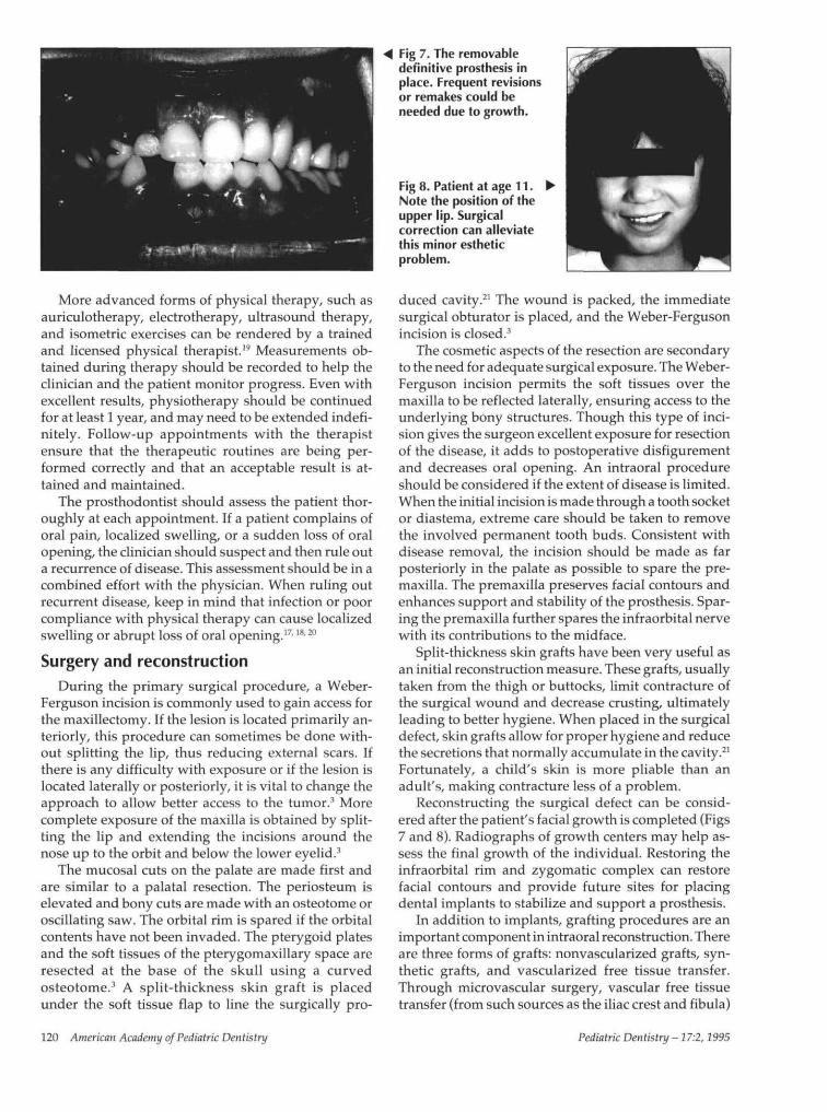
Fig 1. Preoperative panoramic radiograph of pediatric patient scheduled for a maxil-lectomy procedure. Tumor involving left maxillary sinus and hard palate. Patient is inmixed dentition stage. Evaluation of this patient should include pediatric dental consult.
116 American Academy of Pediatric Dentistry Pediatric Dentistry -17:2,1995
history including panoramic, occlusal, and selectedperiapical radiographs. These radiographs not only willindicate the position of the permanent tooth succes-sors, but are useful in diagnosing and treatment plan-ning the maxillectomy procedure. Initial incisions canthen be placed without damaging uninvolved toothbuds. A pediatric dentist also should be consulted as
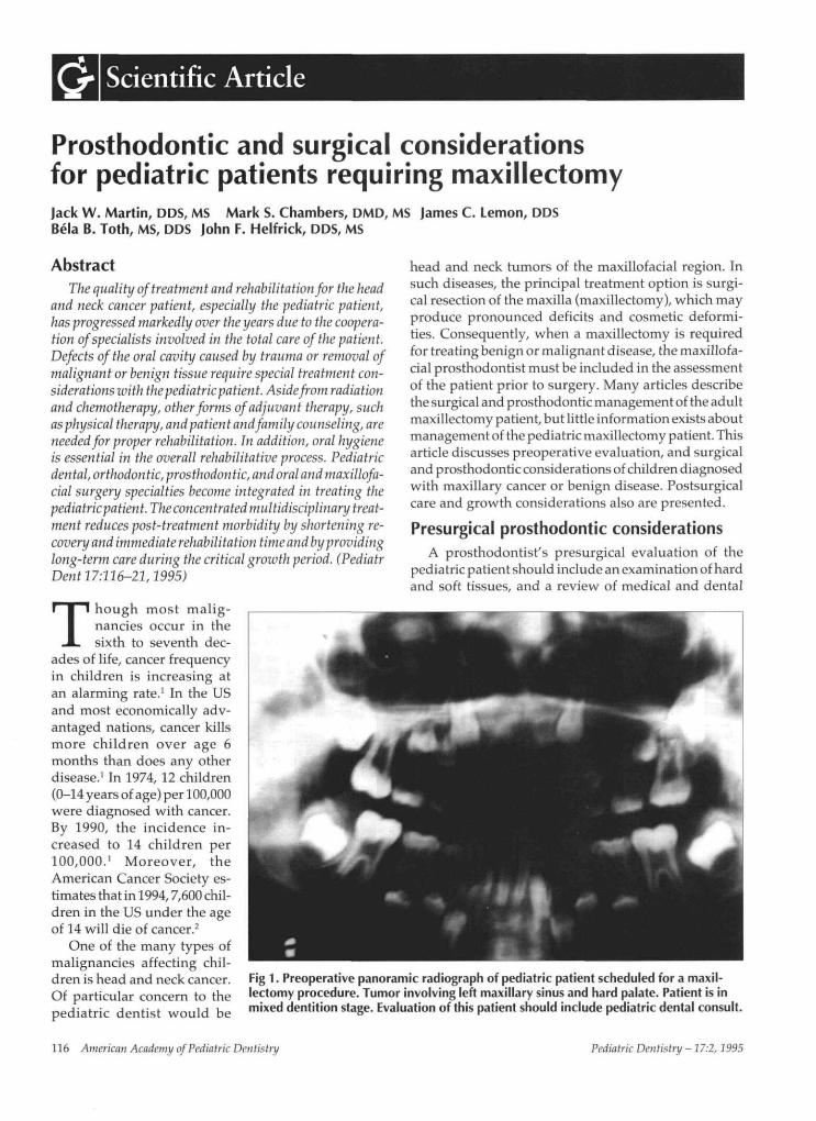
Fig 2. Tumor involving the left maxillary sinusand hard palate.
Fig 3. Tumor was surgically removed by anintraoral approach.
Fig 4. Zygomatic and interdental wires were used toretain the surgical obturator.
part of this assessment when the patient is in the mixeddentition stage to help determine which teeth will besuitable for clasping during the various phases of pros-thetic rehabilitation (Figs 1 and 2). At this stage, pre-liminary impressions of the maxilla and mandibleshould be made and poured in stone to obtain diagnos-tic casts. A surgical obturator is fabricated to restorethe contour of the palate and seal the margins of theintraoral surgical defect.
Parental information prior to surgeryIt is advisable to inform the parents or guardian as
well as the patient (according to the patient's maturityand ability to understand) that there will be fourcritical stages:
1. Surgery2. Surgical packing removal3. Interim4. Definitive rehabilitation (Figs 3 and 4).
The parent should be informed of the requiredpresurgical and postsurgical prosthodontic procedures.The problems associated with a maxillectomy, includ-ing difficulties in speech and swallowing, revisions ofthe surgical obturator prosthesis, and requirements forfollow-up visits during the interim obturator stage,should be discussed with the patient and parents.3-4
This information should be discussed before surgeryand again before surgical pack removal; specifically,problems that will be associated with speech and swal-lowing. Parents should be informed that general anes-thesia or intravenous sedation may be necessary forpack removal and interim obturation procedures. Dur-ing the first 2 to 3 weeks after pack removal, severaloffice visits will be required for revision of the interimobturator to accommodate healing. Initially, the pa-tient and parents should expect some leakage of liq-uids and air around the bulb portion of the obturatorprosthesis. This leakage may cause hypernasal speechand regurgitation of food and fluids through the nose.Depending on the extent of surgery, the clinician cananticipate these problems and discuss them more ap-propriately after the surgical procedure.5
Parents should be informed that adjuvant therapysuch as physiotherapy may be needed to resist a de-crease in mouth opening and assist in eliminating harm-ful habitual oral habits. Decreased oral opening, loss ofinnervation, and facial deformity secondary to amaxillectomy procedure will depend on the extent ofsurgery and adjunctive treatments such as radiationand chemotherapy. Parents should also be counseledon their participation in oral hygiene and examiningthe surgical defect. This parental examination can assistthe surgeon and dentist in early detection, treatment,and even prevention of a number of problems, includ-ing: decreased oral opening, poor oral hygiene, ill-fit-ting prosthesis, and, most important, recurrent disease.
Pediatric Dentistry - 17:2,1995 American Academy of Pediatric Dentistry 117
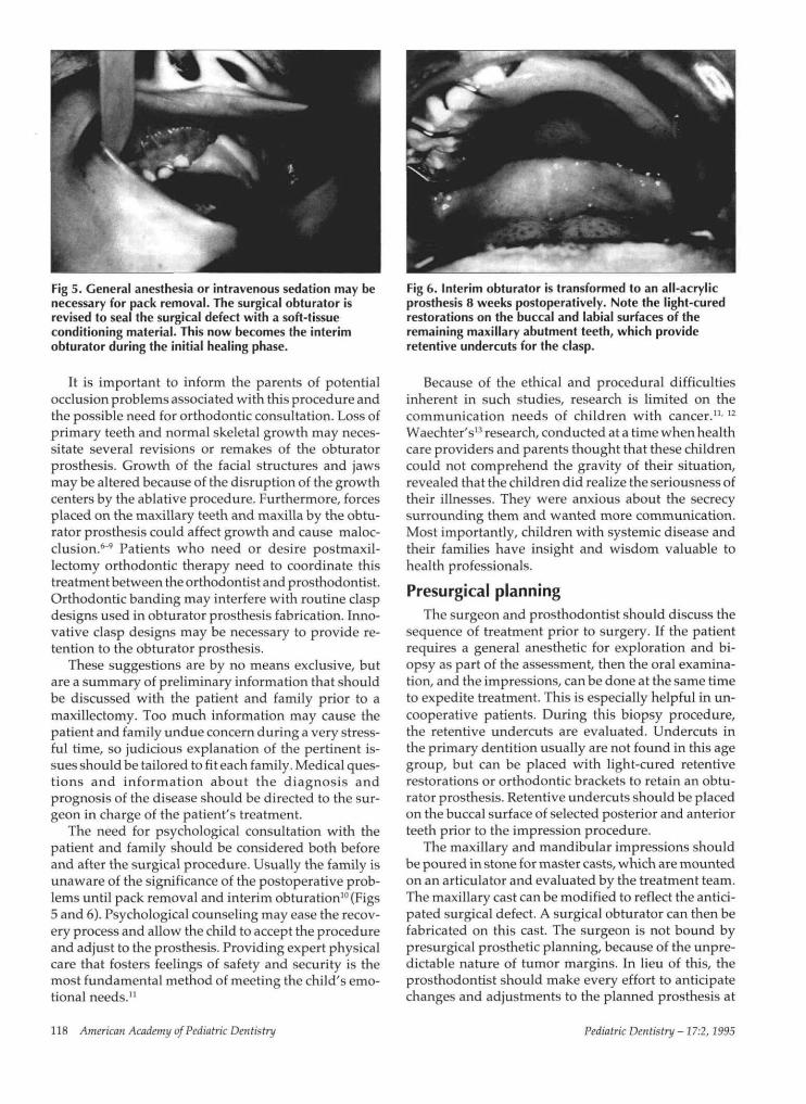
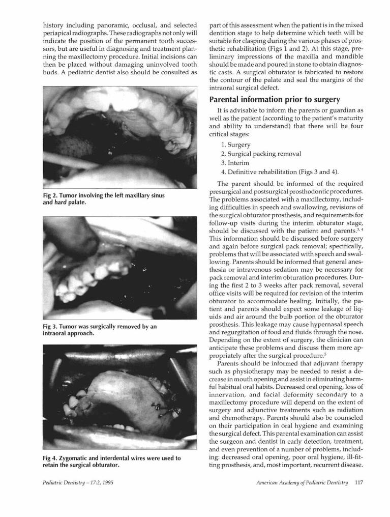
Fig 5. General anesthesia or intravenous sedation may benecessary for pack removal. The surgical obturator isrevised to seal the surgical defect with a soft-tissueconditioning material. This now becomes the interimobturator during the initial healing phase.
It is important to inform the parents of potentialocclusion problems associated with this procedure andthe possible need for orthodontic consultation. Loss ofprimary teeth and normal skeletal growth may neces-sitate several revisions or remakes of the obturatorprosthesis. Growth of the facial structures and jawsmay be altered because of the disruption of the growthcenters by the ablative procedure. Furthermore, forcesplaced on the maxillary teeth and maxilla by the obtu-rator prosthesis could affect growth and cause maloc-clusion.6'9 Patients who need or desire postmaxil-lectomy orthodontic therapy need to coordinate thistreatment between the orthodontist and prosthodontist.Orthodontic banding may interfere with routine claspdesigns used in obturator prosthesis fabrication. Inno-vative clasp designs may be necessary to provide re-tention to the obturator prosthesis.
These suggestions are by no means exclusive, butare a summary of preliminary information that shouldbe discussed with the patient and family prior to amaxillectomy. Too much information may cause thepatient and family undue concern during a very stress-ful time, so judicious explanation of the pertinent is-sues should be tailored to fit each family. Medical ques-tions and information about the diagnosis andprognosis of the disease should be directed to the sur-geon in charge of the patient's treatment.
The need for psychological consultation with thepatient and family should be considered both beforeand after the surgical procedure. Usually the family isunaware of the significance of the postoperative prob-lems until pack removal and interim obturation10 (Figs5 and 6). Psychological counseling may ease the recov-ery process and allow the child to accept the procedureand adjust to the prosthesis. Providing expert physicalcare that fosters feelings of safety and security is themost fundamental method of meeting the child's emo-tional needs.11
Fig 6. Interim obturator is transformed to an all-acrylicprosthesis 8 weeks postoperatively. Note the light-curedrestorations on the buccal and labial surfaces of theremaining maxillary abutment teeth, which provideretentive undercuts for the clasp.
Because of the ethical and procedural difficultiesinherent in such studies, research is limited on thecommunication needs of children with cancer.11-12
Waechter's13 research, conducted at a time when healthcare providers and parents thought that these childrencould not comprehend the gravity of their situation,revealed that the children did realize the seriousness oftheir illnesses. They were anxious about the secrecysurrounding them and wanted more communication.Most importantly, children with systemic disease andtheir families have insight and wisdom valuable tohealth professionals.
Presurgical planningThe surgeon and prosthodontist should discuss the
sequence of treatment prior to surgery. If the patientrequires a general anesthetic for exploration and bi-opsy as part of the assessment, then the oral examina-tion, and the impressions, can be done at the same timeto expedite treatment. This is especially helpful in un-cooperative patients. During this biopsy procedure,the retentive undercuts are evaluated. Undercuts inthe primary dentition usually are not found in this agegroup, but can be placed with light-cured retentiverestorations or orthodontic brackets to retain an obtu-rator prosthesis. Retentive undercuts should be placedon the buccal surface of selected posterior and anteriorteeth prior to the impression procedure.
The maxillary and mandibular impressions shouldbe poured in stone for master casts, which are mountedon an articulator and evaluated by the treatment team.The maxillary cast can be modified to reflect the antici-pated surgical defect. A surgical obturator can then befabricated on this cast. The surgeon is not bound bypresurgical prosthetic planning, because of the unpre-dictable nature of tumor margins. In lieu of this, theprosthodontist should make every effort to anticipatechanges and adjustments to the planned prosthesis at
118 American Academy ofPediatric Dentistry Pediatric Dentistry -17:2,1995
the time of the ablative procedure. Duplicating themaxillary cast may help allow revision of the prosthe-sis if more or less supporting tissues are removed dur-ing surgery.
The laboratory procedures used to fabricate a surgi-cal obturator for a pediatric patient are essentially thesame as for the adult. The obturator protects the surgi-cal wound and acts as a stent to hold the surgical pack-ing in place against the skin graft. Furthermore, it pro-vides oral competence, eliminating the need for anasogastric tube. Denture teeth for the pediatric obtu-rator prosthesis may be selected from a manufacturedmold or be custom made by a sprinkle-on technique, asdescribed by King.14 Holes are drilled strategically intothe surgical prosthesis for interdental wiring of theobturator. Since primary teeth are conical and diastemasare common, other means of fixation include zygo-matic or nasal spine wires to help retain the obturatoroFixing the obturator to bone with bone screws shouldbe a last resort in pediatric patients, because when usedin the alveolar region, this procedure can damage theremaining permanent tooth buds.
Oral hygiene
Oral hygiene is one of the most important aspects ofpostoperative care and cannot be overstressed to thepatient. Initially, the patient -- with parental observa-tion and guidance-- should rinse with a saline solution(1 tsp salt/1 tsp soda dissolved in 16 oz of water) threetimes a day. A commercial irrigation system canbe usedas part of this regimen. The entire surgical defect shouldbe cleansed. Tooth brushing and flossing should beginas soon as the surgical packing has been removed. Thepatient and parents may be apprehensive about resum-ing normal oral hygiene for fear of injuring the surgicalsite, and this fear must be addressed to lessen psycho-logical stress. If the patient is receiving chemotherapy,hematological values should be monitored prior to re-suming normal oral hygiene procedures. When the plate-let count remains at or above 50,000/mm3, the coagula-tion profile is within normal limits, and the absolutephagocyte count is above 500/ram3, routine oral carecan be resumed. Rinsing with commercial mouthwashesis not recommended during the initial healing phasebecause these products usually contain alcohol or phe-nol which can irritate the mucosa.
At the fourth week of healing, a solution of 3% hy-drogen peroxide, diluted 1:1 with water, can be addedto the routine. This mixture used prior to rinsing withthe salt and soda solution can help loosen and removedried mucous crust and desquamated debris in thesurgical defect. A 4x4-cm gauze or wash cloth damp-ened in the peroxide solution can be wrapped aroundthe index finger and used to clean the skin-graftedportion of the defect. Ora-Swab® brushes (Sage Inc,Crystal Lake, IL), which are sponge-tipped applica-tors, may also be used for this purpose. The patient
should be instructed to lean over a sink during irriga-tion to allow the fluid to pass out of the mouth andnasal cavity and into the receptacle. The possibility ofaspirating the solution is an initial concern, but pa-tients quickly learn to protect their airways. The pa-tient should be instructed to brush and floss prior tocleaning the defect. In addition, the parents must betaught to inspect the defect after each session, to helpensure adequate hygiene and optimal health25,16
Physical therapy
Decreased oral opening, loss of innervation, andfacial deformity secondary to a maxillectomy will de-pend on the extent of surgery and adjunctive treat-ments such as radiation and chemotherapy. If themaxillectomy is confined to the alveolus and hard pal-ate, and if the procedure is accomplished intraorally,the postoperative manipulations such as obturator re-visions and oral care will be accomplished with rela-¯ tive ease. However, if the maxillectomy involves aWeber-Ferguson incision and the resection is extendedto include the orbit, zygoma, and pterygoid muscles,the resulting decreased oral opening and facial defor-mity can be severe, and rigorous physical therapy isusually required.
Exercises designed to increase oral opening shouldbe initiated as soon as the patient can tolerate therapy-- usually one week after the surgical obturator andpacking are removed. When the patient is dentate,tongue blades can be inserted between the posteriorteeth until the opening limit is obtained. The oral open-ing is then increased by inserting one tongue blade at atime between those already in place. With each addi-tional tongue blade, opening is increased and the posi-tion is held for several minutes to allow the scar tissueto stretch. Tongue blades are added until a pain thresh-old is reached. Several mouth opening devices, such asa dynamic bite opener and a threaded screw-type ap-pliance, have been discussed in previous articles17,1~ andcan be custom made for each patient. The details in de-sign or adaptation of these devices can make them moreappealing for the pediatric patient. One commercialdevice, the Therabite® mouth opener (Therabite Corp,Bryn Mawr, PA), operates similar to a car jack to openthe mouth and can be custom fitted so that the forces onthe remaining dentition are distributed equally.
No matter what mouth-opening device or techniqueis used, it is important to include as many teeth aspossible in order to distribute the load and reduce thepotential for orthodontic movement of the teeth. Dur-ing the first 4 weeks after surgery, patients should domouth-opening exercises three to four times a day.They can also stretch the scar band by intraorally mas-saging the cheek portion of the scar band with digitalmanipulation. This movement can be combined withextraoral palpation and stretching of the cheek andupper lip, to keep the tissue as pliable as possible.
Pediatric Dentistry- 17:2, 1995 American Academy of Pediatric Dentistry 119
More advanced forms of physical therapy, such asauriculotherapy, electrotherapy, ultrasound therapy,and isometric exercises can be rendered by a trainedand licensed physical therapist.19 Measurements ob-tained during therapy should be recorded to help theclinician and the patient monitor progress. Even withexcellent results, physiotherapy should be continuedfor at least 1 year, and may need to be extended indefi-nitely. Follow-up appointments with the therapistensure that the therapeutic routines are being per-formed correctly and that an acceptable result is at-tained and maintained.
The prosthodontist should assess the patient thor-oughly at each appointment. If a patient complains oforal pain, localized swelling, or a sudden loss of oralopening, the clinician should suspect and then rule outa recurrence of disease. This assessment should be in acombined effort with the physician. When ruling outrecurrent disease, keep in mind that infection or poorcompliance with physical therapy can cause localizedswelling or abrupt loss of oral opening.17-18/ 20
Surgery and reconstructionDuring the primary surgical procedure, a Weber-
Ferguson incision is commonly used to gain access forthe maxillectomy. If the lesion is located primarily an-teriorly, this procedure can sometimes be done with-out splitting the lip, thus reducing external scars. Ifthere is any difficulty with exposure or if the lesion islocated laterally or posteriorly, it is vital to change theapproach to allow better access to the tumor.3 Morecomplete exposure of the maxilla is obtained by split-ting the lip and extending the incisions around thenose up to the orbit and below the lower eyelid.3
The mucosal cuts on the palate are made first andare similar to a palatal resection. The periosteum iselevated and bony cuts are made with an osteotome oroscillating saw. The orbital rim is spared if the orbitalcontents have not been invaded. The pterygoid platesand the soft tissues of the pterygomaxillary space areresected at the base of the skull using a curvedosteotome.3 A split-thickness skin graft is placedunder the soft tissue flap to line the surgically pro-
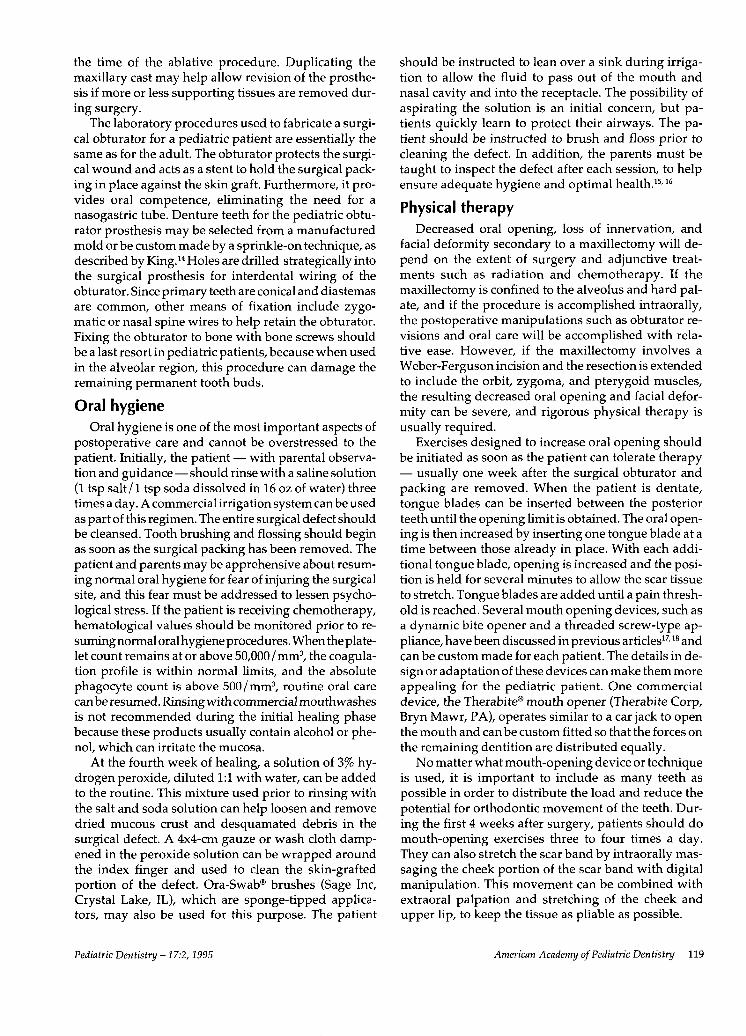
Fig 7. The removabledefinitive prosthesis inplace. Frequent revisionsor remakes could beneeded due to growth.
Fig 8. Patient at age 11.Note the position of theupper lip. Surgicalcorrection can alleviatethis minor estheticproblem.
duced cavity.21 The wound is packed, the immediatesurgical obturator is placed, and the Weber-Fergusonincision is closed.3
The cosmetic aspects of the resection are secondaryto the need for adequate surgical exposure. The Weber-Ferguson incision permits the soft tissues over themaxilla to be reflected laterally, ensuring access to theunderlying bony structures. Though this type of inci-sion gives the surgeon excellent exposure for resectionof the disease, it adds to postoperative disfigurementand decreases oral opening. An intraoral procedureshould be considered if the extent of disease is limited.When the initial incision is made through a tooth socketor diastema, extreme care should be taken to removethe involved permanent tooth buds. Consistent withdisease removal, the incision should be made as farposteriorly in the palate as possible to spare the pre-maxilla. The premaxilla preserves facial contours andenhances support and stability of the prosthesis. Spar-ing the premaxilla further spares the infraorbital nervewith its contributions to the midface.
Split-thickness skin grafts have been very useful asan initial reconstruction measure. These grafts, usuallytaken from the thigh or buttocks, limit contracture ofthe surgical wound and decrease crusting, ultimatelyleading to better hygiene. When placed in the surgicaldefect, skin grafts allow for proper hygiene and reducethe secretions that normally accumulate in the cavity.21
Fortunately, a child's skin is more pliable than anadult's, making contracture less of a problem.
Reconstructing the surgical defect can be consid-ered after the patient's facial growth is completed (Figs7 and 8). Radiographs of growth centers may help as-sess the final growth of the individual. Restoring theinfraorbital rim and zygomatic complex can restorefacial contours and provide future sites for placingdental implants to stabilize and support a prosthesis.
In addition to implants, grafting procedures are animportant component in intraoral reconstruction. Thereare three forms of grafts: nonvascularized grafts, syn-thetic grafts, and vascularized free tissue transfer.Through microvascular surgery, vascular free tissuetransfer (from such sources as the iliac crest and fibula)
120 American Academy ofPediatric Dentistry Pediatric Dentistry -17:2,1995
has revolutionized surgical techniques and offers manynew opportunities for maxillary reconstruction.22-24
Microvascular surgery is a technique used to join anartery and vein from a free tissue transfer flap to anexisting artery and vein in the recipient site. 24 A freetissue transfer flap composed of bone, muscle, associ-ated soft tissue, and skin can be removed from one partof the body, via microvascular surgery, and used torestore supporting tissues resected during ablative can-cer surgery in the head and neck region.22 With this newtechnique, the reconstruction of the maxilla and place-ment of dental endosseous implants can improve facialappearance and prosthesis stability. This rehabilitation,following facial growth, should be coordinated withorthodontic and prosthodontic considerations.2s-2s
SummaryThe pediatric maxillectomy patient poses special
problems for the dentist and surgeon. Prosthodonticand surgical considerations should be discussed withthe patient and parents prior to treatment. Orthodonticand pediatric dental consultations should be planned.Oral hygiene and physical therapy techniques are ofprime importance to the young patient and can be anessential part of the recovery process. The importanceof cooperation between the surgeon and dentist in plan-ning, treatment, and follow-up is crucial to the survivaland rehabilitation of the patient. Psychological consul-tation with the family should be considered both be-fore and after surgery and may be significant in theoverall rehabilitation of the patient.
Dr. Martin is professor, dental oncology; Dr. Chambers is fellow,maxillofacial prosthodontics; Dr. Lemon is associate professor ofdental oncology; Dr. Toth is associate professor, dental oncology;and Dr. Helfrick is professor of oral and maxillofacial surgery,University of Texas, MD Anderson Cancer Center, Houston.
1. Bleyer WA: The impact of childhood cancer on the UnitedStates and the world. In: Murphy GP, Holleb AI, Eds: Ca- ACancer Journal for Clinicians; American Cancer Society,New York, NY, H & W Publishing 40:355-67, 1990.
2. American Cancer Society: Cancer Facts & Figures -- 1993.Atlanta: American Cancer Society, 1993.
3. Beumer JP, Curtis TA, Firtell DN: Restoration of acquiredhard palate defects. In: Maxillofacial rehabilitation:prosthodontic and surgical considerations. St Louis: CVMosby Co, 1979, p 188.
4. Desjardins RP: Early rehabilitative management of themaxillectomy patient. J Prosthet Dent 38:311-38, 1977.
5. King GE: Prosthetic rehabilitation of the nasal and paranasalsinus areas. In: Comprehensive Management of Head andNeck Tumors, Thawley SE, Panje WR, Eds. Philadelphia:WB Saunders Co, 1986.
6. Jaffe N, Toth BB, Hoar RE, Reid HL, Sullivan MP, McNeeseMD: Dental and maxillofacial abnormalities in long termsurvivors of childhood cancer: effect of treatment with che-motherapy and radiation to head and neck. Pediatrics73:816-23, 1984.
7. Dixon AD: Early development of the maxilla. Dent Pract3:331-36, 1953.
8. Dixon AD: The development of the jaws. Dent Pract 9:10-20, 1958.
9. Graber TM: Orthodontics: Principles and Practice. Phila-delphia: WB Saunders Co, 1972, pp 27-125.
10.Gillis RE, Swenson WM, Laney WR: Psychological factorsinvolved in maxillofacial prosthetics. J Prosthet Dent41:183-88, 1979.
11. van Eys J: Clinical research and clinical care: ethical prob-lems in the "War on Cancer," In: Human Values in PediatricHematology/Oncology. Truman JT, van EysJ, Pochedly C,Eds. New York: Praeger, 1986, pp 15-22.
12.Moore IM, Ruccione K: Challenges to conducting researchwith children with cancer. Oncol Nurs Forum 16:587-89,1989.
13.Waechter EH: Children’s awareness of fatal illness. Am JNurs 71:1168-72, 1971.
14.King GE, Frame R: A surgical interim prosthesis. J ProsthetDent 45:108-10, 1981.
15.Toth BB, Hoar R: Oral/dental care for the pediatric oncologypatient. Cancer Bull 34:66-71, 1982.
16.Toth BB, Fleming TJ: Oral/dental considerations for pediat-ric patients receiving anticancer treatment. J Mo Dent AssocMay-June 33-34, 1983.
17.Barrett VJ, Martin JW, Jacob RF, King GE, Sheets JS: Physi-cal therapy techniques in the treatment of the head andneck patient. J Prosthet Dent 59:343-46, 1988.
18.Rocabado M, Johnston BE Jr, Blakney MG: Physical therapyand dentistry: an overview. J Craniomandib Pract 1:46-49,1983.
19.King GE, Scheetz J, Jacob RF, Martin JW: Electrotherapyand hyperbaric oxygen: promising treatments forpostradiation complications. J Prosthet Dent 62:331-34,1989.
20. Rouse PB: The role of physical therapists in support ofmaxillofacial patients. J Prosthet Dent 24:193-97, 1970.
21. Teichgraeber J, Larson DL, Castaneda O, Martin JW: Skingrafts in intraoral reconstruction: a new stenting method.Arch Otolaryngol 110:463, 1984.
22. King GE, Martin JW, Lemon JC, Schusterman MA, ReeceGP: Maxillofacial prosthetic rehabilitation combined withplastic and reconstructive surgery. In: MD AndersonOncology Case Reports & Review. Hickey RC, Ed. 8:1-11,1993.
23. Martin JW, Lemon JC, King GE: Maxillofacial restorationafter tumor ablation. In: Clinics in Plastic Surgery: Headand Neck Reconstruction. Schusterman MA, Ed. 21:87-96,1994.
24. Hidalgo DA: Fibula free flap: a new method of mandiblereconstruction. Plast Reconstr Surg 84:71-79, 1989.
25. King GE, Lemon JC, Martin JW: Multidisciplinary team-work in the treatment and rehabilitation of the head andneck cancer patient. Texas Dent J June:9-12, 1992.
26. Martin JW, Lomba JA, King GE: Maxillofacial prostheticsand oral surgery for the head and neck cancer patient. Can-cer Bull 34:48, 1982.
27. Martin JW, Lemon JC: Prosthetic rehabilitation. In: Headand Neck Surgery-Otolaryngology. Bailey BJ, Ed. Philadel-phia: JB Lippincott Co, 1993, pp 1431-38.
28. King GE, Jacob RF, Martin JW, Fleming TJ, Kramer DC,Udagama A: Prosthetic rehabilitation of the nasal andparanasal sinus areas. In: Comprehensive Management ofHead and Neck Tumors, Vol 1. Thawley SE, Panje WR, Eds.Philadelphia: WB Saunders Co, 1986.
Pediatric Dentistry - 17:2, 1995 American Academy of Pediatric Dentistry 121