Embed Size (px)
Citation preview

Scattering of Neutrons: Basics
Jill TrewhellaUniversity of Sydney
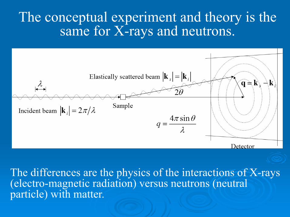
The conceptual experiment and theory is the same for X-rays and neutrons.
The differences are the physics of the interactions of X-rays (electro-magnetic radiation) versus neutrons (neutral particle) with matter.
Fundamentals
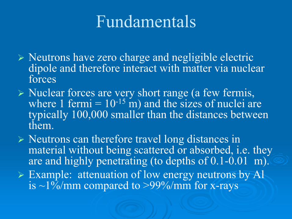
Neutrons have zero charge and negligible electric dipole and therefore interact with matter via nuclear forcesNuclear forces are very short range (a few fermis, where 1 fermi = 10-15 m) and the sizes of nuclei are typically 100,000 smaller than the distances between them.Neutrons can therefore travel long distances in material without being scattered or absorbed, i.e. they are and highly penetrating (to depths of 0.1-0.01 m). Example: attenuation of low energy neutrons by Al is ~1%/mm compared to >99%/mm for x-rays
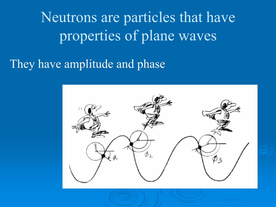
Neutrons are particles that have properties of plane waves
They have amplitude and phase

They can be scattered elastically or inelastically
Inelastic scattering changes both direction and magnitude of the neutron wave vector
Elastic scattering changes direction but not the magnitude of the wave vector
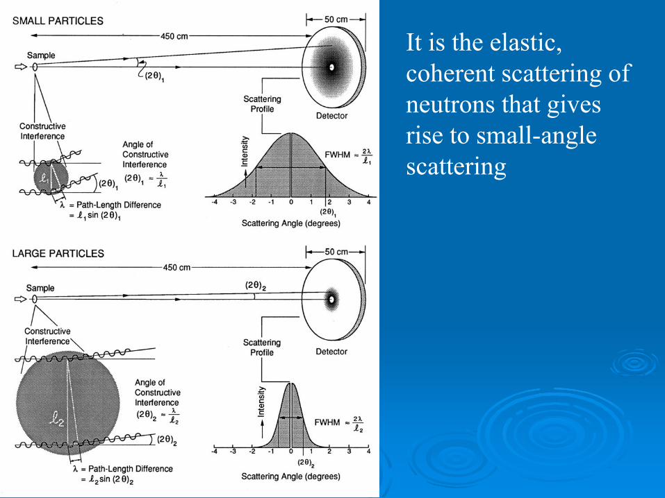
It is the elastic, coherent scattering of neutrons that gives rise to small-angle scattering
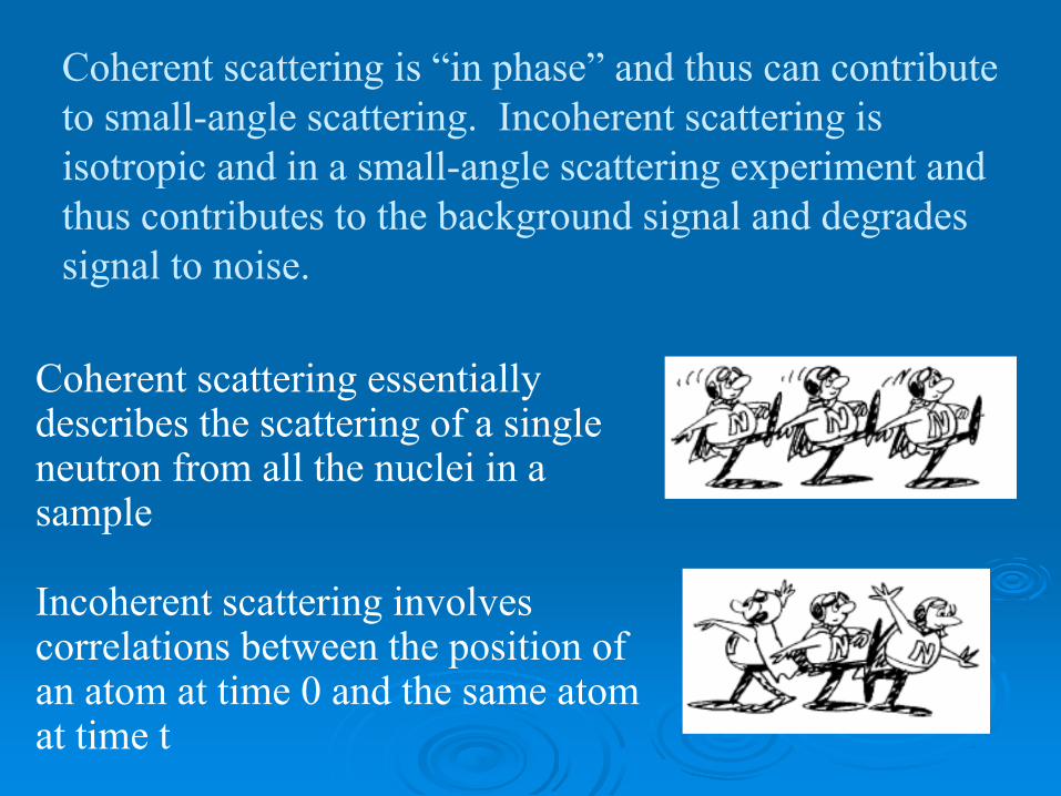
Coherent scattering is “in phase” and thus can contribute to small-angle scattering. Incoherent scattering is isotropic and in a small-angle scattering experiment and thus contributes to the background signal and degrades signal to noise.
Coherent scattering essentially describes the scattering of a single neutron from all the nuclei in a sample
Incoherent scattering involves correlations between the position of an atom at time 0 and the same atom at time t
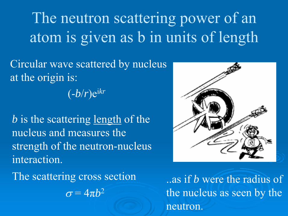
The neutron scattering power of an atom is given as b in units of length
Circular wave scattered by nucleus at the origin is:
(-b/r)eikr
b is the scattering length of the nucleus and measures the strength of the neutron-nucleus interaction. The scattering cross section
σ = 4πb2..as if b were the radius of the nucleus as seen by the neutron.

Neutron scattering lengths for isotopes of the same element can have very different neutron scattering properties

As nuclei are point scattering centers, neutron scattering lengths show no angular dependence
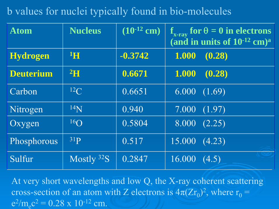
At very short wavelengths and low Q, the X-ray coherent scattering cross-section of an atom with Z electrons is 4π(Zr0)2, where r0 = e2/mec2 = 0.28 x 10⁻¹² cm.
Atom Nucleus (10-12 cm) fx-ray for θ = 0 in electrons (and in units of 10-12 cm)a
Hydrogen 1H -0.3742 1.000 (0.28)
Deuterium 2H 0.6671 1.000 (0.28)
Carbon 12C 0.6651 6.000 (1.69)
Nitrogen 14N 0.940 7.000 (1.97)Oxygen 16O 0.5804 8.000 (2.25)
Phosphorous 31P 0.517 15.000 (4.23)
Sulfur Mostly 32S 0.2847 16.000 (4.5)
b values for nuclei typically found in bio-molecules

I(Q) = ⟨ ∫ | Δρ e-i(q•r) dr]|2 ⟩
where Δρ=ρparticle - ρsolvent.
As average scattering length density ρ is simply the average of the sum of the scattering lengths (b)/unit volume
Because H (1H) and D (2H) have different signs, by manipulating the H/D ratio in a molecule and/or its solvent one can vary the contrast Δρ.
_ _ _
_
_
_

Planning a neutron scattering experiment
Choose your data collection strategy (solvent matching or full contrast variation?)Determine how much sample is neededDecide which subunit to labelWhat deuteration level is needed in the labeling subunitSee MULCh*
http://www.mmb/usyd.edu.au/NCVWeb/
*MULCh, Whitten et al, J. Appl. Cryst. 2008 41, 222-226
MULChModULes for the analysis of neutron Contrast variation data
Contrast, computes neutron contrasts of the components of a complexRg, analyses the contrast dependence of the radius of gyration to yield information relating to the size and disposition of the labelled and unlabeled components in a complexCompost, decomposes the contrast variation data into composite scattering functions containing information on the shape of the labeled and unlabeled components and their dispositions
Solvent matching
Best used when you are interested in the shape of one component in a complex, possibly how it changes upon ligand binding or complex formation. Requires enough of the component to be solvent matched to complete a contrast variation series to determine required %D2O (~4 x 200-300 μL, ~5 mg/ml).Requires 200-300 μL of the labeled complex at 5-10mg/ml.
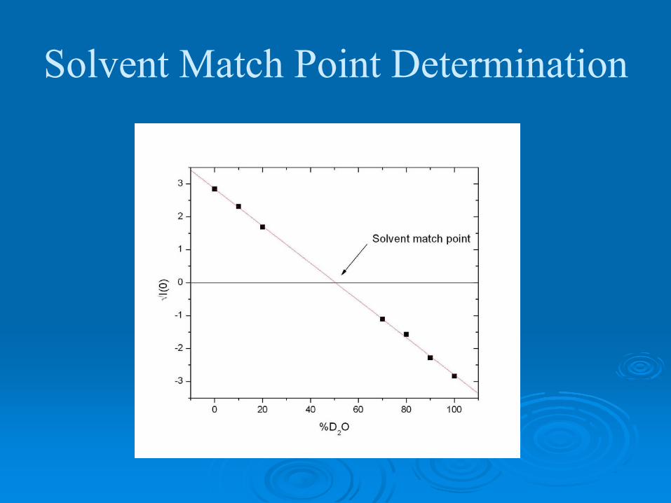
Solvent Match Point Determination
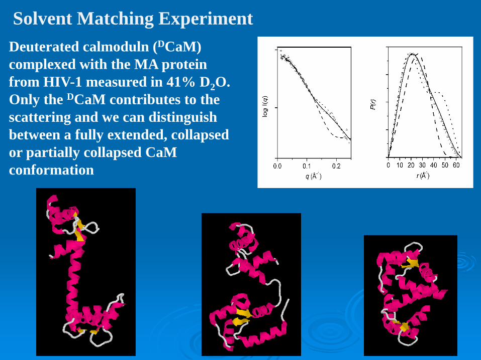
Deuterated calmoduln (DCaM) complexed with the MA protein from HIV-1 measured in 41% D2O. Only the DCaM contributes to the scattering and we can distinguish between a fully extended, collapsed or partially collapsed CaM conformation
Solvent Matching Experiment

Contrast variationTo determine the shapes and dispositions of labeled and unlabelled components in a complexRequires ≥ 5 x 200-300μL (= 1 – 1.5mL) of your labeled complex at ≥ 5 mg/ml .Deuteration level in labeled protein depends upon its size.
Smaller components require higher levels of deuteration to be distinguished.Ideally would like to be able to take data at the solvent match points for the labeled and unlabeled components

The host Ca2+ receptor calmodulin binds the multifunctional MA protein from HIV-1 and unfolds its N-terminal domain in the presence of Ca2+;
removal of Ca2+ results in dissociation and refolding of MA
Taylor et al., Biophys. J.103, 1-9, 2012
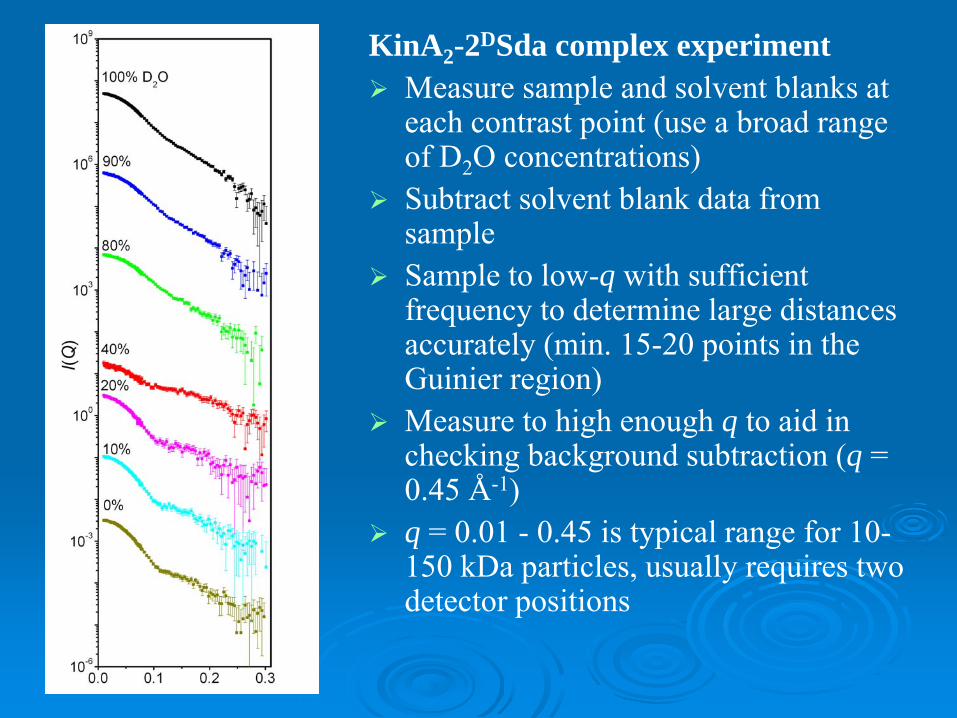
KinA2-2DSda complex experimentMeasure sample and solvent blanks at each contrast point (use a broad range of D2O concentrations)Subtract solvent blank data from sampleSample to low-q with sufficient frequency to determine large distances accurately (min. 15-20 points in the Guinier region) Measure to high enough q to aid in checking background subtraction (q = 0.45 Å-1)q = 0.01 - 0.45 is typical range for 10-150 kDa particles, usually requires two detector positions

Use Rg (from MULCh) for Sturhman analysis
222
ρβ
ρα
Δ−
Δ+= mobs RR
RH = 25.40 ÅRD = 25.3 ÅD = 27.0 Å
Sign of α indicates whether the higher scattering density object is more toward outside (+) or inside (-)
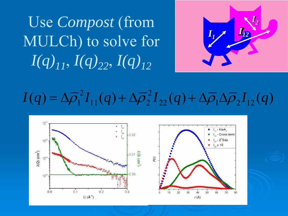
Use Compost (from MULCh) to solve for
I(q)11, I(q)22, I(q)12
I1I12
I2
)()()()( 1221222211
21 qIqIqIqI ρρρρ ΔΔ+Δ+Δ=
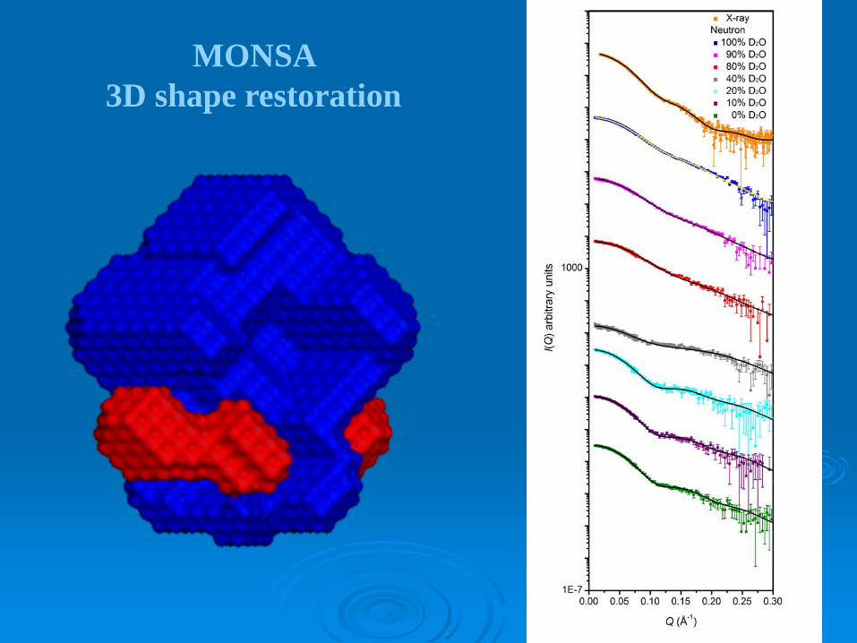
MONSA 3D shape restoration
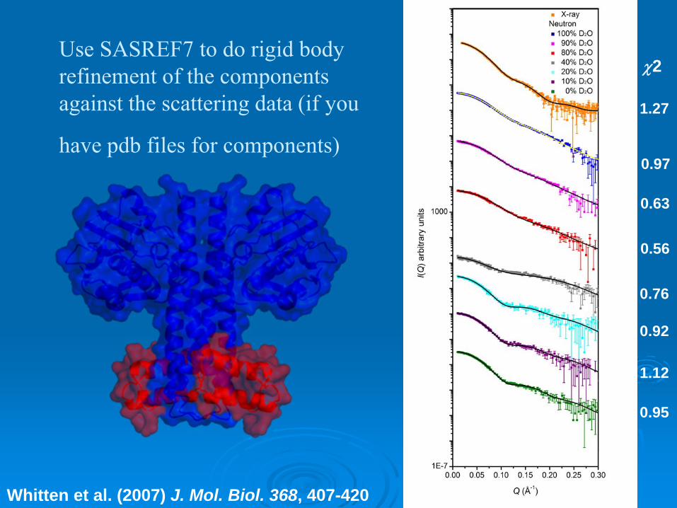
Use SASREF7 to do rigid body refinement of the components against the scattering data (if you
have pdb files for components)
χ2 = 1.27
χ2 = 0.97
χ2 = 0.63
χ2 = 0.56
χ2 = 0.76
χ2 = 0.92
χ2 = 1.12
χ2 = 0.95
χ2
Whitten et al. (2007) J. Mol. Biol. 368, 407-420

B
Jacques et al, J. Mol. Biol. 384, 422, 2008Whitten et al., J. Mol. Biol. 368, 407, 2007
Bacterial histidine kinase (KinA) and its protein inhibitors (Sda and KipI): Neutrons reveal inhibitors bind at the base of the dimerisation domain that
connects to the sensor domains.
Sda Kip I
Sensor domains
Sensor domains
Kin A Kin A
B
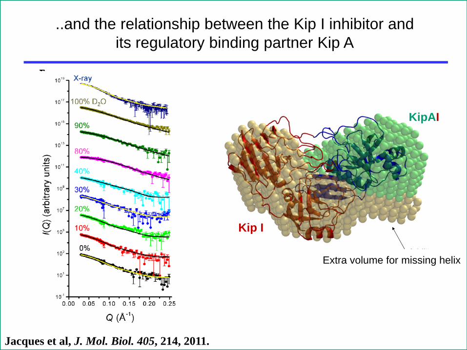
Jacques et al, J. Mol. Biol. 405, 214, 2011.
Kip I
Kip A
..and the relationship between the Kip I inhibitor and its regulatory binding partner Kip A
KipAI
Extra volume for missing helix
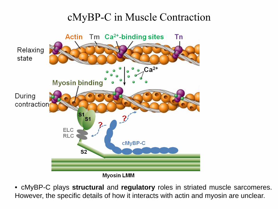
cMyBP-C in Muscle Contraction
• cMyBP-C plays structural and regulatory roles in striated muscle sarcomeres.However, the specific details of how it interacts with actin and myosin are unclear.
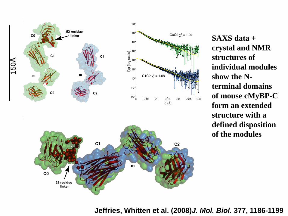
SAXS data + crystal and NMR structures of individual modules show the N-terminal domains of mouse cMyBP-C form an extended structure with a defined disposition of the modules
Jeffries, Whitten et al. (2008)J. Mol. Biol. 377, 1186-1199
150Å
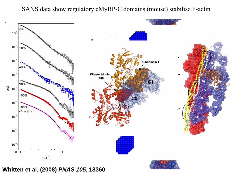
SANS data show regulatory cMyBP-C domains (mouse) stabilise F-actin
Whitten et al. (2008) PNAS 105, 18360
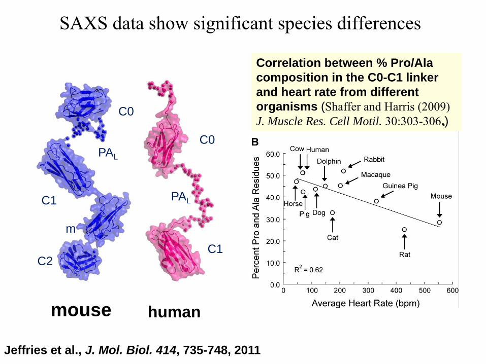
SAXS data show significant species differences
mouse human
C0
C0
C1
C1 PAL
PAL
m
C2
Correlation between % Pro/Ala composition in the C0-C1 linker and heart rate from different organisms (Shaffer and Harris (2009) J. Muscle Res. Cell Motil. 30:303-306.)
Jeffries et al., J. Mol. Biol. 414, 735-748, 2011

2D reconstruction of human C0C1-actin assembly from neutron contrast series consistent with C0 binding with a flexible and extended P/AL
Lu et al., J. Mol. Biol. 413, 908-913, 2011

Incorporation of deuterium up to 86% of the chemically Non-exchangeable protons can be obtained by using D2O as the deuterium source. Complete deuteration can only be obtained by addition of perdeuterated carbon source (glucose or glycerol).
Use mass spec to determine deuteration levels.
The described protocols allow the deuteration content in recombinant proteins to be predicted
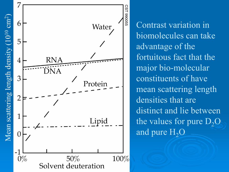
Contrast variation in biomolecules can take advantage of the fortuitous fact that the major bio-molecular constituents of have mean scattering length densities that are distinct and lie between the values for pure D2O and pure H2OM
ean
scat
terin
g le
ngth
den
sity
(1010
cm2 )
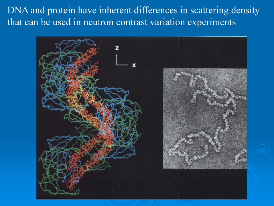
DNA and protein have inherent differences in scattering density that can be used in neutron contrast variation experiments
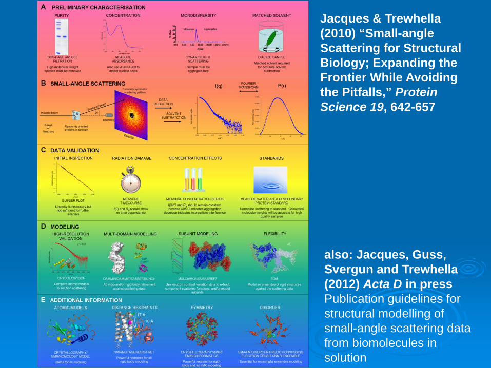
Jacques & Trewhella (2010) “Small-angle Scattering for Structural Biology; Expanding the Frontier While Avoiding the Pitfalls,” Protein Science 19, 642-657
also: Jacques, Guss, Svergun and Trewhella (2012) Acta D in press Publication guidelines for structural modelling of small-angle scattering data from biomolecules in solution
Neutron scattering sample cellsHelma quartz cells (high precision path-length, suprasil) – need lots of them! Banjo-style (280 μL per 1 mm path length) or rectangular (170 μL per 1 mm path length) cells can be usedPath lengths are only good to 1%, so good idea to measure sample and solvent background in the same cell if practical, but experiment logistics may prohibit that, so calibrate cells? High incoherent scattering for 1H means you always want ≤ 1mm 1H2O in the neutron beam to avoid multiple scattering
Andrew Whitten David Jacques
Cy JeffriesYanling Lu
Neutron Ted
John Chow
James Taylor


![Whole Genome Shatqun updates) C] EMBL (Cantiq release) C] EMBL (Coding Sequences) C] Genome Reviews C] EMBL ID/ Accession Mapping C] EMBL MGA Nucleotide sequence databases - subsections](https://img.pdfslide.us/doc/110x75/5ccc4c0c88c993de558c2477/whole-genome-shatqun-updates-c-embl-cantiq-release-c-embl-coding-sequences.jpg)