Embed Size (px)
Citation preview
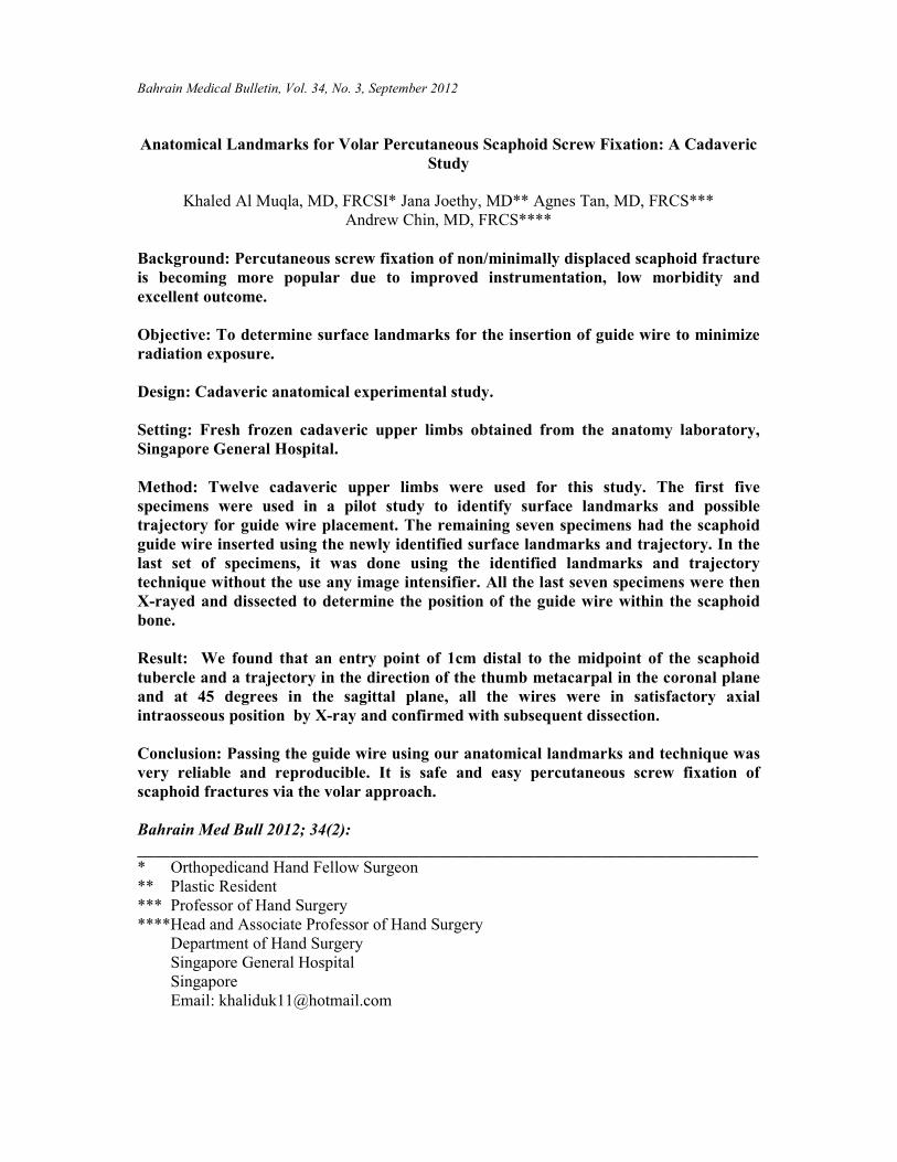
Bahrain Medical Bulletin, Vol. 34, No. 3, September 2012
Anatomical Landmarks for Volar Percutaneous Scaphoid Screw Fixation: A Cadaveric
Study
Khaled Al Muqla, MD, FRCSI* Jana Joethy, MD** Agnes Tan, MD, FRCS***
Andrew Chin, MD, FRCS****
Background: Percutaneous screw fixation of non/minimally displaced scaphoid fracture
is becoming more popular due to improved instrumentation, low morbidity and
excellent outcome.
Objective: To determine surface landmarks for the insertion of guide wire to minimize
radiation exposure.
Design: Cadaveric anatomical experimental study.
Setting: Fresh frozen cadaveric upper limbs obtained from the anatomy laboratory,
Singapore General Hospital.
Method: Twelve cadaveric upper limbs were used for this study. The first five
specimens were used in a pilot study to identify surface landmarks and possible
trajectory for guide wire placement. The remaining seven specimens had the scaphoid
guide wire inserted using the newly identified surface landmarks and trajectory. In the
last set of specimens, it was done using the identified landmarks and trajectory
technique without the use any image intensifier. All the last seven specimens were then
X-rayed and dissected to determine the position of the guide wire within the scaphoid
bone.
Result: We found that an entry point of 1cm distal to the midpoint of the scaphoid
tubercle and a trajectory in the direction of the thumb metacarpal in the coronal plane
and at 45 degrees in the sagittal plane, all the wires were in satisfactory axial
intraosseous position by X-ray and confirmed with subsequent dissection.
Conclusion: Passing the guide wire using our anatomical landmarks and technique was
very reliable and reproducible. It is safe and easy percutaneous screw fixation of
scaphoid fractures via the volar approach.
Bahrain Med Bull 2012; 34(2):
___________________________________________________________________________
* Orthopedicand Hand Fellow Surgeon
** Plastic Resident
*** Professor of Hand Surgery
****Head and Associate Professor of Hand Surgery
Department of Hand Surgery
Singapore General Hospital
Singapore
Email: [email protected]
Fractures of the scaphoid constitute 2-7% of all fractures;they aresecond to distal radial
fractures1. The tiny blood supply combined with the demanding functional requirements
could lead to delayed healing1.Eighty to ninety percent involvethe scaphoid
1. The incidence is
approximately 5 in every 10,000 in Western countries1. Percutaneous fixation of
non/minimally displaced scaphoid fracture is becoming more popular due to improved
instrumentation, low morbidity, excellent results in term of healing, avoidance of prolonged
immobilization and possibly lower costs2-5.
The aim of this cadaveric study is to find simple and reliable surface anatomical landmarks to
pass the guide wire for the cannulated screw through a volar approach.
METHOD
Twelve cadaveric specimens were used. In the first 5 specimens, the guide wire (1.2 mm,
non-threaded) was passed with the aid of an Image Intensifier through volar approach in
multiple projections to make sure that the wire was in axial intraossous position within the
scaphoid bone. In all the specimens, the wrist was maintained in neutral position to
standardize our technique. We used these 5 specimens to find a reference anatomical
landmark for the entry point and possible trajectory for the guide wire. After identifying the
above-mentioned landmark and trajectory, it was used for passing the guide wire in the
subsequent 7 specimens without using any image intensification. All the 7 specimens were
then X-rayed and dissected to check the position of the guide wires.
RESULT
The starting point for the passage of the guide wire from the volar side was 10 mm distal to
midpoint of the scaphoid tubercle (range 8-12 mm) was found in the first group.
The radial ulnar (coronal) direction was parallel to the thumb metacarpal bone (range+/- 5
degrees) with both the thumb carpometacarpal and the wrist joint in neutral position. The
volar dorsal trajectory (sagittal) was around 45 degrees (range 40-50 degrees) with neutral
joint position maintained, see figure 1. We used this anatomical landmark (i.e. 0 degrees in
coronal plane and 45 degrees in sagittal of the thumb metacarpal) for the next 7 specimens
without using image intensifier, see figure 2. X-rays of these specimens showed that all the
wires were in acceptable position within the bone except for one which was slightly ulnar in
position, a second wire was passed radial to it and in the same sagittal (volar dorsal)
direction. A subsequent X-ray showed good position in the coronal (radial ulnar) direction.
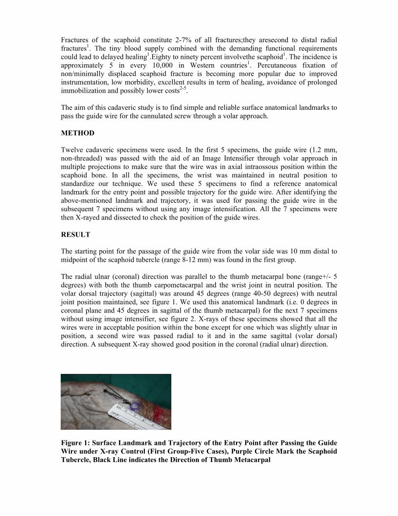
Figure 1: Surface Landmark and Trajectory of the Entry Point after Passing the Guide
Wire under X-ray Control (First Group-Five Cases), Purple Circle Mark the Scaphoid
Tubercle, Black Line indicates the Direction of Thumb Metacarpal
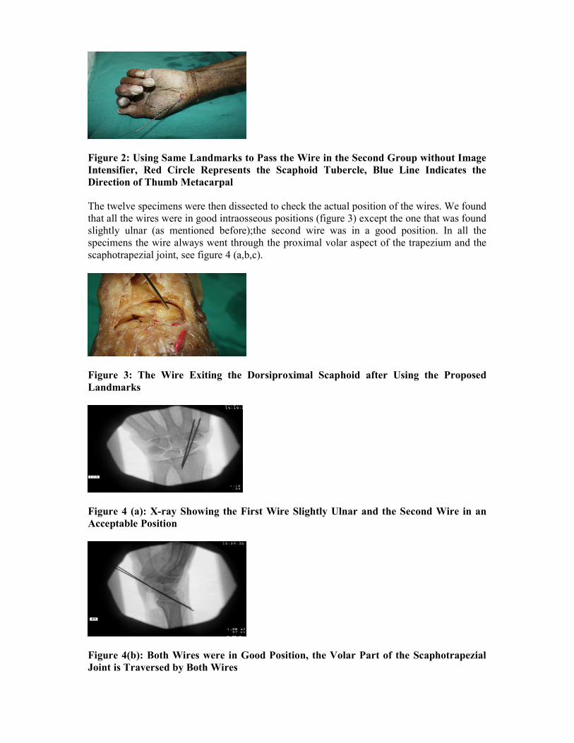
Figure 2: Using Same Landmarks to Pass the Wire in the Second Group without Image
Intensifier, Red Circle Represents the Scaphoid Tubercle, Blue Line Indicates the
Direction of Thumb Metacarpal
The twelve specimens were then dissected to check the actual position of the wires. We found
that all the wires were in good intraosseous positions (figure 3) except the one that was found
slightly ulnar (as mentioned before);the second wire was in a good position. In all the
specimens the wire always went through the proximal volar aspect of the trapezium and the
scaphotrapezial joint, see figure 4 (a,b,c).
Figure 3: The Wire Exiting the Dorsiproximal Scaphoid after Using the Proposed
Landmarks
Figure 4 (a): X-ray Showing the First Wire Slightly Ulnar and the Second Wire in an
Acceptable Position
Figure 4(b): Both Wires were in Good Position, the Volar Part of the Scaphotrapezial
Joint is Traversed by Both Wires
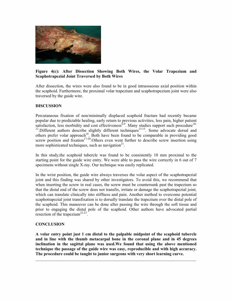
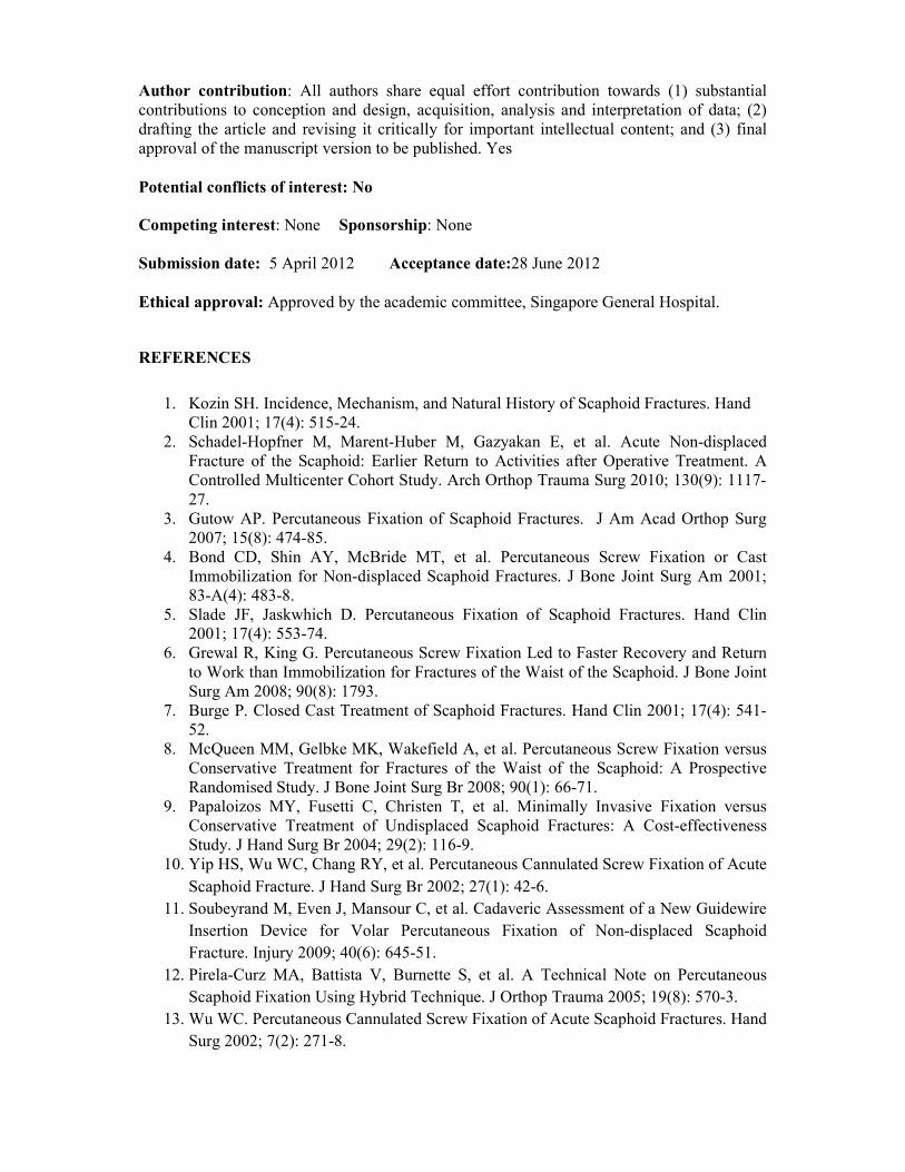
Figure 4(c): After Dissection Showing Both Wires, the Volar Trapezium and
Scaphotrapezial Joint Traversed by Both Wires
After dissection, the wires were also found to be in good intraosseous axial position within
the scaphoid. Furthermore, the proximal volar trapezium and scaphotrapezium joint were also
traversed by the guide wire.
DISCUSSION
Percutaneous fixation of non/minimally displaced scaphoid fracture had recently became
popular due to predictable healing, early return to previous activities, less pain, higher patient
satisfaction, less morbidity and cost effectiveness6-9. Many studies support such procedure
10-
12.Different authors describe slightly different techniques
13-15. Some advocate dorsal and
others prefer volar approach16. Both have been found to be comparable in providing good
screw position and fixation17-20
.Others even went further to describe screw insertion using
more sophisticated techniques, such as navigation21.
In this study,the scaphoid tubercle was found to be consistently 10 mm proximal to the
starting point for the guide wire entry. We were able to pass the wire correctly in 6 out of 7
specimens without single X-ray. Our technique was easily replicated.
In the wrist position, the guide wire always traverses the volar aspect of the scaphotrapezial
joint and this finding was shared by other investigators. To avoid this, we recommend that
when inserting the screw in real cases, the screw must be countersunk past the trapezium so
that the distal end of the screw does not transfix, irritate or damage the scaphotrapezial joint,
which can translate clinically into stiffness and pain. Another method to overcome potential
scaphotrapezial joint transfixation is to dorsally translate the trapezium over the distal pole of
the scaphoid. This maneuver can be done after passing the wire through the soft tissue and
prior to engaging the distal pole of the scaphoid. Other authors have advocated partial
resection of the trapezium22-23
.
CONCLUSION
A volar entry point just 1 cm distal to the palpable midpoint of the scaphoid tubercle
and in line with the thumb metacarpal bone in the coronal plane and in 45 degrees
inclination in the sagittal plane was used.We found that using the above mentioned
technique the passage of the guide wire was easy, reproducible and with high accuracy.
The procedure could be taught to junior surgeons with very short learning curve.
___________________________________________________________________________
Author contribution: All authors share equal effort contribution towards (1) substantial
contributions to conception and design, acquisition, analysis and interpretation of data; (2)
drafting the article and revising it critically for important intellectual content; and (3) final
approval of the manuscript version to be published. Yes
Potential conflicts of interest: No
Competing interest: None Sponsorship: None
Submission date: 5 April 2012 Acceptance date:28 June 2012
Ethical approval: Approved by the academic committee, Singapore General Hospital.
REFERENCES
1. Kozin SH. Incidence, Mechanism, and Natural History of Scaphoid Fractures. Hand
Clin 2001; 17(4): 515-24.
2. Schadel-Hopfner M, Marent-Huber M, Gazyakan E, et al. Acute Non-displaced
Fracture of the Scaphoid: Earlier Return to Activities after Operative Treatment. A
Controlled Multicenter Cohort Study. Arch Orthop Trauma Surg 2010; 130(9): 1117-
27.
3. Gutow AP. Percutaneous Fixation of Scaphoid Fractures. J Am Acad Orthop Surg
2007; 15(8): 474-85.
4. Bond CD, Shin AY, McBride MT, et al. Percutaneous Screw Fixation or Cast
Immobilization for Non-displaced Scaphoid Fractures. J Bone Joint Surg Am 2001;
83-A(4): 483-8.
5. Slade JF, Jaskwhich D. Percutaneous Fixation of Scaphoid Fractures. Hand Clin
2001; 17(4): 553-74.
6. Grewal R, King G. Percutaneous Screw Fixation Led to Faster Recovery and Return
to Work than Immobilization for Fractures of the Waist of the Scaphoid. J Bone Joint
Surg Am 2008; 90(8): 1793.
7. Burge P. Closed Cast Treatment of Scaphoid Fractures. Hand Clin 2001; 17(4): 541-
52.
8. McQueen MM, Gelbke MK, Wakefield A, et al. Percutaneous Screw Fixation versus
Conservative Treatment for Fractures of the Waist of the Scaphoid: A Prospective
Randomised Study. J Bone Joint Surg Br 2008; 90(1): 66-71.
9. Papaloizos MY, Fusetti C, Christen T, et al. Minimally Invasive Fixation versus
Conservative Treatment of Undisplaced Scaphoid Fractures: A Cost-effectiveness
Study. J Hand Surg Br 2004; 29(2): 116-9.
10. Yip HS, Wu WC, Chang RY, et al. Percutaneous Cannulated Screw Fixation of Acute
Scaphoid Fracture. J Hand Surg Br 2002; 27(1): 42-6.
11. Soubeyrand M, Even J, Mansour C, et al. Cadaveric Assessment of a New Guidewire
Insertion Device for Volar Percutaneous Fixation of Non-displaced Scaphoid
Fracture. Injury 2009; 40(6): 645-51.
12. Pirela-Curz MA, Battista V, Burnette S, et al. A Technical Note on Percutaneous
Scaphoid Fixation Using Hybrid Technique. J Orthop Trauma 2005; 19(8): 570-3.
13. Wu WC. Percutaneous Cannulated Screw Fixation of Acute Scaphoid Fractures. Hand
Surg 2002; 7(2): 271-8.
14. Geurts GF, Van Riet RP, Meermans G, et al. Volar Percutaneous Transtrapezial
Fixation of Scaphoid Waist Fractures: Surgical Technique. Acta Orthop Belg 2012;
78(1): 121-5.
15. Zlotolow DA, Knutsen E, Yeo J. Optimization of Volar Percutaneous Screw Fixation
for Scaphoid Waist Fractures Using Traction, Positioning, Imaging, and
Angiocatheter Guide. J Hand Surg Am 2011; 36(5): 916-21.
16. Chan KW, McAdams TR. Central Screw Placement in Percutaneous Screw Scaphoid
Fixation: A Cadaveric Comparison of Proximal and Distal Techniques. J Hand Surg
Am 2004; 29(1): 74-9.
17. Kim JK, Kim JO, Lee SY. Volar Percutaneous Screw Fixation for Scaphoid Waist
Delayed Union. Clin Orthop Relat Res 2010; 468(4):1066-71.
18. Levitz S, Ring D. Retrograde (Volar) Scaphoid Screw Insertion- A Quantitative
Computed Tomographic Analysis. J Hand Surg Am 2005; 30(3): 543-8.
19. Polsky MB, Kozin SH, Porter ST, et al. Scaphoid Fractures: Dorsal Versus Volar
Approach. Orthopedics 2002; 25(8): 817-9.
20. Drac P, Manak P, Cizmar I, et al. A Palmar Percutaneous Volar Versus a Dorsal
Limited Approach for Treatment of Non-and Minimally Displaced Scaphoid Waist
Fractures: An Assessment of Functional Outcome and Complications. Acta Chir
Orthop Traumatol Cech 2010; 77(2): 143-8.
21. Walsh E, Crisco JJ, Wolfe SW. Computer-assisted Navigation for Volar Percutaneous
Scaphoid Placement. J Hand Surg Am 2009; 34(9): 1722-8.
22. Leventhal EL, Wolfe SW, Walsh EF, et al. A Computational Approach to the Optimal
Screw Axis Location and Orientation in the Scaphoid Bone. J Hand Surg Am 2009;
34(4): 677-84.
23. Merrell G, Slade J. Technique for Percutaneous Fixation of Displaced and Non-
displaced Acute Scaphoid Fractures and Non-unions. J Hand Surg Am 2008; 33(6):
966-73.