Embed Size (px)
Citation preview
Journal of Pre-Clinical and Clinical Research, 2013, Vol 7, No 1, 63-65www.jpccr.euCASE REPORT
Scalping degloving foot injury as a di�cult therapeutic problemTomasz Kulesza1, Leszek Jankiewicz1, Jacek Sompor1, Andrzej Prystupa2
1 Department of Traumatology and Emergency Medicine, Medical University, Lublin, Poland 2 Department of Internal Medicine, Medical University, Lublin, Poland
Kulesza T, Jankiewicz L, Sompor J, Prystupa A. Scalping degloving foot injury as a difficult therapeutic problem. J Pre-Clin Clin Res. 2013; 7(1): 63–65.
AbstractThe case is presented of a rare case of scalping foot injury of the degloving type, treated at the Clinic of Traumatology and Emergency Medicine at the Medical University in Lublin, Poland. The successful treatment used the simplest reconstructive technique involving the utilization of an intermediate thickness skin graft, which was taken from the detached skin of the feet. This technique should be taken into consideration in this era of using new and sophisticated methods of treating physical defects.
Key wordsscalping, degloving, foot injury
INTRODUCTION
Serious complex injuries of the lower limbs still remain a great challenge for surgeons. Among them, special attention is needed for injuries with extensive, scalping detachment of skin, known as ‘degloving’ in English literature. Injuries of this type can be de�ned as avulsive-abrasive detachment of skin from the underlying fascia, muscle and bone tissue, which causes damage to blood vessels supplying the skin [1, 2, 3]. �ey are most frequently caused by complex abrasive, compressive and tearing deforming forces which are transmitted to the limbs during high energy trauma in tra�c, agricultural and industrial accidents. Injuries of this type interrupt large vessels and damage the capillary network of the subcutaneous tissue. �e resulting swelling and haematoma cause the detached skin to move away from the deeper underlying tissue which a�ects the formation of ischaemia [2].
�e aim of this study was to present a patient who su�ered a rare scalping degloving foot injury.
CASE REPORT
A steelworker was injured a�er his foot was caught in moving conveyor belt rollers. As a result of the trauma, the patient su�ered total scalping of the foot without metatarsal and tarsal fractures (Fig. 1). �e skin in the subcutaneous tissue layer was torn o�, exposing the fascia, and delivered with the patient without being cooled during transportation. �e limb was rated 5 points on the MESS scale [4]. Surgery was performed as a matter of urgency. Surgical toilet of the wounds with the necessary amputation of the distal phalanges of the toes due to the total exposure of bone was performed. Gra�s of intermediate thickness were taken from the detached foot skin of both dorsal and plantar surfaces and sutured in the defect. In this way, the entire wound was
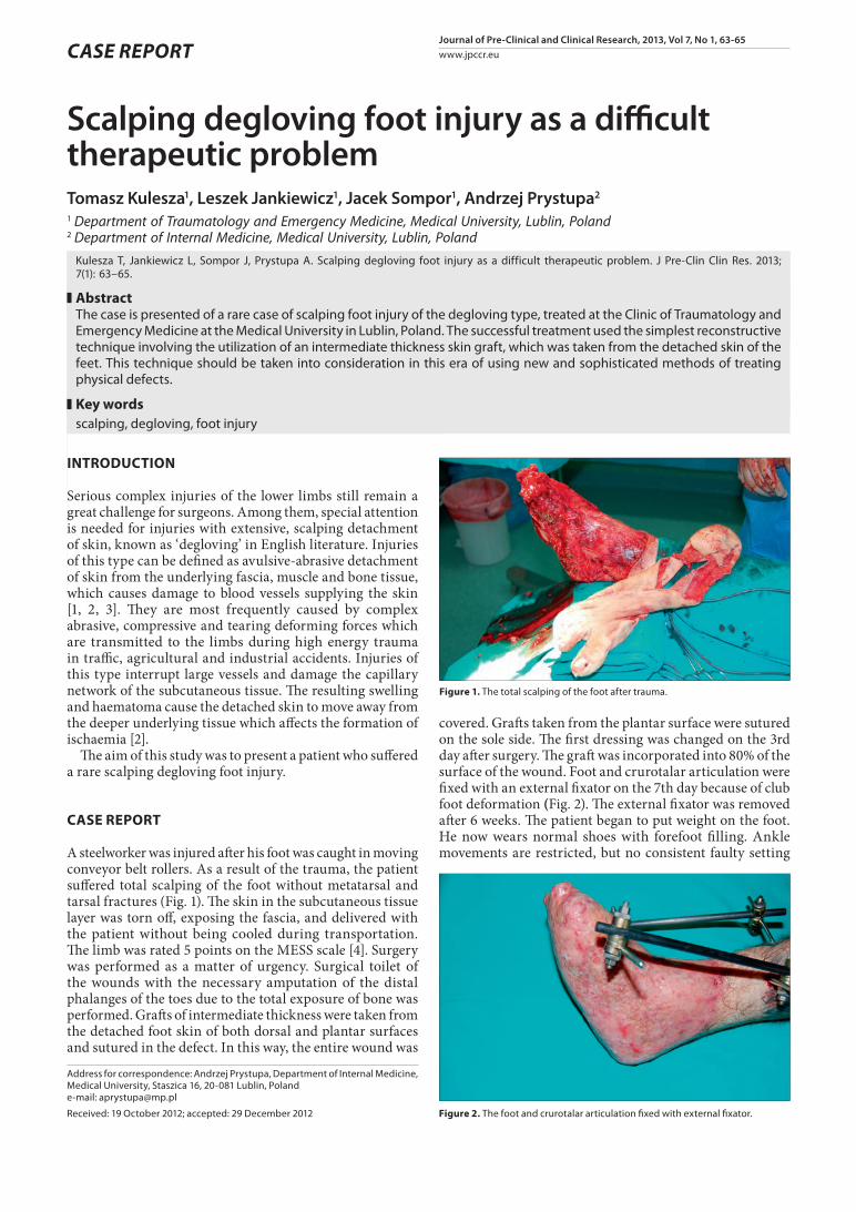
covered. Gra�s taken from the plantar surface were sutured on the sole side. �e �rst dressing was changed on the 3rd day a�er surgery. �e gra� was incorporated into 80% of the surface of the wound. Foot and crurotalar articulation were �xed with an external �xator on the 7th day because of club foot deformation (Fig. 2). �e external �xator was removed a�er 6 weeks. �e patient began to put weight on the foot. He now wears normal shoes with forefoot �lling. Ankle movements are restricted, but no consistent faulty setting
Figure 1. The total scalping of the foot after trauma.
Figure 2. The foot and crurotalar articulation �xed with external �xator.
Address for correspondence: Andrzej Prystupa, Department of Internal Medicine, Medical University, Staszica 16, 20-081 Lublin, Polande-mail: [email protected]
Received: 19 October 2012; accepted: 29 December 2012
Journal of Pre-Clinical and Clinical Research, 2013, Vol 7, No 1Tomasz Kulesza, Leszek Jankiewicz, Jacek Sompor, Andrzej Prystupa. Scalping degloving foot injury as a di�cult therapeutic problem
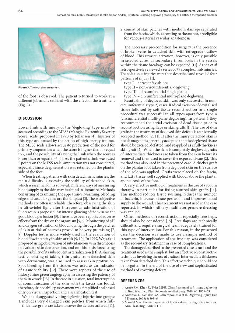
of the foot is observed. �e patient returned to work at a di�erent job and is satis�ed with the e�ect of the treatment (Fig. 3).
DISCUSSION
Lower limb with injury of the ‘degloving’ type must be accessed according to the MESS (Mangled Extremity Severity Score) scale, proposed in 1990 by Johansen [4]. Injuries of this type are caused by the action of high-energy trauma. �e MESS scale allows accurate prediction of the need for primary amputation when the score is higher than or equal to 7, and the possibility of saving the limb when the score is lower than or equal to 6 [4]. As the patient’s limb was rated 5 points on the MESS scale, amputation was not considered, especially since deep sensation was retained on the plantar side of the foot.
When treating patients with skin detachment injuries, the main di�culty is assessing the viability of detached skin, which is essential for its survival. Di�erent ways of measuring blood supply to the skin may be found in literature. Methods consisting of examining the appearance, warming, bleeding edge and vascular game are the simplest [3]. �ese subjective methods are o�en unreliable; therefore, observing the skin in ultraviolet light a�er intravenous administration of �uorescein is proposed. An intense glowing of the skin meant good blood perfusion [3]. �ere have been reports of adverse e�ects from the dye on the organism [5, 6]. Monitoring of pH and oxygen saturation of blood �owing through the patches of skin at risk of necrosis proved to be very promising [7, 8]. Doppler test is more widely used in the evaluation of blood �ow intensity in skin at risk [9, 10]. In 1997, Wajkakul proposed using observation of subcutaneous vein thrombosis to evaluate skin demarcation, and on this basis forecasting the possibility of its subsequent arterialization [11]. A shaving test, consisting of taking thin gra�s from detached skin with dermatome, was also used to assess skin protrusion. Spot bleeding from the tissues was used as an indicator of tissue viability [12]. �ere were reports of the use of indocyanine green angiography in assessing the patency of the skin vessels [13]. In the case in question, total interruption of communication of the skin with the fascia was found; therefore, skin viability assessment was simpli�ed and based only on visual inspection and a scari�cation test.
Waikakul suggests dividing degloving injuries into groups:1. includes very damaged skin patches from which full-
thickness gra�s are taken to cover the defects su�ered [11].
2. consist of skin patches with medium damage separated from the fascia, which, according to the author, are eligible for venous-arterial vascular anastomosis.
�e necessary pre-condition for surgery is the presence of broken veins in detached skin with retrograde out�ow of blood. �is revascularization, however, is only possible in selected cases, as secondary thrombosis in the vessels within the tissue breakage can be expected [11]. Arnez et al retrospectively reviewed a series of 79 complex limb injuries. �e so�-tissue injuries were then described and revealed four patterns of injury [1].
type I – abrasion/avulsion;type II – non-circumferential degloving;type III – circumferential single plane;type IV – circumferential multi-plane degloving.Resuturing of degloved skin was only successful in non-
circumferential (type 2) cases. Radical excision of devitalised tissue followed by so�-tissue reconstruction in a single procedure was successful in all types apart from type 4 (circumferential multi-plane degloving). In pattern 4 they recommended the serial excision of dead tissue prior to reconstruction using �aps or skin gra�s [1]. �e use of skin gra�s in the treatment of degloved skin defects is a universally accepted method [2, 11]. If a�er the injury detached skin is not damaged it is generally accepted that the degloved tissue should be excised, defatted, and reapplied as a full-thickness skin gra� [2]. When the skin is completely degloved, gra�s of intermediate thickness are taken from the skin before its removal and then used to cover the exposed tissue [2]. �is method was also used in the presented case. A thicker gra� on the plantar foot taken from degloved skin on the surface of the sole was applied. Gra�s were placed on the fascia, and fatty tissue well supplied with blood, above the plantar aponeurosis of the foot.
A very e�ective method of treatment is the use of vacuum therapy, in particular for �xing sutured skin gra�s [14]. �is method reduces tissue swelling, reduces the number of bacteria, increases tissue perfusion and improves blood supply to the wound. �is treatment was not used in the case reported, in which ordinary multilayer pressure dressing was applied.
Other methods of reconstruction, especially free �aps, should also be considered [15]. Free �aps are technically di�cult and require a surgical team adequately trained for this type of intervention. For this reason, in the presented case the decision was made to use a simple method of treatment. �e application of the free �ap was considered as the secondary treatment in case of complications.
�e damage described in the presented case is rare and the treatment used is the simplest, but an e�ective reconstructive technique involving the use of gra�s of intermediate thickness taken from detached skin. �is e�ective technique should not be forgotten in the era of the use of new and sophisticated methods of covering defects.
REFERENCES
1. Arnez ZM, Khan U, Tyler MPH. Classi�cation of so�-tissue degloving in limb trauma. J Plast Reconstr Aesthet Surg. 2010; 63: 1865–69.
2. Antoniou D, Kyriakidis A, Zaharopoulos A et al. Degloving injury. Eur J Trauma. 2005; 6: 593–6.
3. Mandel MA. �e management of lower extremity degloving injuries. Ann Plast Surg. 1981; 6: 1–5.
64
Figure 3. The foot after treatment.

Journal of Pre-Clinical and Clinical Research, 2013, Vol 7, No 1Tomasz Kulesza, Leszek Jankiewicz, Jacek Sompor, Andrzej Prystupa. Scalping degloving foot injury as a di�cult therapeutic problem
4. Johansen K, Daines M, Howey T, et al. Objective criteria accurately predict amputation following lower extremity trauma. J Trauma. 1990; 30: 568–73.
5. Graham BH, Gordon L, Alpert BS, et al. Serial quantitative skin surface �uorescence: a new method for postoperative monitoring of vascular perfusion in revascularized digits. J Hand Surg. 1985; 10: 226–30.
6. Casanova R, Iribarren O, Grotting JC, et al. Clinical evaluation of �ap viability with a dermal surface �uorometer. Ann Plast Surg. 1988; 20: 112–6.
7. Sera�n D, Lesesne CB,Mullen RY, et al. Transcutaneous PO2 monitoring for assessing viability and predicting survival of skin �aps: experimental and clinical correlations. J Microsurg. 1981; 2: 165–78.
8. Hirigoyen MB, Blackwell KE, Zhang WX, et al. Continuous tissue oxygen tension measurement as a monitor of free- �ap viability. Plast Reconstr Surg. 1997; 99: 763–73.
9. Solomon GA, Yeremchuk MJ, Mansen PN. Doppler ultrasound surface monitoring of both arterial and venous �ow in clinical free tissue transfers. J Reconstr Microsurg. 1986; 3: 39–41.
10. Jones BM, Mayou BJ. �e laser Doppler �owmeter for microvascular monitoring: a preliminary report. Br J Plast Surg. 1988; 35: 147–9.
11. Waikakul S. Revascularisation of degloving injuries of the limbs. Injury 1997; 28: 271–4.
12. Ziv I, Zeligowski AA, Elyashuv O, et al. Immediate care of crush injuries and compartment syndromes with the split-thickness skin excision. Clin Orthop. 1990; 256: 224–8.
13. Kamolz LP, Andel H, Auer T, et al. Evaluation of skin perfusion by use of indocyanine green video angiography: rational design and planning of trauma surgery. J Trauma. 2006; 61: 635–41.
14. Josty IC, Ramaswamy R, Laing JHE. Vacuum assisted closure: an alternative strategy in the management of degloving injuries of the foot. Br J Plast Surg. 2001; 54: 363–5.
15. Swartz WM, Mears DC. �e role of free-tissue transfers in lower- extremity reconstruction. Plast Reconstr Surg. 1985; 76: 364–73.
65