Embed Size (px)
Citation preview
INTERNATIONAL JOURNAL OF CONTEMPORARY MEDICAL RESEARCH Volume 2 | Issue 3|
522 IJCMR
ABSTRACT Introduction: In this modern era, soft tissue injuries of the face are becoming more frequent. Surgeons are facing patients for reconstruction of mutilating lacerations and massive degloving injuries. Degloving generally occurs from high - energy trauma. Such injuries demand meticulous care; inadequate management may lead to grotesque deformity, with its inevitable physiological and psychological consequences. Case report: This paper describes an extreme case of traumatic facial degloving injury and the decisions made regarding the prevention of necrosis and infection by surgical debridement and timely repair of the vital soft tissue structures. Conclusion: Facial degloving injury is a complex reconstructive challenge which may require a staged approach for optimal cosmetic and functional outcome. Proper planning, surgical procedure, meticulous and proper alignment of tissues and post-operative care gave us good aesthetic and functional outcome with a satisfied patient. Keywords: Degloving injuries, reconstruction, scar, healing. How to cite this article: Pulkit Khandelwal, Vikas Dhupar. Complex Traumatic Facial Degloving Injury - Challenges And Management Outcome – A Case Report. International Journal of Contemporary Medical Research 2015;2(3): 522-524
Source of Support: Nil Conflict of Interest: None INTRODUCTION Soft-tissue injuries that result from a shearing or stripping force are termed degloving injuries. It involves the separation or detachment of the skin and subcutaneous tissue from the bones, compromising the adjacent fascia, muscles, blood vessels and
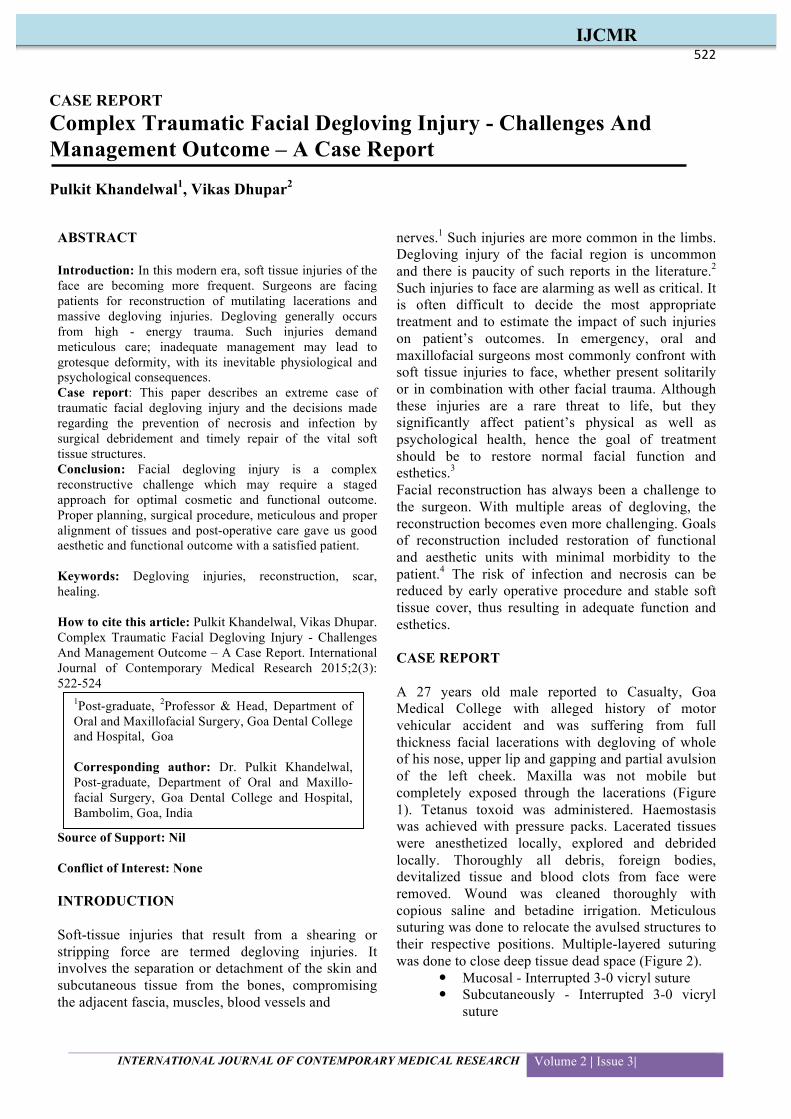
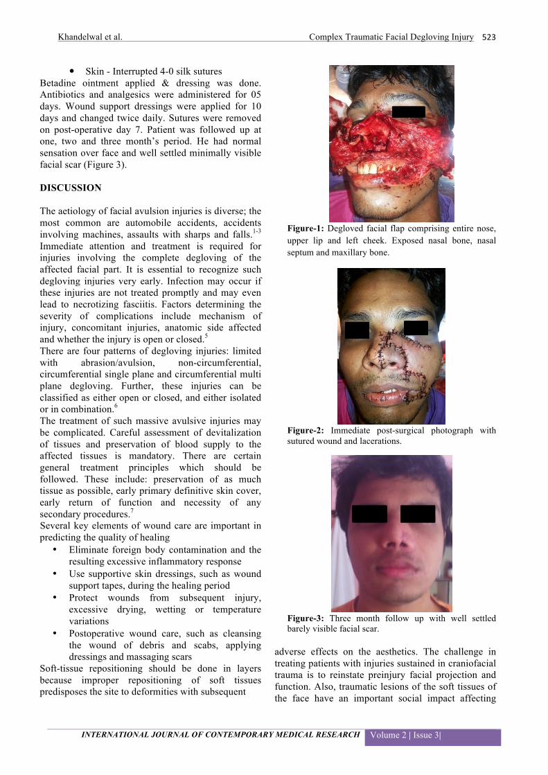
nerves.1 Such injuries are more common in the limbs. Degloving injury of the facial region is uncommon and there is paucity of such reports in the literature.2 Such injuries to face are alarming as well as critical. It is often difficult to decide the most appropriate treatment and to estimate the impact of such injuries on patient’s outcomes. In emergency, oral and maxillofacial surgeons most commonly confront with soft tissue injuries to face, whether present solitarily or in combination with other facial trauma. Although these injuries are a rare threat to life, but they significantly affect patient’s physical as well as psychological health, hence the goal of treatment should be to restore normal facial function and esthetics.3 Facial reconstruction has always been a challenge to the surgeon. With multiple areas of degloving, the reconstruction becomes even more challenging. Goals of reconstruction included restoration of functional and aesthetic units with minimal morbidity to the patient.4 The risk of infection and necrosis can be reduced by early operative procedure and stable soft tissue cover, thus resulting in adequate function and esthetics. CASE REPORT A 27 years old male reported to Casualty, Goa Medical College with alleged history of motor vehicular accident and was suffering from full thickness facial lacerations with degloving of whole of his nose, upper lip and gapping and partial avulsion of the left cheek. Maxilla was not mobile but completely exposed through the lacerations (Figure 1). Tetanus toxoid was administered. Haemostasis was achieved with pressure packs. Lacerated tissues were anesthetized locally, explored and debrided locally. Thoroughly all debris, foreign bodies, devitalized tissue and blood clots from face were removed. Wound was cleaned thoroughly with copious saline and betadine irrigation. Meticulous suturing was done to relocate the avulsed structures to their respective positions. Multiple-layered suturing was done to close deep tissue dead space (Figure 2).
� Mucosal - Interrupted 3-0 vicryl suture � Subcutaneously - Interrupted 3-0 vicryl
suture
CASE REPORT Complex Traumatic Facial Degloving Injury - Challenges And Management Outcome – A Case Report Pulkit Khandelwal1, Vikas Dhupar2
1Post-graduate, 2Professor & Head, Department of Oral and Maxillofacial Surgery, Goa Dental College and Hospital, Goa Corresponding author: Dr. Pulkit Khandelwal, Post-graduate, Department of Oral and Maxillo- facial Surgery, Goa Dental College and Hospital, Bambolim, Goa, India
Khandelwal et al. Complex Traumatic Facial Degloving Injury
INTERNATIONAL JOURNAL OF CONTEMPORARY MEDICAL RESEARCH Volume 2 | Issue 3|
523
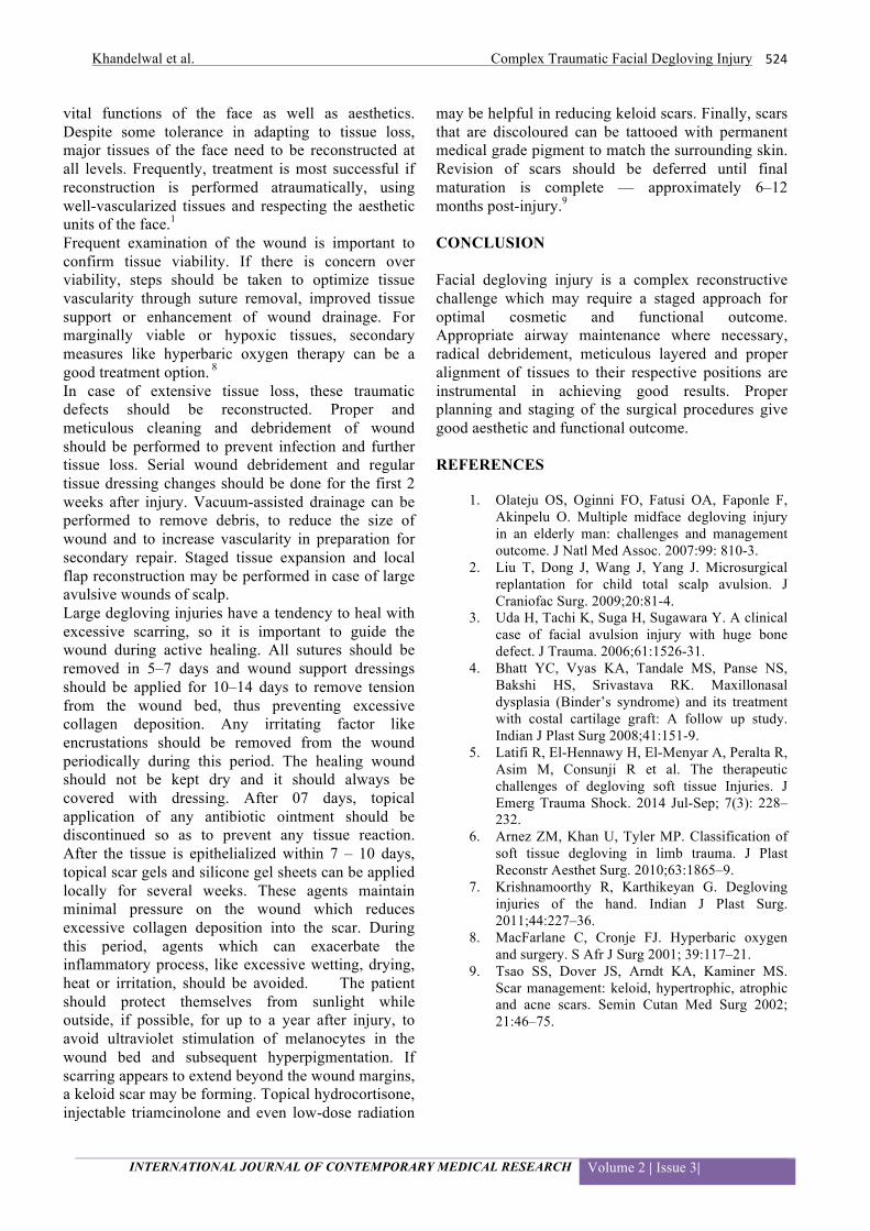
� Skin - Interrupted 4-0 silk sutures Betadine ointment applied & dressing was done. Antibiotics and analgesics were administered for 05 days. Wound support dressings were applied for 10 days and changed twice daily. Sutures were removed on post-operative day 7. Patient was followed up at one, two and three month’s period. He had normal sensation over face and well settled minimally visible facial scar (Figure 3). DISCUSSION The aetiology of facial avulsion injuries is diverse; the most common are automobile accidents, accidents involving machines, assaults with sharps and falls.1-3 Immediate attention and treatment is required for injuries involving the complete degloving of the affected facial part. It is essential to recognize such degloving injuries very early. Infection may occur if these injuries are not treated promptly and may even lead to necrotizing fasciitis. Factors determining the severity of complications include mechanism of injury, concomitant injuries, anatomic side affected and whether the injury is open or closed.5 There are four patterns of degloving injuries: limited with abrasion/avulsion, non-circumferential, circumferential single plane and circumferential multi plane degloving. Further, these injuries can be classified as either open or closed, and either isolated or in combination.6 The treatment of such massive avulsive injuries may be complicated. Careful assessment of devitalization of tissues and preservation of blood supply to the affected tissues is mandatory. There are certain general treatment principles which should be followed. These include: preservation of as much tissue as possible, early primary definitive skin cover, early return of function and necessity of any secondary procedures.7 Several key elements of wound care are important in predicting the quality of healing
• Eliminate foreign body contamination and the resulting excessive inflammatory response
• Use supportive skin dressings, such as wound support tapes, during the healing period
• Protect wounds from subsequent injury, excessive drying, wetting or temperature variations
• Postoperative wound care, such as cleansing the wound of debris and scabs, applying dressings and massaging scars
Soft-tissue repositioning should be done in layers because improper repositioning of soft tissues predisposes the site to deformities with subsequent
Figure-1: Degloved facial flap comprising entire nose, upper lip and left cheek. Exposed nasal bone, nasal septum and maxillary bone.
Figure-2: Immediate post-surgical photograph with sutured wound and lacerations.
Figure-3: Three month follow up with well settled barely visible facial scar.
adverse effects on the aesthetics. The challenge in treating patients with injuries sustained in craniofacial trauma is to reinstate preinjury facial projection and function. Also, traumatic lesions of the soft tissues of the face have an important social impact affecting
Khandelwal et al. Complex Traumatic Facial Degloving Injury
INTERNATIONAL JOURNAL OF CONTEMPORARY MEDICAL RESEARCH Volume 2 | Issue 3|
524
vital functions of the face as well as aesthetics. Despite some tolerance in adapting to tissue loss, major tissues of the face need to be reconstructed at all levels. Frequently, treatment is most successful if reconstruction is performed atraumatically, using well-vascularized tissues and respecting the aesthetic units of the face.1 Frequent examination of the wound is important to confirm tissue viability. If there is concern over viability, steps should be taken to optimize tissue vascularity through suture removal, improved tissue support or enhancement of wound drainage. For marginally viable or hypoxic tissues, secondary measures like hyperbaric oxygen therapy can be a good treatment option. 8 In case of extensive tissue loss, these traumatic defects should be reconstructed. Proper and meticulous cleaning and debridement of wound should be performed to prevent infection and further tissue loss. Serial wound debridement and regular tissue dressing changes should be done for the first 2 weeks after injury. Vacuum-assisted drainage can be performed to remove debris, to reduce the size of wound and to increase vascularity in preparation for secondary repair. Staged tissue expansion and local flap reconstruction may be performed in case of large avulsive wounds of scalp. Large degloving injuries have a tendency to heal with excessive scarring, so it is important to guide the wound during active healing. All sutures should be removed in 5–7 days and wound support dressings should be applied for 10–14 days to remove tension from the wound bed, thus preventing excessive collagen deposition. Any irritating factor like encrustations should be removed from the wound periodically during this period. The healing wound should not be kept dry and it should always be covered with dressing. After 07 days, topical application of any antibiotic ointment should be discontinued so as to prevent any tissue reaction. After the tissue is epithelialized within 7 – 10 days, topical scar gels and silicone gel sheets can be applied locally for several weeks. These agents maintain minimal pressure on the wound which reduces excessive collagen deposition into the scar. During this period, agents which can exacerbate the inflammatory process, like excessive wetting, drying, heat or irritation, should be avoided. The patient should protect themselves from sunlight while outside, if possible, for up to a year after injury, to avoid ultraviolet stimulation of melanocytes in the wound bed and subsequent hyperpigmentation. If scarring appears to extend beyond the wound margins, a keloid scar may be forming. Topical hydrocortisone, injectable triamcinolone and even low-dose radiation
may be helpful in reducing keloid scars. Finally, scars that are discoloured can be tattooed with permanent medical grade pigment to match the surrounding skin. Revision of scars should be deferred until final maturation is complete — approximately 6–12 months post-injury.9
CONCLUSION Facial degloving injury is a complex reconstructive challenge which may require a staged approach for optimal cosmetic and functional outcome. Appropriate airway maintenance where necessary, radical debridement, meticulous layered and proper alignment of tissues to their respective positions are instrumental in achieving good results. Proper planning and staging of the surgical procedures give good aesthetic and functional outcome. REFERENCES
1. Olateju OS, Oginni FO, Fatusi OA, Faponle F, Akinpelu O. Multiple midface degloving injury in an elderly man: challenges and management outcome. J Natl Med Assoc. 2007:99: 810-3.
2. Liu T, Dong J, Wang J, Yang J. Microsurgical replantation for child total scalp avulsion. J Craniofac Surg. 2009;20:81-4.
3. Uda H, Tachi K, Suga H, Sugawara Y. A clinical case of facial avulsion injury with huge bone defect. J Trauma. 2006;61:1526-31.
4. Bhatt YC, Vyas KA, Tandale MS, Panse NS, Bakshi HS, Srivastava RK. Maxillonasal dysplasia (Binder’s syndrome) and its treatment with costal cartilage graft: A follow up study. Indian J Plast Surg 2008;41:151-9.
5. Latifi R, El-Hennawy H, El-Menyar A, Peralta R, Asim M, Consunji R et al. The therapeutic challenges of degloving soft tissue Injuries. J Emerg Trauma Shock. 2014 Jul-Sep; 7(3): 228–232.
6. Arnez ZM, Khan U, Tyler MP. Classification of soft tissue degloving in limb trauma. J Plast Reconstr Aesthet Surg. 2010;63:1865–9.
7. Krishnamoorthy R, Karthikeyan G. Degloving injuries of the hand. Indian J Plast Surg. 2011;44:227–36.
8. MacFarlane C, Cronje FJ. Hyperbaric oxygen and surgery. S Afr J Surg 2001; 39:117–21.
9. Tsao SS, Dover JS, Arndt KA, Kaminer MS. Scar management: keloid, hypertrophic, atrophic and acne scars. Semin Cutan Med Surg 2002; 21:46–75.