Embed Size (px)
Citation preview
Botanica Marina 54 (2011): 147–157 � 2011 by Walter de Gruyter • Berlin • New York. DOI 10.1515/BOT.2011.014
2010/056
Article in press - uncorrected proof
Sargassum polyceratium (Phaeophyceae, Fucaceae) surface
molecule activity towards fouling organisms and embryonic
development of benthic species
Marie Thabard1,2,*, Olivier Gros3, Claire Hellio2 andJean-Philippe Marechal1
1 Observatoire du Milieu Marin Martiniquais (OMMM),3 avenue Condorcet, 97200 Fort de France, Martinique,French West Indies, e-mail: [email protected] School of Biological Sciences, King Henry Building,University of Portsmouth, Portsmouth PO1 2DY, UK3 UMR-CNRS 7138 Systematique-Adaptation-Evolution,equipe ‘‘Biologie de la mangrove’’, Universite des Antilleset de la Guyane, UFR des Sciences Exactes et Naturelles,departement de Biologie, B.P. 592. 97159 Pointe-a-PitreCedex, Guadeloupe, French West Indies
* Corresponding author
Abstract
Coral reefs have undergone profound ecological changesover recent decades. Areas formerly covered by scleractiniancoral species are now often overgrown by macroalgae. InMartinique (West Indies), this phenomenon has lead to thecolonisation of numerous coral reefs by algae, amongstwhich Sargassum is one of the most prominent. This studyfocuses on potential defence molecules produced by Sargas-sum polyceratium. The hexane dipping method wasemployed to extract surface molecules on fresh material, andtheir bioactivities were assessed against bacteria (marine andestuarine), and marine tropical invertebrates wan annelid(Pseudonereis sp.), a bivalve (Codakia orbicularis) and a seaurchin (Diadema antillarum)x. Extracts were active againstall microorganisms tested (MICs150 or 300 mg ml-1), earlystages of development in Pseudonereis sp. (MICs100 mgml-1) and embryos of C. orbicularis and D. antillarum(MICs5 mg ml-1), suggesting the production of defencecompounds by S. polyceratium.
Keywords: bacteria; embryonic development; hexanedipping; Sargassum polyceratium; toxicity.
Introduction
Studies on production of marine natural products (MNPs)and their activities have increased over the past 20 years.Particular attention has been given to the production of sec-ondary metabolites by macroalgae, microalgae, invertebrates,Cyanobacteria, octocorals, sponges and ascidians (Hay and
Fenical 1996, Paul and Puglisi 2004, Paul et al. 2007, Hellioet al. 2009). Algae produce secondary metabolites with awide range of biological activities including antifungal, anti-bacterial, antibiotic, antifouling (AF), UV radiation protec-tion, feeding deterrence, inhibition of competitors, gameteattraction and inhibition of larval settlement and develop-ment (Hay and Fenical 1988, Paul et al. 1988, Hay and Feni-cal 1996, Hellio et al. 2001, 2002, 2005, Steinberg and deNys 2002, Birrell 2003, Paul and Puglisi 2004, Paul et al.2007, Plouguerne et al. 2010a).
Several examples demonstrate that specific structures loca-lised on the surfaces of algae release active compounds intothe environment (Dworjanyn et al. 1999, De Nys and Stein-berg 2002); Vesicular physodes located on the surfaces ofphaeophyceans contain phlorotannins (Ragan and Glombitza1986). The role of phlorotannins as macroalgal defencemechanism has been extensively described in the literature,but yet remains not fully understood. These compounds havebeen described as UV screens, herbivore deterrents and anti-microbial agents, but are also known to play a role in primarymetabolism (Paul and Puglisi 2004). Moreover, fluorescencemicroscopy, combined with chemical analyses, has demon-strated that Delisea pulchra (Grev.) Mont. (Rhodophyceae)releases AF compounds (halogenated furanones) from glandcells located on its surface (Dworjanyn et al. 1999, De Nysand Steinberg 2002).
The ‘‘corps en cerise’’ found on cortical cells of Lauren-cia snyderae E.Y. Dawon (Rhodophyceae) may be a primarylocation of halogenated MNPs (Young et al. 1980). However,even though it has been stated that the ‘‘corps en cerise’’are main reserves for halogenated compounds in the algaLaurencia obtusa Lamour and store high concentrations ofbromine and chlorine (Salgado et al. 2008), they are notfound at the surface of the alga and no structure linking themto the algal surface have been found (De Nys et al. 1998).
Sargassum species have been extensively analysed fortheir allelochemical activities for several reasons: 1) some ofthem are lightly fouled in the field, 2) the genus is one ofthe most conspicuous algae in numerous areas, especially thetropics (Ang 1986, De Ruyter van Stevenick and Breeman1987, Littler et al. 1993, Lapointe 1997, Engelen et al. 2001),3) some species have invaded many parts of the world (Plou-guerne et al. 2006). Antibacterial, anti-algal, anti-fungal andanti-invertebrate activities have been demonstrated in S.muticum (Yendo) Fensholt samples (Hellio et al. 2002, Plou-guerne et al. 2008, 2010b, Marechal and Hellio 2011), S.wightii Greville ex J. Agardh and S. johnshonii V.J. Chapman(Sastry and Rao 1994).
AUTHOR’S COPY | AUTORENEXEMPLAR
AUTHOR’S COPY | AUTORENEXEMPLAR
148 M. Thabard et al.: Sargassum polyceratium surface molecule bioactivity
Article in press - uncorrected proof
Sieburth and Conover (1965) found antibiotic activity inphlorotannins extracted from S. vestitum (R. Brown ex Turn-er) C. Agardh and S. natans (Linnaeus) Gaillon, and Tanakaand Asakawa (1988) found antialgal activity in extracts fromS. horneri (Turner) C. Agardh. S. vulgare C. Agardh extractsfrom Brazil had AF activity against microalgae and musselsettlement (Plouguerne et al. 2010a). S. muricatum and S.tenerrimum J. Agardh induce modifications of swimmingactivity in two-day-old larvae of Platygyra daedalea Ellis etSolander (coral) and decrease their proportional settlement,indicating chemical defences (Diaz-Pulido et al. 2010).
Sargassum polyceratium W.R. Taylor, one of the mostabundant macroalgae of Martinique tropical reefs (M. Tha-bard, unpublished data), has, however, never been investi-gated. Sargassum species have been recorded since the 1970son the Atlantic coast of the island (Battistini 1978). Coralreef colonisation by macroalgae has induced significantchanges in the structure and diversity of communities. As anexample, S. polyceratium and S. hystrix J. Agardh have sup-planted hermatypic coral species on the Jamaican north coast(Lapointe 1997, Lapointe and Thacker 2002). Algal canopiesare known to affect understorey species (Eckman and Dug-gins 1991) and may interfere with invertebrate larval recruit-ment processes (Pawlik 1992, Birrell 2003, Titlyanov et al.2005). To survive the environmental pressures they are sub-jected to, and to successfully colonise new areas, macroalgaehave developed several strategic mechanisms, including bothspecific morphological characteristics and production ofdeterrents to avoid epibiont overgrowth (Littler and Littler1980, Littler et al. 1983a,b, Paul and Puglisi 2004).
In a survey conducted in Martinique in 2007 (M. Thabardunpublished data), the sea urchin Diadema antillarum Phi-lippi was found to be almost absent from the Sargassum area,but a sea urchin population had developed nearby, formingdistinct belts. D. antillarum, which is considered as a key-stone herbivore species on Caribbean coral reefs (Knowlton2001), suffered mass mortality in 1983 in this region (Lessioset al. 1983, 1984). This induced profound changes on coralreefs, and is thought to have played a major role in the com-munity composition shift observed nowadays in the Carib-bean due to reduced grazing pressure on macroalgae (Hughes1994). Diadema recovery is proving to be a very slow proc-ess in the Caribbean although its reproductive biology seemsto indicate that the species should recover rapidly (Lessios1995). There are several hypotheses (too few adults, patho-gens remaining in water, etc.) for this phenomenon (Lessios2005). It may be that fast colonisation by Sargassum sp. oncoral reefs in the 1980s (Littler et al. 1993) is in part respon-sible for this slow recovery process as it is possible that theseaweed prevents the larval recruitment process.
The present study focuses on potential antibacterial anddeterrent activities of Sargassum polyceratium and their pos-sible interactions with other tropical species. In order to per-form a broad spectrum analysis of the toxic activity of MNPsextracted from S. polyceratium surface, three marine tropicalinvertebrates were tested, one sea urchin, one bivalve andone worm, representing organisms from different phyla. Invitro effects of S. polyceratium extracts towards bacteria andembryos of marine invertebrates were investigated.
Materials and methods
Algal collection site
Martinique is a volcanic island located in the eastern Car-ibbean Sea (Lesser Antilles) between latitudes 148509N and148239N and at mean longitude of 628129W. It has a landarea of 1128 km2 and is bordered by the Caribbean Sea tothe west and the Atlantic Ocean to the east. The climate isdefined by distinct dry and wet periods, the dry season last-ing from December to July and the wet season from Augustto early December.
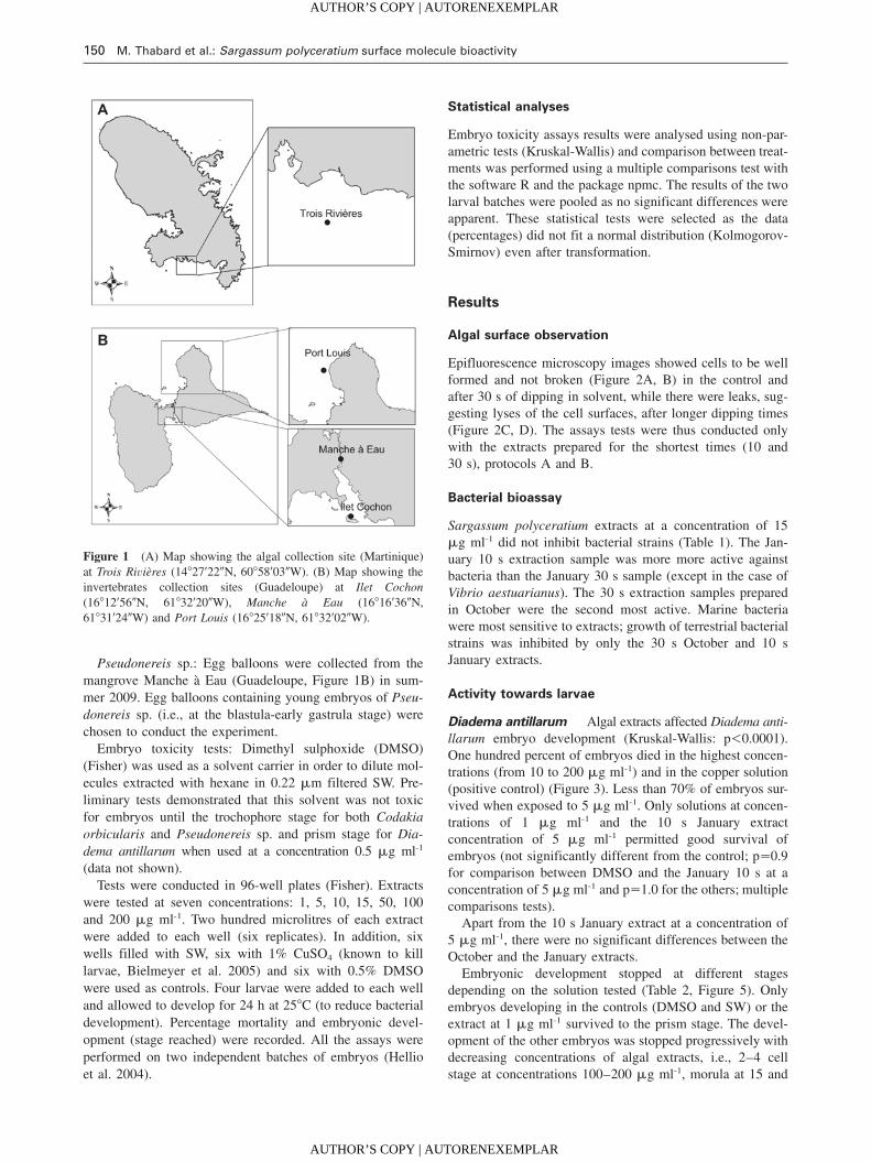
The site chosen for this study is a fringing coral reef tothe south of Martinique, close to the mouth of Trois Rivieresriver. This site was chosen for the dominant presence of Sar-gassum polyceratium.
Study organism
Sargassum polyceratium is commonly found in moderatelyturbulent habitats from the lower intertidal zone to depthsover 50 m (Littler and Littler 2000, Engelen 2004). It has atough crowded thallus and dense branches reaching to100 cm. The main axes roughened with small spines may benumerous. The blades measure 3–8 mm in width and1.5–2.0 cm in length. The holdfast is strong and disc like.The importance of recruitment vs. regeneration was modeledand demonstrated to vary with population, year and distur-bance (Engelen et al. 2005).
Algal extractions
Sargassum polyceratium samples were collected in October2008 and January 2009 (wet and dry seasons). Thalli wereremoved with their holdfasts by breaking away pieces ofsubstratum, thus reducing stress on the algae; stress may beresponsible for secondary metabolite production. The sam-ples were collected by SCUBA diving at 18 m depth. Thealgae were cleaned of epiphytes, rinsed with seawater (SW)and transported to the laboratory in a container filled withclean SW. The fresh samples were soaked in hexane (Fisher,Loughborough, UK) in the ratio 1 l hexane kg-1 wet weightS. polyceratium (De Nys et al. 1998). All the extractionswere performed in the dark as some secondary metabolites,such as the polyphenolics are known to react to light. Thegoal of using the hexane dipping method was to extract mol-ecules produced both by the alga at its surface and by itsassociated biofilm in order to obtain the whole range ofMNPs that might interact with eukaryotic and/or prokaryoticorganisms present in the algal environment.
Two protocols were used:
• Protocol A (October 2008 samples, rainy season), Sar-gassum polyceratium thalli were dipped for 30 s in hex-ane for preliminary tests.
• Protocol B (January 2009 samples, dry season) was devel-oped based on results of both tests conducted on 30 sextracts (October) and the observations of algal surfacesfor breaks (see results). This amended protocol was usedto test for the effect of dipping time on extract efficiency.
AUTHOR’S COPY | AUTORENEXEMPLAR
AUTHOR’S COPY | AUTORENEXEMPLAR
M. Thabard et al.: Sargassum polyceratium surface molecule bioactivity 149
Article in press - uncorrected proof
This was done by dipping algae in hexane for two dif-ferent periods (10 s and 30 s), with the algal surfaceremaining intact.
Observation of algal surface
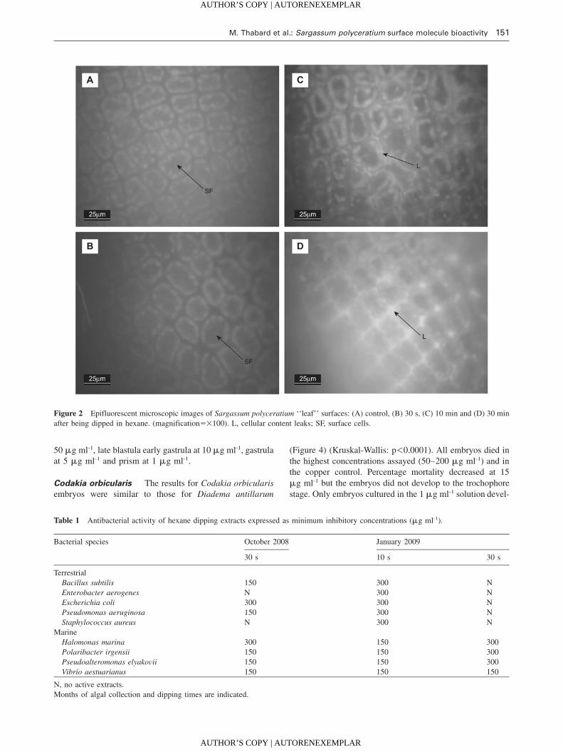
The objective of our experiments was to select the moleculespresent at the surface of Sargassum polyceratium only. Thehexane dipping method was shown to break some algal sur-face cells when thalli were dipped for more than 30 s (DeNys et al. 1998). The algal surface was therefore checkedfor breaks that would result in leaks of cell contents in thallidipped for 30 s, 10 min and 30 min to determine the bestdipping time. As UV excitation of plant leaves is known toinduce two distinct types of fluorescence, damage caused tothe surface cells of S. polyceratium by hexane dipping wasinvestigated by epifluorescent microscopy (Nikon, Tokyo,Japan, Eclipse 80i microscope; Filter FITC 494 nm excita-tion/514 nm emission; Nikon Dxm1200F camera) using aprotocol adapted from Cerocic et al. (1999).
Bioassays
Bacteria Culture of bacteria: Four marine bacterial strainswere used: Halomonas marina (Cobet et al.) Dobson etFranzmann (ATCC 25374), Pseudoalteromonas elyakovii(Ivanova et al.) Sawabe et al. (ATCC 700519), Polaribacterirgensii Gosink et al. (ATCC 700398) and Vibrio aestuaria-nus Tilson et Seidler (ATCC 35048). These bacteria werechosen because they are typical marine fouling bacteria(Plouguerne et al. 2010b). Pseudoalteromonas elyakovii andVibrio aestuarianus are also known to cause infections inmarine organisms, such as molluscs, crustacean, fishes oralgae, the latter being thought responsible for the summermortality of Crassostrea gigas Thunberg (Labreuche et al.2005). Marine bacteria were cultivated with marine broth(5% tryptone, Oxoid, Basingstoke, UK, diluted in SW) andincubated at 308C to allow development (Plouguerne et al.2008).
Five terrestrial bacterial strains known to be present inestuaries and coastal environment (Mokrini et al. 2008) wereused: Bacillus subtilis (Ehrenberg) Cohn (NCIMB 1026),Enterobacter aerogenes Hormaeche et Edwards (ATCC13048), Escherichia coli (Migula) Castellani et Chalmers (B81), Pseudomonas aeruginosa (Schroeter) Migula (NCIMB10390) and Staphylococcus aureus Rosenbach (NCIMB8625). B. subtilis and S. aureus are Gram-positive bacteria.The remainder are Gram-negative. Terrestrial bacteria werecultivated on a nutrient broth (CM0067, No. 2, Nutrientmedia Powder Oxoid 25 g l-1) and incubated at 308C. Bio-logical activities of extracts were evaluated following themethod of Amsterdam (1996).
Antibacterial assays: Aliquots of 100 ml of each hexaneextract were poured in six wells of 96 well plates (Fisher)for each bacterial assay following the protocols of Plou-guerne et al. (2010b). The solutions were tested at three con-centrations: 15, 150 and 300 mg ml-1. In addition, six wellsfree of extracts and six wells containing hexane were usedas controls. The plates were first dried in a flow cabinet to
evaporate the solvent and then left for 15 min in a UV cab-inet for sterilization.
The optical densities (OD) of bacterial stock cultures weremeasured at 630 nm for every sample to determine the quan-tity of solution required to obtain 1 mOD (mili optical den-sity). Then, 100 ml of bacterial solutions were added underaseptic conditions and the plates were incubated for 48 h at308C for bacterial growth. Activity was obtained comparingthe controls and the wells containing the extracts. Solutionswere considered to be active when bacteria did not grow infour, five or six wells. Bacterial growth was noted by thepresence of a cloudy solution. One plate was used for eachstrain to limit the cross-contamination risk.
Invertebrates Organisms: The toxicity of the extractswas tested against larvae of Codakia orbicularis L., Diademaantillarum and Pseudonereis sp. These organisms are tropicaland represent typical species from three marine ecosystems(seagrass bed, reef and mangrove). Their spawning and earlylarval development has been described previously (Gros etal. 1997, Eckert 1998). For both C. orbicularis and D. anti-llarum, spawns were induced in the laboratory under con-trolled conditions, while Pseudonereis sp. embryos werecollected from the wild.
Codakia orbicularis is a tropical bivalve mollusc distrib-uted from Florida to Brazil (Abbott 1974). Adult C. orbi-cularis (between 40 and 60 mm shell length) were collectedby hand from seagrass beds in Ilet Cochon (Guadeloupe,Figure 1B) in July 2009. Fertilization was induced followingthe method described by Gros et al. (1997). Adults werecleaned with a brush and spawning was induced by injectionof 0.3 ml of a 4 mM serotonin solution in 0.22 mm filteredSW into the visceral mass. Sperm and oocytes were mixedin a 1 l cylinder until the appearance of two-cell embryos.Fertilization occurred under constant aeration to hold eggsin suspension as they are slightly negatively buoyant. C.orbicularis embryonic development follows the generaldevelopment of bivalves (Gros et al. 1997). Appearance ofthe first polar body (indicating fertilization) is not alwaysvisible under a dissecting microscope, thus the two cellsembryos were chosen to ensure the fertilization had occurred,and these were used in toxicity experiments.
Diadema antillarum: The black spined sea urchin D. anti-llarum was selected for experiments. Adults were collectedon the shore at Port-Louis (Guadeloupe, Figure 1B) duringsummer 2009. Urchins were acclimated in the laboratory(258C) for a week and fed on agar pellets containing a mix-ture of algae (including Ulva lactuca L. and Sargassum sp.)following the protocol of Pereira et al. (2003). After a week,the urchins were transferred to another tank containing 298Cfiltered SW. Thermal shocks (3–58C) that induce spawningof D. antillarum (M. Moe, personal communication) wereapplied over a few minutes. Both male and female gameteswere pipetted from this tank and diluted in 10 l of 0.22 mmfiltered SW (258C) to induce the fertilization. The eggs areslightly negatively buoyant, thus aeration was used to keepthem suspended. Embryos at the two cells stage wfertilization(To)q1hx were chosen for experiments.
AUTHOR’S COPY | AUTORENEXEMPLAR
AUTHOR’S COPY | AUTORENEXEMPLAR
150 M. Thabard et al.: Sargassum polyceratium surface molecule bioactivity
Article in press - uncorrected proof
Figure 1 (A) Map showing the algal collection site (Martinique)at Trois Rivieres (148279220N, 608589030W). (B) Map showing theinvertebrates collection sites (Guadeloupe) at Ilet Cochon(168129560N, 618329200W), Manche a Eau (168169360N,618319240W) and Port Louis (168259180N, 618329020W).
Pseudonereis sp.: Egg balloons were collected from themangrove Manche a Eau (Guadeloupe, Figure 1B) in sum-mer 2009. Egg balloons containing young embryos of Pseu-donereis sp. (i.e., at the blastula-early gastrula stage) werechosen to conduct the experiment.
Embryo toxicity tests: Dimethyl sulphoxide (DMSO)(Fisher) was used as a solvent carrier in order to dilute mol-ecules extracted with hexane in 0.22 mm filtered SW. Pre-liminary tests demonstrated that this solvent was not toxicfor embryos until the trochophore stage for both Codakiaorbicularis and Pseudonereis sp. and prism stage for Dia-dema antillarum when used at a concentration 0.5 mg ml-1
(data not shown).Tests were conducted in 96-well plates (Fisher). Extracts
were tested at seven concentrations: 1, 5, 10, 15, 50, 100and 200 mg ml-1. Two hundred microlitres of each extractwere added to each well (six replicates). In addition, sixwells filled with SW, six with 1% CuSO4 (known to killlarvae, Bielmeyer et al. 2005) and six with 0.5% DMSOwere used as controls. Four larvae were added to each welland allowed to develop for 24 h at 258C (to reduce bacterialdevelopment). Percentage mortality and embryonic devel-opment (stage reached) were recorded. All the assays wereperformed on two independent batches of embryos (Hellioet al. 2004).
Statistical analyses
Embryo toxicity assays results were analysed using non-par-ametric tests (Kruskal-Wallis) and comparison between treat-ments was performed using a multiple comparisons test withthe software R and the package npmc. The results of the twolarval batches were pooled as no significant differences wereapparent. These statistical tests were selected as the data(percentages) did not fit a normal distribution (Kolmogorov-Smirnov) even after transformation.
Results
Algal surface observation
Epifluorescence microscopy images showed cells to be wellformed and not broken (Figure 2A, B) in the control andafter 30 s of dipping in solvent, while there were leaks, sug-gesting lyses of the cell surfaces, after longer dipping times(Figure 2C, D). The assays tests were thus conducted onlywith the extracts prepared for the shortest times (10 and30 s), protocols A and B.
Bacterial bioassay
Sargassum polyceratium extracts at a concentration of 15mg ml-1 did not inhibit bacterial strains (Table 1). The Jan-uary 10 s extraction sample was more more active againstbacteria than the January 30 s sample (except in the case ofVibrio aestuarianus). The 30 s extraction samples preparedin October were the second most active. Marine bacteriawere most sensitive to extracts; growth of terrestrial bacterialstrains was inhibited by only the 30 s October and 10 sJanuary extracts.
Activity towards larvae
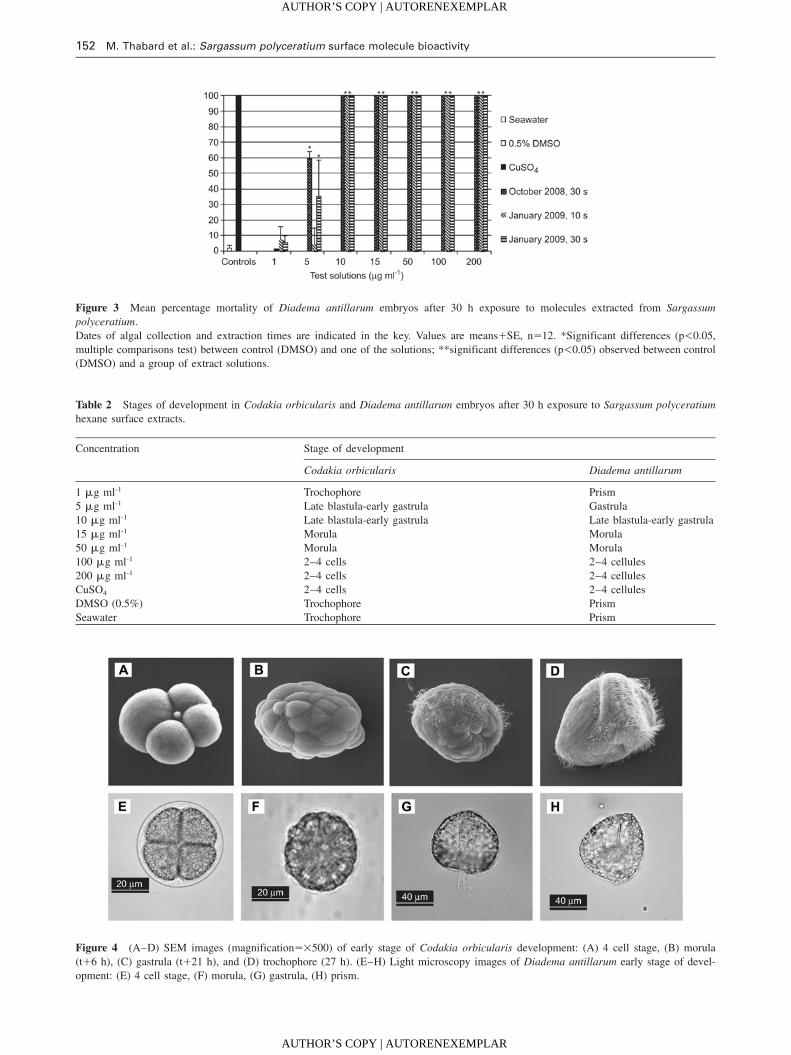
Diadema antillarum Algal extracts affected Diadema anti-llarum embryo development (Kruskal-Wallis: p-0.0001).One hundred percent of embryos died in the highest concen-trations (from 10 to 200 mg ml-1) and in the copper solution(positive control) (Figure 3). Less than 70% of embryos sur-vived when exposed to 5 mg ml-1. Only solutions at concen-trations of 1 mg ml-1 and the 10 s January extractconcentration of 5 mg ml-1 permitted good survival ofembryos (not significantly different from the control; ps0.9for comparison between DMSO and the January 10 s at aconcentration of 5 mg ml-1 and ps1.0 for the others; multiplecomparisons tests).
Apart from the 10 s January extract at a concentration of5 mg ml-1, there were no significant differences between theOctober and the January extracts.
Embryonic development stopped at different stagesdepending on the solution tested (Table 2, Figure 5). Onlyembryos developing in the controls (DMSO and SW) or theextract at 1 mg ml-1 survived to the prism stage. The devel-opment of the other embryos was stopped progressively withdecreasing concentrations of algal extracts, i.e., 2–4 cellstage at concentrations 100–200 mg ml-1, morula at 15 and
AUTHOR’S COPY | AUTORENEXEMPLAR
AUTHOR’S COPY | AUTORENEXEMPLAR
M. Thabard et al.: Sargassum polyceratium surface molecule bioactivity 151
Article in press - uncorrected proof
Figure 2 Epifluorescent microscopic images of Sargassum polyceratium ‘‘leaf’’ surfaces: (A) control, (B) 30 s, (C) 10 min and (D) 30 minafter being dipped in hexane. (magnifications=100). L, cellular content leaks; SF, surface cells.
Table 1 Antibacterial activity of hexane dipping extracts expressed as minimum inhibitory concentrations (mg ml-1).
Bacterial species October 2008 January 2009
30 s 10 s 30 s
TerrestrialBacillus subtilis 150 300 NEnterobacter aerogenes N 300 NEscherichia coli 300 300 NPseudomonas aeruginosa 150 300 NStaphylococcus aureus N 300 N
MarineHalomonas marina 300 150 300Polaribacter irgensii 150 150 300Pseudoalteromonas elyakovii 150 150 300Vibrio aestuarianus 150 150 150
N, no active extracts.Months of algal collection and dipping times are indicated.
50 mg ml-1, late blastula early gastrula at 10 mg ml-1, gastrulaat 5 mg ml-1 and prism at 1 mg ml-1.
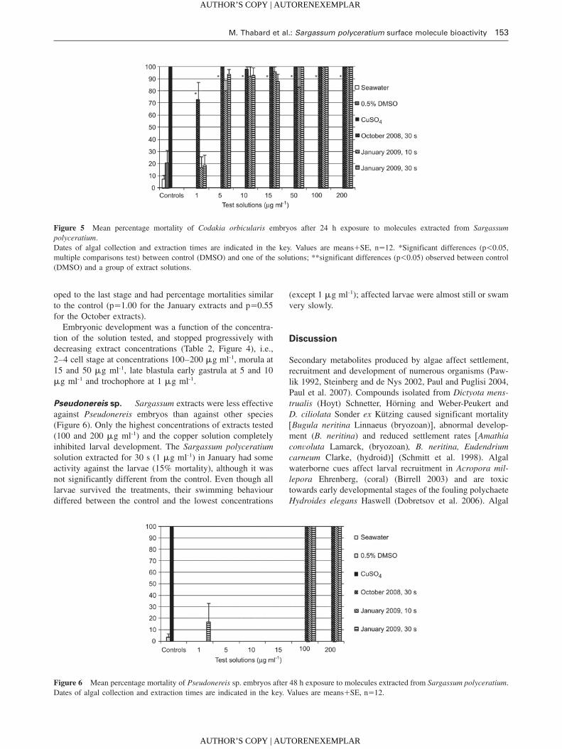
Codakia orbicularis The results for Codakia orbicularisembryos were similar to those for Diadema antillarum
(Figure 4) (Kruskal-Wallis: p-0.0001). All embryos died inthe highest concentrations assayed (50–200 mg ml-1) and inthe copper control. Percentage mortality decreased at 15mg ml-1 but the embryos did not develop to the trochophorestage. Only embryos cultured in the 1 mg ml-1 solution devel-
AUTHOR’S COPY | AUTORENEXEMPLAR
AUTHOR’S COPY | AUTORENEXEMPLAR
152 M. Thabard et al.: Sargassum polyceratium surface molecule bioactivity
Article in press - uncorrected proof
Figure 3 Mean percentage mortality of Diadema antillarum embryos after 30 h exposure to molecules extracted from Sargassumpolyceratium.Dates of algal collection and extraction times are indicated in the key. Values are meansqSE, ns12. *Significant differences (p-0.05,multiple comparisons test) between control (DMSO) and one of the solutions; **significant differences (p-0.05) observed between control(DMSO) and a group of extract solutions.
Table 2 Stages of development in Codakia orbicularis and Diadema antillarum embryos after 30 h exposure to Sargassum polyceratiumhexane surface extracts.
Concentration Stage of development
Codakia orbicularis Diadema antillarum
1 mg ml-1 Trochophore Prism5 mg ml-1 Late blastula-early gastrula Gastrula10 mg ml-1 Late blastula-early gastrula Late blastula-early gastrula15 mg ml-1 Morula Morula50 mg ml-1 Morula Morula100 mg ml-1 2–4 cells 2–4 cellules200 mg ml-1 2–4 cells 2–4 cellulesCuSO4 2–4 cells 2–4 cellulesDMSO (0.5%) Trochophore PrismSeawater Trochophore Prism
Figure 4 (A–D) SEM images (magnifications=500) of early stage of Codakia orbicularis development: (A) 4 cell stage, (B) morula(tq6 h), (C) gastrula (tq21 h), and (D) trochophore (27 h). (E–H) Light microscopy images of Diadema antillarum early stage of devel-opment: (E) 4 cell stage, (F) morula, (G) gastrula, (H) prism.
AUTHOR’S COPY | AUTORENEXEMPLAR
AUTHOR’S COPY | AUTORENEXEMPLAR
M. Thabard et al.: Sargassum polyceratium surface molecule bioactivity 153
Article in press - uncorrected proof
Figure 5 Mean percentage mortality of Codakia orbicularis embryos after 24 h exposure to molecules extracted from Sargassumpolyceratium.Dates of algal collection and extraction times are indicated in the key. Values are meansqSE, ns12. *Significant differences (p-0.05,multiple comparisons test) between control (DMSO) and one of the solutions; **significant differences (p-0.05) observed between control(DMSO) and a group of extract solutions.
Figure 6 Mean percentage mortality of Pseudonereis sp. embryos after 48 h exposure to molecules extracted from Sargassum polyceratium.Dates of algal collection and extraction times are indicated in the key. Values are meansqSE, ns12.
oped to the last stage and had percentage mortalities similarto the control (ps1.00 for the January extracts and ps0.55for the October extracts).
Embryonic development was a function of the concentra-tion of the solution tested, and stopped progressively withdecreasing extract concentrations (Table 2, Figure 4), i.e.,2–4 cell stage at concentrations 100–200 mg ml-1, morula at15 and 50 mg ml-1, late blastula early gastrula at 5 and 10mg ml-1 and trochophore at 1 mg ml-1.
Pseudonereis sp. Sargassum extracts were less effectiveagainst Pseudonereis embryos than against other species(Figure 6). Only the highest concentrations of extracts tested(100 and 200 mg ml-1) and the copper solution completelyinhibited larval development. The Sargassum polyceratiumsolution extracted for 30 s (1 mg ml-1) in January had someactivity against the larvae (15% mortality), although it wasnot significantly different from the control. Even though alllarvae survived the treatments, their swimming behaviourdiffered between the control and the lowest concentrations
(except 1 mg ml-1); affected larvae were almost still or swamvery slowly.
Discussion
Secondary metabolites produced by algae affect settlement,recruitment and development of numerous organisms (Paw-lik 1992, Steinberg and de Nys 2002, Paul and Puglisi 2004,Paul et al. 2007). Compounds isolated from Dictyota mens-trualis (Hoyt) Schnetter, Horning and Weber-Peukert andD. ciliolata Sonder ex Kutzing caused significant mortalitywBugula neritina Linnaeus (bryozoan)x, abnormal develop-ment (B. neritina) and reduced settlement rates wAmathiaconvoluta Lamarck, (bryozoan), B. neritina, Eudendriumcarneum Clarke, (hydroid)x (Schmitt et al. 1998). Algalwaterborne cues affect larval recruitment in Acropora mil-lepora Ehrenberg, (coral) (Birrell 2003) and are toxictowards early developmental stages of the fouling polychaeteHydroides elegans Haswell (Dobretsov et al. 2006). Algal
AUTHOR’S COPY | AUTORENEXEMPLAR
AUTHOR’S COPY | AUTORENEXEMPLAR
154 M. Thabard et al.: Sargassum polyceratium surface molecule bioactivity
Article in press - uncorrected proof
MNPs are often species-specific and can have an effect ondifferent larval development stages. Dictyota spp. and Lau-rencia sp. are toxic against larvae of H. elegans and B. neri-tina; Padina sp. and Halimeda sp. inhibit their larvalsettlement; Hypnea sp. and Ulva sp. stimulate larval settle-ment and Sargassum spp. have no effect (Walters et al.1996).
Our results demonstrate that Sargassum polyceratium sur-face molecules significantly affect embryonic developmentof three tropical invertebrates and bacterial growth. Theseorganisms are not all in direct contact with S. polyceratium(except Diadema antillarum), but they represent species fromthree invertebrate phyla and thus give some indication ofpotential toxic activity of Sargassum towards several typesof organisms. In the embryo toxicity assays, the activeextracts concentrations varied by species tested. Both Coda-kia orbicularis and D. antillarum had similar responsesalthough C. orbicularis was expected to be more resistantbecause of the large envelope surrounding its embryos (Groset al. 1997). Pseudonereis embryos were the most resistantto the extracts. In the control, C. orbicularis embryos devel-oped in 24 h to the trochophore stage and D. antillarumembryos developed in 30 h to the prism stage, consistentwith previously reported values (Gros et al. 1997, Eckert1998). However, S. polyceratium surface compound treat-ments had progressive effects (according to increasing con-centration) on embryonic development, suggesting theextracts block egg cleavage. The highest concentrationsblocked development at the 2–4 cells stage, the medium con-centrations allowed development until the morula stage andthe lowest concentrations until the blastula, gastrula andfinally trochophore or prism stages (respectively for C. orbi-cularis and D. antillarum). Similarly, Paul and Fenical(1986), showed that caulerpyne blocks the cleavage of devel-oping sea urchin eggs. The division of Paracentrotus lividusLamarck (urchin) embryos was inhibited by algal extracts(Martin and Uriz 1993). Dihydrorhipocephalin, aldehyde,udoteal, petiodal, dihydroudoteal, rhipocephalin, halimeda-trial, halimeda tetraacetate (4,9-diacetoxy-udoteal) isolatedfrom siphonous green algal species (Halimeda spp., Penicil-lus spp., Rhipocephalus phoenix Ellis et Solander, Udoteaspp.) are toxic against developing sea urchin eggs, sperm andlarvae (Paul and Fenical 1986), with ED100 levels (lowestconcentration leading to 100% inhibition of cell division)ranging from 0.2 to 16 mg ml-1. Concentrations of MNPsextracted from siphonous algae have higher activity againstboth Codakia and Diadema than the solutions tested in thisstudy (ED100 ranging between 50 and 100 mg ml-1). How-ever, the tests performed by Paul and Fenical (1986) wereperformed with isolated secondary metabolites and not crudeextracts. It is possible that the active compounds tested inthis study were present in the crude extract at very low con-centration (mg ml-1). On the other hand, some molecules actsynergistically such that a pool of molecules is required toinduce a biological effect (Hay 1996). Further analyticalchemistry tests involving molecule purification and analyti-cal chemistry analyses (GC-MS and NMR) will give furtherinsights into the chemical nature of the bioactive compoundsand their activity levels.
Molecules extracted at the algal surface with hexane arenon-polar. Sargassum species produce polyphenols (polarmetabolites), but some non-polar extracts, such as S. vulgarehexane extracts, are highly active towards the developmentof microalgae, suggesting production of active non-polar sec-ondary metabolites by this alga (Plouguerne et al. 2010b).So far, no methods allowing the extraction of polar mole-cules located at the surface of the algae have been developedand the activity observed here thus represents only a portionof the possible MNPs present at the surface of S.polyceratium.
There were no differences in toxic activity towards embry-os between the 30 s extracts in October and January. How-ever, antibacterial activity was different between these twoextracts, suggesting a possible seasonality in surface mole-cule activities. Temperate algal extracts have a seasonal var-iation in molecular composition, antimicrobial activity, andAF activity (Steinberg and Van Altena 1992, Hellio et al.2004, Marechal et al. 2004, Plouguerne 2006). The Octoberextracts were from the rainy season, while those from Jan-uary were collected in the dry season. Hellio et al. (2004)demonstrated that S. muticum secondary metabolite activityvaries with seasons and is higher during the summer monthswhen fouling pressure (including fouling by bacteria) is mostintense. It is thus possible that macroalgae develop specificmechanisms for protection in the rainy season when bacterialconcentrations are high in coastal environments (Futch et al.2010). In the present study, the levels at which extracts wereactive towards bacteria were high (150–300 mg ml-1) incomparison with other algal crude extracts. Crude extractsobtained from seaweeds collected in Brittany were activewhen concentrations ranged between 24 and 96 mg ml-1
(Hellio et al. 2001) and between 0.1 and 100 mg ml-1 (Plou-guerne et al. 2008). Marine bacteria were the most sensitivestrains, suggesting that defence strategies of S. polyceratiumare specific. Such targeted defence strategies have beendescribed for other algal species (Paul and Puglisi 2004).
Bioactivity of Sargassum polyceratium extracts wasmarked; however, we do not know whether the active mol-ecules were produced by Sargassum polyceratium or by itsassociated biofilm. Secondary metabolite isolation fromalgae can be confounded by associated microorganisms. Asthis study focused on surface molecules only, it was impos-sible to clean the macroalgal surface from microepiphytesusing existing methods, such as ethanol, without breakingthe surface cells (De Nys et al. 1998). The extract obtainedtherefore corresponds to Sargassum and/or its associated bio-film. Numerous bacteria living in SW produce active sec-ondary metabolites (Jensen and Fenical 1994). Moreover,there are host-specific associations between algae and bac-teria, and algae may control associated bacteria (Lachnit etal. 2009). The bacterial biofilm may in turn confer a protec-tion to the host alga through production of secondarymetabolites.
Sargassum polyceratium surface extracts inhibited bacte-rial growth and embryo development of three tropical marineinvertebrates one of which was Diadema antillarum, a trop-ical herbivorous keystone species that controls macroalgal
AUTHOR’S COPY | AUTORENEXEMPLAR
AUTHOR’S COPY | AUTORENEXEMPLAR
M. Thabard et al.: Sargassum polyceratium surface molecule bioactivity 155
Article in press - uncorrected proof
populations. Testing surface molecules was a first investi-gation step and further work will be carried out to relate thenatural compounds to their possible ecological role. Theseresults require more work focused on 1) concentrations pro-duced on the algal surface, 2) the source of production (algaor biofilm, or both) and 3) the release of compounds into thewater column, a process that might interact with embryonicdevelopment of organisms surrounding Sargassum. We arecurrently performing tests on molecules present in the watersin which algae are immersed (conditioned water) in order toassess the activity of these cues against tropical invertebratemarine larvae in the same environment.
Acknowledgements
This work was financially supported by the Ministere de l’Ecologie,de l’Energie, du Developpement Durable et de la Mer (IFRECORprogram), Ministere de l’Outremer, the European Community(FEDER), the Regional Council of Martinique and the School ofBiological Sciences of the University of Portsmouth (UK).
References
Abbott, R.T. 1974. American seashells. Van Nostrand ReinholdCompany (New York). pp. 663.
Amsterdam, D. 1996. Susceptibility testing of antimicrobials in liq-uid media. In: (V. Loman, ed) Antibiotics in Laboratory Medi-cine 4th edition. Williams and Wilkins, Baltimore, MD. pp52–111.
Ang, P.O. 1986. Analysis of the vegetation structure of a Sargassumcommunity in the Philippines. Mar. Ecol Prog Ser. 28: 9–19.
Battistini, R. 1978. Les recifs coralliens de la Martinique: compa-raison avec ceux au sud-ouest de l’Ocean Indien. Cah.O.R.S.T.O.M. ser. Oceanogr. 16: 157–177.
Bielmeyer, G.K., K.V. Brix, T.R. Capo and M. Grosell. 2005. Theeffects of metals on embryo-larval and adult life stages of thesea urchin, Diadema antillarum. Aquat. Toxicol. 74: 254–263.
Birrell, C.L. 2003. Influences of benthic algae on coral settlementand post-settlement survival: implications for the recovery ofdisturbed and degraded reefs. Masters thesis, James Cook Uni-versity, Townsville. pp. 132.
Cerocic, Z.G., G. Samson, F. Morales, N. Tremblay and I. Moya.1999. Ultraviolet-induced fluorescence for plant monitoringpresent state and prospects. Agronomie 19: 543–578.
De Nys, R. and P.D. Steinberg. 2002. Linking marine biology andbiotechnology. Curr. Op. Biotechnol. 13: 244–248.
De Nys, R., S.A. Dworjanyn and P.D. Steinberg. 1998. A new meth-od for determining surface concentrations of marine naturalproducts on seaweeds. Mar. Ecol. Prog. Ser. 162: 79–87.
De Ruyter van Stevenick, E.D. and A.M. Breeman. 1987. Popula-tion dynamics of a tropical intertidal and deep-water populationof Sargassum polyceratium (Phaeophyta). Aquat. Bot. 29:139–156.
Diaz-Pulido, G., S. Harii, L.J. McCook and O. Hoegh-Guldberg.2010. The impact of benthic algae on the settlement of a reef-building coral. Coral Reefs 29: 203–208.
Dobretsov, S., H.U. Dahms, T. Harder and P.Y. Qian. 2006. Alle-lochemical defense against epibiosis in the macroalga Caulerparacemosa var. turbinata. Mar. Ecol. Prog. Ser. 318: 165–175.
Dworjanyn, S.A., R. de Nys and P.D. Steinberg. 1999. Localisationand surface quantification of secondary metabolites in the redalga Delisea pulchra. Mar. Biol. 133: 727–736.
Eckert, G.L. 1998. Larval development, growth and morphology ofthe sea urchin Diadema antillarum. Bull. Mar. Sci. 63: 443–451.
Eckman, J.E. and D.O. Duggins. 1991. Life and death beneath mac-rophyte canopies: effects of understorey kelps on growth ratesand survival of marine, benthic suspension feeders. Oecologia87: 473–487.
Engelen, A.H. 2004. Flexibility without compromises. Populationbiology of the brown seaweed Sargassum polyceratium aroundthe island of Curacao. PhD thesis, University of Groningen. pp¸180.
Engelen, A.H., J.L. Olsen, A.M. Breeman and W.T. Stam. 2001.Genetic differentiation in Sargassum polyceratium (Fucales:Phaeophyceae) around the island of Curacao (Netherlands Antil-¸les). Mar. Biol. 139: 267–277.
Engelen, A.H., A.M. Breeman, J.L. Olsen, W.T. Stam and P. Aberg.2005. Life history flexibility allows Sargassum polyceratium topersist in different environments subjected to stochastic distur-bance events. Coral Reefs 24: 670–680.
Futch, J.C., D.W. Griffin and E.K. Lipp. 2010. Human enteric virus-es in groundwater indicate offshore transport of human sewageto coral reefs of the Upper Florida Keys. Environ. Microbiol.12: 964–974.
Gros, O., L. Frenkiel and M. Moueza. 1997. Embryonic, larval, andpost-larval development in the symbiotic clam Codakia orbi-cularis (Bivalvia: Lucinidae). Invert. Biol. 116: 86–101.
Hay, M.E. 1996. Marine chemical ecology: what’s known andwhat’s next? J. Exp. Mar. Biol. Ecol. 200: 103–134.
Hay, M.E. and W. Fenical. 1988. Marine plant-herbivore interac-tions: the ecology of chemical defense. Ann. Rev. Ecol. Syst. 19:111–145.
Hay, M. and W. Fenical. 1996. Chemical ecology and marine bio-diversity: insights and products from the sea. Oceanogr. 9:10–20.
Hellio, C., D. De La Broise, L. Dufosse, Y. Le Gal and N. Bour-gougnon. 2001. Inhibition of marine bacteria by extracts ofmacroalgae: potential use for environmentally friendly antifou-ling paints. Mar. Environ. Res. 52: 231–247.
Hellio, C., J.P. Berge, C. Beaupoil, Y. Le Gal and N. Bourgougnon.2002. Screening of marine algal extracts for anti-settlementactivities against microalgae and macroalgae. Biofouling 18:205–215.
Hellio, C., C. Simon-Colin, A.S. Clare and E. Deslandes. 2004.Isethionic acid and floridoside isolated from the red alga, Gra-teloupia turuturu, inhibit settlement of Balanus amphitritecyprid Larvae. Biofouling 20: 139–145.
Hellio, C., M. Tsoukatou, J.P. Marechal, N. Aldred, C. Beaupoil,A.S. Clare, C. Vagias and V. Roussis. 2005. Inhibitory effectsof Mediterranean sponge extracts and metabolites on larval set-tlement of the barnacle Balanus amphitrite. Mar. Biotechnol. 7:297–305.
Hellio, C., J.P. Marechal, B.A.P. Da Gama, R.C. Pereira and A.S.Clare. 2009. Natural marine products with antifouling activities.In: (C. Hellio and D.M.Y. Yebra, eds.) Advances in marine anti-fouling coatings and technologies. Woodshead Publishing, Cam-bridge, UK, pp. 572–622.
Hughes, T.P. 1994. Catastrophes, phase-shifts, and large-scale deg-radation of a Caribbean coral reef. Science 265: 1547–1551.
Jensen, P.R. and W. Fenical. 1994. Strategies for the discovery ofsecondary metabolites from marine bacteria: ecological perspec-tives. Ann. Rev. Microbiol. 48: 559–584.
AUTHOR’S COPY | AUTORENEXEMPLAR
AUTHOR’S COPY | AUTORENEXEMPLAR
156 M. Thabard et al.: Sargassum polyceratium surface molecule bioactivity
Article in press - uncorrected proof
Knowlton, N. 2001. Sea urchin recovery from mass mortality: newhope for Caribbean coral reefs? Proc. Natl. Acad. Sci. USA 98:4822–4824.
Labreuche, Y., P. Soudant, M. Gonzalves, C. Lambert and J.L. Nico-las. 2005. Effects of the extracellular products from the patho-genic Vibrio aestuarianus strain 01/32 on lethality and cellularimmune responses of the oyster Crassostrea gigas. Develop.Comp. Immunol. 30: 367–379.
Lachnit, T., M. Bluemel, J.F. Imhoff and M. Wahl. 2009. Specificepibactrial communities on macroalgae: phylogeny matters morethan habitat. Aquat. Biol. 5: 181–186.
Lapointe, B.E. 1997. Nutrient thresholds for bottom-up control ofmacroalgal blooms on coral reefs in Jamaica and southeast Flor-ida. Limnol. Oceanogr. 42: 1119–1131.
Lapointe, B.E. and K. Thacker. 2002. Community-based water qual-ity and coral reef monitoring in the Negril marine park, Jamaica:Land-based nutrient inputs and their ecological consequences.In: (J.W. Porter and K.G. Porter, eds.) The Everglades, FloridaBay and coral reefs of the Florida Keys an ecosystem source-book. CRC Press, Boca Raton, FL. pp. 939–963.
Lessios, H.A. 1995. Diadema antillarum 10 years after mass mor-tality: still rare, despite help from a competitor. Proc. Roy. Soc.B. 259: 331–337.
Lessios, H.A. 2005. Diadema antillarum populations in Panamatwenty years following mass mortality. Coral Reefs 24:125–127.
Lessios, H.A., P.W. Glynn and D.R. Robertson. 1983. Mass mor-talities of Coral reef organisms. Science 222: 715–715.
Lessios, H.A., D.R. Robertson and J.D. Cubit. 1984. Spread of Dia-dema mass mortality through the Caribbean. Science 226:335–337.
Littler, M.M. and D.S. Littler. 1980. The evolution of thallus formand survival strategies in benthic marine macroalgae: field andlaboratory tests of a functional form model. Am. Nat. 116:25–44.
Littler, D.S. and M.M. Littler. 2000. Caribbean reef plants, an iden-tification guide to the reef plants of the Caribbean, Bahamas,Florida and Gulf of Mexico. Offshore Graphics Inc. Washington,DC. pp. 542.
Littler, M.M., D.S. Littler and P.R. Taylor. 1983a. Evolutionary strat-egies in a tropical barrier reef system-functional-form-groups ofmarine macroalgae. J. Phycol. 19: 229–237.
Littler, M.M., P.R. Taylor and D.S. Littler. 1983b. Algal resistanceto herbivory on a Caribbean barrier reef. Coral Reefs 2:111–118.
Littler, M.M., D.S. Littler and B.E. Lapointe. 1993. Modification oftropical reef community structure due to cultural eutrophication:the southwest coast of Martinique. Proc. 7th Int. Coral ReefSymp. 1: 335–343.
Marechal, J.P. and C. Hellio. 2011. Antifouling activity against bar-nacle cypris larvae: Do target species matter (AmphibalanusAmphitrite and Semibalanus balanoides)? Int. Biodet. Biodeg.65: 92–101.
Marechal, J.P., G. Culiolib, C. Hellio, H. Thomas-Guyonc, M.E.Callow, A.S. Clare and A. Ortalo-Magne. 2004. Seasonal vari-ation in antifouling activity of crude extracts of the brown algaBifurcaria bifurcata (Cystoseiraceae) against cyprids of Balanusamphitrite and the marine bacteria Cobetia marina and Pseu-doalteromonas haloplanktis. J. Exp. Mar. Biol. Ecol. 313:47–62.
Martin, D. and M.J. Uriz. 1993. Chemical bioactivity of Mediter-ranean benthic organisms against embryos and larvae of marineinvertebrates. J. Exp. Mar. Biol. Ecol. 173: 11–27.
Mokrini, R., M. Ben Mesaoud, M. Daoudi, C. Hellio, J.P. Marechal,M. El Hattab, A. Ortalo-Magne, L. Piovetti and C. Culioli. 2008.Meroditerpenoids and derivatives from the brown alga Cysto-seira baccata and their antifouling Properties J. Nat. Prod. 71:1806–1811.
Paul, V.J. and W. Fenical. 1986. Chemical defense in tropical greenalgae, order Caulerpales. Mar. Ecol. Prog. Ser. 34: 157–169.
Paul, V.J. and M.P. Puglisi. 2004. Chemical mediation of interac-tions among marine organisms. Nat. Prod. Rep. 21: 189–209.
Paul, V.J., C.R. Wylie and B.R. Sanger. 1988. Effects of algal chem-ical defenses toward different coral-reef herbivorous fishes: apreliminary study. Proc. 6th Int. Coral Reef Symp. 3: 73–78.
Paul, V.J., K.E. Arthur, R. Ritson-Williams, C. Ross and K. Sharp.2007. Chemical defenses: from compounds to communities.Biol. Bull. 213: 226–251.
Pawlik, J.R. 1992. Chemical ecology of the settlement of benthicmarine invertebrates. Oceanogr. Mar. Biol. Annu. Rev. 30:273–335.
Pereira, R.C., B.A.P. Da Gama, V.L. Teixeira and Y. Yoneshigue-Valentin. 2003. Ecological roles of natural products of the Bra-zilian red seaweed Laurencia obtusa. Brazilian J. Biol. 63:665–672.
Plouguerne, E. 2006. Etude ecologique et chimique de deux alguesintroduites sur les cotes bretonnes, Grateloupia turuturu Yamadaet Sargassum muticum (Yendo) Fensholt: nouvelles ressourcesbiologiques de composes a activite antifouling. PhD thesis, Uni-versity of Brittany. pp. 251.
Plouguerne, E., K. Le Lann, S. Connan, G. Jechoux, E. Deslandesand V. Stiger-Pouvreau. 2006. Spatial and seasonal variation indensity, reproductive status, length and phenolic content of theinvasive brown macroalgae Sargassum muticum (Yendo) Fens-holt along the coast of Western Brittany (France). Aquat. Bot.85: 337–344.
Plouguerne, E., C. Hellio, E. Deslandes, B. Veron and V. Stiger-Pouvreau. 2008. Anti-microfouling activities in extracts of twoinvasive algae: Grateloupia turuturu and Sargassum muticum.Bot. Mar. 51: 202–208.
Plouguerne, E., C. Hellio, C. Cesconetto, M. Thabard, K. Mason,B. Veron, R.C. Pereira and B.A.P. da Gama. 2010a. Antifoulingactivity as a function of population variation in Sargassum vul-gare from the littoral of Rio de Janeiro (Brazil). J Appl. Phycol.22: 717–724.
Plouguerne, E., E. Ioannou, P. Georgantea, C. Vagias, V. Roussis,C. Hellio, E. Kraffe and V. Stiger-Pouvreau. 2010b. Anti-micro-fouling activity of lipidic metabolites from the invasive brownalga Sargassum muticum (Yendo) Fensholt. Mar. Biotechnol. 12:52–61.
Ragan, M.A. and K.W. Glombitza. 1986. Phlorotannins, brown algalpolyphenols. Prog. Phycol. Res. 4: 129–241.
Salgado, L.T., N.B. Viana, L.R. Andrade, R.N. Leal, B.A.P. daGama, M. Attias, R.C. Pereira and G.M. Amado Filho. 2008.Intra-cellular storage, transport and exocytosis of halogenatedcompounds in marine red alga Laurencia obtusa. J. Struct. Biol.162: 345–355.
Sastry, V.M.V.S. and G.R.K. Rao. 1994. Antibacterial substancesfrom marine algae: successive extraction using benzene, chlo-roform and methanol. Bot. Mar. 37: 357–360.
Schmitt, T.M., N. Lindquist and M.E. Hay. 1998. Seaweed second-ary metabolites as antifoulants: Effects of Dictyota spp. diter-penes on survivorship, settlement, and development of marineinvertebrate larvae. Chemoecology 8: 125–131.
Sieburth, J. and J.T. Conover. 1965. Sargassum tannin, an antibioticwhich retards fouling. Nature 208: 52–53.
AUTHOR’S COPY | AUTORENEXEMPLAR
AUTHOR’S COPY | AUTORENEXEMPLAR
M. Thabard et al.: Sargassum polyceratium surface molecule bioactivity 157
Article in press - uncorrected proof
Steinberg, P.D. and R. de Nys. 2002. Chemical mediation of colo-nization of seaweed surfaces. J. Phycol. 38: 621–629.
Steinberg, P.D. and I. Van Altena. 1992. Tolerance of marine inver-tebrate herbivores to brown algal phlorotannins in temperateAustralasia. Ecol. Monogr. 62: 189–222.
Tanaka, N. and A. Asakawa. 1988. Allelopathic effect of mucilagereleased from a brown alga Sargassum horneri on marine dia-toms. Nippon Suisan Gakkaishi. 54: 1711–1714.
Titlyanov, E.A., T.V. Titlyanova, I.M. Yakovleva, Y. Nakano and R.Bhagooli. 2005. Regeneration of artificial injuries on scleracti-nian corals and coral/algal competition of newly formed sub-strate. J. Exp. Mar. Biol. Ecol. 323: 27–42.
Walters, L.J., M.G. Hadfield and C.M. Smith. 1996. Waterbornechemical compounds in tropical macroalgae: positive and neg-ative cues for larval settlement. Mar. Biol. 126: 383–393.
Young, D., B. Howard and W. Fenical. 1980. Subcellular localiza-tion of brominated secondary metabolites in the red alga Lau-rencia synderae. J. Phycol. 16: 182–185.
Received 13 May, 2010; accepted 22 November, 2010; online first15 March, 2011
AUTHOR’S COPY | AUTORENEXEMPLAR
AUTHOR’S COPY | AUTORENEXEMPLAR