Embed Size (px)
Citation preview
8/11/2019 Santhi PDF 2013
http://slidepdf.com/reader/full/santhi-pdf-2013 1/9
Oncogenic microRNAs as biomarkers of oral tumorigenesis and minimal
residual disease
W.S. Santhi a, R. Prathibha a, Sona Charles a, K.G. Anurup a, G. Reshmi a, Surya Ramachandran a, V.T. Jissa a,Paul Sebastian b, M. Radhakrishna Pillai a,⇑
a Cancer Research Program, Rajiv Gandhi Centre for Biotechnology, Thiruvananthapuram 695 014, Kerala, Indiab Division of Surgical Oncology, Regional Cancer Centre, Thiruvananthapuram 695 011, Kerala, India
a r t i c l e i n f o
Article history:
Received 31 May 2012
Received in revised form 3 January 2013
Accepted 6 January 2013
Available online xxxx
Keywords:
MicroRNAs
Oral tumor
Minimal residual disease
Biomarker
Leukoplakia
s u m m a r y
Objectives: Classical diagnostic methods are not sensitive enough in detecting oral lesions that may pro-
gress to cancer and in assessing minimal residual disease (MRD) in oral surgical margins. Altered expres-
sion of microRNAs (miRNAs) contributes to human cancer, including oral cancer. Although there are
many studies on microRNAs in oral cancer, there is no reported study comparing the expression of
microRNAs during oral tumor progression and in oral surgical margins.
Materials and methods: This study analyzed the expression of 72 miRNAs that were reported (till June
2011) to be differentially expressed in oral cancer, during phases of oral cancer progression and in oral
surgical margins.
Results: Of the 72 miRNAs analyzed, four (hsa-miR-125a, hsa-miR-184, hsa-miR16 and hsa-miR-96)
showed a common pattern of expression in both sets of tissues. We further analyzed the downstream tar-
get genes of hsa-miR-16 BCL2 and CCND1. The in silico network analysis of these four microRNAs and
their target genes revealed presence of genes involved in tumor progression and transcription factors.
Conclusions: The findings suggest that the combinatorial regulation by these miRNAs and their target
transcription factors might play a substantial role in oral tumorigenesis. Here we report for the first time
that a decreased expression of hsa-miR-125a, hsa-miR-184 and hsa-miR-16 and an increased expression
of hsa-miR-96 could be useful in predicting oral tumorigenesis and importantly in the detection of MRD
and decision-making process for postoperative treatment modalities.
2013 Elsevier Ltd. All rights reserved.
Introduction
MicroRNAs (miRNAs) are a group of endogenous, non-coding,
18-24 nucleotide length, single-stranded RNAs that mediate gene
expression at the post-transcriptional level through mRNA degra-
dation or translational repression.1 It is estimated that the human
genome may have more than 1000 miRNAs. Although they account
for only a minor fraction of the expressed genome, miRNAs areessential regulators of diverse cellular processes, including prolif-
eration, differentiation, apoptosis, survival, motility, invasion and
morphogenesis. Most of the miRNA genes are in cancer-associated
genomic regions or in fragile sites.2 Many miRNAs are implicated
as proto-oncogenes or as tumor suppressors and are aberrantly ex-
pressed in various cancers,3 including Oral Squamous Cell Carci-
noma (OSCC). By modulating oncogenic and tumor suppressor
pathways miRNAs could contribute to tumorigenesis. Recent stud-
ies have identified aberrant miRNA expression profiles in OSCC tis-
sues and/or cell lines compared with matched normal controls.1
Studies by Lu et al.4 have shown that miRNA expression profiles
can be used in cancer diagnosis and also in human cancer
classification.
A high incidence of oral cancer is observed in the Indian subcon-tinent, which accounts for a third of the world burden.5 Oral cancer
is the commonest cancer in India and accounts for the most cancer-
related deaths among men in India. The oral cancer incidence in
Kerala is one among the highest in India. Malignancy of the oral
cavity is often preceded by premalignant lesions, the most com-
mon of which is leukoplakia. The molecular mechanisms leading
to and involved in the malignant transformation of normal oral tis-
sue and leukoplakia are still poorly understood. The annual per-
centage of malignant transformation of leukoplakia varies in
different parts of the world owing to the difference in the use of
tobacco and dietary habits. Despite advances made in the manage-
ment of oral cancer over the last few decades, the survival rate of
1368-8375/$ - see front matter 2013 Elsevier Ltd. All rights reserved.http://dx.doi.org/10.1016/j.oraloncology.2013.01.001
⇑ Corresponding author. Tel.: +91 471 2347973; fax: +91 471 2348096.
E-mail addresses: [email protected] (W.S. Santhi), [email protected]
(R. Prathibha), [email protected] (S. Charles), [email protected]
(K.G. Anurup), [email protected] (G. Reshmi), [email protected]
(S. Ramachandran), [email protected] (V.T. Jissa), [email protected]
(P. Sebastian), [email protected] (M. Radhakrishna Pillai).
Oral Oncology xxx (2013) xxx–xxx
Contents lists available at SciVerse ScienceDirect
Oral Oncology
j o u r n a l h o m e p a g e : w w w . e l s e v i e r . c o m / l o c a t e / o r a l o n c o l o g y
Please cite this article in press as: Santhi WS et al. Oncogenic microRNAs as biomarkers of oral tumorigenesis and minimal residual disease. Oral Oncol
(2013), http://dx.doi.org/10.1016/j.oraloncology.2013.01.001
8/11/2019 Santhi PDF 2013
http://slidepdf.com/reader/full/santhi-pdf-2013 2/9
patients remains unacceptably low. The most important factor in
surgical management of oral cancer is the completeness of surgical
removal of tumor. The success rate of many cancer treatments is
largely dependent on the often microscopically undetectable tu-
mor cells that remain in the body (Minimal Residual Disease or
MRD). Classical diagnostic modalities such as histopathology and
radiology are not always sensitive enough to detect these small
numbers of cells that remain in the body. The increased local–re-
gional recurrences and metastasis of oral cancer, even after treat-
ment points to the importance of molecular markers in the
detection of oral tumor progression and in the assessment of com-
pleteness of surgery. The genetic alterations harbored by tumor
cells can be used as molecular markers for the detection of oral le-
sions that has the capacity for tumor progression and also in the
assessment of MRD in oral surgical margins.
Development of malignancy results from the disruption of fine
tuned signaling homeostasis for proliferation, accompanied by
malfunctional signals for differentiation, cell cycle and apoptosis.6
miRNAs preferentially regulate genes that have a high transcrip-
tional regulation complexity7 and that preferentially target down-
stream genes in cellular signaling flows.8
Complexity of cancer progression mechanisms in higher organ-
isms is thought to also be achieved through controlled and coordi-
nated gene regulatory networks of miRNAs. A miRNA-gene
regulatory network provides a global perspective on the miRNAs
that are dysregulated in cancer as well as the pattern of dysregula-
tion, namely, upregulation or downregulation.9 Several miRNAs are
differentially expressed in various cancers, suggesting common al-
tered regulatory pathways. The effect of miRNAs on cell pathology
and physiology is complex because their activity is exerted in a
one-to-many fashion, and a single messenger can be controlled
by more than one miRNA.10 The cancer miRNA-gene network
throws up several new and interesting biological insights which
will become evident when studied in the global perspective.
Recent work has revealed intriguing changes in the global state
of miRNA expression in cancers including oral cancer. Despite
remarkable recent progress in understanding of functional mecha-nisms of miRNA in various human cancers, the mechanisms, tar-
gets and extent of miRNA-mediated gene expression in oral
cancer is unclear. Studies have compared expression of various
miRNAs in oral cancer and normal oral tissues.11–14 Few have
examined miRNA expression in oral precancerous lesions.15,16
However no previous study has reported expression of miRNAs
during oral tumor progression and in oral surgical margins. This
investigation looked into the expression profile of all reported miR-
NAs (till June 2011) deregulated in oral cancer in two sets of tis-
sues, one involved in oral tumorigenesis and the other in oral
surgical margins. Furthermore, we constructed a bilayered regula-
tory network formed between the miRNAs (that showed the simi-
lar pattern of expression in the two sets of samples) and their
target genes. To the best of our knowledge, this is the first studycomparing the expression profiles of miRNAs in oral tumorigenesis
and in the assessment of MRD in oral surgical margins besides
looking at a computational network analysis. Understanding the
functions of miRNAs provides new insights on the molecular basis
of oral cancer as well as new biomarkers for diagnosis, classifica-
tion, therapy and prognosis evaluation.
Materials and methods
Identification of miRNAs involved in oral cancer
OrCa-dB (www.rgcb.res.in/orcadb)17 is a specialized database
that contains all experimentally determined human oral cancer re-lated information and their interactomics along with clinical data
till June 2011. This database revealed 72 miRNAs that are differen-
tially expressed in human oral cancer.
Computational analysis
The targets of these miRNAs were retrieved from Tarbase data-
base (diana.cslab.ece.ntua.gr/tarbase/). We classified identified
genes by function with DAVID (Database for Annotation, Visualiza-
tion and Integrated Discovery; National Institute of Allergy and
Infections, National Institutes of Health) available at (http://davi-
d.abcc.ncifcrf.gov/) and sorted by Gene Ontology (GO) Consortium
terms. The subgroups assigned were enriched with a set of genes
participating in apoptosis and cancer progression. Most of the
genes in this list were found to be targeted by the four miRNAs
hsa-miR-125a, hsa-miR-184, hsa-miR16 and hsa-miR-96. The
molecular interactions of miRNA-gene regulatory network for the
clustered genes were constructed using Cytoscape Network Analy-
sis Tool. These four miRNAs were chosen for our further analysis.
Study subjects and tissue specimens
Tissue samples for this study were obtained at the Regional
Cancer Centre, Thiruvanthapuram. The Institutional Review Board
and Human Ethics Committee of Regional Cancer Centre, Thiru-
vanthapuram and Rajiv Gandhi Centre for Biotechnology, Thiru-
vanthapuram both approved the study. Informed consent was
obtained from all study subjects. Oral cancer patients, who re-
ceived any preoperative chemotherapy or radiotherapy, were ex-
cluded from the study.
Two groups of tissue specimens were analyzed. The first group
included tissues from various phases of oral cancer progression,
namely 31 normal oral mucosa, 49 leukoplakia and 84 oral cancer
tissues. The second group included tissues from 84 patients under-
going surgical resection as the primary treatment modality for oral
cancer. In this set, three types of samples were obtained from the
same patient, which included the primary tumor, surgical margin
(tissue taken from the actual surgical margin) and an extra margintissue (taken 2 cm away from the actual surgical margin). Normal
oral mucosa was obtained from persons undergoing oral and max-
illofacial reconstructive surgery. None of the leukoplakia patients
had received any treatment. Histologically the leukoplakias were
all dysplastic and had moderate to severe dysplasia. The cancer tis-
sues were all Squamous Cell Carcinomas. The normal tissues were
matched for age and smoking status. The surgical margin tissues
were histologically mild to moderate dysplastic. The extra margin
tissues were histologically normal oral tissues and were compara-
ble with the normal oral mucosa of the first group of tissues. The
tissue specimens for qRT-PCR were collected in RNAlater (Ambion,
USA) and stored at 80 C till use while the specimens for immu-
nohistochemical analysis were collected in 10% buffered formalin.
Total RNA isolation
Histopathological assessment was first done on the tissue spec-
imens to conform the pathological status of the tissues. Total RNA
was isolated from tissues using mirVana miRNA Isolation Kit (Ap-
plied Biosystems, USA) following manufactures protocol. In brief,
tissue was disrupted in a denaturing lysis buffer. The samples were
then subjected to Acid-Phenol:Chloroform extraction which pro-
vided a robust front-end purification that also removes most
DNA. For isolation of total RNA, samples were treated with ethanol,
and passed through a Filter Cartridge containing a glass-fiber filter
which immobilizes the RNA. The filter was then washed a few
times, and finally RNA was eluted with a low ionic-strength solu-
tion supplied with the isolation kit. The RNA concentrations weredetermined with a NanoDrop instrument (Thermo Scientific, USA).
2 W.S. Santhi et al. / Oral Oncology xxx (2013) xxx–xxx
Please cite this article in press as: Santhi WS et al. Oncogenic microRNAs as biomarkers of oral tumorigenesis and minimal residual disease. Oral Oncol
(2013), http://dx.doi.org/10.1016/j.oraloncology.2013.01.001
8/11/2019 Santhi PDF 2013
http://slidepdf.com/reader/full/santhi-pdf-2013 3/9
Reverse transcription and quantitative RT-PCR of miRNAs and mRNAs
Quantitative RT-PCR analyses for miRNAs and mRNAs were per-
formed using TaqMan miRNA and mRNA assays. All reagents and
primers were obtained from Applied Biosystems, USA. Reverse
transcriptase reactions and real-time PCR were performed accord-
ing to the manufacturer’s protocols. This technique, unlike the con-
ventional assay of random-primed RT, consists of using a target-
specific stem-loop structure and a reverse-transcriptase primer fol-
lowed by a specific real-time assay. cDNA was synthesized on a
15ll RT reaction mix containing 100 mM dNTP, MultiScribe Re-
verse Transcriptase (50 U/ll), RT buffer, RNase Inhibitor, RNA sam-
ple (10 ng/15 ll) and specific 5X TaqMan MicroRNA RT Primers for
miRNA analysis, while random primers were used for mRNA anal-
ysis. The RT product was diluted 15 times. The qRT-PCR reactions
were carried out in a 20 ll reaction mixture containing TaqMan
2X Universal Master Mix (No AmpErase UNG), 20X TaqMan Micr-
oRNA Assay or 20XTaqMan mRNA Assay and diluted cDNA. RNU
44 was used as endogenous control for miRNAs and GAPDH for
mRNAs. Real-Time PCR were performed in a 7500 Real Time PCR
System (Applied Biosystems, USA). Relative expression was calcu-
lated using the comparative threshold cycle (Ct) method.
Immunohistochemistry
Immunohistochemical reactions were carried out as described
by us earlier.18 Briefly, sections were deparaffinized in xylene
and rehydrated through graded alcohol to water. Endogenous per-
oxidase activity was blocked with 1.5% H2O2 in methanol for
15 min. Sections were then incubated with 3% bovine serum albu-
min (BSA) for 30 min followed by incubation at 4 C overnight with
primary antibodies specific for bcl-2 and cyclin d1 (Santa Cruz Bio-
technology, USA) at dilutions 1:50. The reactions were visualized
using Super Sensitive Polymer-HRP Detection System (BioGenex,
USA) following manufacturer’s protocol. The sections were coun-
terstained with Mayer’s hematoxylin, dehydrated in graded alco-hol, cleared in xylene and mounted.
Statistical analysis
Data analysis was done using Intercooled Stata 8.02 software
package. mRNA and miRNA expression along tissues involved in
oral surgical margins and along oral tumor progression were ana-
lyzed by spearman rank correlation coefficient test. Analysis show-
ing a confidence interval above 95% ( p < 0.05) was considered
significant.
Results
Our main objective for this study was to identify deregulated
microRNA expression associated with both oral cancer progression
and in oral surgical margins and further network analysis to eluci-
date the underlying miRNA-Target gene regulatory network in can-
cer progression and apoptosis. Two sets of tissues were used in this
study; the first set included tissues from various phases of tumor
progression (including normal oral mucosa, premalignant leuko-
plakia and oral cancer tissues) and the second set that included tu-
mor, surgical margin and an extra margin tissue taken from the
same patient for assessing MRD in oral surgical margins. For
qRT-PCR, in case of tissues involved in oral cancer progression, nor-
mal oral mucosa was taken as the calibrator for calculating the fold
change and in the evaluation of oral surgical margins, the extramargin tissues from that set were used as calibrator.
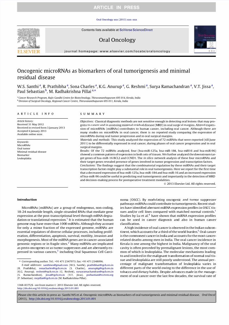
In silico network analysis
Computational functional clustering and target prediction tools
revealed the presence of 17 validated genes which were the targets
of hsa-miR-184, hsa-miR-96, hsa-miR-125a and hsa-miR-16 in-
volved in cancer progression and apoptosis. The regulatory net-
work formed between these four miRNAs and the target genes
was then constructed (Fig. 1). Out of these 17 target genes 3 were
transcription factors.
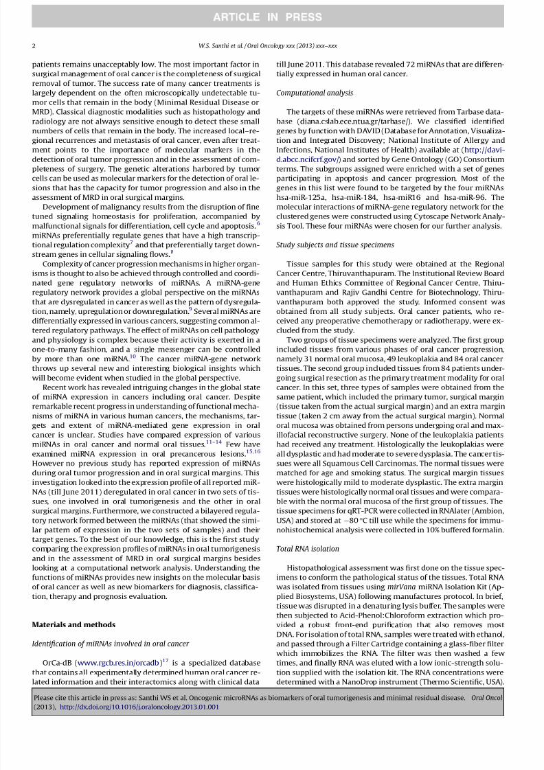
Expression of miRNAs during oral tumor progression and in oral
surgical margins
In this study we considered those miRNAs that were found to
have a differential expression both in oral tumor progression and
in oral surgical margins. Only four miRNAs (miR-125a, miR-184,
miR-16, and miR-96) fell into this criterion from both the groups
of tissues. In the first set we analyzed 16 normal oral mucosa, 28
leukoplakia and 56 oral cancer tissues. The expression of miR-
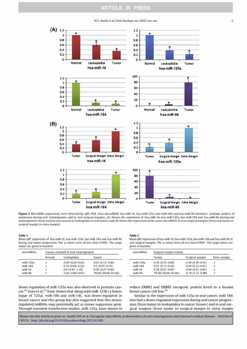
125a, miR-184 and miR-16 decreased where as miR-96 increased
with oral tumorigenesis (Fig. 2 and Table. 1). The second group in-
cluded 56 sets of tissues (each set including oral tumor, surgicalmargin and an extra margin tissue). The expression of these
microRNAs were found to be reversed in the second set of tissues.
miR-125a, miR-184 and miR-16 expression increased in oral surgi-
cal margins while miR-96 decreased (Fig. 2 and Table 2)
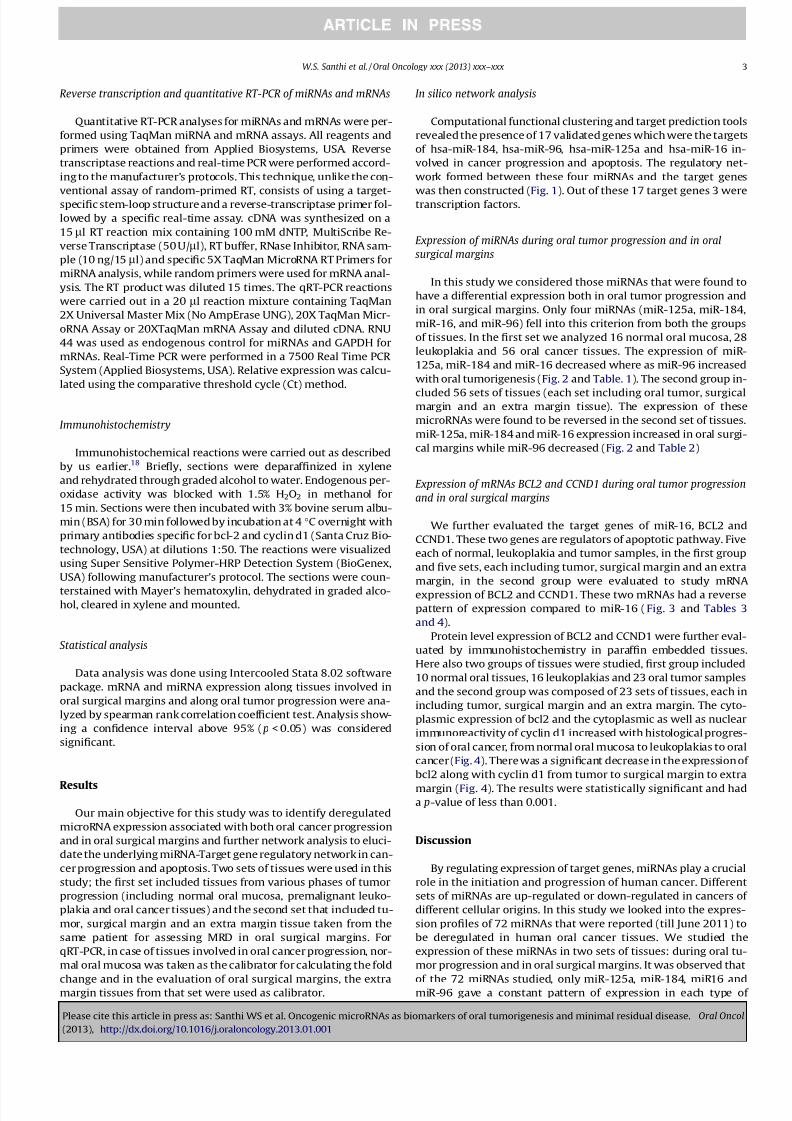
Expression of mRNAs BCL2 and CCND1 during oral tumor progression
and in oral surgical margins
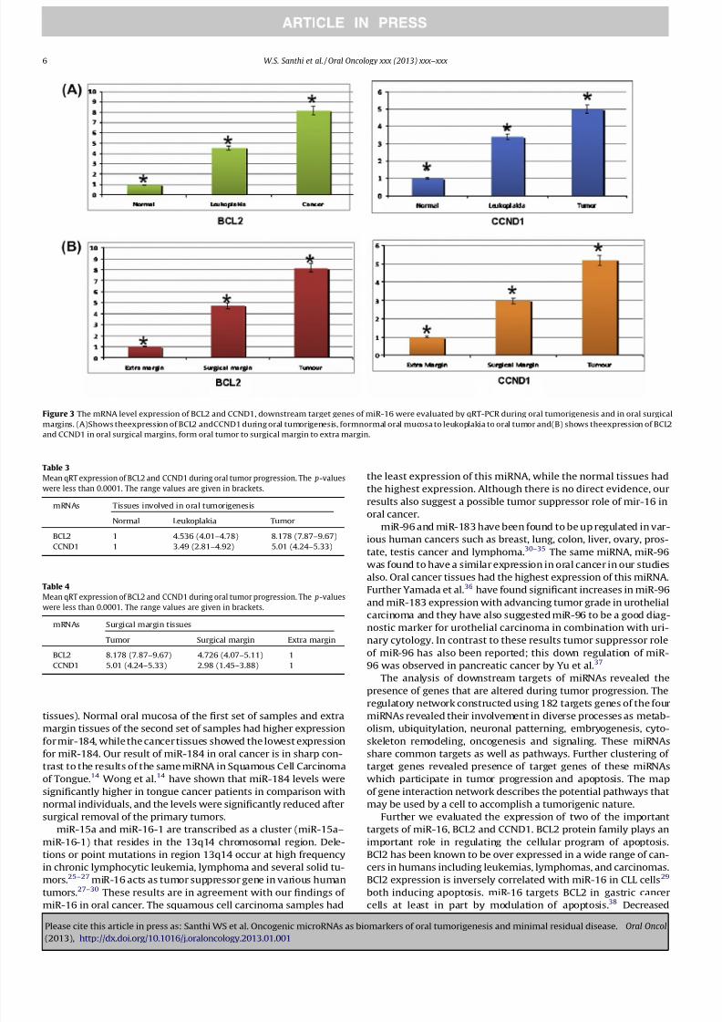
We further evaluated the target genes of miR-16, BCL2 and
CCND1. These two genes are regulators of apoptotic pathway. Five
each of normal, leukoplakia and tumor samples, in the first group
and five sets, each including tumor, surgical margin and an extra
margin, in the second group were evaluated to study mRNA
expression of BCL2 and CCND1. These two mRNAs had a reverse
pattern of expression compared to miR-16 (Fig. 3 and Tables 3
and 4).
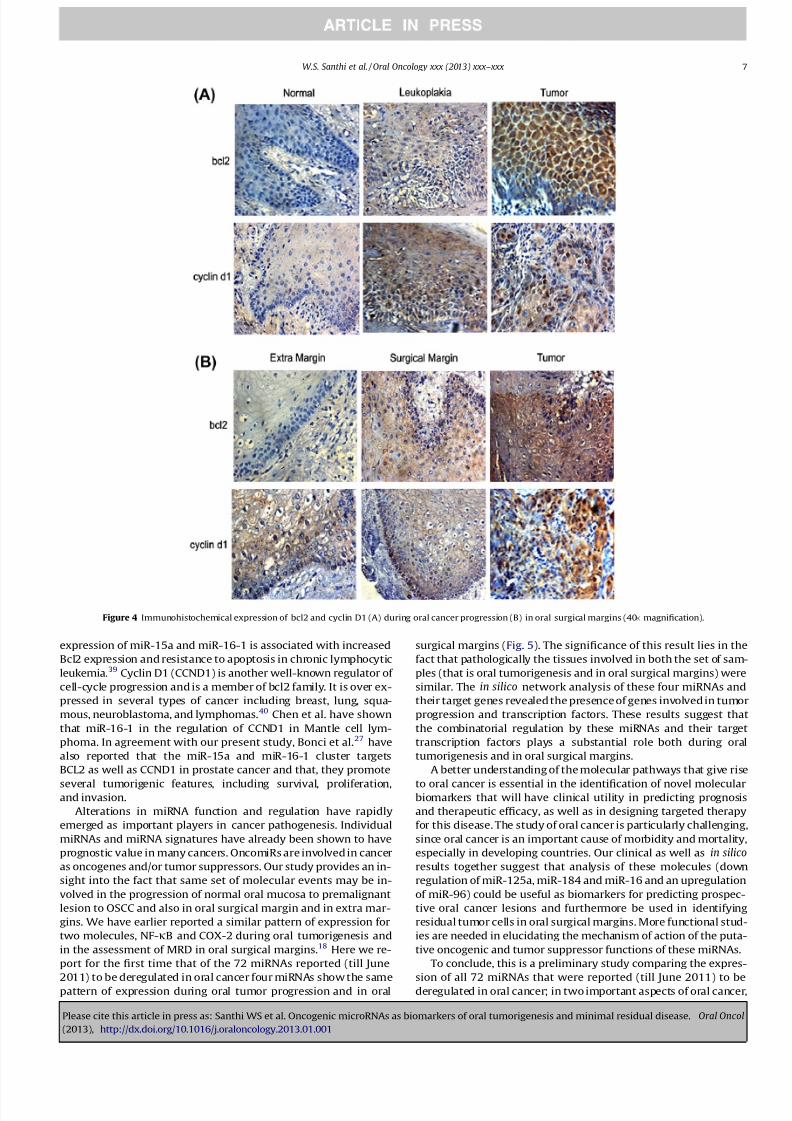
Protein level expression of BCL2 and CCND1 were further eval-
uated by immunohistochemistry in paraffin embedded tissues.
Here also two groups of tissues were studied, first group included
10 normal oral tissues, 16 leukoplakias and 23 oral tumor samples
and the second group was composed of 23 sets of tissues, each in
including tumor, surgical margin and an extra margin. The cyto-
plasmic expression of bcl2 and the cytoplasmic as well as nuclear
immunoreactivity of cyclin d1 increased with histological progres-
sion of oral cancer, from normal oral mucosa to leukoplakias to oral
cancer (Fig. 4). There was a significant decrease in the expression of
bcl2 along with cyclin d1 from tumor to surgical margin to extra
margin (Fig. 4). The results were statistically significant and hada p-value of less than 0.001.
Discussion
By regulating expression of target genes, miRNAs play a crucial
role in the initiation and progression of human cancer. Different
sets of miRNAs are up-regulated or down-regulated in cancers of
different cellular origins. In this study we looked into the expres-
sion profiles of 72 miRNAs that were reported (till June 2011) to
be deregulated in human oral cancer tissues. We studied the
expression of these miRNAs in two sets of tissues: during oral tu-
mor progression and in oral surgical margins. It was observed that
of the 72 miRNAs studied, only miR-125a, miR-184, miR16 andmiR-96 gave a constant pattern of expression in each type of
W.S. Santhi et al. / Oral Oncology xxx (2013) xxx–xxx 3
Please cite this article in press as: Santhi WS et al. Oncogenic microRNAs as biomarkers of oral tumorigenesis and minimal residual disease. Oral Oncol
(2013), http://dx.doi.org/10.1016/j.oraloncology.2013.01.001
8/11/2019 Santhi PDF 2013
http://slidepdf.com/reader/full/santhi-pdf-2013 4/9
tissues involved in oral tumor progression and in oral surgical
margins.
In this study the first set of samples evaluated were pathologi-cally squamous cell carcinomas, dysplasia and normal oral mucosa,
and the second set of samples was clinically tumor, surgical margin
and extra margin tissues. The oral cancer tissues of both sets were
pathologically squamous cell carcinomas, while the surgical mar-
gin tissues had varying degree of dysplasia similar to the leukopla-
kia tissues of the first set. The extra margin tissues of the second
set of samples were histologically normal oral tissues resembling
the normal oral mucosa in the first set. Like the normal oral muco-
sa tissues, the apparently normal looking extra margin tissues had
the highest expression of the miRNAs, miR-125a, miR-184 and
miR-16, while least expression was observed in OSCC tissues. On
the other hand miR-96 showed a reverse pattern of expression,
the highest expression was observed in oral cancer tissues mean
while the normal oral tissues and the extra margin tissues showedmaximum expression.
MiRNAs are differentially expressed in various cancer cells com-
pared with normal cells. In the first set of tissues (during oral tu-
mor progression), it was observed that miR-125a, miR-184 andmiR-16 decreased with the progression of oral tumor. In this sec-
ond set of samples (those involved in oral surgical margins),
miR-125a, miR-184, miR-16 had a statistically significant increase
in its expression form tumor to surgical margin to extra margin tis-
sues. Down regulation of miRNAs is more common than up-regu-
lation in cancers.19,20,4,21 The global down-regulation of miRNAs
in cancer cells has been suggested to reflect the lower differentia-
tion stage of the tumor cells compared with normal cells.4 In both
sets of tissues least expression of miRNAs miR-125a, miR-184,
miR-16 were observed in oral cancer tissues which suggested a tu-
mor suppressor role for these miRNAs.
Park et al.12 have found that two salivary miRNAs, miR-200a
and miR-125a, which were present at lower levels in oral squa-
mous cell carcinoma patient saliva than in healthy controls. Iden-tical to our results of miR-125a in oral squamous cell carcinoma,
Figure1 Gene regulatory network formedbetween thedifferentially expressedmicroRNAs andtheir targetgenes involved in tumorigenesis. Thetargets of miRNAswhich are
involved in the process of tumorigenesis were identified and a network was constructed. This network consists of three potential transcription factors that are involved in
cancer progression. The network was constructed using Cytoscape 2.8.2.
4 W.S. Santhi et al. / Oral Oncology xxx (2013) xxx–xxx
Please cite this article in press as: Santhi WS et al. Oncogenic microRNAs as biomarkers of oral tumorigenesis and minimal residual disease. Oral Oncol
(2013), http://dx.doi.org/10.1016/j.oraloncology.2013.01.001
8/11/2019 Santhi PDF 2013
http://slidepdf.com/reader/full/santhi-pdf-2013 5/9
down regulation of miR-125a was also observed in prostate can-
cer.22 Iorio et al.23 have shown that along with miR-125b (a homo-
logue of 125a), miR-10b and miR-145, was down-regulated in
breast cancer and this group has also suggested that this down-
regulated miRNAs may potentially act as tumor suppressor gene.Through transient transfection studies, miR-125a, have shown to
reduce ERBB2 and ERBB3 oncogenic protein levels in a human
breast cancer cell line.24
Similar to the expression of miR-125a in oral cancer, miR-184
also had a down-regulated expression during oral cancer progres-
sion (form tumor to leukoplakia to cancer tissues) and in oral sur-gical margins (from tumor to surgical margin to extra margin
Figure 2 MicroRNA expressions were detected by qRT–PCR. Four microRNAs hsa-miR-16, hsa-miR-125a, has-miR-184 and hsa-miR-96 showed a common pattern of
expression during oral tumorigenesis and in oral surgical margins. (A) Shows the expression of hsa-miR-16, hsa-miR-125a, has-miR-184 and hsa-miR-96 during oral
tumorigenesis (form normal oral mucosa to leukoplakia to oral tumor) and (B) shows the expression of the same microRNAs in oral surgical margins (form oral tumor to
surgical margin to extra margin).
Table 1
Mean qRT expression of hsa-miR-16, hsa-miR-125a, has-miR-184 and hsa-miR-96
during oral tumor progression. The p-values were all less than 0.0001. The range
values are given in brackets.
microRNAs Tissues involved in oral tumorigenesis
Normal Leukoplakia Tumor
miR-125a 1 0.39 (0.24–0.42) 0.23 (0.15–0.40)
miR-184 1 0.14 (0.06–0.22) 0.1 (0.07–0.19)
miR-16 1 0.6 (0.47–1.10) 0.38 (0.27–0.45)
miR-96 1 5.42 (3.89–6.67) 78.64 (56.04–81.66)
Table 2
Mean qRT expression of hsa-miR-16, hsa-miR-125a, has-miR-184 and hsa-miR-96 in
oral surgical margins. The p-values were all less than 0.0001. The range values are
given in brackets.
microRNAs Surgical margin tissues
Tumor Surgical margin Extra margin
miR-125a 0.23 (0.15–0.40) 0.39 (0.18–0.51) 1
miR-184 0.21 (0.11–0.34) 0.28 (0.12–0.41) 1
miR-16 0.38 (0.27–0.45) 0.58 (0.51–0.65) 1
miR-96 78.64 (56.04–81.66) 6.19 (5.11–6.89) 1
W.S. Santhi et al. / Oral Oncology xxx (2013) xxx–xxx 5
Please cite this article in press as: Santhi WS et al. Oncogenic microRNAs as biomarkers of oral tumorigenesis and minimal residual disease. Oral Oncol
(2013), http://dx.doi.org/10.1016/j.oraloncology.2013.01.001
8/11/2019 Santhi PDF 2013
http://slidepdf.com/reader/full/santhi-pdf-2013 6/9
tissues). Normal oral mucosa of the first set of samples and extramargin tissues of the second set of samples had higher expression
for mir-184, while the cancer tissues showed the lowest expression
for miR-184. Our result of miR-184 in oral cancer is in sharp con-
trast to the results of the same miRNA in Squamous Cell Carcinoma
of Tongue.14 Wong et al.14 have shown that miR-184 levels were
significantly higher in tongue cancer patients in comparison with
normal individuals, and the levels were significantly reduced after
surgical removal of the primary tumors.
miR-15a and miR-16-1 are transcribed as a cluster (miR-15a–
miR-16-1) that resides in the 13q14 chromosomal region. Dele-
tions or point mutations in region 13q14 occur at high frequency
in chronic lymphocytic leukemia, lymphoma and several solid tu-
mors.25–27 miR-16 acts as tumor suppressor gene in various human
tumors.27–30
These results are in agreement with our findings of miR-16 in oral cancer. The squamous cell carcinoma samples had
the least expression of this miRNA, while the normal tissues had
the highest expression. Although there is no direct evidence, our
results also suggest a possible tumor suppressor role of mir-16 in
oral cancer.
miR-96 and miR-183 have been found to be up regulated in var-
ious human cancers such as breast, lung, colon, liver, ovary, pros-tate, testis cancer and lymphoma.30–35 The same miRNA, miR-96
was found to have a similar expression in oral cancer in our studies
also. Oral cancer tissues had the highest expression of this miRNA.
Further Yamada et al.36 have found significant increases in miR-96
and miR-183 expression with advancing tumor grade in urothelial
carcinoma and they have also suggested miR-96 to be a good diag-
nostic marker for urothelial carcinoma in combination with uri-
nary cytology. In contrast to these results tumor suppressor role
of miR-96 has also been reported; this down regulation of miR-
96 was observed in pancreatic cancer by Yu et al.37
The analysis of downstream targets of miRNAs revealed the
presence of genes that are altered during tumor progression. The
regulatory network constructed using 182 targets genes of the four
miRNAs revealed their involvement in diverse processes as metab-olism, ubiquitylation, neuronal patterning, embryogenesis, cyto-
skeleton remodeling, oncogenesis and signaling. These miRNAs
share common targets as well as pathways. Further clustering of
target genes revealed presence of target genes of these miRNAs
which participate in tumor progression and apoptosis. The map
of gene interaction network describes the potential pathways that
may be used by a cell to accomplish a tumorigenic nature.
Further we evaluated the expression of two of the important
targets of miR-16, BCL2 and CCND1. BCL2 protein family plays an
important role in regulating the cellular program of apoptosis.
BCl2 has been known to be over expressed in a wide range of can-
cers in humans including leukemias, lymphomas, and carcinomas.
BCl2 expression is inversely correlated with miR-16 in CLL cells29
both inducing apoptosis. miR-16 targets BCL2 in gastric cancercells at least in part by modulation of apoptosis.38 Decreased
Figure 3 The mRNA level expression of BCL2 and CCND1, downstream target genes of miR-16 were evaluated by qRT-PCR during oral tumorigenesis and in oral surgical
margins. (A)Shows theexpression of BCL2 andCCND1 during oral tumorigenesis, formnormal oral mucosa to leukoplakia to oral tumor and(B) shows theexpression of BCL2
and CCND1 in oral surgical margins, form oral tumor to surgical margin to extra margin.
Table 3
Mean qRT expression of BCL2 and CCND1 during oral tumor progression. The p-values
were less than 0.0001. The range values are given in brackets.
mRNAs Tissues involved in oral tumorigenesis
Normal Leukoplakia Tumor
BCL2 1 4.536 (4.01–4.78) 8.178 (7.87–9.67)CCND1 1 3.49 (2.81–4.92) 5.01 (4.24–5.33)
Table 4
Mean qRT expression of BCL2 and CCND1 during oral tumor progression. The p-values
were less than 0.0001. The range values are given in brackets.
mRNAs Surgical margin tissues
Tumor Surgical margin Extra margin
BCL2 8.178 (7.87–9.67) 4.726 (4.07–5.11) 1
CCND1 5.01 (4.24–5.33) 2.98 (1.45–3.88) 1
6 W.S. Santhi et al. / Oral Oncology xxx (2013) xxx–xxx
Please cite this article in press as: Santhi WS et al. Oncogenic microRNAs as biomarkers of oral tumorigenesis and minimal residual disease. Oral Oncol
(2013), http://dx.doi.org/10.1016/j.oraloncology.2013.01.001
8/11/2019 Santhi PDF 2013
http://slidepdf.com/reader/full/santhi-pdf-2013 7/9
expression of miR-15a and miR-16-1 is associated with increased
Bcl2 expression and resistance to apoptosis in chronic lymphocytic
leukemia.39 Cyclin D1 (CCND1) is another well-known regulator of
cell-cycle progression and is a member of bcl2 family. It is over ex-
pressed in several types of cancer including breast, lung, squa-
mous, neuroblastoma, and lymphomas.40 Chen et al. have shown
that miR-16-1 in the regulation of CCND1 in Mantle cell lym-
phoma. In agreement with our present study, Bonci et al.27 have
also reported that the miR-15a and miR-16-1 cluster targets
BCL2 as well as CCND1 in prostate cancer and that, they promote
several tumorigenic features, including survival, proliferation,and invasion.
Alterations in miRNA function and regulation have rapidly
emerged as important players in cancer pathogenesis. Individual
miRNAs and miRNA signatures have already been shown to have
prognostic value in many cancers. OncomiRs are involved in cancer
as oncogenes and/or tumor suppressors. Our study provides an in-
sight into the fact that same set of molecular events may be in-
volved in the progression of normal oral mucosa to premalignant
lesion to OSCC and also in oral surgical margin and in extra mar-
gins. We have earlier reported a similar pattern of expression for
two molecules, NF-jB and COX-2 during oral tumorigenesis and
in the assessment of MRD in oral surgical margins.18 Here we re-
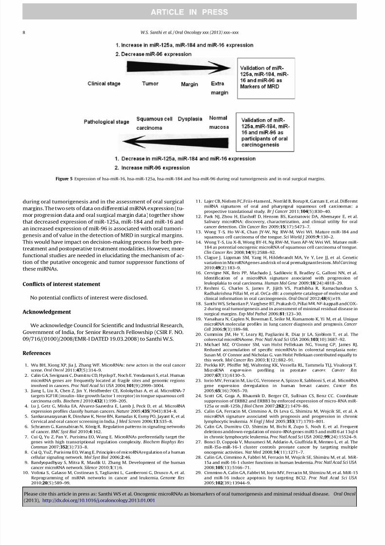
port for the first time that of the 72 miRNAs reported (till June
2011) to be deregulated in oral cancer four miRNAs show the samepattern of expression during oral tumor progression and in oral
surgical margins (Fig. 5). The significance of this result lies in the
fact that pathologically the tissues involved in both the set of sam-
ples (that is oral tumorigenesis and in oral surgical margins) were
similar. The in silico network analysis of these four miRNAs and
their target genes revealed the presence of genes involved in tumor
progression and transcription factors. These results suggest that
the combinatorial regulation by these miRNAs and their target
transcription factors plays a substantial role both during oral
tumorigenesis and in oral surgical margins.
A better understanding of the molecular pathways that give rise
to oral cancer is essential in the identification of novel molecularbiomarkers that will have clinical utility in predicting prognosis
and therapeutic efficacy, as well as in designing targeted therapy
for this disease. The study of oral cancer is particularly challenging,
since oral cancer is an important cause of morbidity and mortality,
especially in developing countries. Our clinical as well as in silico
results together suggest that analysis of these molecules (down
regulation of miR-125a, miR-184 and miR-16 and an upregulation
of miR-96) could be useful as biomarkers for predicting prospec-
tive oral cancer lesions and furthermore be used in identifying
residual tumor cells in oral surgical margins. More functional stud-
ies are needed in elucidating the mechanism of action of the puta-
tive oncogenic and tumor suppressor functions of these miRNAs.
To conclude, this is a preliminary study comparing the expres-
sion of all 72 miRNAs that were reported (till June 2011) to bederegulated in oral cancer; in two important aspects of oral cancer,
Figure 4 Immunohistochemical expression of bcl2 and cyclin D1 (A) during oral cancer progression (B) in oral surgical margins (40 magnification).
W.S. Santhi et al. / Oral Oncology xxx (2013) xxx–xxx 7
Please cite this article in press as: Santhi WS et al. Oncogenic microRNAs as biomarkers of oral tumorigenesis and minimal residual disease. Oral Oncol
(2013), http://dx.doi.org/10.1016/j.oraloncology.2013.01.001
8/11/2019 Santhi PDF 2013
http://slidepdf.com/reader/full/santhi-pdf-2013 8/9
during oral tumorigenesis and in the assessment of oral surgical
margins. The two sets of data on differential miRNA expression (tu-
mor progression data and oral surgical margin data) together show
that decreased expression of miR-125a, miR-184 and miR-16 and
an increased expression of miR-96 is associated with oral tumori-
genesis and of value in the detection of MRD in surgical margins.
This would have impact on decision-making process for both pre-
treatment and postoperative treatment modalities. However, more
functional studies are needed in elucidating the mechanism of ac-
tion of the putative oncogenic and tumor suppressor functions of
these miRNAs.
Conflicts of interest statement
No potential conflicts of interest were disclosed.
Acknowledgement
We acknowledge Council for Scientific and Industrial Research,
Government of India, for Senior Research Fellowship (CSIR F. NO.
09/716/(0100)/2008/EMR-I DATED 19.03.2008) to Santhi W.S.
References
1. Wu BH, Xiong XP, Jia J, Zhang WF. MicroRNAs: new actors in the oral cancer
scene. Oral Oncol 2011;47(5):314–9.
2. Calin GA, Sevignani C, Dumitru CD, HyslopT, Noch E, Yendamuri S, etal. Human
microRNA genes are frequently located at fragile sites and genomic regionsinvolved in cancers. Proc Natl Acad Sci USA 2004;101(9):2999–3004.
3. Jiang L, Liu X, Chen Z, Jin Y, Heidbreder CE, Kolokythas A, et al. MicroRNA-7
targets IGF1R (insulin–like growth factor 1 receptor) in tongue squamous cell
carcinoma cells. Biochem J 2010;432(1):199–205.
4. Lu J, Getz G, Miska EA, Alvarez-Saavedra E, Lamb J, Peck D, et al. MicroRNA
expression profiles classify human cancers. Nature 2005;435(7043):834–8.
5. Sankaranarayanan R, Dinshaw K, Nene BN, Ramadas K, Esmy PO, Jayant K, et al.
Cervical and oral cancer screening in India. J Med Screen 2006;13:S35–8.
6. Schramm G, Kannabiran N, König R. Regulation patterns in signaling networks
of cancer. BMC Syst Biol 2010;4:162.
7. Cui Q, Yu Z, Pan Y, Purisima EO, Wang E. MicroRNAs preferentially target the
genes with high transcriptional regulation complexity. Biochem Biophys ResCommun 2007;352(3):733–8.
8. Cui Q, YuZ, Purisima EO, Wang E. Principles of microRNAregulation of a human
cellular signaling network. Mol Syst Biol 2006;2:46.
9. Bandyopadhyay S, Mitra R, Maulik U, Zhang M. Development of the human
cancer microRNA network. Silence 2010;1(1):6.
10. Volinia S, Galasso M, Costinean S, Tagliavini L, Gamberoni G, Drusco A, et al.
Reprogramming of miRNA networks in cancer and leukemia. Genome Res2010;20(5):589–99.
11. Lajer CB, Nielsen FC,Friis-HansenL, Norrild B, Borup R, Garnæs E, et al. Different
miRNA signatures of oral and pharyngeal squamous cell carcinomas: a
prospective translational study. Br J Cancer 2011;104(5):830–40.
12. Park NJ, Zhou H, Elashoff D, Henson BS, Kastratovic DA, Abemayor E, et al.
Salivary microRNA: discovery, characterization, and clinical utility for oral
cancer detection. Clin Cancer Res 2009;15(17):5473–7.
13. Wong T-S, Ho W-K, Chan JY-W, Ng RW-M, Wei WI. Mature miR-184 and
squamous cell carcinoma of the tongue. Sci World J 2009;9:130–2.
14. Wong T-S, Liu X-B, Wong BY-H, Ng RW-M, Yuen AP-W, Wei WI. Mature miR-
184 as potential oncogenic microRNA of squamous cell carcinoma of tongue.
Clin Cancer Res 2008;14(9):2588–92.
15. Clague J, Lippman SM, Yang H, Hildebrandt MA, Ye Y, Lee JJ, et al. Genetic
variation in MicroRNAgenes andrisk of oral premalignantlesions. Mol Carcinog 2010;49(2):183–9.
16. Cervigne NK, Reis PP, Machado J, Sadikovic B, Bradley G, Galloni NN, et al.
Identification of a microRNA signature associated with progression of
leukoplakia to oral carcinoma. Human Mol Gene 2009;18(24):4818–29.
17. Reshmi G, Charles S, James P, Jijith VS, Prathibha R, Ramachandran S,
Radhakrishna Pillai M, et al. OrCa-dB: a complete catalogue of molecular and
clinical information in oral carcinogenesis. Oral Oncol 2012;48(6):e19.
18. Santhi WS, Sebastian P, Varghese BT, Prakash O, Pillai MR. NF-kappaB andCOX-
2 during oral tumorigenesis and in assessment of minimal residual disease in
surgical margins. Exp Mol Pathol 2006;81:123–30.
19. Yanaihara N, Caplen N, Bowman E, Seike M, Kumamoto K, Yi M, et al. Unique
microRNA molecular profiles in lung cancer diagnosis and prognosis. Cancer Cell 2006;9(3):189–98.
20. Cummins JM, He Y, Leary RJ, Pagliarini R, Diaz Jr LA, Sjoblom T, et al. The
colorectal microRNAome. Proc Natl Acad Sci USA 2006;103(10):3687–92.
21. Michael MZ, O’Connor SM, van Holst Pellekaan NG, Young GP, James RJ.
Reduced accumulation of specific microRNAs in colorectal neoplasia note:
Susan M. O’ Connor and Nicholas G. van Holst Pellekaan contributed equally to
this work. Mol Cancer Res 2003;1(12):882–91.
22. Porkka KP, Pfeiffer MJ, Waltering KK, Vessella RL, Tammela TLJ, Visakorpi T.
MicroRNA expression profiling in prostate cancer. Cancer Res2007;67(13):6130–5.
23. Iorio MV, Ferracin M, Liu CG, Veronese A, Spizzo R, Sabbioni S, et al. MicroRNAgene expression deregulation in human breast cancer. Cancer Res2005;65(16):7065–70.
24. Scott GK, Goga A, Bhaumik D, Berger CE, Sullivan CS, Benz CC. Coordinate
suppression of ERBB2 and ERBB3 by enforced expression of micro-RNA miR-
125a or miR-125b. J Biol Chem 2007;282(2):1479–86.
25. Calin GA, Ferracin M, Cimmino A, Di Leva G, Shimizu M, Wojcik SE, et al. A
microRNA signature associated with prognosis and progression in chronic
lymphocytic leukemia. N Engl J Med 2005;353(17):1793–801.
26. Calin GA, Dumitru CD, Shimizu M, Bichi R, Zupo S, Noch E, et al. Frequent
deletions anddown-regulation of micro-RNA genes miR15 and miR16 at 13q14
in chronic lymphocytic leukemia. Proc Natl Acad Sci USA 2002;99(24):15524–9.
27. Bonci D, Coppola V, Musumeci M, Addario A, Giuffrida R, Memeo L, et al. The
miR-15a-miR-16-1 cluster controls prostate cancer by targeting multiple
oncogenic activities. Nat Med 2008;14(11):1271–7.
28. Calin GA, Cimmino A, Fabbri M, Ferracin M, Wojcik SE, Shimizu M, et al. MiR-
15a and miR-16-1 cluster functions in human leukemia. Proc Natl Acad Sci USA2008;105(13):5166–71.
29. Cimmino A, Calin GA, Fabbri M, Iorio MV, Ferracin M, Shimizu M, et al. MiR-15
and miR-16 induce apoptosis by targeting BCL2. Proc Natl Acad Sci USA2005;102(39):13944–9.
Figure 5 Expression of hsa-miR-16, hsa-miR-125a, hsa-miR-184 and hsa-miR-96 during oral tumorigenesis and in oral surgical margins.
8 W.S. Santhi et al. / Oral Oncology xxx (2013) xxx–xxx
Please cite this article in press as: Santhi WS et al. Oncogenic microRNAs as biomarkers of oral tumorigenesis and minimal residual disease. Oral Oncol
(2013), http://dx.doi.org/10.1016/j.oraloncology.2013.01.001

8/11/2019 Santhi PDF 2013
http://slidepdf.com/reader/full/santhi-pdf-2013 9/9
30. Guttilla IK, White BA. Coordinate regulation of FOXO1by miR-27a, miR-96, and
miR-182 in breast cancer cells. J Biol Chem 2009;284(35):23204–16.
31. Lehmann U, Streichert T, Otto B, Albat C, Hasemeier B, Christgen H, et al.
Identification of differentially expressed microRNAs in human male breast
cancer. BMC Cancer 2010;10(1):109.
32. Wang G, Mao W, Zheng S. MicroRNA-183 regulates Ezrin expression in lung
cancer cells. FEBS Lett 2008;582(25–26):3663–8.
33. Sarver A, French A, Borralho P, Thayanithy V, Oberg AL, Silverstein KA, et al.
Human colon cancer profiles show differential microRNA expression depending
on mismatch repair status and are characteristic of undifferentiated
proliferative states. BMC Cancer 2009;9(1):401.34. Li J, Fu H, Xu C, Tie Y, Xing R, Zhu J, et al. MiR-183 inhibits TGF-beta1-induced
apoptosis by downregulation of PDCD4 expression in human hepatocellular
carcinoma cells. BMC Cancer 2010;10(1):354.
35. Myatt SS, Wang J, Monteiro LJ, Christian M, Ho KK, Fusi L, et al. Definition of
microRNAs that repress expression of the tumor suppressor gene FOXO1 in
endometrial cancer. Cancer Res 2010;70(1):367–77.
36. Yamada Y, Enokida H, Kojima S, Kawakami K, Chiyomaru T, Tatarano S, et al.
MiR-96 and miR-183 detection in urine serve as potential tumor markers of
urothelial carcinoma: correlation with stage and grade, and comparison with
urinary cytology. Cancer Sci 2011;102(3):522–9.
37. Yu S, Lu Z, Liu C, Meng Y, Ma Y, Zhao W, et al. MiRNA-96 suppresses KRAS and
functions as a tumor suppressor gene in pancreatic cancer. Cancer Res2010;70(14):6015–25.
38. Xia L, Zhang D, Du R, Pan Y, Zhao L, et al. MiR-15b and miR-16 modulate
multidrug resistance by targeting BCL2 in human gastric cancer cells. Int J Cancer 2008;123:372–9.
39. Aqeilan RI, Calin GA, Croce CM. MiR-15a and miR-16-1 in cancer: discovery,function and future perspectives. Cell Death Differ 2010;17:215–20.
40. Chen RW, Bemis LT, Amato CM, Myint H, Tran H, Birks DK, et al. Truncation in
CCND1 mRNA alters miR-16-1 regulation in mantle cell lymphoma. Blood2008;112:822–9.
W.S. Santhi et al. / Oral Oncology xxx (2013) xxx–xxx 9
Please cite this article in press as: Santhi WS et al. Oncogenic microRNAs as biomarkers of oral tumorigenesis and minimal residual disease. Oral Oncol


![[XLS] Pradesh.xlsx · Web viewCHAPARALA CHANDU CHAPARALA VIJAYA LAKSHMI 11-120 SANTHI NILAYAM, CHAPARALA VARI STREET AYAPPA NAGAR EVANGELIN LILIAN YEEDA 9440794*** ****usrao@gmail.com](https://img.pdfslide.us/doc/110x75/5acff9ff7f8b9a4e7a8d573d/xls-pradeshxlsxweb-viewchaparala-chandu-chaparala-vijaya-lakshmi-11-120-santhi.jpg)