Embed Size (px)
Citation preview
www.wjpps.com Vol 6, Issue 10, 2017.
1202
Sathiya et al. World Journal of Pharmacy and Pharmaceutical Sciences
SAFETY EVALUATION OF SIDDHA FORMULATION AKIL KATTAI
CHOORANAM BY ACUTE AND SUB-ACUTE TOXICITY STUDIES IN
WISTAR RATS
X. Helen Sathiya*1, N. Anbu
2, P. Parthibhan
3 and K. Kanakavalli
4
1P.G. Scholar, Post Graduate Department of Maruthuvam, Government Siddha Medical
College, Arumbakkam, Chennai 600 106, Tamil Nadu, India.
2Professor, HOD, Post Graduate Dept. of Maruthuvam, Government Siddha Medical College,
Arumbakkam, Chennai 600 106, Tamil Nadu, India.
3Joint Director, Indian Medicine and Homeopathy, Chennai 600 106, Tamil Nadu, India.
4Principal, Government Siddha Medical College, Arumbakkam, Chennai 600 106, Tamil
Nadu, India.
ABSTRACT
According to the recent regulatory guidelines preclinical toxicity
evaluation of the siddha formulations is mandatory to ascertain the
possibility of adverse event in humans upon short and long term usage
of the drugs. The main aim of the present research work is to evaluate
the safety of the traditional siddha formulation Akil Kattai Chooranam
(AKC) and to establish the toxicity profiling by acute and sub-acute
toxicity studies in accordance with OECD guidelines. In short term
acute toxicity study the drug AKC was administered in single doss of
2000 / kg b.w (p.o). Potential drug toxicity related to C.N.S, A.N.S and
C.V.S were observed up to 14 days. In sub-acute toxicity study the
drug AKC was administered at two dose level such as low and dose of
200 and 400 mg / kg b.w (p.o) for four weeks, Results obtained from
the acute and sub-acute study reveals that the drug AKC doesn’t reveal any significant
change in body weight, behavior and mortality in treated rats. Throughout the study period no
sign of toxicity was registered. Further it was observed that AKC at both the dose level did
not modify the weight index, food and water intake in treated animals. There is no significant
change in hematological, biochemical and histopathological observation of animals treated
with AKC at both the dose level of 200 and 400 mg / kg b.w (p.o) when compare to control
*Corresponding Author
Dr. X. Helen Sathiya
P.G.Scholar, Post Graduate
Department of Maruthuvam,
Government Siddha Medical
College, Arumbakkam,
Chennai 600 106, Tamil
Nadu, India.
Article Received on
15 August 2017,
Revised on 04 Sept. 2017,
Accepted on 25 Sept. 2017
DOI: 10.20959/wjpps201710-10295
WORLD JOURNAL OF PHARMACY AND PHARMACEUTICAL SCIENCES
SJIF Impact Factor 6.647
Volume 6, Issue 10, 1202-1225 Research Article ISSN 2278 – 4357
www.wjpps.com Vol 6, Issue 10, 2017.
1203
Sathiya et al. World Journal of Pharmacy and Pharmaceutical Sciences
group animals. From the result it was concluded that the siddha preparation AKC offers wide
margin of safety in tested rodents and further long term usage of drugs will be considered as
safe for the ailment of various disease.
KEYWORD: Siddha, Akil Kattai Chooranam, OECD, Toxicity profiling, Acute study, Sub-
acute.
1. INTRODUCTION
Toxicity profiling of siddha preparations are become highly essential in order to prove the
safety of the formulation upon short and long term administration in humans. Results of
toxicity study render some useful information to the investigators with respect to the effect of
the drug on CNS, CVS, ANS and other metabolic organs. It provides a base for fixation of
dose to the pharmacological study. Toxicity study further reveals the information about LD50
and also the dose which causes lethal effect and also safe therapeutic dose.
The OECD Guidelines for the testing of chemicals are a collection of the most relevant
internationally agreed testing methods used by government, industry, and independent
laboratories to characterize potential hazards of new and existing chemical substances and
chemical preparations, mixtures. They cover tests for the physico-chemical properties of
chemicals, human health effects, environmental effects, and degradation and accumulation in
the environment.[1]
Animals have been used as models for centuries to predict what chemicals and environmental
factors would do to humans.[2]
Siddha preparation which is not purified properly and also
preparations with contaminants like pesticides, organic chemicals, and heavy metals exert
their toxic effect on the hematopoietic system and may render anemia, neutropenia, leukemia,
thrombocytopenia, mutagenesis, apoptosis, myelotoxicity, anemia, immunomodulation etc.
Exposure to benzene and its metabolites leads to myelodysplastic syndromes, leukemia,
lymphomas and bone marrow aplasia. Mercury and chrome affect the immune system by
immunosuppression and by evoking autoimmune reactions. Dithiocarbamates are suspected
to induce leukemia. An analysis of the pathophysiology of individual substances reveal
universal toxic mechanisms.[3]
Liver a major organ for drug metabolism predominantly exposed to the adverse effect of the
toxic chemicals. Wide of range of toxicants disturbs the liver function includes insecticides,
www.wjpps.com Vol 6, Issue 10, 2017.
1204
Sathiya et al. World Journal of Pharmacy and Pharmaceutical Sciences
metabolites, heavy metals, pesticides etc. Hepatotoxicity nature of the drugs characterized by
changes in liver function test ascertained by estimation of SGOPT and SGPT level. Liver of
limited capacity of self-healing/ regeneration but chronic administration of certain chemicals
cause abnormal liver function which in turn hinders the physiology of metabolism. Toxic
nature of compounds causes’ pathological damage in the liver was manifested by sinusoidal
congestion (SC), RBC deposition in the vein and inflammatory response. Although drug
biotransformation generally parallels a detoxication process, the bioactivation is frequently
the major cause of hepatocyte injury.[4]
With regard to the bioactivation of drugs, attention
has been mostly paid to Phase I metabolites of drugs.
Renal toxicity of the drug will be evaluated by serum creatinine and blood urea nitrogen
level. According to the literature there is a remarkable nephrotoxicity caused by specific
medicinal herbs.[5]
Adverse unadulterated herb reactions resulting in notable kidney
manifestations are usually caused by an abuse of the substance or ignorance regarding the
herb's intended administration. The most common example involves ephedra (Ephedra sinica
spp.), which contains ephedrine.
The main aim of the present investigation is to evaluate the safety of the siddha formulation
Akil Kattai Chooranam in rodents at fixed dose level by acute and sub-acute toxicity studies
in accordance with OECD guidelines.
2. MATERIALS AND METHODS
The formulation AKC comprises of the following herbs as its ingredients.
2.1. Ingredients
1. Chandanam (Santalum album) -17.5g.
2. Agil kattai (Aquilaria agallocha Roxb) -17.5g.
3. Elam (Elettaria cardamomm) -17.5g.
4. Lavangappattai (Cinnamomum verum) -17.5g.
5. Kirambu (Syzygium aromaticum) -17.5g.
6. Sombu (Pimpinella anisum) -17.5g.
7. Ati-maduram (Glycyrrhiza glabra) -17.5g.
8. Karboki vitthu (Psoralea corylifolia) -17.5g.
9. Vetpalai arisi (Wrightia –tinctoria) -17.5g.
10. Thettran vithai (Strychnos potatorum) -17.5g.
www.wjpps.com Vol 6, Issue 10, 2017.
1205
Sathiya et al. World Journal of Pharmacy and Pharmaceutical Sciences
11. Arugam vear (Cynodon dactylon) - 17.5g.
12. Chitramutti (Pavonia Zeylanica) -17.5g.
13. Karkandu – Quantity Sufficient
2.2. Source of raw drugs
The required raw drugs are procured from a well reputed indigenous drug shop. The raw
drugs taken for study will be authenticated by the Botanist of Medicinal botany department,
Govt. Siddha Medical College, Chennai.
2.3. Preparation[6]
Take each ingredient about 17.5 gm. and made it to dry in the sun light, after that it was
ground and powdered. Then equal Quantity of karkandu was added and bottled up.
2.4. Drug Storage
The trial drug is stored in clean dry air tight container and it is dispensed to the patients in air
tight bottle.
Vehicle : Butter.
Dose : 1gm, twice a day.
Duration : 48 Days.
2.5. Toxicological Profiling of Akil Kattai Chooranam
2.5.1. Animal
Healthy adult Wistar albino rat weighing between 170-200 g were used for the study. The
animals were housed in poly propylene cages and were kept in well ventilated with 100%
fresh air by air handling unit (AHU). A 12 light / dark cycle were maintained. Room
temperature was maintained between 22 + 20C
and relative humidity 50–65%. They were
provided with food (Sai feeds, Bangalore, India) and water ad libitum. All the animals were
acclimatized to the laboratory for 7 days prior to the start of the study. The experimental
protocol was approved by The Institutional Animal Ethics Committee of Sathyabama
University, Chennai, Tamil Nadu, India. SU/CLATR/IAEC/IV/016/2016.
2.5.2. Acute toxicity Study
The animals were fasted overnight (12- 16 hrs) with free access to water. The study was
conducted with single oral administration of study drug Akil Kattai Chooranam (AKC)
www.wjpps.com Vol 6, Issue 10, 2017.
1206
Sathiya et al. World Journal of Pharmacy and Pharmaceutical Sciences
2000mg/kg (p.o). The animals were observed continuously for first 72 h and then 14 days for
emerging signs of behavioral changes, body weight changes and for mortality.
Occurrence of toxicity in animals were observed continuously for the first 4 to 24 h and
observed periodically for the next 14 days. Observation includes the change in skin, fur, eyes
and mucus membrane. Appearance of C.N.S,C.V.S and A.N.S related toxicity such as
tremors, convulsions, sedation, steric behavior, respiratory distress, cardiovascular collapse,
response to sensory stimuli, salivation, diarrhea, lethargy, sleep, coma and mortality were
observed with special attention.[7]
Body weight was recorded periodically. At the end of the experiment all animals were
subjected for gross necropsy and observed for pathological changes.
2.5.3. Sub-Acute toxicity Study
The animals were randomly divided into control group and drug treated groups for two
different doses viz. low dose (200 mg/kg b.w) and high dose (400 mg/kg b.w). The animals
were administrated with the study drug once daily for 28 days. The animals in group I
(control group) received normal saline 5 ml/kg b.w. The animals in group II received low
dose of Akil Kattai Chooranam 200 mg/kg b.w (p.o) and group III received high dose of Akil
Kattai Chooranam 400 mg/kg b.w (p.o).
The rats were weighed periodically and observed for signs of toxicity pertains to C.N.S,
C.V.S, A.N.S including behavioral changes, food - water intake and morphological changes.
At the end of 28th
day, the animals were fasted for overnight with free access to water. On
29th
day the animals were sacrificed with excess anesthesia. Blood samples were collected
from aorta and stored in EDTA (ethylenediamine –tetra actate) for Hematological analysis
and for serum generation for biochemical analysis. The vital organs including heart, brain,
lungs, spleen, kidneys, liver, stomach, testes, and ovary were harvested and carefully
examined for gross lesions. The organs were preserved in 10% formalin for histopathological
assessment and interpretation.[8]
2.5.4. Hematological analysis
Blood samples were analyzed using established procedures and automated Bayer Hematology
analyzer. Parameters evaluated include Packed Cell Volume (PCV), Red Blood Cells (RBC)
count, White blood cell count (WBC), Platelet Count, Hemoglobin (Hb), Mean cell

www.wjpps.com Vol 6, Issue 10, 2017.
1207
Sathiya et al. World Journal of Pharmacy and Pharmaceutical Sciences
Haemoglobin Concentration (MCHC), Mean Red Cell Volume (MCV), Mean Cell
Hemoglobin (MCH), Mean platelet volume (MPV), Neutrophils, Eosinophil’s, Basophils,
Lymphocytes and Monocytes.
2.5.5. Biochemical analysis[9]
Serum samples were analyzed for High Density Lipoprotein (HDL), Low density Lipoprotein
(LDL), Very low density Lipoprotein (VLDL), Triglycerides (TGL), Total Cholesterol,
Blood urea nitrogen (BUN), Creatinine, Albumin, Total Protein, Glucose, Uric acid,
Aspartate Transaminase (AST), Alanine amino Transaminase (ALT) and Alkaline
Phosphatase (ALP) using Mind ray auto analyzer model BS 120.
2.5.6. Histopathological evaluation[10]
Organs included of heart, brain, lungs, spleen, kidneys, liver, stomach, testes and ovary.
Histological slides of organs were made and observed under the microscope. The
pathological observations of cross section of these organs were performed on gross and
microscopic bases. Histological examinations were performed on the preserved tissues with
particular emphasis on those which showed gross pathological changes.
2.5.7. Statistical analysis[11]
The statistical analysis will be carried by one way ANOVA (GRAPH PAD PRISM 5
computer program). Results were expressed as mean ± standard error. A statistical
comparison was carried out using the Dunnet’s test for the control and treatment group. P-
values less than 0.05 were set as the level of significance.
3. RESULTS
3.1. Effect of AKC on clinical signs of Female rats in Acute Oral Toxicity Study
The dose of AKC used for acute toxicity study is 200mg/kg is about 2 folds higher than the
normal therapeutic dose. No mortality observed at this dose level, further no significant
change with respect to clinical signs on acute toxicity observed for (24-48 h) and a long
period (14 days). The results were tabulated in Table 1.
3.2. Effect of AKC on Body weight of Female rats in acute toxicity study
No significant change was observed in body weight of female rats treated with AKC at the
dose of 2000mg/ kg. The results were tabulated in Table 2.
www.wjpps.com Vol 6, Issue 10, 2017.
1208
Sathiya et al. World Journal of Pharmacy and Pharmaceutical Sciences
3.3. Effect of AKC on clinical signs of male and female rats in Sub-acute oral toxicity
study.
No significant toxicity was observed in rats during the 28 consecutive days of treatment via
oral route with AKC at low and high dose of 200 and 400 mg/ kg b.w. The results were
tabulated in Table 3.
3.4. Effect of AKC on Body weight rats in Sub-acute oral toxicity study
No significant toxicity was observed in rats during the 28 consecutive days of treatment via
oral route with AKC at low and high dose of 200 and 400 mg/ kg b.w. The results were
tabulated in Table 4.
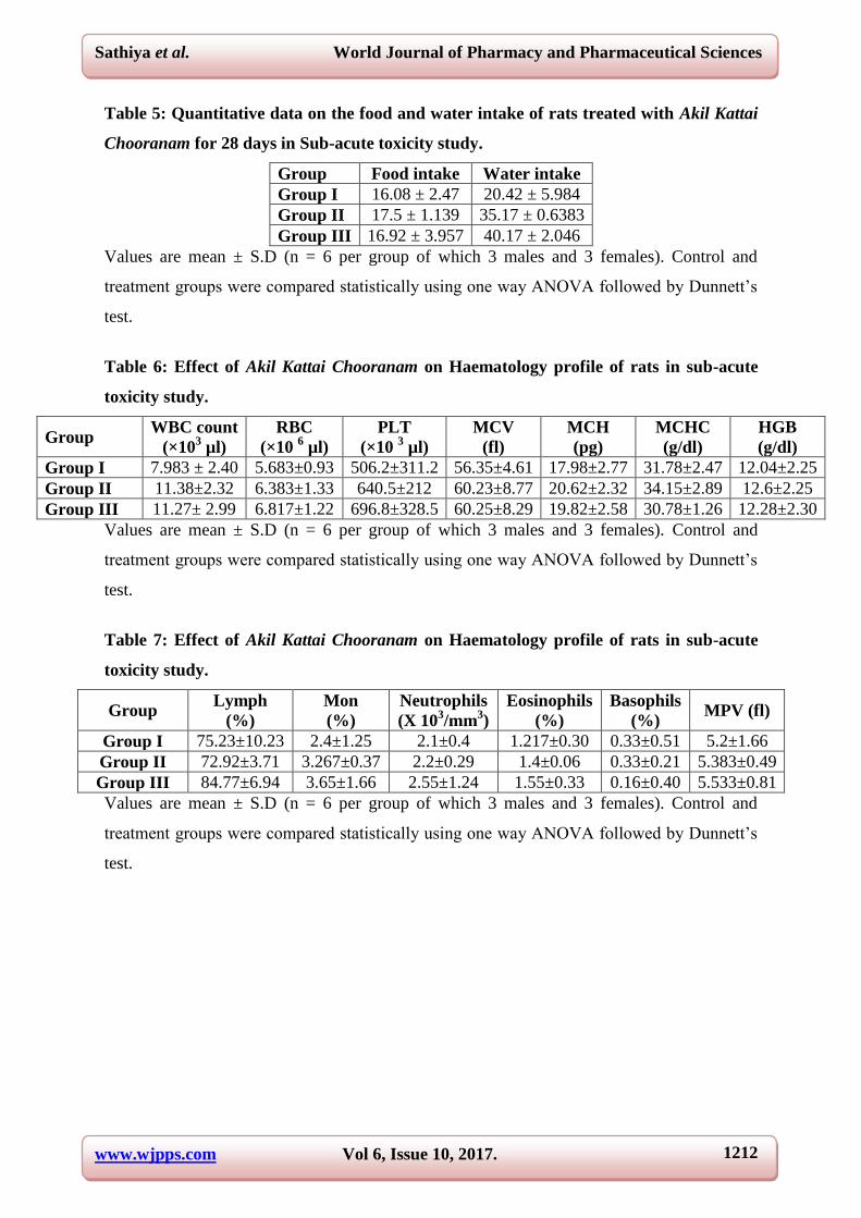
3.5. Effect of AKC on food and water intake of rats in Sub-acute oral toxicity study
No significant change was observed in body weight of both male and female rats treated with
AKC at the dose of 200 and 400mg / kg b.w. The results were tabulated in Table 5.
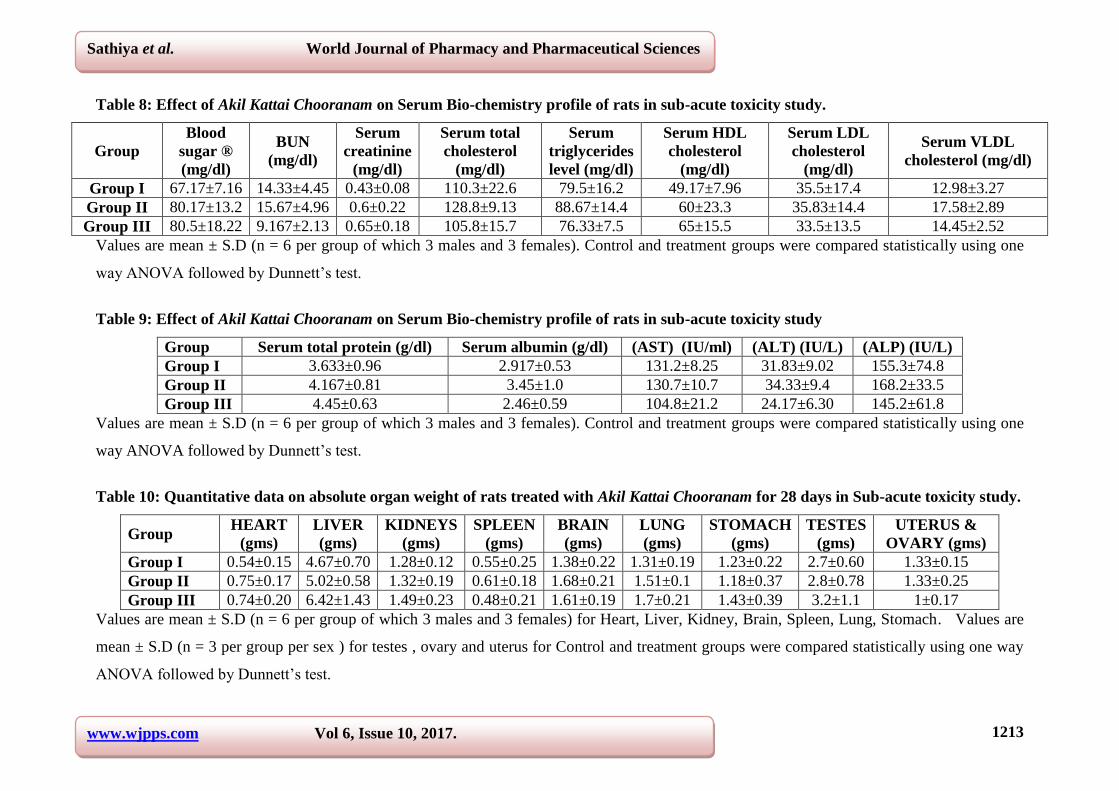
3.6. Effect of AKC on Hematological and Biochemical parameters of male and female
rats in Sub-acute oral toxicity study
No statistically significant differences were recorded in hematological and biochemical
parameters of rats treated with AKC at the dose of 200 and 400mg / kg b.w. The results were
tabulated in Table 6-9.
3.7. Effect of AKC on Histopathological changes of male and female rat in Sub-acute
oral toxicity study
No abnormality were detected in the histopathological analysis of organs (Kidney, Heart,
Liver, Brain, Lung, Spleen and Stomach) retrieved from the rats treated with low and high
dose of AKC.
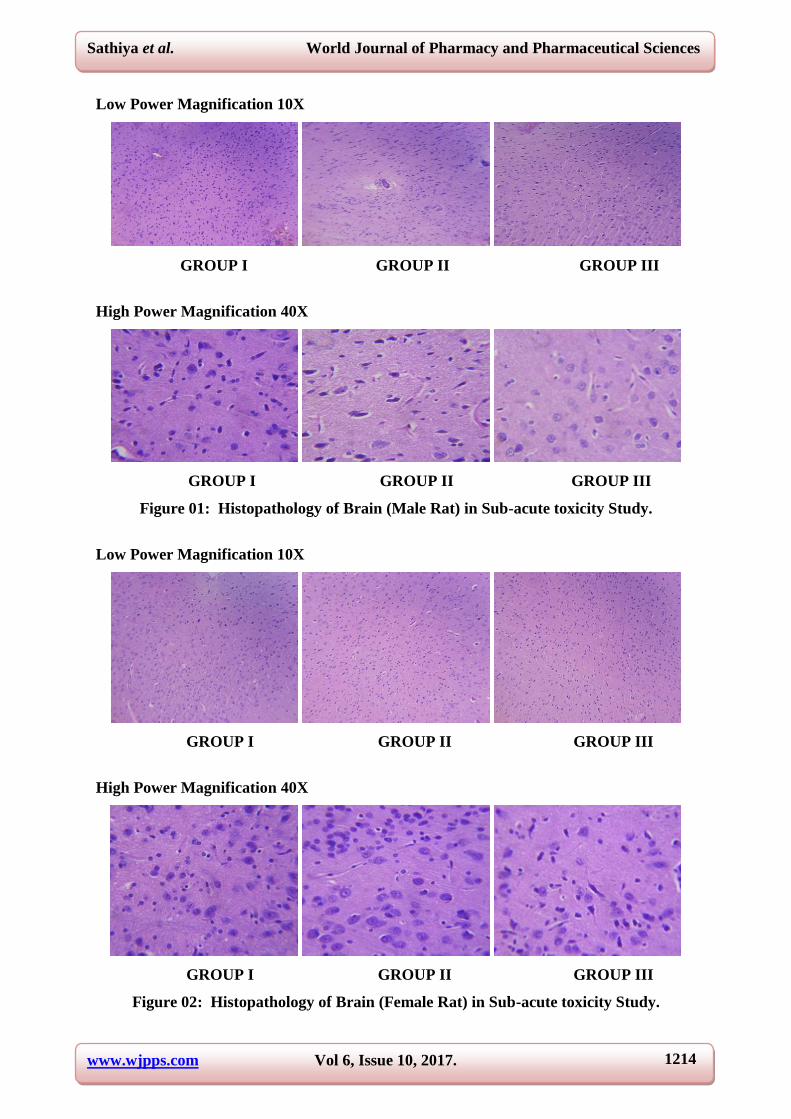
Histology of brain reveals regular marginal alignment on the neurons with promising
histology. Neurons is very intact and there were no signs of edema or degeneration were
observed in sample belongs to group I,II and III. As shown in figure 01 and 02.Microscopic
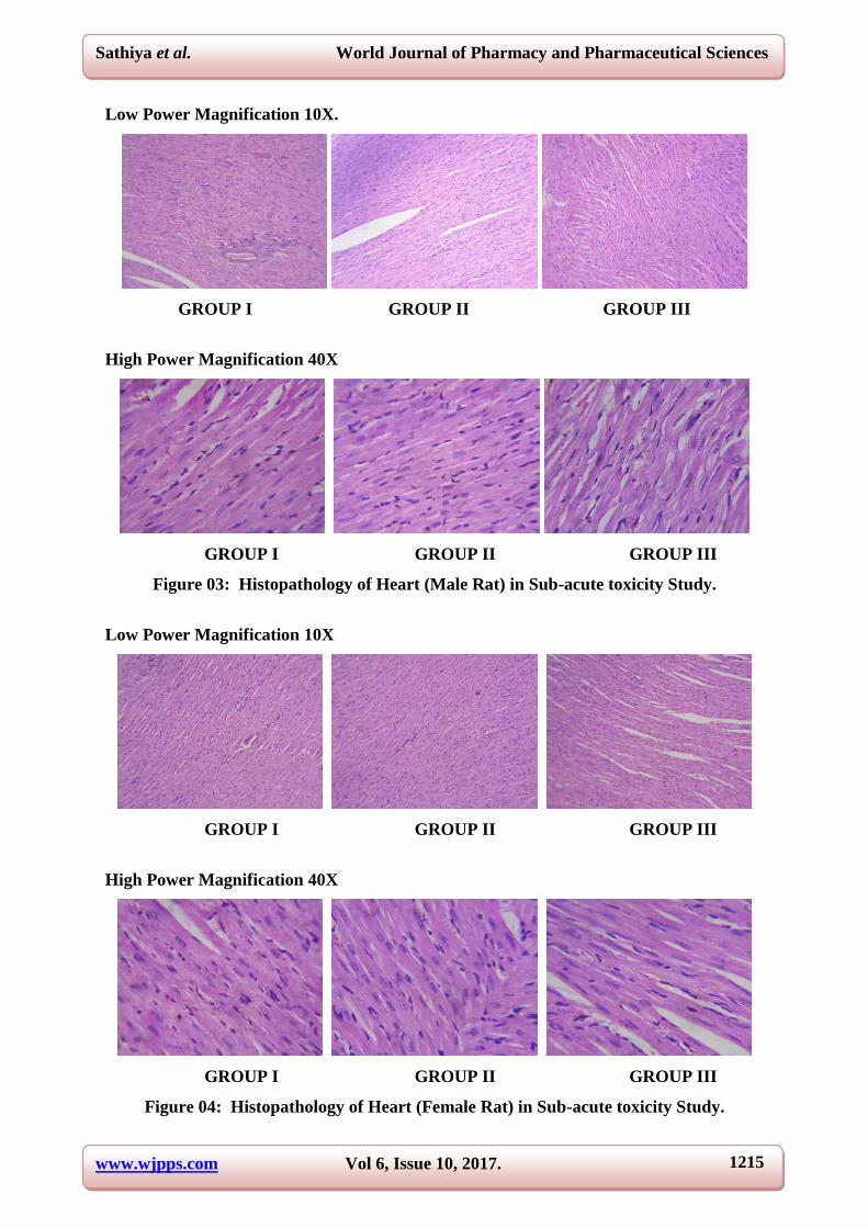
observation of heart projects prominent nucleus with regular arrangement of fibres. No
evidence of pyknotic nucleus were observed in samples belongs to group I, II and III. As
shown in figure 03 and 04.
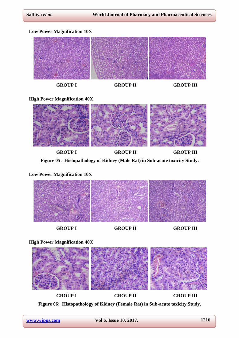
Appearance of proximal and distal convolutes tubules of kidney was normal with no evidence
of atrophy. Lumen of distal convolutes tubule and collecting duct was normal in sample

www.wjpps.com Vol 6, Issue 10, 2017.
1209
Sathiya et al. World Journal of Pharmacy and Pharmaceutical Sciences
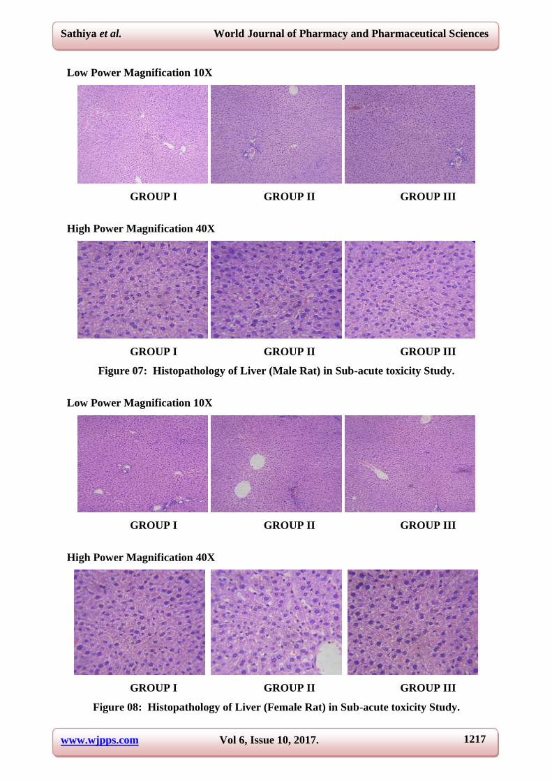
belongs to group I,II and III. As shown in figure 05 and 06.In liver centrilobular zone appears
normal with stable network of hepatocytes. Rare appearance of Kupffer cells with no
evidence of phagocytosis in intracytoplasmic region were observed in sample belongs to
group I, II and III. As shown in figure 07 and 08.
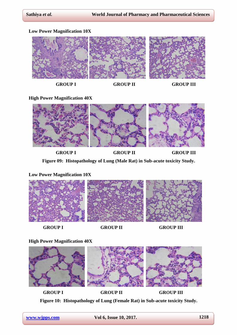
Histopathology of lung shows no signs of airway secretion and bronchial secretion. Bronchial
blood vessels and connective tissue appears normal with no signs of pulmonary edema. In the
stomach Intracytoplasmic zone of mucosa appears normal. As shown in figure 09 and
10.Histology of spleen shown appearance of LF – lymphoid follicle; PALS – periarterial
lymphoid sheath was normal with no significant signs of enlargement were observed in
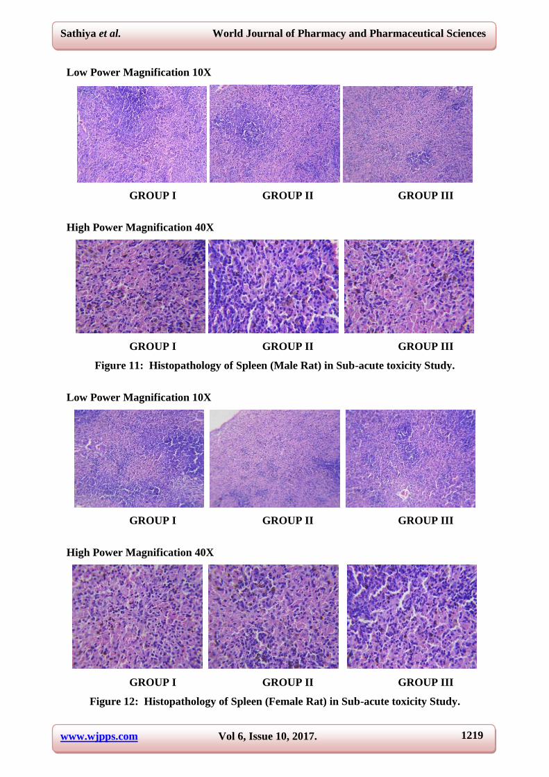
sample belongs to group I, II and III. As shown in figure 11 and 12.
Light microscopic observation stomach reveals normal histology of gastric wall composed of
normal mucosa, muscularismucosa, submucosa, muscularispropiria and adventitia. No signs
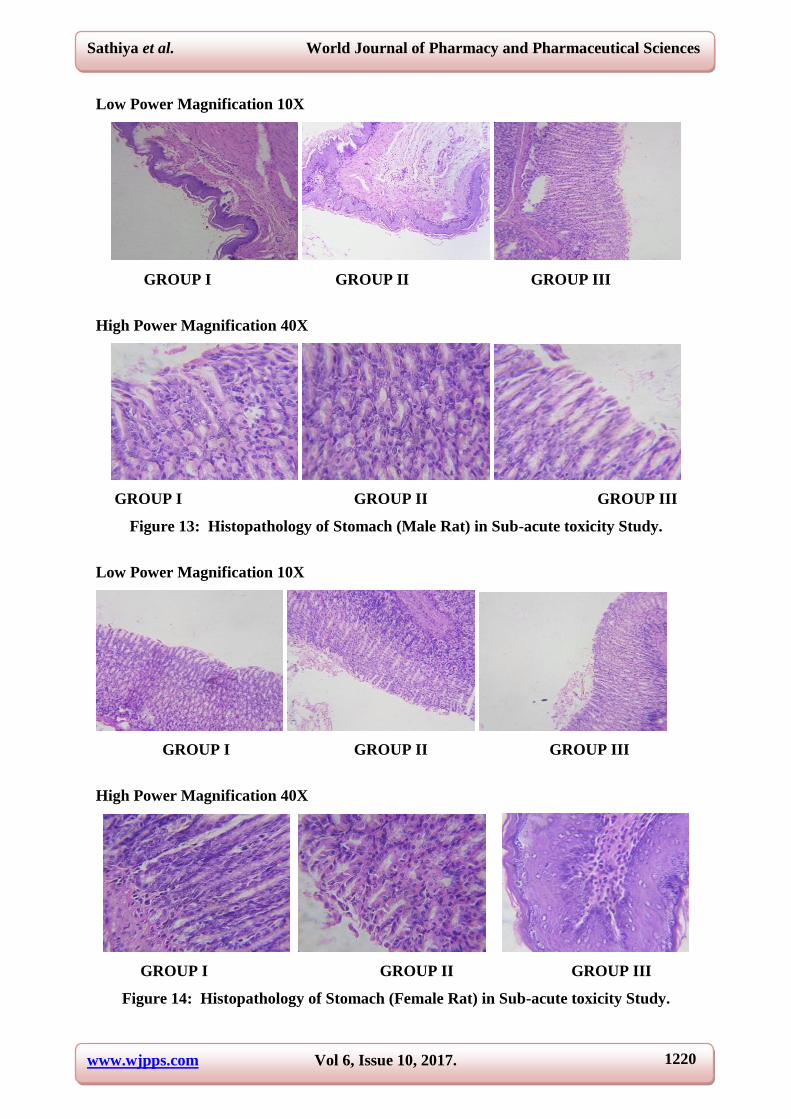
of ulceration were observed in sample belongs to group I, II and III. As shown in figure 13
and 14. In testes presence of mature somatic cells project the perfect histomorphology of
testicular cells in this group. Primary spermatocytes with large centered nucleus and dense
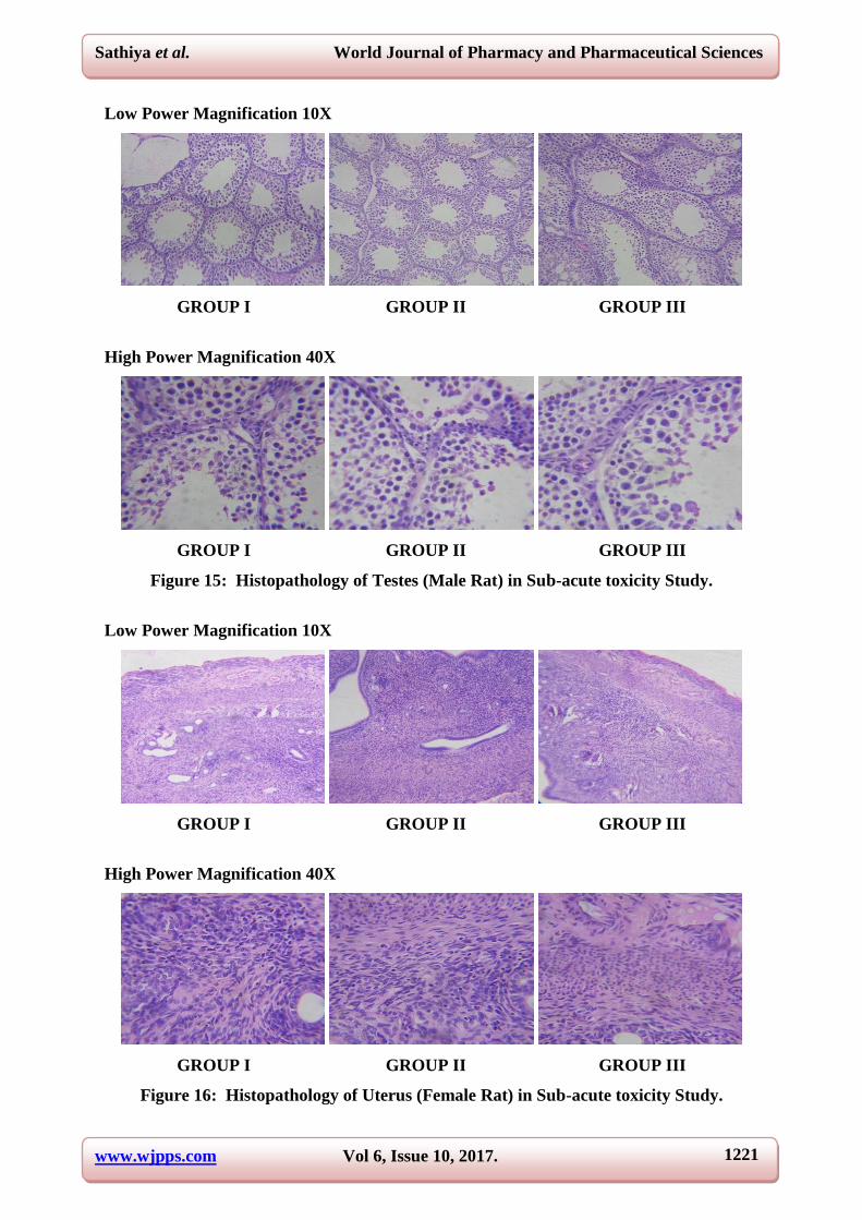
chromatin were observed in sample belongs to group I,II and III. As shown in figure 15.
Uterus of female rats shown normal appearance of endometrium, myometrium and uterine
glands. Arrangement of stratum basale, functionale and surface epithelium seems normal in
samples belongs to group I,II and III. As shown in figure 16. Histopathological analysis of
ovary showing normal corpus luteum (CL) and Primordial follicles with few mature ovarian
follicles with no signs of abnormality. Appearance of antral follicle, primary oocyte and
secondary follicles are normal in sample belong to group I,II and III. As shown in figure 17.
Table 1: Effect of Akil Kattai Chooranam on clinical signs of Female rats in Acute Oral
Toxicity Study.
Parameter Group I
Clinical Signs Parameters for the duration of 14 days Test Drug 2000mg/ Kg
Number of animals observed 6 Female
Lacrimation Absence
Salivation Absence
Animal appearance Normal
Tonic Movement Absence
Clonic Movement Absence
Laxative action Absence
Touch Response Normal
www.wjpps.com Vol 6, Issue 10, 2017.
1210
Sathiya et al. World Journal of Pharmacy and Pharmaceutical Sciences
Response to Sound Normal Response
Response to Light Normal Response
Mobility Normal Response
Respiratory Distress Nil
Skin Color Normal
Stereotype behavior Absence
Piloerection Absence
Limb Paralysis Absence
Posture Normal
Open field behavior Normal
Gait Balancing Normal
Freezing Behaviour Absent
Sings of Stress and Anxiety None Observed
Muscular coordination Normal
Muscle grip Normal
Sedation Absence
Social Behavior Normal
Urine Analysis No Abnormality
Urine Colour Yellowish
Urine pH 7
Urine -Glucose Absence
Urine -Ketones Absence
Urine- Bilirubin Absence
Urine-Blood Cells Negative
Urine - Pus cells Negative
Mortality Nil
Table 2: Effect of Akil Kattai Chooranam on Body weight of Female rats in acute toxicity
study.
Group Before Treatment Weight in Gms After Treatment Weight in Gms
Group I 181.8 ± 6.94 184.5 ± 7.86
Values are mean ± S.D (n = 6 per group). Statistically using one way ANOVA followed by
Dunnett’s test.
Table 3: Effect of Akil Kattai Chooranam on clinical signs of male and female rats in
Sub-acute oral toxicity study.
Parameter Group I Group II Group III
Clinical Signs Parameters for
28 days Control
Test Drug
200mg/ Kg
Test Drug
400mg/ Kg
Number of animals observed 3 Male and 3
Female
3 Male and 3
Female
3 Male and 3
Female
Lacrimation Absence Absence Absence
Salivation Absence Absence Absence
Animal appearance Normal Normal Normal
Tonic Movement Absence Absence Absence
Clonic Movement Absence Absence Absence
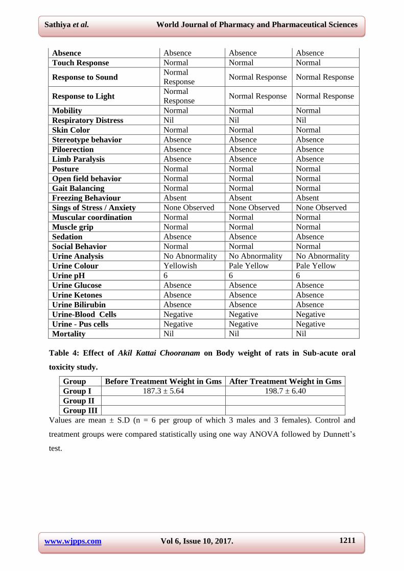
www.wjpps.com Vol 6, Issue 10, 2017.
1211
Sathiya et al. World Journal of Pharmacy and Pharmaceutical Sciences
Absence Absence Absence Absence
Touch Response Normal Normal Normal
Response to Sound Normal
Response Normal Response Normal Response
Response to Light Normal
Response Normal Response Normal Response
Mobility Normal Normal Normal
Respiratory Distress Nil Nil Nil
Skin Color Normal Normal Normal
Stereotype behavior Absence Absence Absence
Piloerection Absence Absence Absence
Limb Paralysis Absence Absence Absence
Posture Normal Normal Normal
Open field behavior Normal Normal Normal
Gait Balancing Normal Normal Normal
Freezing Behaviour Absent Absent Absent
Sings of Stress / Anxiety None Observed None Observed None Observed
Muscular coordination Normal Normal Normal
Muscle grip Normal Normal Normal
Sedation Absence Absence Absence
Social Behavior Normal Normal Normal
Urine Analysis No Abnormality No Abnormality No Abnormality
Urine Colour Yellowish Pale Yellow Pale Yellow
Urine pH 6 6 6
Urine Glucose Absence Absence Absence
Urine Ketones Absence Absence Absence
Urine Bilirubin Absence Absence Absence
Urine-Blood Cells Negative Negative Negative
Urine - Pus cells Negative Negative Negative
Mortality Nil Nil Nil
Table 4: Effect of Akil Kattai Chooranam on Body weight of rats in Sub-acute oral
toxicity study.
Group Before Treatment Weight in Gms After Treatment Weight in Gms
Group I 187.3 ± 5.64 198.7 ± 6.40
Group II
Group III
Values are mean ± S.D (n = 6 per group of which 3 males and 3 females). Control and
treatment groups were compared statistically using one way ANOVA followed by Dunnett’s
test.
www.wjpps.com Vol 6, Issue 10, 2017.
1212
Sathiya et al. World Journal of Pharmacy and Pharmaceutical Sciences
Table 5: Quantitative data on the food and water intake of rats treated with Akil Kattai
Chooranam for 28 days in Sub-acute toxicity study.
Group Food intake Water intake
Group I 16.08 ± 2.47 20.42 ± 5.984
Group II 17.5 ± 1.139 35.17 ± 0.6383
Group III 16.92 ± 3.957 40.17 ± 2.046
Values are mean ± S.D (n = 6 per group of which 3 males and 3 females). Control and
treatment groups were compared statistically using one way ANOVA followed by Dunnett’s
test.
Table 6: Effect of Akil Kattai Chooranam on Haematology profile of rats in sub-acute
toxicity study.
Group WBC count
(×103 µl)
RBC
(×10 6 µl)
PLT
(×10 3 µl)
MCV
(fl)
MCH
(pg)
MCHC
(g/dl)
HGB
(g/dl)
Group I 7.983 ± 2.40 5.683±0.93 506.2±311.2 56.35±4.61 17.98±2.77 31.78±2.47 12.04±2.25
Group II 11.38±2.32 6.383±1.33 640.5±212 60.23±8.77 20.62±2.32 34.15±2.89 12.6±2.25
Group III 11.27± 2.99 6.817±1.22 696.8±328.5 60.25±8.29 19.82±2.58 30.78±1.26 12.28±2.30
Values are mean ± S.D (n = 6 per group of which 3 males and 3 females). Control and
treatment groups were compared statistically using one way ANOVA followed by Dunnett’s
test.
Table 7: Effect of Akil Kattai Chooranam on Haematology profile of rats in sub-acute
toxicity study.
Group Lymph
(%)
Mon
(%)
Neutrophils
(X 103/mm
3)
Eosinophils
(%)
Basophils
(%) MPV (fl)
Group I 75.23±10.23 2.4±1.25 2.1±0.4 1.217±0.30 0.33±0.51 5.2±1.66
Group II 72.92±3.71 3.267±0.37 2.2±0.29 1.4±0.06 0.33±0.21 5.383±0.49
Group III 84.77±6.94 3.65±1.66 2.55±1.24 1.55±0.33 0.16±0.40 5.533±0.81
Values are mean ± S.D (n = 6 per group of which 3 males and 3 females). Control and
treatment groups were compared statistically using one way ANOVA followed by Dunnett’s
test.
www.wjpps.com Vol 6, Issue 10, 2017.
1213
Sathiya et al. World Journal of Pharmacy and Pharmaceutical Sciences
Table 8: Effect of Akil Kattai Chooranam on Serum Bio-chemistry profile of rats in sub-acute toxicity study.
Group
Blood
sugar ®
(mg/dl)
BUN
(mg/dl)
Serum
creatinine
(mg/dl)
Serum total
cholesterol
(mg/dl)
Serum
triglycerides
level (mg/dl)
Serum HDL
cholesterol
(mg/dl)
Serum LDL
cholesterol
(mg/dl)
Serum VLDL
cholesterol (mg/dl)
Group I 67.17±7.16 14.33±4.45 0.43±0.08 110.3±22.6 79.5±16.2 49.17±7.96 35.5±17.4 12.98±3.27
Group II 80.17±13.2 15.67±4.96 0.6±0.22 128.8±9.13 88.67±14.4 60±23.3 35.83±14.4 17.58±2.89
Group III 80.5±18.22 9.167±2.13 0.65±0.18 105.8±15.7 76.33±7.5 65±15.5 33.5±13.5 14.45±2.52
Values are mean ± S.D (n = 6 per group of which 3 males and 3 females). Control and treatment groups were compared statistically using one
way ANOVA followed by Dunnett’s test.
Table 9: Effect of Akil Kattai Chooranam on Serum Bio-chemistry profile of rats in sub-acute toxicity study
Group Serum total protein (g/dl) Serum albumin (g/dl) (AST) (IU/ml) (ALT) (IU/L) (ALP) (IU/L)
Group I 3.633±0.96 2.917±0.53 131.2±8.25 31.83±9.02 155.3±74.8
Group II 4.167±0.81 3.45±1.0 130.7±10.7 34.33±9.4 168.2±33.5
Group III 4.45±0.63 2.46±0.59 104.8±21.2 24.17±6.30 145.2±61.8
Values are mean ± S.D (n = 6 per group of which 3 males and 3 females). Control and treatment groups were compared statistically using one
way ANOVA followed by Dunnett’s test.
Table 10: Quantitative data on absolute organ weight of rats treated with Akil Kattai Chooranam for 28 days in Sub-acute toxicity study.
Group HEART
(gms)
LIVER
(gms)
KIDNEYS
(gms)
SPLEEN
(gms)
BRAIN
(gms)
LUNG
(gms)
STOMACH
(gms)
TESTES
(gms)
UTERUS &
OVARY (gms)
Group I 0.54±0.15 4.67±0.70 1.28±0.12 0.55±0.25 1.38±0.22 1.31±0.19 1.23±0.22 2.7±0.60 1.33±0.15
Group II 0.75±0.17 5.02±0.58 1.32±0.19 0.61±0.18 1.68±0.21 1.51±0.1 1.18±0.37 2.8±0.78 1.33±0.25
Group III 0.74±0.20 6.42±1.43 1.49±0.23 0.48±0.21 1.61±0.19 1.7±0.21 1.43±0.39 3.2±1.1 1±0.17
Values are mean ± S.D (n = 6 per group of which 3 males and 3 females) for Heart, Liver, Kidney, Brain, Spleen, Lung, Stomach. Values are
mean ± S.D (n = 3 per group per sex ) for testes , ovary and uterus for Control and treatment groups were compared statistically using one way
ANOVA followed by Dunnett’s test.
www.wjpps.com Vol 6, Issue 10, 2017.
1214
Sathiya et al. World Journal of Pharmacy and Pharmaceutical Sciences
Low Power Magnification 10X
GROUP I GROUP II GROUP III
High Power Magnification 40X
GROUP I GROUP II GROUP III
Figure 01: Histopathology of Brain (Male Rat) in Sub-acute toxicity Study.
Low Power Magnification 10X
GROUP I GROUP II GROUP III
High Power Magnification 40X
GROUP I GROUP II GROUP III
Figure 02: Histopathology of Brain (Female Rat) in Sub-acute toxicity Study.
www.wjpps.com Vol 6, Issue 10, 2017.
1215
Sathiya et al. World Journal of Pharmacy and Pharmaceutical Sciences
Low Power Magnification 10X.
GROUP I GROUP II GROUP III
High Power Magnification 40X
GROUP I GROUP II GROUP III
Figure 03: Histopathology of Heart (Male Rat) in Sub-acute toxicity Study.
Low Power Magnification 10X
GROUP I GROUP II GROUP III
High Power Magnification 40X
GROUP I GROUP II GROUP III
Figure 04: Histopathology of Heart (Female Rat) in Sub-acute toxicity Study.
www.wjpps.com Vol 6, Issue 10, 2017.
1216
Sathiya et al. World Journal of Pharmacy and Pharmaceutical Sciences
Low Power Magnification 10X
GROUP I GROUP II GROUP III
High Power Magnification 40X
GROUP I GROUP II GROUP III
Figure 05: Histopathology of Kidney (Male Rat) in Sub-acute toxicity Study.
Low Power Magnification 10X
GROUP I GROUP II GROUP III
High Power Magnification 40X
GROUP I GROUP II GROUP III
Figure 06: Histopathology of Kidney (Female Rat) in Sub-acute toxicity Study.
www.wjpps.com Vol 6, Issue 10, 2017.
1217
Sathiya et al. World Journal of Pharmacy and Pharmaceutical Sciences
Low Power Magnification 10X
GROUP I GROUP II GROUP III
High Power Magnification 40X
GROUP I GROUP II GROUP III
Figure 07: Histopathology of Liver (Male Rat) in Sub-acute toxicity Study.
Low Power Magnification 10X
GROUP I GROUP II GROUP III
High Power Magnification 40X
GROUP I GROUP II GROUP III
Figure 08: Histopathology of Liver (Female Rat) in Sub-acute toxicity Study.
www.wjpps.com Vol 6, Issue 10, 2017.
1218
Sathiya et al. World Journal of Pharmacy and Pharmaceutical Sciences
Low Power Magnification 10X
GROUP I GROUP II GROUP III
High Power Magnification 40X
GROUP I GROUP II GROUP III
Figure 09: Histopathology of Lung (Male Rat) in Sub-acute toxicity Study.
Low Power Magnification 10X
GROUP I GROUP II GROUP III
High Power Magnification 40X
GROUP I GROUP II GROUP III
Figure 10: Histopathology of Lung (Female Rat) in Sub-acute toxicity Study.
www.wjpps.com Vol 6, Issue 10, 2017.
1219
Sathiya et al. World Journal of Pharmacy and Pharmaceutical Sciences
Low Power Magnification 10X
GROUP I GROUP II GROUP III
High Power Magnification 40X
GROUP I GROUP II GROUP III
Figure 11: Histopathology of Spleen (Male Rat) in Sub-acute toxicity Study.
Low Power Magnification 10X
GROUP I GROUP II GROUP III
High Power Magnification 40X
GROUP I GROUP II GROUP III
Figure 12: Histopathology of Spleen (Female Rat) in Sub-acute toxicity Study.
www.wjpps.com Vol 6, Issue 10, 2017.
1220
Sathiya et al. World Journal of Pharmacy and Pharmaceutical Sciences
Low Power Magnification 10X
GROUP I GROUP II GROUP III
High Power Magnification 40X
GROUP I GROUP II GROUP III
Figure 13: Histopathology of Stomach (Male Rat) in Sub-acute toxicity Study.
Low Power Magnification 10X
GROUP I GROUP II GROUP III
High Power Magnification 40X
GROUP I GROUP II GROUP III
Figure 14: Histopathology of Stomach (Female Rat) in Sub-acute toxicity Study.
www.wjpps.com Vol 6, Issue 10, 2017.
1221
Sathiya et al. World Journal of Pharmacy and Pharmaceutical Sciences
Low Power Magnification 10X
GROUP I GROUP II GROUP III
High Power Magnification 40X
GROUP I GROUP II GROUP III
Figure 15: Histopathology of Testes (Male Rat) in Sub-acute toxicity Study.
Low Power Magnification 10X
GROUP I GROUP II GROUP III
High Power Magnification 40X
GROUP I GROUP II GROUP III
Figure 16: Histopathology of Uterus (Female Rat) in Sub-acute toxicity Study.

www.wjpps.com Vol 6, Issue 10, 2017.
1222
Sathiya et al. World Journal of Pharmacy and Pharmaceutical Sciences
Low Power Magnification 10X
GROUP I GROUP II GROUP III
High Power Magnification 40X
GROUP I GROUP II GROUP III
Figure 17: Histopathology of Ovary (Female Rat) in Sub-acute toxicity Study.
4. DISCUSSION
In modern society, toxicology has become an important element in environmental and
occupational health. This is because many organizations, governmental and non-
governmental, utilize information from toxicology to evaluate and regulate hazards in the
workplace and nonoccupational environment. As part of prevention strategies, toxicology is
invaluable, since it is the source of information on potential hazards in the absence of
widespread human exposures. Toxicological methods are also widely used by industry in
product development, to provide information useful in the design of specific molecules or
product formulations.[12]
NOEL (NOAEL) means the no observed (adverse) effect level, or the highest dose that does
not cause a toxic effect. To establish a NOEL requires multiple doses, a large population and
additional information to make sure that absence of a response is not merely a statistical
phenomenon. LOEL is the lowest observed effective dose on a dose-response curve, or the
lowest dose that causes an effect.[13]
www.wjpps.com Vol 6, Issue 10, 2017.
1223
Sathiya et al. World Journal of Pharmacy and Pharmaceutical Sciences
Herbal medicines have been used for thousands of years, and herb preparations are
commercially available for the folk remedies or for the promotion of health. In contrast to
chemical drugs, herbs are regarded to be non-toxic and safe, because of their natural origin.
But accumulating clinical data claim the toxicity of herbal medicine. The results of the acute
toxicity profiling of the drug AKC reveals no significant change in body weight, behavioral
and mortality at the dose of 2000mg/kg.
The liver is considerably the most important target for toxicity caused by drugs. This
susceptibility is a consequence of the functional features of the liver and their role in the
metabolic elimination of most drugs. Therefore, evaluation of potential hepatotoxicity
represents a critical step in the development of new drugs. The liver is very active in
metabolising foreign compounds and, although biotransformation reactions generally parallel
detoxification processes, the formation of reactive metabolites is relatively frequent. Thus,
drug-induced hepatotoxicity can be due to the administered compound itself or to metabolites
formed by hepatic metabolism.[14]
In sub-acute toxicity study treatment with AKC at the dose level of 200 and 400mg/kg did
not reveal any significant change in body weight, food and water intake in both male and
female rats. Further there is no alteration in AST, ALT and ALP level of treated animals
which denotes the hepato protective nature of the drug.
Blood a fluid connective tissue involved in supply of nutrient’s and also drug to the target
organs often exposed to the ill effect of certain cytotoxic drugs. Most of the siddha
preparation seems to be safe with hematopoietic system but certain drugs and chemicals
which particularly have tendency to disturb the bone marrow and causes hemolysis disturbs
the cardiovascular physiology. The hemo toxic nature of the drug exerted by the fluctuation
in blood cell count in particular to RBC and WBC cells. Low hemoglobin content reflects the
low level of RBC which in turn affects the oxygen carrying capacity of the blood. At the end
of the most of the toxicity studies the blood collected from the animal before sacrifice will be
subjected to whole blood analysis and also to serological analysis.
Results of hematological and serological profiling of AKC at the dose of 200 and 400mg/kg
did not shown any signs of hematopoietic toxicity and there is no change in the blood and
serum parameters of the treated animals.
www.wjpps.com Vol 6, Issue 10, 2017.
1224
Sathiya et al. World Journal of Pharmacy and Pharmaceutical Sciences
5. CONCLUSION
From the results of the present toxicology study of the trial drug AKC there was a generation
of evidence based data that shows the drug is relatively non-toxic, causes no apparent organ
damage or mortality in both the short term and long term administration on study animals.
Hence from the results, it was concluded that the drug AKC was safe and reveals no signs of
significant toxicity for long term treatment for the chronic disease condition.
6. REFERENCES
1. OECD. 2003. Descriptions of Selected Key Generic Terms Used in Chemical Hazard
Risk Assessment. Joint Project with IPCS on the Harmonisation of Hazard Risk
Assessment Terminology. OECD Series on Testing and Assessment No. 44. Environment
Directorate, Joint Meeting of the Chemicals Committee and the Working Party on
Chemicals, Pesticides and Biotechnology. ENV/JM/MONO (2003)15. Paris: OECD.
2. Rozman K. Disposition of xenobiotics: Species differences. Toxicol. Pathol, 1988; 16:
123–129.
3. Pyszel A. Effect of metals, benzene, pesticides and ethylene oxide on the haematopoietic
system. Med Pr., 2005; 56: 249-55.
4. Ioannides C, Lewis DF. Cytochromes P450 in the bioactivation of chemicals. Curr Top
Med Chem., 2004; 4: 1767-1788.
5. Jha V, Chugh KS. Nephropathy associated with animal, plant, and chemical toxins in the
tropics. Sem Nephrol, 2003; 23: 49–65.
6. Mohan RC, Agasthiyar Paripooranam 400. Thamarai Noolagam, Chennai-26, Edition-
2012: 111.
7. OECD guideline for testing of chemicals. Guideline 423, 17th December 2001.
8. OECD Guide lines 407 for testing of chemicals. Repeated dose 28-Day Oral Toxicity
Study in Rodents, 2008; 2- 8.
9. Jain N, Sharma P, Sharma N, Joshi S C. Haemato-biochemical profile following sub
acute toxicity of malathion in male albino rats. Pharmacologyonline, 2009; 2: 500–506.
10. Suvarna SK, Layton C, Bancroft JD. Bancroft’s theory and practice of histological
techniques. 7th edn, Churchill Livingstone, London, 2013.
11. Visweswara Rao. Biostatistics, A manual of statistic methods for use in Health, Nutrition
and Anthropology, Rajkamal Electrical press, Delhi, 2007; 226-312.
12. US department of Health and human services. Asbestos Toxicity. Case Studies in
Environmental Medicine, 2005.
www.wjpps.com Vol 6, Issue 10, 2017.
1225
Sathiya et al. World Journal of Pharmacy and Pharmaceutical Sciences
13. Wolfgang Dekant. Evaluation of Toxic Effects. Ullmanns Encyclopedia of Industrial
Chemistry, 2011.
14. Gomez-Lechon MJ. In Vitro Evaluation of Potential Hepatotoxicity Induced by Drugs.
Current Pharmaceutical Design, 2010; 16: 01-15.